Case Report Tophaceous Gout of the Patella: A Report of Two Cases
|
|
|
- Evan McKinney
- 5 years ago
- Views:
Transcription
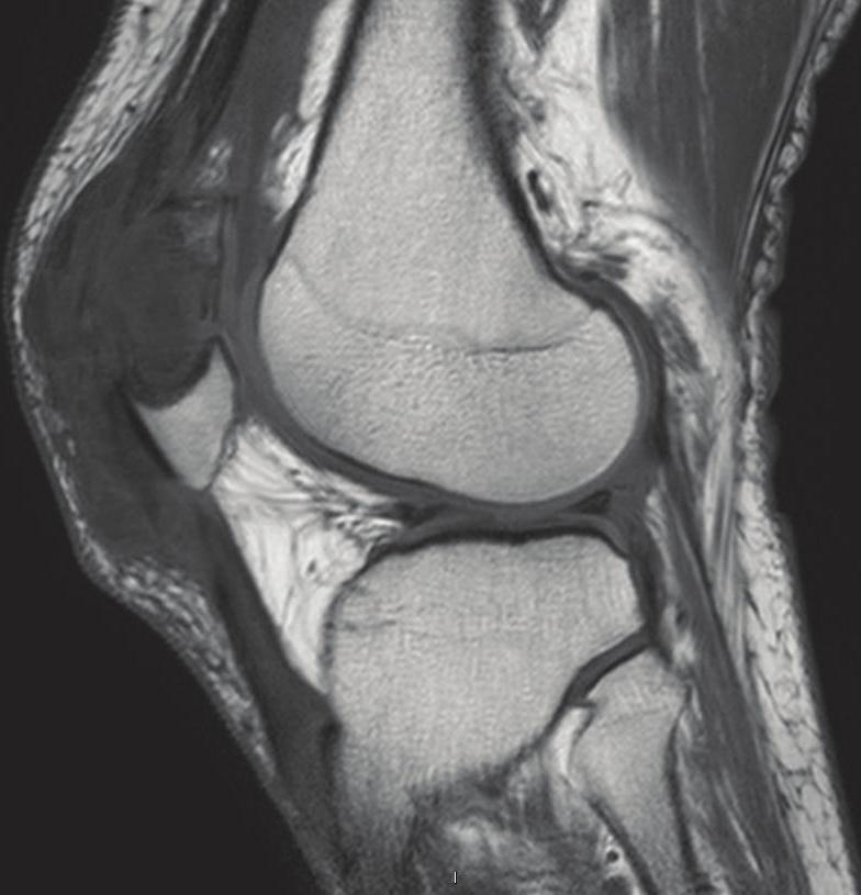
1 Case Reports in Rheumatology Volume 2012, Article ID , 6 pages doi: /2012/ Case Report Tophaceous Gout of the Patella: A Report of Two Cases Graeme Hopper, 1 Sanjay Gupta, 1 Sarath Bethapudi, 2 David Ritchie, 2 Elaine MacDuff, 3 and Ashish Mahendra 1 1 Trauma and Orthopaedics, Glasgow Royal Infirmary, 84 Castle Street, Glasgow G4 0SF, UK 2 Radiology, Glasgow Western Infirmary, Dumbarton Road, Glasgow G11 6NT, UK 3 Pathology, Glasgow Western Infirmary, Dumbarton Road, Glasgow G11 6NT, UK Correspondence should be addressed to Graeme Hopper, hopperg@doctors.org.uk Received 3 October 2012; Accepted 19 October 2012 Academic Editors: R. Cevik, U. Gresser, and M. A. Hunt Copyright 2012 Graeme Hopper et al. This is an open access article distributed under the Creative Commons Attribution License, which permits unrestricted use, distribution, and reproduction in any medium, provided the original work is properly cited. Introduction. Tophaceous gout of the patella is rare and may masquerade as a tumour or tumour-like condition. Cases.Wereport two patients with gout involving the patella, one complicated by a pathological fracture and the other occurring in a bipartite patella in a young adult. Discussion. Typical imaging appearances and measurement of serum urate will usually confirm the diagnosis but, occasionally, the serum urate level may be normal in active gout and in such cases, a biopsy will be required. Conclusion. Gout of the patella may masquerade as a tumour or tumour-like condition and it is important to consider gout in the differential diagnosis. 1. Introduction Gout is a relatively common crystal arthropathy caused by chronic hyperuricaemia resulting in the deposition of monosodium urate crystals around joints and tendons. In chronic tophaceous gout, crystals may be deposited in tendons and may cause osseous erosions at their bony attachments. Involvement of the extensor tendon of the knee is not an uncommon site for gout but involvement of the patella is rare and underreported in the literature [1 13]. For patients who present with a patellar lesion, the diagnosis of gout might not be suspected and patients given a provisional diagnosis of infection, tumour, metabolic disease, and degenerative or inflammatory arthropathy. We report two patients with gout involving the patella and discuss the clinical, imaging, and pathological features, differential diagnosis, and management. Patients gave informed consent prior to inclusion in this paper and each author certifies that there is no actual or potential conflict of interests in relation to this paper. 2. Case Reports 2.1. Case 1. A 70-year-old gentleman presented to his general practitioner with a two-month history of pain and swelling involving the anterior aspect of his right knee. He had no past medical history of note and no history of arthropathy or predisposing factors of gout. Radiographs of his right knee showed a faintly and partially mineralised soft tissue mass mainly overlying the superior aspect of the patella as well as a well-defined lytic lesion with marginal sclerosis in the mid patella (Figures 1(a) and 1(b)). Unfortunately, the patient was lost to follow up until he represented to the accident and emergency a year later, having injured his right knee. Radiographs of the knee showed a slightly displaced, comminuted pathological fracture of the patella as well as the previously noted partially mineralised soft tissue mass overlying the patella. The fracture was treated conservatively with a splint (Figures 1(c) and 1(d)). MRimaging confirmed the pathological fracture as well as the prominent prepatellar mass infiltrating the distal quadriceps and proximal patellar tendons (Figures 1(e) 1(h)). The elevated serumurate levels (1.04 mmol/l, normal range <0.40 mmol/l) confirmed active gout but a biopsy was felt justified in view of the unusual site and the need to exclude a coexisting tumour. Subsequent histological examination revealed aggregates of amorphous eosinophilic material surrounded by a palisade of histiocytes and giant cells in keeping with tophaceous gout (Figures 1(i) and 1(j)). This gentleman was referred

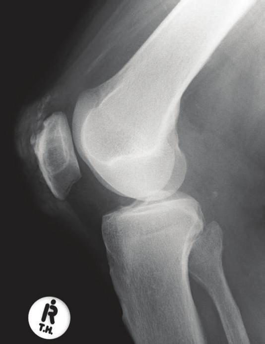
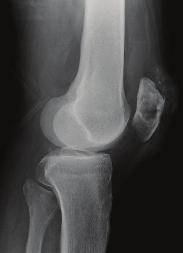
2 2 Case Reports in Rheumatology (a) (b) (c) (d) (e) (f) (g) (h) Figure 1: Continued.
. Note the well-defined lytic lesion with marginal sclerosis (arrowheads) in the patella and prepatellar soft tissue swelling.")
. The faint calcification within the distal quadriceps is again noted (black arrow).")
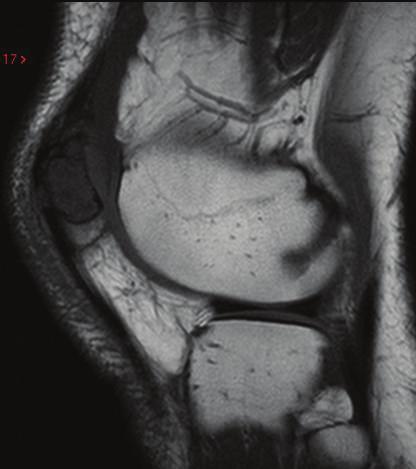
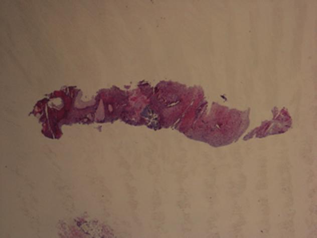
3 Case Reports in Rheumatology 3 (i) (j) Figure 1: (a) AP and (b) lateral radiographs of right knee demonstrate diffuse faint calcification involving the insertion of the quadriceps tendon at the upper pole of the patella (black arrow). Note the well-defined lytic lesion with marginal sclerosis (arrowheads) in the patella and prepatellar soft tissue swelling. (c) AP and (d) lateral radiographs of the right knee a year later show a comminuted pathological fracture of the mid patella involving the lytic lesion (white arrows) with associated joint effusion (star). The faint calcification within the distal quadriceps is again noted (black arrow). Sagittal (e) T1-WSE and (f) T2-WGE and coronal (g) PDSE and (h) STIR MR images confirm a comminuted pathological fracture of the mid and upper patella (black arrows) involving the patellar lesion (white arrows) that displays nonspecific features. However, there is a prominent inhomogeneous soft tissue mass in the prepatellar region, some of which displays low signal intensity (SI) on all sequences that corresponds to the mineralisation noted on the radiographs (arrowheads). (i) Core biopsy at low power and (j) core biopsy at high power, stained with H&E. There are aggregates of amorphous eosinophilic material surrounded by a palisade of histiocytes and giant cells in keeping with tophaceous gout. back to his GP for appropriate medical management of gout with colchicine and allopurinol. He was reviewed one year later in the oncology clinic, his symptoms had improved significantly, and radiographs demonstrated satisfactory healing of his fracture Case 2. A 34-year-old gentleman presented to the orthopaedic department with a two-month history of anterior knee pain and swelling involving his left knee. He had no past medical history of note, no history of arthritis or predisposing factors of gout such as obesity, high blood pressure, high cholesterol levels, and heavy alcohol use. Radiographs of his left knee showed a well-defined slightly expansile, lytic lesion involving the patella (Figures 2(a) and 2(b)). MR imaging showed a slightly inhomogeneous lesion involving the superolateral aspect of the patella with soft tissue infiltration around the patellar attachment of the lateral retinaculum (Figures 2(c) 2(g)). Serum urate levels were slightly raised at 0.54 mmol/l. An ultrasound-guided biopsy of this lesion was subsequently arranged to confirm the diagnosis. Histological examination revealed amorphous material consistent with gout (Figures 2(h) and 2(i)). This gentleman was referred back to his GP for appropriate medical management of gout with colchicine and allopurinol and has had no reported adverse outcomes. 3. Discussion Tophaceous gout of the patella was first reported in 1955 by Peloquin [10]. They presented a patient who was found to have erosion of the patellar cortex at surgery. Greenberg described the first reported pathological fracture of the patella due to underlying gout in 1986 [5]. Further pathological fractures of the patella due to gout have been published in[1, 2, 11]. Other reports include a nonunion of a patellar fracture as a result of gout [4] and a painful bipartite patella secondary to a gouty tophus [3]. Rechtet al. reported seven patients with gouty tophi of the patella and concluded that osteolytic lesions of the superolateral portion of the patella with an associated soft tissue mass should raise the possibility of gout [13]. Reber et al. reported three cases of patellar gout, one in a bipartite patella [12]. The classical history and examination findings of gout include previous attacks of gouty arthritis, tophaceous deposits, swelling of the joint, and occasional pain. Our reported patients did not have any history of gout but presented with a two-month history of pain and swelling involving the knee. Routine blood tests should include evaluation of serum urate and raised levels will confirm the diagnosis. However, occasionally, the serum urate may be normal during acute gout and in such cases evaluation of synovial fluid or biopsy may be required [14]. Radiographs of the extensor complex of the knee may show soft tissue swelling and mineralisation as in Case 1 but involvement of the patella is rare and raises the possibility of other pathology including tumours, infection, other arthropathies, and metabolic disorders [7 9, 15]. Other arthropathies, including rheumatoid arthritis and seronegative inflammatory arthropathies, osteoarthritis, and calcium pyrophosphate disease are often associated with subarticular changes including geodes and cysts but neither patient gave a history of a known arthropathy and there was no other evidence of arthropathy on the imaging studies of the affected knee joints. The differential also includes bone tumours but tumour-like lesions of the patella are more common than tumours and benign tumours more common than malignant tumours. In patients less than 40 years of age, chondroblastoma and giant cell tumour of bone are the most common diagnoses whereas over the age of 40 years, the most common diagnoses are gout and metastatic disease [15]. Radiologically, there is often difficulty in differentiating benign from malignant lesions. The relative small size of the patella, lack of physeal zones, and absence of periosteum make radiological assessment more difficult. Mineralisation in a patellar lesion may be found with gout and various


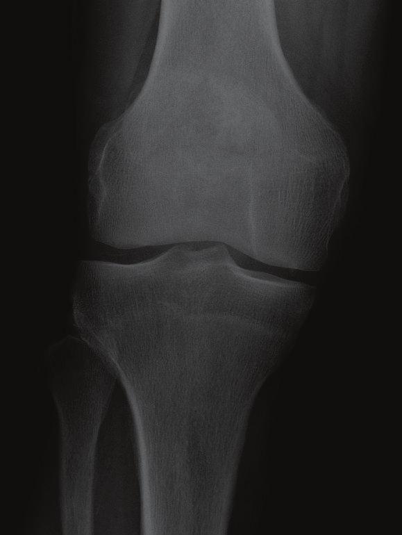
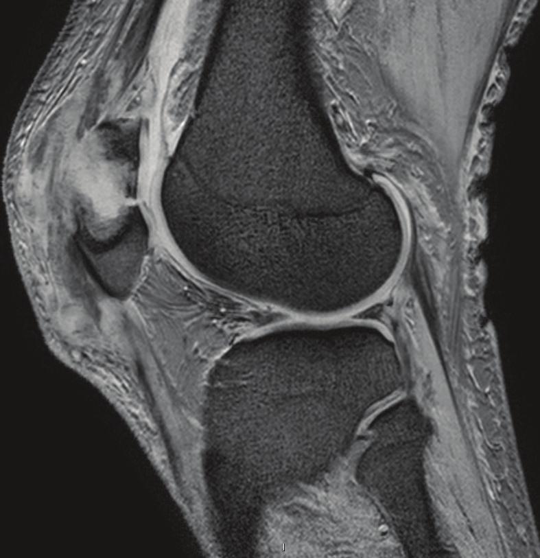
4 4 Case Reports in Rheumatology (a) (b) (c) (d) (e) (f) (g) (h) Figure 2: Continued.
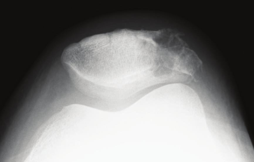
 and knee joint effusion (star). On the skyline view, there is also evidence of a bipartite patella (black arrows).")
 and reactive joint effusion (white arrow).")
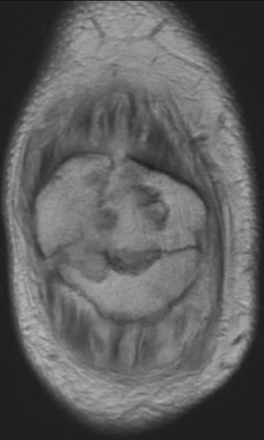
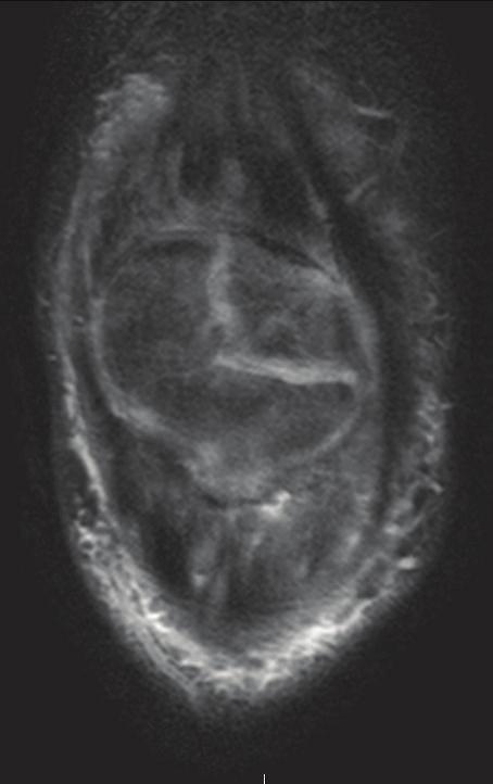
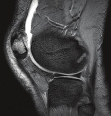
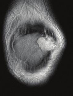
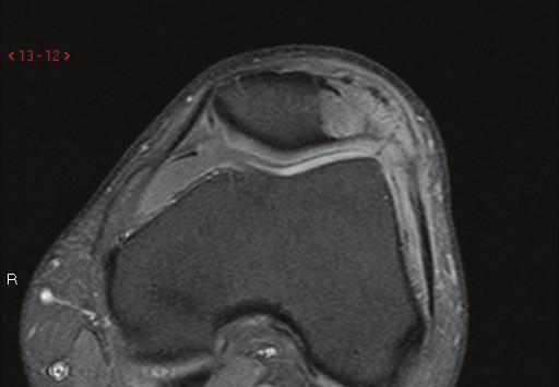
5 Case Reports in Rheumatology 5 (i) (j) Figure 2: (a) Lateral and (b) sky line view radiographs of the patella demonstrate a well-defined slightly expansile lytic lesion with sclerotic margin in the mid lateral aspect of the patella (arrowheads) and knee joint effusion (star). On the skyline view, there is also evidence of a bipartite patella (black arrows). Sagittal (c) T1-WSE and (d) T2-WGE and (e) coronal T2-WSE MR images demonstrate a well-defined mass in the patella (black arrows) that displays intermediate SI on T1-W and moderately increased SI on T2-W MR images. Note the prepatellar bursitis (chevrons) and reactive joint effusion (white arrow). Axial T1-WSE fat saturated (f) before and (g) after gadolinium MRI images, show avid enhancement of the patellar lesion (black arrows), involvement of the adjacent lateral parapatellar soft tissues and lateral retinaculum (arrowhead), and enhancing reactive synovitis (chevrons). (h) Core biopsy at low power, (i) core biopsy at medium power, and (j) core biopsy at high power stained with H&E. There are aggregates of amorphous eosinophilic material with a surrounding foreign body giant cell and histiocyte reaction consistent with gout. neoplasms including chondroblastoma, osteoid osteoma, and osteoblastoma, osteosarcoma but these tumours tend to occur in the second decades and gout would be most unusual in this age group unless there was a history of renal disease or genetic disorders associated with hyperuricaemia. A focal lesion in the patella may also be due to acute haematogenous osteomyelitis but this again is usually found in the immature patella and is very rare in adulthood [15]. An expansile lytic lesion should suggest either a giant cell tumour or brown tumour due to hyperparathyroidism and both of these diagnoses were considered in Case 2. Gout in a patient of 34 years is unusual and may be associated with obesity, high blood pressure, high cholesterol levels, and heavy alcohol abuse but none of these predisposing factors were present. Although the serum urate level was slightly elevated in Case 2, a biopsy was felt justified in view of the patient s relatively young age. Soft tissue involvement may occur with malignant lesions, including metastases, lymphomas, and sarcomas as well as gout but the presence of mineralisation within the soft tissue mass adjacent to the patella and in particular, the extensor tendon is more suggestive of gout. Furthermore, the marginal sclerosis around the intrapatellar lesions on the radiographs is a benign feature in keeping with a slow growing lesion as opposed to a malignant lesion that would usually have a more ill-defined zone of transition between lesion and normal bone. Pigmented villo-nodular synovitis (PVNS) and synovial osteochondromatosis (SOC) could also be considered in the differential diagnosis as these conditions may involve tendon sheaths and can cause intraosseous erosions with marginal sclerosis. However, mineralisation is not a feature of PVNS and involvement of the extensor tendon of the knee in SOC is very rare. Cross-sectional imaging may be helpful in lesion characterisation and staging. CT is very sensitive at detecting mineralisation and cortical destruction and MR imaging is the most accurate imaging technique for assessing soft tissue extent. The MR signal characteristics of gouty tophi are nonspecific with low/intermediate signal intensity (SI) on T1-weighting and variable SI on T2-weighting. Low SI foci on T2-weighting may be due to haemosiderin deposition or mineralisation, as in Case 1. Although the serum urate level was also elevated in Case 1, a biopsy was felt justified in view of the unusual site and the need to exclude a coexisting tumour. The pathological features of tophaceous gout are characteristic. Macroscopic examination reveals a nodular chalky appearance. Polarised microscopy of a fresh imprint demonstrates positively birefringent needle shaped crystals. These crystals are lost during processing for H&E examination. Instead, histologically, there are nodular aggregates of amorphous material surrounded by a palisade of histiocytes and multinucleated giant cells. In conclusion, gout of the patella is a rare but recognised condition. We have presented two cases, one complicated by a pathological fracture and other occurring in bipartite patella in a male of 34 years. Gout of the patella may masquerade as a tumour or tumour-like condition and it is important to consider gout in the differential diagnosis. In patients with raised serum urate levels and typical imaging appearances, a biopsy is probably not required but, occasionally, serum urate may be normal in active gout and a cytological or histological proof will be required in such cases. References [1] A. J. Aboulafia, B. Prickett, and L. Giltman, Displaced pathological patella fracture due to gout, Orthopedics, vol. 22, no. 5, pp , [2] P. Engelhardt and F. Buschor, Pathological fracture of the patella secondary to gout, a case report, Zeitschrift fur Rheumatologie, vol. 52, no. 4, pp , [3]H.Enomoto,N.Nagosi,E.Okada,N.Ota,S.Iwabu,and S. Kamiishi, Hemilaterally symptomatic bipartite patella
6 6 Case Reports in Rheumatology associated with bone erosions arising from a gouty tophus: a case report, Knee, vol. 13, no. 6, pp , [4] R. Espinosa-Morales and A. Escalante, Gout presenting as non-union of a patellar fracture, Rheumatology, vol. 24, no. 7, pp , [5] D. C. Greenberg, Pathological fracture of the patella secondary to gout. A case report, Bone and Joint Surgery A, vol. 68, no. 8, pp , [6] M. J. Kransdorf, R. P. Moser, T. N. Vinh, J. Aoki, and J. J. Callaghan, Primary tumors of the patella: a review of 42 cases, Skeletal Radiology, vol. 18, no. 5, pp , [7] R. L. Linscheid and D. C. Dahlin, Unusual lesions of the patella, Bone and Joint Surgery A,vol.48,no.7,pp , [8] M. Mercuri and R. Casadei, Patellar tumors, Clinical Orthopaedics and Related Research, no. 389, pp , [9] P. Melloni, R. Valls, M. Yuguero, and M. Larrosa, Unusual imaging manifestations of intraosseous tophaceous gout of the patella, Rheumatology, vol. 32, no. 5, pp , [10] L. U. Peloquin, Graham JH. Gout of the patella, report of a case, The New England Medicine, vol. 253, no. 22, pp , [11] M. D. Price, R. F. Padera, M. B. Harris, and M. S. Vrahas, Pathologic fracture of the patella from a gouty tophus, Clinical Orthopaedics and Related Research, no. 445, pp , [12] P. Reber, X. Crevoisier, and B. Noesberger, Unusual localisation of tophaceous gout: a report of four cases and review of the literature, Archives of Orthopaedic and Trauma Surgery, vol. 115, no. 5, pp , [13] M. P. Recht, F. Seragini, J. Kramer, M. K. Dalinka, K. Hurtgen, and D. Resnick, Isolated or dominant lesions of the patella in gout: a report of seven patients, Skeletal Radiology, vol. 23, no. 2, pp , [14] N. Schlesinger, J. M. Norquist, and D. J. Watson, Serum urate during acute gout, Rheumatology, vol. 36, no. 6, pp , [15] J. Singh, S. L. James, H. M. Kroon, K. Woertler, S. E. Anderson, and A. M. Davies, Tumour and tumour-like lesions of the patella a multicentre experience, European Radiology, vol. 19, no. 3, pp , 2009.


7 MEDIATORS of INFLAMMATION The Scientific World Journal Gastroenterology Research and Practice Diabetes Research International Endocrinology Immunology Research Disease Markers Submit your manuscripts at BioMed Research International PPAR Research Obesity Ophthalmology Evidence-Based Complementary and Alternative Medicine Stem Cells International Oncology Parkinson s Disease Computational and Mathematical Methods in Medicine AIDS Behavioural Neurology Research and Treatment Oxidative Medicine and Cellular Longevity
Case Report Multiple Giant Cell Tumors of Tendon Sheath Found within a Single Digit of a 9-Year-Old
 Case Reports in Orthopedics Volume 2016, Article ID 1834740, 4 pages http://dx.doi.org/10.1155/2016/1834740 Case Report Multiple Giant Cell Tumors of Tendon Sheath Found within a Single Digit of a 9-Year-Old
MRI XR, CT, NM. Principal Modality (2): Case Report # 2. Date accepted: 15 March 2013
: Case Report # 2. Date accepted: 15 March 2013") Radiological Category: Musculoskeletal Principal Modality (1): Principal Modality (2): MRI XR, CT, NM Case Report # 2 Submitted by: Hannah Safia Elamir, D.O. Faculty reviewer: Naga R. Chinapuvvula, M.D.
Radiological Category: Musculoskeletal Principal Modality (1): Principal Modality (2): MRI XR, CT, NM Case Report # 2 Submitted by: Hannah Safia Elamir, D.O. Faculty reviewer: Naga R. Chinapuvvula, M.D.
Gout. Crystal deposition disease: Imaging perspectives. Crystal associated arthropathies. Clinical Stages of Gout 07/06/60
 Crystal associated arthropathies Crystal deposition disease: Imaging perspectives Warapat Virayavanich, MD Ramathibodi hospital, Mahidol University Commonly seen arthropathy MSU (gout) CPPD HADD Uncommon
Case Report Bilateral Distal Femoral Nailing in a Rare Symmetrical Periprosthetic Knee Fracture
 Case Reports in Orthopedics, Article ID 745083, 4 pages http://dx.doi.org/10.1155/2014/745083 Case Report Bilateral Distal Femoral Nailing in a Rare Symmetrical Periprosthetic Knee Fracture Marcos Carvalho,
Spondyloarthritis: A Gouty Display
 Spondyloarthritis: A Gouty Display Preetam Gongidi 1*, Shawn Gough-Fibkins 2 1. Nova Southeastern University College of Osteopathic Medicine, Fort Lauderdale, FL, USA 2. Broward General Medical Center,
Baris Beytullah Koc, 1 Martijn Schotanus, 1 Bob Jong, 2 and Pieter Tilman Introduction. 2. Case Presentation
 Case Reports in Orthopedics Volume 2016, Article ID 7898090, 4 pages http://dx.doi.org/10.1155/2016/7898090 Case Report The Role of Dynamic Contrast-Enhanced MRI in a Child with Sport-Induced Avascular
Pigmented Villonodular Synovitis PVNS
 February 2002 Pigmented Villonodular Synovitis PVNS Amy Gillis, Harvard Medical School Year III 47 year old female Our Patient Right hip pain since age 20 No history of trauma Diagnosed with DJD of R hip
Case Report Medial Radial Head Dislocation Associated with a Proximal Olecranon Fracture: A Bado Type V?
 Case Reports in Surgery, Article ID 723756, 4 pages http://dx.doi.org/10.1155/2014/723756 Case Report Medial Radial Head Dislocation Associated with a Proximal Olecranon Fracture: A Bado Type V? Neil Segaren,
A case of extensive synovial involvement by tophaceous gout
 A case of extensive synovial involvement by tophaceous gout Nausheen Khan, MB BS, FCRad (D) Irma van de Werke, MB ChB, FRCR Farzanah Ismail, MB ChB, FCRad (D) Department of Radiology, Kalafong Hospital,
Case Report Denosumab Chemotherapy for Recurrent Giant-Cell Tumor of Bone: A Case Report of Neoadjuvant Use Enabling Complete Surgical Resection
 Case Reports in Oncological Medicine Volume 2013, Article ID 496351, 4 pages http://dx.doi.org/10.1155/2013/496351 Case Report Denosumab Chemotherapy for Recurrent Giant-Cell Tumor of Bone: A Case Report
COPYRIGHT 2004 BY THE JOURNAL OF BONE AND JOINT SURGERY, INCORPORATED
 84 COPYRIGHT 2004 BY THE JOURNAL BONE AND JOINT SURGERY, INCORPORATED Radiographic Evaluation of Pathological Bone Lesions: Current Spectrum of Disease and Approach to Diagnosis BY BENJAMIN G. DOMB, MD,
Case Report Internal Jugular Vein Thrombosis in Isolated Tuberculous Cervical Lymphadenopathy
 Volume 2016, Article ID 5184196, 4 pages http://dx.doi.org/10.1155/2016/5184196 Case Report Internal Jugular Vein Thrombosis in Isolated Tuberculous Cervical Lymphadenopathy Sanjay Khaladkar, Avadhesh
Clinical Study Primary Malignant Tumours of Bone Following Previous Malignancy
 Sarcoma Volume 2008, Article ID 418697, 4 pages doi:10.1155/2008/418697 Clinical Study Primary Malignant Tumours of Bone Following Previous Malignancy J. T. Patton, S. M. M. Sommerville, and R. J. Grimer
Case Report Osteolysis of the Greater Trochanter Caused by a Foreign Body Granuloma Associated with the Ethibond Suture after Total Hip Arthroplasty
 Hindawi Volume 2017, Article ID 6082302, 4 pages https://doi.org/10.1155/2017/6082302 Case Report Osteolysis of the Greater Trochanter Caused by a Foreign Body Granuloma Associated with the Ethibond Suture
Case Report Traumatic Haemorrhagic Cervical Lymphadenopathy with Underlying Infectious Mononucleosis
 Hindawi Case Reports in Radiology Volume 2017, Article ID 3097414, 4 pages https://doi.org/10.1155/2017/3097414 Case Report Traumatic Haemorrhagic Cervical Lymphadenopathy with Underlying Infectious Mononucleosis
Case Report A Rare Case of Traumatic Bilateral Fibular Head Fractures
 Case Reports in Medicine Volume 2010, Article ID 920568, 4 pages doi:10.1155/2010/920568 Case Report A Rare Case of Traumatic Bilateral Fibular Head Fractures Anastasios Chytas, Antonios Spyridakis, John
Case Report An Undescribed Monteggia Type 3 Equivalent Lesion: Lateral Dislocation of Radial Head with Both-Bone Forearm Fracture
 Case Reports in Orthopedics Volume 2016, Article ID 8598139, 5 pages http://dx.doi.org/10.1155/2016/8598139 Case Report An Undescribed Monteggia Type 3 Equivalent Lesion: Lateral Dislocation of Radial
ELENI ANDIPA General Hospital of Athens G. Gennimatas
 ELENI ANDIPA General Hospital of Athens G. Gennimatas Technological advances over the last years have caused a dramatic improvement in ultrasound quality and resolution An established imaging modality
Case Report Reverse Segond Fracture Associated with Anteromedial Tibial Rim and Tibial Attachment of Anterior Cruciate Ligament Avulsion Fractures
 Hindawi Case Reports in Orthopedics Volume 2017, Article ID 9637153, 4 pages https://doi.org/10.1155/2017/9637153 Case Report Reverse Segond Fracture Associated with Anteromedial Tibial Rim and Tibial
SPECT/CT in knee pain
 in knee pain Dr Lu Suat Jin MB, M.Med, FRCR, FAMS Department of Diagnostic Imaging National University Hospital, Singapore Tc-99m methylene diphosphonate (MDP) Sensitivity Detect bone abnormalities early
Case Report Double-Layered Lateral Meniscus in an 8-Year-Old Child: Report of a Rare Case
 Case Reports in Orthopedics Volume 2016, Article ID 5263248, 4 pages http://dx.doi.org/10.1155/2016/5263248 Case Report Double-Layered Lateral Meniscus in an 8-Year-Old Child: Report of a Rare Case Susumu
Case Report Intra-Articular Entrapment of the Medial Epicondyle following a Traumatic Fracture Dislocation of the Elbow in an Adult
 Hindawi Case Reports in Orthopedics Volume 2018, Article ID 5401634, 6 pages https://doi.org/10.1155/2018/5401634 Case Report Intra-Articular Entrapment of the Medial Epicondyle following a Traumatic Fracture
The Radiology Assistant : Bone tumor - ill defined osteolytic tumors and tumor-like lesions
 Bone tumor - ill defined osteolytic tumors and tumor-like lesions Henk Jan van der Woude and Robin Smithuis Radiology department of the Onze Lieve Vrouwe Gasthuis, Amsterdam and the Rijnland hospital,
Case Report Renal Cell Carcinoma Metastatic to Thyroid Gland, Presenting Like Anaplastic Carcinoma of Thyroid
 Case Reports in Urology Volume 2013, Article ID 651081, 4 pages http://dx.doi.org/10.1155/2013/651081 Case Report Renal Cell Carcinoma Metastatic to Thyroid Gland, Presenting Like Anaplastic Carcinoma
Case Report A Rare Cutaneous Adnexal Tumor: Malignant Proliferating Trichilemmal Tumor
 Case Reports in Medicine Volume 2015, Article ID 742920, 4 pages http://dx.doi.org/10.1155/2015/742920 Case Report A Rare Cutaneous Adnexal Tumor: Malignant Proliferating Trichilemmal Tumor Omer Alici,
Bone Tumors Clues and Cues
 William Herring, M.D. 2002 Bone Tumors Clues and Cues In Slide Show mode, advance the slides by pressing the spacebar All Photos Retain the Copyright of their Authors Clues by Appearance of Lesion Patterns
Kanji Mori, Kazuya Nishizawa, Akira Nakamura, and Shinji Imai. 1. Introduction. 2. Case Presentation
 Case Reports in Orthopedics Volume 2015, Article ID 301858, 4 pages http://dx.doi.org/10.1155/2015/301858 Case Report Atraumatic Occult Odontoid Fracture in Patients with Osteoporosis-Associated Thoracic
Case Report Painful Os Peroneum Syndrome: Underdiagnosed Condition in the Lateral Midfoot Pain
 Case Reports in Radiology Volume 2016, Article ID 8739362, 4 pages http://dx.doi.org/10.1155/2016/8739362 Case Report Painful Os Peroneum Syndrome: Underdiagnosed Condition in the Lateral Midfoot Pain
Case Report A Unique Case of Left Second Supernumerary and Left Third Bifid Intrathoracic Ribs with Block Vertebrae and Hypoplastic Left Lung
 Volume 2013, Article ID 620120, 4 pages http://dx.doi.org/10.1155/2013/620120 Case Report A Unique Case of Left Second Supernumerary and Left Third Bifid Intrathoracic Ribs with Block Vertebrae and Hypoplastic
Ultrasound Evaluation of Masses
 Ultrasound Evaluation of Masses Jon A. Jacobson, M.D. Professor of Radiology Director, Division of Musculoskeletal Radiology University of Michigan Disclosures: Consultant: Bioclinica Advisory Panel: GE,
Case Report A Case Report of Isolated Cuboid Nutcracker Fracture
 Case Reports in Orthopedics Volume 2016, Article ID 3264172, 5 pages http://dx.doi.org/10.1155/2016/3264172 Case Report A Case Report of Isolated Cuboid Nutcracker Fracture Takaaki Ohmori, 1,2 Shinichi
Case Report Multiple Tophaceous Gout of Hand with Extensor Tendon Rupture
 Hindawi Case Reports in Orthopedics Volume 2017, Article ID 7201312, 4 pages https://doi.org/10.1155/2017/7201312 Case Report Multiple Tophaceous Gout of Hand with Extensor Tendon Rupture Haruki Tobimatsu,
Giant-cell tumor of the tendon sheath: when must we suspect it?
 Giant-cell tumor of the tendon sheath: when must we suspect it? Poster No.: C-0538 Congress: ECR 2014 Type: Educational Exhibit Authors: C. Santos Montón, J. M. Alonso Sánchez, D. C. Cuellar, P. A. Chaparro
The Radiology Assistant : Bone tumor - well-defined osteolytic tumors and tumor-like lesions
 Bone tumor - well-defined osteolytic tumors and tumor-like lesions Henk Jan van der Woude and Robin Smithuis Radiology department of the Onze Lieve Vrouwe Gasthuis, Amsterdam and the Rijnland hospital,
Arthritis due to deposition diseases: differential diagnosis in conventional radiology
 Arthritis due to deposition diseases: differential diagnosis in conventional radiology Poster No.: C-1599 Congress: ECR 2016 Type: Educational Exhibit Authors: A. P. Pissarra, R. R. Domingues Madaleno,
Eisuke Nomura, Hisatada Hiraoka, and Hiroya Sakai. 1. Introduction. 2. Case Report
 Case Reports in Orthopedics Volume 2016, Article ID 1026861, 5 pages http://dx.doi.org/10.1155/2016/1026861 Case Report Spontaneous Recurrent Hemarthrosis of the Knee: A Report of Two Cases with a Source
Clinical Study Rate of Improvement following Volar Plate Open Reduction and Internal Fixation of Distal Radius Fractures
 SAGE-Hindawi Access to Research Advances in Orthopedics Volume 2011, Article ID 565642, 4 pages doi:10.4061/2011/565642 Clinical Study Rate of Improvement following Volar Plate Open Reduction and Internal
Case Report Combined Effect of a Locking Plate and Teriparatide for Incomplete Atypical Femoral Fracture: Two Case Reports of Curved Femurs
 Case Reports in Orthopedics Volume 2015, Article ID 213614, 5 pages http://dx.doi.org/10.1155/2015/213614 Case Report Combined Effect of a Locking Plate and Teriparatide for Incomplete Atypical Femoral
Case Report A Rare Case of Near Complete Regression of a Large Cervical Disc Herniation without Any Intervention Demonstrated on MRI
 Case Reports in Radiology, Article ID 832765, 4 pages http://dx.doi.org/10.1155/2014/832765 Case Report A Rare Case of Near Complete Regression of a Large Cervical Disc Herniation without Any Intervention
Urgent Cases and Foreign Bodies
 Urgent Cases and Foreign Bodies Catherine J. Brandon, MD, MS University of Michigan Ann Arbor, MI, USA Introduction: Patients added on to the schedule from the emergency department or as urgent add-on
Message of the Month for GPs June 2013
 Message of the Month for GPs June 2013 Dr Winn : Consultant Musculoskeletal Radiologist, Manchester Royal Infirmary Imaging of the musculoskeletal system Musculoskeletal pain is a common problem in the
Case Report Patellofemoral Joint Replacement and Nickel Allergy: An Unusual Presentation
 Case Reports in Orthopedics Volume 2015, Article ID 635082, 4 pages http://dx.doi.org/10.1155/2015/635082 Case Report Patellofemoral Joint Replacement and Nickel Allergy: An Unusual Presentation Farhan
Case Report Sequential MR Images and Radiographs of Epiphyseal Osteomyelitis in the Distal Femur of an Infant
 Case Reports in Radiology Volume 2013, Article ID 672815, 4 pages http://dx.doi.org/10.1155/2013/672815 Case Report Sequential MR Images and Radiographs of Epiphyseal Osteomyelitis in the Distal Femur
Crystal Deposition Disease and Psoriatic Arthritis
 74 Crystal Deposition Disease and Psoriatic Arthritis Philip J. O Connor, MRCP, FRCR, FFSEM (UK) 1,2 1 Department of Radiology, Leeds Teaching Hospitals, Chapel Allerton Hospital, Leeds, United Kingdom
Ultrasound of Mid and Hindfoot Pathology
 Ultrasound of Mid and Hindfoot Pathology Levon N. Nazarian, M.D. Professor of Radiology Thomas Jefferson University Hospital Disclosures None relevant to this presentation Educational Objective Following
GIANT CELL TUMOR OF LOWER END OF FEMUR IN A SKELETALLY IMMATURE-A RARE CASE
 GIANT CELL TUMOR OF LOWER END OF FEMUR IN A SKELETALLY IMMATURE-A RARE CASE *Surojit Mondal 1, Aniket Chowdhury 2 and Goutam Bandyopadhyay 3 1 Department of Orthopaedics, B.S.Medical College, Bankura,
Figuring out the "fronds"-synovial proliferative disorders of the knee.
 Figuring out the "fronds"-synovial proliferative disorders of the knee. Poster No.: C-1209 Congress: ECR 2014 Type: Educational Exhibit Authors: S. Sivasubramanian; Tamil Nadu/IN Keywords: Imaging sequences,
CASE STUDY: PRO-DENSE Injectable Regenerative Graft Used to Backfill a Bone Cavity Following Resection of a Giant Cell Tumor
 : PRO-DENSE Used to Backfill a Bone Cavity Following Resection of a Giant Cell Tumor Contributed by: Matthew J. Seidel, MD* Lauren A. Schwartz, NP Scottsdale, AZ *Dr. Seidel is a paid consultant for Wright
TUBERCULOUS OSTEOMYELITIS OF PATELLA: A CASE REPORT Babu B. Hundekar 1
 TUBERCULOUS OSTEOMYELITIS OF PATELLA: A Babu B. Hundekar 1 HOW TO CITE THIS ARTICLE: Babu B. Hundekar. Tuberculous Osteomyelitis of Patella: A Case Report. Journal of Evolution of Medical and Dental Sciences
Mandana Moosavi 1 and Stuart Kreisman Background
 Case Reports in Endocrinology Volume 2016, Article ID 6471081, 4 pages http://dx.doi.org/10.1155/2016/6471081 Case Report A Case Report of Dramatically Increased Thyroglobulin after Lymph Node Biopsy in
A 24 year old male patient presented with a swelling on the dorsal aspect of left foot since 3 years. He was operated thrice before, outside, for
 A 24 year old male patient presented with a swelling on the dorsal aspect of left foot since 3 years. He was operated thrice before, outside, for same. Came to us with recurrence since last one year with
Case Report Sacral Emphysematous Osteomyelitis Caused by Escherichia coli after Arthroscopy of the Knee
 Case Reports in Orthopedics Volume 2016, Article ID 1961287, 4 pages http://dx.doi.org/10.1155/2016/1961287 Case Report Sacral Emphysematous Osteomyelitis Caused by Escherichia coli after Arthroscopy of
Gastric Signet-Ring Cell Carcinoma: Unilateral Lower Extremity Lymphoedema as the Presenting Feature
 Clinical Image TheScientificWorldJOURNAL (2007) 7, 1189 1192 ISSN 1537-744X; DOI 10.1100/tsw.2007.199 Gastric Signet-Ring Cell Carcinoma: Unilateral Lower Extremity Lymphoedema as the Presenting Feature
Tumours and tumour-like lesions of the patella : A report of eight cases
 Acta Orthop. Belg., 2008, 74, 391-396 ORIGINAL STUDY Tumours and tumour-like lesions of the patella : A report of eight cases Yener SAGLIK, Yusuf YILDIZ, Kerem BASARIR, Engin TEZEN, Derviş GÜNER From Ankara
GIANT CELL TUMOUR OF PATELLA
 GIANT CELL TUMOUR OF PATELLA Pages with reference to book, From 279 To 281 Younus Soomro, Asim Hussain ( Department of Orthopaedics, Civil Hospital and Dow Medical College, Karachi. ) The giant cell tumour
Olecranon lesions: Radiographic Appearances with Cross Sectional Imaging Correlation
 Olecranon lesions: Radiographic Appearances with Cross Sectional Imaging Correlation Poster No.: P-0107 Congress: ESSR 2014 Type: Educational Poster Authors: U. Kularatne, N. Evans, S. L. J. James ; Nottingham/UK,
Case Report Successful Closed Reduction of a Lateral Elbow Dislocation
 Case Reports in Orthopedics Volume 2016, Article ID 5934281, 5 pages http://dx.doi.org/10.1155/2016/5934281 Case Report Successful Closed Reduction of a Lateral Elbow Dislocation Kenya Watanabe, Takuma
Imaging of Ankle and Foot pain
 Imaging of Ankle and Foot pain Pramot Tanutit, M.D. Department of Radiology Faculty of Medicine, Prince of Songkla University 1 Outlines Plain film: anatomy Common causes of ankle and foot pain Exclude:
Gout and Hyperuricemia
 Gout and Hyperuricemia 100 Access publication Sep. 2016 Computed tomography manifestation of gouty arthritis Hu Yabin 1, Yang Qing 1, Cao Qiang 2, Ren Jianan 1, Yang Qing * Objective: Of the most commonly
OSTEOPHYTOSIS OF THE FEMORAL HEAD AND NECK
 908 RDIOLOGIC VIGNETTE OSTEOPHYTOSIS OF THE FEMORL HED ND NECK DONLD RESNICK Osteophytes are frequently considered the most characteristic abnormality of degenerative joint disease. In patients with osteoarthritis,
MUSCULOSKELETAL RADIOLOGY
 MUSCULOSKELETAL RADOLOGY SECTON www.cambridge.org Achilles tendonopathy/rupture Characteristics Describes pathology of the combined tendon of the gastro-soleus complex, which inserts onto the calcaneum.
Disclosures. Giant Cell Rich Tumors of Bone. Outline. The osteoclast. Giant cell rich tumors 5/21/11
 Disclosures Giant Cell Rich Tumors of Bone Andrew Horvai, MD, PhD Associate Clinical Professor, Pathology This lecture discusses "off label" uses of a number of pharmaceutical agents. The speaker is describing
Bilateral Renal Angiomyolipomas with Invasion of the Renal Vein: A Case Report
 Case Study TheScientificWorldJOURNAL (2008) 8, 145 148 TSW Urology ISSN 1537-744X; DOI 10.1100/tsw.2008.29 Bilateral Renal Angiomyolipomas with Invasion of the Renal Vein: A Case Report C. Blick, N. Ravindranath,
Research Article Relationship between Pain and Medial Meniscal Extrusion in Knee Osteoarthritis
 Advances in Orthopedics Volume 2015, Article ID 210972, 4 pages http://dx.doi.org/10.1155/2015/210972 Research Article Relationship between Pain and Medial Meniscal Extrusion in Knee Osteoarthritis Hiroaki
Disclosures. Ultrasonography. Conventional radiography (XR) Magnetic resonance imaging. Conventional CT 10/27/2013
 Magnetic resonance imaging. Conventional CT 10/27/2013") What I can see in my gout patient that I couldn t before: the role of advanced imaging in gout diagnosis and monitoring Disclosures ND is supported by the Health Research Council of New Zealand I have
What I can see in my gout patient that I couldn t before: the role of advanced imaging in gout diagnosis and monitoring Disclosures ND is supported by the Health Research Council of New Zealand I have
MRI of Pediatric Ankle and Foot. Mahesh Thapa, MD Associate Professor Seattle Children s University of Washington School of Medicine
 MRI of Pediatric Ankle and Foot Mahesh Thapa, MD Associate Professor Seattle Children s University of Washington School of Medicine Disclosures Under contract with Lippincott Williams and Wilkins (LWW)
Case Report Detached Anterior Horn of the Medial Meniscus Mimicking a Parameniscal Cyst
 Case Reports in Orthopedics Volume 2015, Article ID 706241, 4 pages http://dx.doi.org/10.1155/2015/706241 Case Report Detached Anterior Horn of the Medial Meniscus Mimicking a Parameniscal Cyst Shoji Fukuta,
GIANT CELL TUMOR OF TENDON SHEATH A CYTO HISTO CORRELATION
 GIANT CELL TUMOR OF TENDON SHEATH A CYTO HISTO CORRELATION Dr.S.SRIKANTH, Assistant Professor.Dept of Patholgy. Dr.SMITHA VADANA, Resident.Dept of pathology. Dr.R.SUHELA. Resident.Dept Of Pathology. Prathima
Acute gout and the accident and emergency department
 Archives of Emergency edicine, 1984, 2, 89-95 Acute gout and the accident and emergency department R. H. HARDY AND B. NATION Department of Accident and Emergency edicine, Hereford General Hospital, and
Case Report Pediatric Transepiphyseal Seperation and Dislocation of the Femoral Head
 Case Reports in Orthopedics Volume 2013, Article ID 703850, 4 pages http://dx.doi.org/10.1155/2013/703850 Case Report Pediatric Transepiphyseal Seperation and Dislocation of the Femoral Head Mehmet Elmadag,
MARK D. MURPHEY MD, FACR. Physician-in-Chief, AIRP. Chief, Musculoskeletal Imaging
 ALPHABET SOUP AND CYSTIC LESIONS OF THE BONE MARK D. MURPHEY MD, FACR Physician-in-Chief, AIRP Chief, Musculoskeletal Imaging ALPHABET SOUP AND CYSTIC LESIONS OF THE BONE Giant cell tumor (GCT) Unicameral
Case Report Intracranial Capillary Hemangioma in the Posterior Fossa of an Adult Male
 Case Reports in Radiology Volume 2016, Article ID 6434623, 4 pages http://dx.doi.org/10.1155/2016/6434623 Case Report Intracranial Capillary Hemangioma in the Posterior Fossa of an Adult Male Jordan Nepute,
Case Report A Case of Nonunion Avulsion Fracture of the Anterior Tibial Eminence
 Case Reports in Orthopedics Volume 2016, Article ID 9648473, 5 pages http://dx.doi.org/10.1155/2016/9648473 Case Report A Case of Nonunion Avulsion Fracture of the Anterior Tibial Eminence Satoru Atsumi,
Primary bone tumors > metastases from other sites Primary bone tumors widely range -from benign to malignant. Classified according to the normal cell
 Primary bone tumors > metastases from other sites Primary bone tumors widely range -from benign to malignant. Classified according to the normal cell counterpart and line of differentiation. Among the
Research Article Stromal Expression of CD10 in Invasive Breast Carcinoma and Its Correlation with ER, PR, HER2-neu, and Ki67
 SAGE-Hindawi Access to Research International Breast Cancer Volume 20, Article ID 47957, 4 pages doi:0.406/20/47957 Research Article Stromal Expression of CD0 in Invasive Breast Carcinoma and Its Correlation
Why? Ultrasound of the Foot. Ultrasound of the Foot. General Rules. Plantar Fascia. Plantar Fasciitis 18/09/2018
 Ultrasound of the Foot Why? Ultrasound of the Foot Plantar fasciitis Plantar fascia fibromatosis Morton s neuroma Intermetatarsal bursitis Adventitial bursitis Plantar plate tears MTP joint synovitis Ganglia
Health Sciences 1111 Module 17 Skeletal System Lab 17. View the Video Reconstructive Hand Surgery and answer the questions on your worksheet.
 Health Sciences 1111 Module 17 Skeletal System Lab 17 View the Video Reconstructive Hand Surgery and answer the questions on your worksheet. Anatlab o On campus students: Double-click on the Anatlab icon
Case Report Giant Cell Tumor of Bone: Documented Progression over 4 Years from Its Origin at the Metaphysis to the Articular Surface
 Volume 2016, Article ID 9786925, 5 pages http://dx.doi.org/10.1155/2016/9786925 Case Report Giant Cell Tumor of Bone: Documented Progression over 4 Years from Its Origin at the Metaphysis to the Articular
Fluid-fluid levels in bone tumors: A pictorial review
 Fluid-fluid levels in bone tumors: A pictorial review Poster No.: C-578 Congress: ECR 2009 Type: Educational Exhibit Topic: Musculoskeletal Authors: L. Figueroa Nasra, C. Martín Hervás, M. Tapia-Viñé,
CLINICAL PRESENTATION AND RADIOLOGY QUIZ QUESTION
 Donald L. Renfrew, MD Radiology Associates of the Fox Valley, 333 N. Commercial Street, Suite 100, Neenah, WI 54956 12/01/2012 Radiology Quiz of the Week # 101 Page 1 CLINICAL PRESENTATION AND RADIOLOGY
Case Report Calcific Tendonitis of the Longus Colli Muscle: A Noninfectious Cause of Retropharyngeal Fluid Collection
 Case Reports in Otolaryngology, Article ID 286190, 4 pages http://dx.doi.org/10.1155/2014/286190 Case Report Calcific Tendonitis of the Longus Colli Muscle: A Noninfectious Cause of Retropharyngeal Fluid
Radiology-Pathology Conference
 July 31, 2009 Radiology-Pathology Conference Daniel T Ginat, M.D., M.S. Sharlin Johnykutty,, M.D. Presentation material is for education purposes only. All rights reserved. 2009 URMC Radiology Page 1 of
Case Report Müllerian Remnant Cyst as a Cause of Acute Abdomen in a Female Patient with Müllerian Agenesis: Radiologic and Pathologic Findings
 Volume 2016, Article ID 6581387, 4 pages http://dx.doi.org/10.1155/2016/6581387 Case Report üllerian Remnant Cyst as a Cause of Acute Abdomen in a Female Patient with üllerian Agenesis: Radiologic and
Case Report A Case of Cystic Basal Cell Carcinoma Which Shows a Homogenous Blue/Black Area under Dermatoscopy
 Volume 20, Article ID 450472, 4 pages doi:0.55/20/450472 Case Report A Case of Cystic Basal Cell Carcinoma Which Shows a Homogenous Blue/Black Area under Dermatoscopy Akihiro Yoneta, Kohei Horimoto, Keiko
Case Report A Rare Case of Progressive Palsy of the Lower Leg Caused by a Huge Lumbar Posterior Endplate Lesion after Recurrent Disc Herniation
 Case Reports in Orthopedics Volume 2016, Article ID 5963924, 4 pages http://dx.doi.org/10.1155/2016/5963924 Case Report A Rare Case of Progressive Palsy of the Lower Leg Caused by a Huge Lumbar Posterior
Review Course «Musculoskeletal Oncology» October 6, 2011 UNIKLINIK BALGRIST. Imaging of Bone and Soft Tissue. Tumors
 Imaging of Bone and Soft Tissue Tumors Approach from a radiologist s point of view Florian Buck Radiology Radio- Radio- Oncologist Oncologist Orthopedist Orthopedist Patient Management Oncologist Oncologist
Case Report Complex Form Variant of Dysembryoplastic Neuroepithelial Tumor of the Cerebellum
 Case Reports in Pathology Volume 2012, Article ID 718651, 4 pages doi:10.1155/2012/718651 Case Report Complex Form Variant of Dysembryoplastic Neuroepithelial Tumor of the Cerebellum Jesús Vaquero, 1,
Case Report Tortuous Common Carotid Artery: A Report of Four Cases Observed in Cadaveric Dissections
 Case Reports in Otolaryngology Volume 2016, Article ID 2028402, 4 pages http://dx.doi.org/10.1155/2016/2028402 Case Report Tortuous Common Carotid Artery: A Report of Four Cases Observed in Cadaveric Dissections
Research Article A Clinicopathological Analysis of Soft Tissue Sarcoma with Telangiectatic Changes
 Sarcoma Volume 2015, Article ID 740571, 5 pages http://dx.doi.org/10.1155/2015/740571 Research Article A Clinicopathological Analysis of Soft Tissue Sarcoma with Telangiectatic Changes Hiroshi Kobayashi,
Salisbury Foundation Trust Radiology Department Referral Guidelines for Primary Care: Musculoskeletal Imaging
 Salisbury Foundation Trust Radiology Department Referral Guidelines for Primary Care: Musculoskeletal Imaging These guidelines have been issued in conjunction with the Royal College of Radiology referral
Topics. Musculoskeletal Infection Extremities. Detection of Infection. Role of Imaging in Extremity Infection. Detection of Infection
 Topics Musculoskeletal Infection Extremities Nuttaya Pattamapaspong M.D. Department of Radiology, Faculty of Medicine, Chiang Mai University, Chiang Mai, Thailand Role of imaging in extremity infection
Radiologic manifestations of gout in MRI
 Radiologic manifestations of gout in MRI Poster No.: C-1053 Congress: ECR 2012 Type: Authors: Keywords: DOI: Educational Exhibit D. Gorostiza Laborda, A. Urresola Olabarrieta, B. Canteli Padilla, F. Perez-Ruiz,
MRI of the Knee: Part 4 - normal variants that may simulate disease. Mark Anderson, M.D. University of Virginia
 MRI of the Knee: Part 4 - normal variants that may simulate disease Mark Anderson, M.D. University of Virginia discuss the most common normal variants in the pediatric knee that may simulate pathology
Index. Note: Page numbers of article titles are in boldface type.
 Magn Reson Imaging Clin N Am 12 (2004) 185 189 Index Note: Page numbers of article titles are in boldface type. A Acromioclavicular joint, MR imaging findings concerning, 161 Acromion, types of, 77 79
GOUT & PSEUDOGOUT OPSC 2018 HOWARD L. FEINBERG, D.O., F.A.C.O.I.., F.A.C.R.
 GOUT & PSEUDOGOUT OPSC 2018 HOWARD L. FEINBERG, D.O., F.A.C.O.I.., F.A.C.R. Everything in excess is opposed by nature Eunuchs do not take the gout, nor become bald. GOUT Hyperuricemia is not gout Gout
Case Report Marked Subchondral Bandlike Osteopenia on Radiography after Trauma and Inactivity: A Report of four Cases
 Case Reports in Orthopedics Volume 2013, Article ID 234278, 4 pages http://dx.doi.org/10.1155/2013/234278 Case Report Marked Subchondral Bandlike Osteopenia on Radiography after Trauma and Inactivity:
CLINICAL PRESENTATION AND RADIOLOGY QUIZ QUESTION
 Donald L. Renfrew, MD Radiology Associates of the Fox Valley, 333 N. Commercial Street, Suite 100, Neenah, WI 54956 11/17/2012 Radiology Quiz of the Week # 99 Page 1 CLINICAL PRESENTATION AND RADIOLOGY
Intra-articular soft tissue masses of the knee: An imaging review of biopsy proven diagnoses
 Intra-articular soft tissue masses of the knee: An imaging review of biopsy proven diagnoses Poster No.: P-0114 Congress: ESSR 2014 Type: Scientific Poster Authors: A. Kirwadi 1, S. Raniga 2, R. Hargunani
Radiologic Pathologic Correlation of Intraosseous Lipomas. Tim Propeck 1, Mary Anne Bullard 1, John Lin 1, Kei Doi 2, William Martel 1
 Downloaded from www.ajronline.org by 148.251.232.83 on 04/10/18 from IP address 148.251.232.83. opyright RRS. For personal use only; all rights reserved Radiologic Pathologic orrelation of Intraosseous
Giant cell tumor of the patella: An uncommon cause of anterior knee pain
 MOLECULAR AND CLINICAL ONCOLOGY 3: 207-211, 2015 Giant cell tumor of the patella: An uncommon cause of anterior knee pain TATSUYA SHIBATA 1, JUN NISHIO 1, TAIKI MATSUNAGA 1, MIKIKO AOKI 2, HIROSHI IWASAKI