Enterprise Interest None
|
|
|
- Stuart Henderson
- 5 years ago
- Views:
Transcription

1 Enterprise Interest None


2 Pauline M. Chou, M.D Professor of Pathology Ann and Robert H. Lurie Children s Hospital Lurie Cancer Center Northwestern University Feinberg School of Medicine, Chicago European Congress, Sept. 2018
3 Introduction Needle biopsies are underutilized in pediatric tumors in the US The reason is multifactorial: Pediatric tumors are rare and diagnostically challenging as many have overlapping morphologic features With few exceptions, most pediatric pathologists are not trained in cytopathology. In addition, COG protocols requires large tissue samples and discourages needle biopsies Sköldenberg EG, et al. J Pediatr Surg Silverman JF, et al. Am J Clin Pathol Drut R, et al. J Pediatr Hematol Oncol Monaco SE, Teot LA. Cancer Cytopathol 2014
4 Introduction Recently there are a number of reports attributing to the safety and diagnostic accuracy of needle biopsies in bone and soft tissue lesions including children Current trend is to approach diagnosis using minimal invasive techniques We are required to do more with less Khalbuss et al 2010; Interiano RB, et al Yu G. et al; Diagnostic Cytopathol 2017 Ziv E., et al. Cancer J 2016
5 Molecular medicine: key points Childhood tumors are different. Childhood tumors tend to show more genomic changes such as copy number alterations, gene amplifications, chromosomal breaks and gene fusions Tumors typically have small mutational burden but develop along several evolutionary trajectories within the same tumor or that occur across metastasis or during relapse These findings underscore the importance of needle biopsies as recurrent or metastatic tumors tend to be genetically distinct from primary tumors Hence for personalized treatment strategy, we anticipate more needle biopsies in future. training in cyto-histopathology and molecular pathology Ziv. 2016, Karrlson 2018 Triche T. the Pathologist
6 Data from our past experience 185 biopsies were performed between 1993 and were for initial diagnosis of which 47% were for primary tumor, 29% to rule out metastasis and 24% at follow-up to rule out relapse/infection Overall satisfactory results in 82% 18% unsatisfactory-includes erroneous diagnosis at ROSE or had additional surgical biopsy (including those that were driven by clinicians without allowing time to perform ancillary studies 4 false negative (IMT, Fibrosarcoma, hemangioendothelioma and LCH) and 1 false positive (Nephroblastomatosis) Tucker R, Chou PM. Lab invest 79:6p,
 *Others: mediastinum, parotid, kidney, liver and lymph node Unsat 2 bone sampling error, and one")
7 In contrast We performed 123 biopsies for primary diagnosis in 2015, 153 in 2016 (30%) and 171 in 2017 (18% increased) *Others: mediastinum, parotid, kidney, liver and lymph node Unsat 2 bone sampling error, and one spleen IMT
8 Diagnosis is key Certain tumors are more prevalent in children but adult type malignancies can also be seen as well Establishing correct diagnosis require a team approach: clinical colleagues, radiologists, surgeons, oncologists Key cyto-histologic and immunohistochemical features and ancillary molecular studies

 Subsequent MRI showed soft tissue mass concerning for")
9 Case #1 8 year old male Presented with a 1-year history of R heel pain No other symptoms Diagnosis: plantar fasciitis Later seen at an Ortho clinic Xray R foot was read as abnormal (likely osteomyelitis or primary bone tumor) Subsequent MRI showed soft tissue mass concerning for malignancy


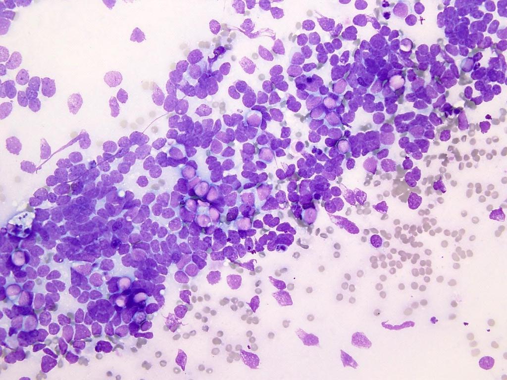
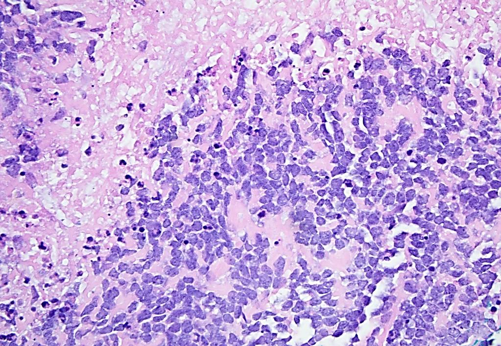
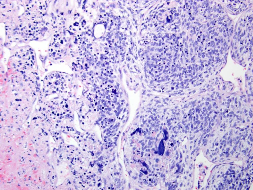
10 Hypercellular smears with monotonous hyperchromatic cells and scant occasional vacuolated cytoplasm (best on Diff Quik) The nuclei have evenly dispersed fine chromatin with indistinct nucleoli
11
12
13 Tumor histology IHC ES/PNET Small round cells CD99, NSE or neural, Cytokeratins +/- Alveolar RMS Mesenchymal chondrosarcoma Synovial sarcoma, monophasic Alveolar/solid, wreath-like Giant cells Small cells, mottled calcifications, cartilage Oval cells, molding focal whorled pattern Desmin, myogenin, CD99 positive or negative Sox 9, CD99, desmin CD99, EMA, CK, TLE-1, ATF2 Neuroblastoma Neuropil, ganglion cell Phox2B, synaptophysin, NB84, CD99- Desmoplastic SRBC Malignant rhabdoid tumor Nests of tumor cells+ desmoplastic stroma Rhabdoid like with perinuclear inclusions, vesicular nuclei Myogenic, neural epithelial, CD99 +/- Vimentin, EMA, CK, SMA, CD99, synaptophysin, BAF47 Undifferentiated sarcoma Primitive oval to round cells occasionally spindled No specific lineage, CD99 diffuse or patchy positivity
14 EWSR1 is implicated in numerous tumors 14
15 Neoplasm Genetic abnormality Chromosome alterations Fusion gene Ewing family Ewing-like sarcoma Undifferentiated round cell sarcoma Desmoplastic round cell tumor Mesenchymal chondrosarcoma Alveolar rhabdomyosarcoma Extraskeletal myxoid chondrosarcoma EWSR1 to ETS fusion FUS to ETS fusion BCOR-CCNB3 EWSR1-NFATc2 translocation t(11;22)(q24;q12); t(21;22)(q22;q21); t(7;22)(q22;q24) t(16;21)(p11;q22); t(2;16)(q35;q11) inv(x)(p11.4p11.2) Complex rearrangement leading to amplification of translocated segment with ring chromosome formation EWSR1-FLI1 EWSR1-ERG EWSR1-ETV1 FUS-ERG FUS-FEV BCOR-CCNB3 EWSR1-NFATc2 CIC-DUX4 t(4;19)(q35;q13) t(10;19)(q26;q13) CIC-DUX4 EWSR1-WT1 t(11;22)(p13;q12) EWSR1-WT1 HEY1-NCOA2 fusion inv(8)(q13;q21) HEY1-NCOA2 FOXOA1 fusion t(2;13)(q35;q14) PAX3-FOXOA1 PAX7-FOXOA1 NR4A3 fusion to TET t(9;22)(q22;q12) t(9;17)(q22;q11) EWSR1-NR4A3 TAF2N-NR4A3 Synovial sarcoma SYT fusion t(x;18)(p11;q11) SYT-SSX1, SSX2 Katjtiar et al
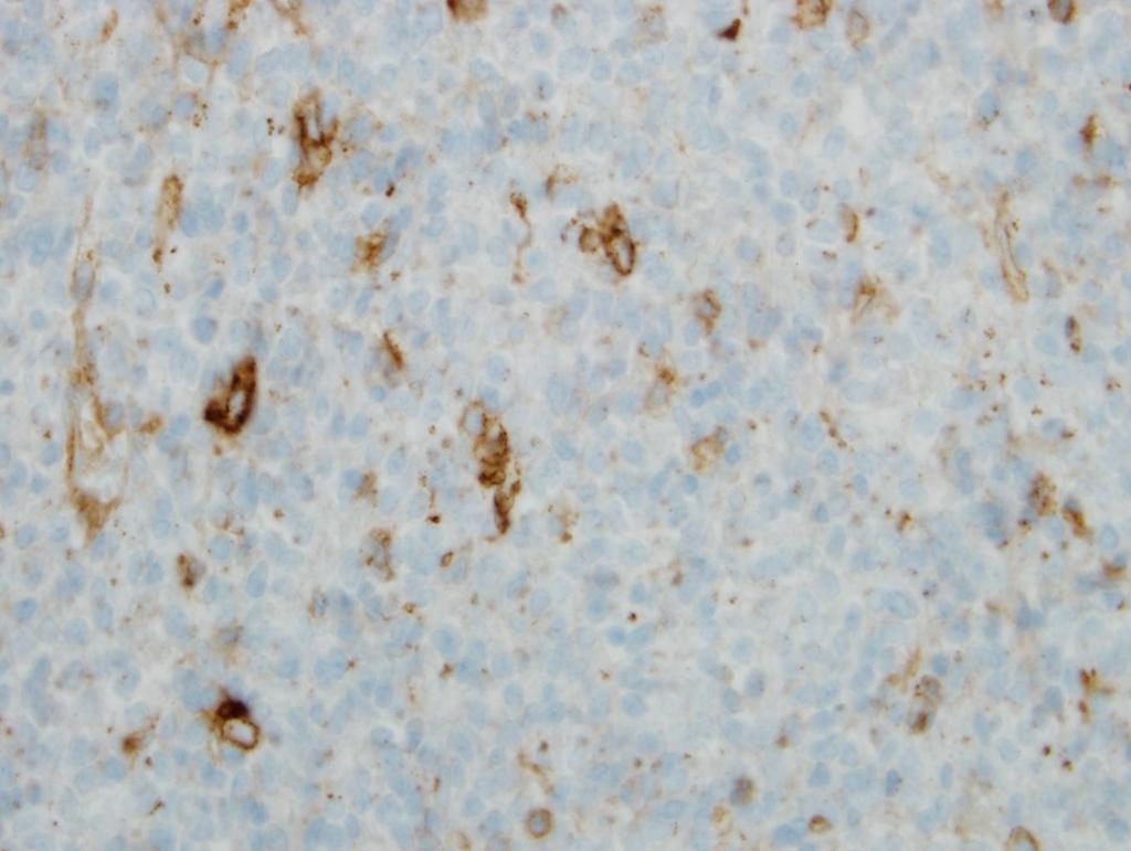
16 Our case was diffusely CD99 positive Negative for all other markers FISH for EWSR1 and RT/PCR for the most common translocation: EWSR1-FLI1 and EWSR1-ERG, EWSR1-WT1 and CIC-DUX were negative Additional study: a complex rearrangement of EWSR1 leading to amplification with ring chromosome formation DIAGNOSIS: EWING-LIKE SARCOMA
17 Triaging for spindle cell tumor Often more challenging While most are benign, sarcomas still account for up to 7% of malignancies in young children Touch imprints: establish cell lineage, namely vascular, myofibroblastic/fibroblastic or myogenic Specific diagnosis: team approach incorporating histology, clinical parameters and imaging studies

18 Case 2: 4 month old infant with an axillary mass, present since birth. She had no significant medical history. Physical examination: A 2 cm smooth, well-defined soft mass on the right lateral chest wall at lower border of the axilla with no tenderness, redness or drainage from the area. Ultrasound examination revealed a cyst in the subcutaneous region, consistent with lymphangioma.

19 During the procedure copious amount of blood was drained causing a decreased in size: supportive of a vascular lesion Touch imprints: Monotonous spindle cells Nuclei: elongated with rounded ends, finely dispersed chromatin Nucleoli inconspicuous

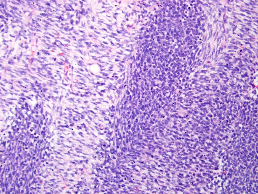
20 Cellular spindle cells with few admixed inflammatory cells (lymphocytes and plasma cells). The spindle cells were mildly pleomorphic without particular architectural arrangement. Mitotic figures were rare. 20
21 List of IHC and fusion transcripts Spindle cell tumors IHC Genetics Diagnosis Alk ALK IMT CD34, Vimentin COL1A1-PDGFB t(17;22)(q21;q13) DFSP CD68, CD99, desmin EWSR1-ATF/ EWSR1-CREB1 t(12;22), (2:22) Angiomatoid fibrous histiocytoma Vimentin ETV6-NTRK3 t(12;15)(p13;q25) Infantile fibrosarcoma Bcl2, cytokeratins, EMA, CD99 Vimentin, MUC4 SYT-SSX1/SSX2/SSX4 t(x;18) FUS-CREB3L2 t(7;16) and FUS- CREB3L1 t(11;16) Synovial Sarcoma Low grade fibromyxoid sarcoma 21
22 ETV6-NTRK3: 69bp 22
23 Infantile Fibrosarcoma IFS: a mimicker of vascular malformation Most IFS show recurrent ETV6/NTRK3 gene fusions, sensitive to chemotherapy with an overall favorable clinical outcome Recent studies have shown the ETV6/NTRK3 fusion negative IFS, or IFS-like tumors in older children have BRAF gene rearrangements or related fusions. A number of unclassifiable spindle cell sarcomas have been associated with NTRK1 IFS that lacks ETV6/NTRK3 may benefit from testing for BRAF, NTRK1 and Met. Hu et al., 2015; Kao Y-C, et al


24 Case 3. 5 year-old with history of pneumonia and left pleural effusion She was found to have a 7.5 x 6.5 x 7cm solid mass in LUL
25
 3. Synovial sarcoma (after 7 years) 4.")
 Myogenin Diagnosis: Pleuropulmonary Blastoma type III")
26 Desmin Positive: Vimentin Desmin Myogenin, MyoD1 Negative: Epithelial markers Differential diagnosis 1. Embryonal Rhabdomyosarcoma 2. Ewing s Sarcoma (chest wall based) 3. Synovial sarcoma (after 7 years) 4. Pleuro-pulmonary blastoma (type III) Molecular study was negative for FOXOA1, EWSR and SYT Evaluation for DICER1: germline DICER1 (pathogenic variant) Myogenin Diagnosis: Pleuropulmonary Blastoma type III
27 Tumor syndromes A major challenge, particularly germane to pediatric pathologist is that tumor development may precede recognition of a syndrome With few exceptions (Beckwith-Wiedemann, hemihypertrophy), most patients lack recognizable phenotype Consequence of diagnosing TPS: adjust chemotherapy, surveillance program for screening other associated malignancies, genetic counselling for family members Postema et al. Pediatr Blood Cancer 2017
28 Childhood tumors with high probability of being part of predisposition syndrome Tumor Associated syndromes Incidence % Adrenocortical Ca Li-Fraumeni S., Beckwith-Wiedemann S 52, 7 ATRT Rhabdoid predisposition Syndrome 24 Choroid plexus Ca Li-Fraumeni Syndrome 55 CMML, JMML Neurofibromatosis type 1 14, 12 Desmoid tumor Familial adenomatous polyposis 14 Hemangioblastoma Von Hippel Lindau Syndrome 33 Hepatoblastoma Familial adenomatous polyposis 6 Malignant melanoma Xeroderma pigmentosum 7 MPNST Neurofibromatosis type 1 25 Medulloblastoma (<3) Li-Fraumeni S (Gorlin S) 5, 4 Meningioma Neurofibromatosis type 2 12 Myelodysplastic S GATA2 spectrum, Fanconi anemia, NF1 7, 7, 2 Nephroblastoma BWS, WAGR S, Isolated hemihyperplasia, Denys-Drash Syndrome 5, 3, 3, 2 Postema et al
29 Childhood tumors with high probability of being part of predisposition syndrome Tumor Associated syndromes Incidence % Adrenocortical Ca Li-Fraumeni S., Beckwith-Wiedemann S 52, 7 ATRT Rhabdoid predisposition Syndrome 24 Choroid plexus Ca Li-Fraumeni Syndrome 55 CMML, JMML Neurofibromatosis type 1 14, 12 Desmoid tumor Familial adenomatous polyposis 14 Hemangioblastoma Von Hippel Lindau Syndrome 33 Hepatoblastoma Familial adenomatous polyposis 6 Malignant melanoma Xeroderma pigmentosum 7 MPNST Neurofibromatosis type 1 25 Medulloblastoma (<3) Li-Fraumeni S (Gorlin S) 5, 4 Meningioma Neurofibromatosis type 2 12 Myelodysplastic S GATA2 spectrum, Fanconi anemia, NF1 7, 7, 2 Nephroblastoma BWS, WAGR S, Isolated hemihyperplasia, Denys-Drash Syndrome 5, 3, 3, 2 Postema et al
30 Childhood tumors with high probability of being part of predisposition syndrome Tumor Associated syndromes % Optic nerve glioma Neurofibromatosis type 1 48 Phaeochromocytoma/ paraganglioma Pineoblastoma, pituitary blastoma, pleuropulmonary blastoma Pituitary adenoma Von Hippel Lindau Syndrome, Familial PC/PGL, MEN, NF type 1 34, 30, 5, 3 PPB family tumor and dysplasia syndrome 29, 82, 66 Familial isolated pituitary adenomas, MEN type i Retinoblastoma, unilateral Retinoblastoma tumor syndrome 13 Rhabdomyosarcoma (<3y) Li-Fraumeni Syndrome 5 Spinal ependymoma Neurofibromatosis type 2, Li-Fraumeni S 9, 6 Osteosarcoma Sarcoma predisposition Li-Fraumeni S, Retinoblastoma, NF1, Rothmund-Thompson S, Werner s S, Bloom S, RAPADILINO S, Diamond-Blackfan anemia DNA repair machinery 17, 7 12, 10, x8, 32, 10, 3, 7-13, 3rpt Postema et al 2017, Jackson et al 2016, Benna et al
31 Childhood tumors with high probability of being part of predisposition syndrome Tumor Associated syndromes % Optic nerve glioma Neurofibromatosis type 1 48 Phaeochromocytoma/ paraganglioma Pineoblastoma, pituitary blastoma, pleuropulmonary blastoma Pituitary adenoma Von Hippel Lindau Syndrome, Familial PC/PGL, MEN, NF type 1 34, 30, 5, 3 PPB family tumor and dysplasia syndrome 29, 82, 66 Familial isolated pituitary adenomas, MEN type i Retinoblastoma, unilateral Retinoblastoma tumor syndrome 13 Rhabdomyosarcoma (<3y) Li-Fraumeni Syndrome 5 Spinal ependymoma Neurofibromatosis type 2, Li-Fraumeni S 9, 6 Osteosarcoma Sarcoma predisposition Li-Fraumeni S, Retinoblastoma, NF1, Rothmund-Thompson S, Werner s S, Bloom S, RAPADILINO S, Diamond-Blackfan anemia DNA repair machinery 17, 7 12, 10, x8, 32, 10, 3, 7-13, 3rpt Postema et al 2017, Jackson et al 2016, Benna et al
32 Tumors associated with PPB family tumor and dysplasia syndrome Benign Cystic nephroma Multinodular goitre Nasal Chondromesenchymal hamartoma Ovarian stromal tumors: Sertoli Leydig cell tumors, juvenile granulosa cell and gynandroblastomas Malignant Ciliary body medulloepithelioma Thyroid carcinoma (PTC) Botryoid embryonal sarcoma (the uterine cervix, ovary, bladder, biliary tree) Pineoblastoma Pituitary blastoma Rare: Wilms, DICER-related renal sarcoma Schultz 2014, de Kock 2015, Doros 2017, PPB registry website

33 Bone tumors Two most common bone tumors are Ewing s sarcoma and osteosarcoma. Our challenge is to obtain adequate, quality material for precise diagnosis Find alternative for decal solution-fresh tissue, EDTA, FFPE 33
34 Future Directions tions Precision medicine starts with pathology- I would add with pediatric pathology A number of advances have been made and a number of precision trials are available globally Basket trials in relapsed/refractory cancers across multiple histologies (results of sequencing used to identify actionable variants), disease-specific precision trials in newly diagnosed patients or disease-specific umbrella trials in relapsed disease Other advances: liquid biopsies and cell free DNA Next gen pathologists : training in cyto-histopathology and molecular pathology Forrest 2018, Fassan M. Arch Pathol Lab Med 2018 Triche T. the Pathologist COG web 34

35 35
Disclosures. An update on ancillary techniques in the diagnosis of soft tissue tumors. Ancillary techniques. Introduction
 Disclosures An update on ancillary techniques in the diagnosis of soft tissue tumors. I have nothing to disclose. Andrew Horvai, MD, PhD Clinical Professor, Pathology Introduction Ancillary techniques
Klinisch belang van chromosomale translocatie detectie in sarcomen
 Translocations in sarcomas Klinisch belang van chromosomale translocatie detectie in sarcomen Judith V.M.G. Bovée, M.D., Ph.D. Department of Pathology Leiden University Medical Center RNA binding DNA binding
Tumores de células pequeñas, redondas y azules: diagnóstico diferencial cuando el tiempo apremia
 Tumores de células pequeñas, redondas y azules: diagnóstico diferencial cuando el tiempo apremia Sílvia Bagué Servei de Patologia Hospital de Sant Pau Barcelona Soft tissue sarcomas Heterogeneous group
Financial disclosures
 An update on immunohistochemical markers in mesenchymal neoplasms By Konstantinos Linos MD, FCAP, FASDP Assistant Professor of Pathology Geisel School of Medicine at Dartmouth Dartmouth-Hitchcock Medical
Cutaneous Mesenchymal Neoplasms with EWSR1 Rearrangement
 Cutaneous Mesenchymal Neoplasms with EWSR1 Rearrangement By Konstantinos Linos MD, FCAP, FASDP Bone, Soft Tissue and Dermatopathology Assistant Professor of Pathology Dartmouth-Hitchcock Medical Center
Case 2. Dr. Sathima Natarajan M.D. Kaiser Permanente Medical Center Sunset
 Case 2 Dr. Sathima Natarajan M.D. Kaiser Permanente Medical Center Sunset History 24 year old male presented with a 3 day history of right flank pain, sharp in nature Denies fever, chills, hematuria or
Molecular pathology in soft tissue tumors. Sylvia Höller Pathologie
 Molecular pathology in soft tissue tumors Sylvia Höller Pathologie When do we perform molecular testing? Morphology and IHC are not clearly fitting with an entity some translocations are entity specific
Surgical Pathology Evening Specialty Conference USCAP 2015
 Surgical Pathology Evening Specialty Conference USCAP 2015 John R. Goldblum, M.D. Chairman, Department of Pathology, Cleveland Clinic Professor of Pathology, Cleveland Clinic Lerner College of Medicine
Diplomate of the American Board of Pathology in Anatomic and Clinical Pathology
 A 33-year-old male with a left lower leg mass. Contributed by Shaoxiong Chen, MD, PhD Assistant Professor Indiana University School of Medicine/ IU Health Partners Department of Pathology and Laboratory
Disclosure of Relevant Financial Relationships
 Ewing and Ewing like sarcomas Using Genetic Signatures in Refining Small Blue Round Cell Tumor Classification Cristina Antonescu, MD Department of Pathology Disclosure of Relevant Financial Relationships
Molecular Genetics of Paediatric Tumours. Gino Somers MBBS, BMedSci, PhD, FRCPA Pathologist-in-Chief Hospital for Sick Children, Toronto, ON, CANADA
 Molecular Genetics of Paediatric Tumours Gino Somers MBBS, BMedSci, PhD, FRCPA Pathologist-in-Chief Hospital for Sick Children, Toronto, ON, CANADA Financial Disclosure NanoString - conference costs for
Financial disclosures
 Cutaneous Mesenchymal Neoplasms with EWSR1 Rearrangement By Konstantinos Linos MD, FCAP, FASDP Bone, Soft Tissue and Dermatopathology Assistant Professor of Pathology Dartmouth-Hitchc Geisel School of
Shared Care & Survival CTYA SSCRG (Childhood Cancer Research Group)
") Shared Care & Survival CTYA SSCRG (Childhood Cancer Research Group) January 2013 The NCIN is a UK-wide initiative, working to drive improvements in standards of cancer care and clinical outcomes by improving
Shared Care & Survival CTYA SSCRG (Childhood Cancer Research Group) January 2013 The NCIN is a UK-wide initiative, working to drive improvements in standards of cancer care and clinical outcomes by improving
Rhabdomyomas and Rhabdomyosarcomas (RMS) David M. Parham, MD Chief of Anatomic Pathology
 David M. Parham, MD Chief of Anatomic Pathology") Rhabdomyomas and Rhabdomyosarcomas (RMS) David M. Parham, MD Chief of Anatomic Pathology Tumors of skeletal muscle: Rhabdomyomas and rhabdomyosarcomas Embryonal muscle 2 3 4 5 6 7 8 Rhabdomyoma Benign
Rhabdomyomas and Rhabdomyosarcomas (RMS) David M. Parham, MD Chief of Anatomic Pathology Tumors of skeletal muscle: Rhabdomyomas and rhabdomyosarcomas Embryonal muscle 2 3 4 5 6 7 8 Rhabdomyoma Benign
Aspen conference on pediatric disease. July through August Bone and Soft Tissue Update. David M. Parham, MD. Rhabdomyoma and rhabdomyosarcoma
 Aspen conference on pediatric disease July through August 2014 Bone and Soft Tissue Update David M. Parham, MD Rhabdomyoma and rhabdomyosarcoma Embryonic rhabdomyogenesis is a highly conserved process
Recent Advances In Select Round Cell Sarcomas
 Recent Advances In Select Round Cell Sarcomas Rajiv M. Patel, M.D. Associate Professor of Pathology & Dermatology University of Michigan, Ann Arbor, MI rajivpat@med.umich.edu 1 Translocation Associated
Financial disclosures
 Mesenchymal Neoplasms with Melanocytic Differentiation By Konstantinos Linos MD, FCAP, FASDP Bone, Soft Tissue and Dermatopathology Assistant Professor of Pathology Dartmouth-Hitchcock Medical Center Geisel
Problem 1: Differential of Neuroendocrine Carcinoma 3/23/2017. Disclosure of Relevant Financial Relationships
 Differential of Neuroendocrine Carcinoma Alain C. Borczuk,MD Weill Cornell Medicine Disclosure of Relevant Financial Relationships USCAP requires that all faculty in a position to influence or control
Lung Tumor Cases: Common Problems and Helpful Hints
 Lung Tumor Cases: Common Problems and Helpful Hints Brandon T. Larsen, MD, PhD Senior Associate Consultant Department of Laboratory Medicine and Pathology Mayo Clinic Arizona Arizona Society of Pathologists
Immunohistochemistry in Bone and Soft Tissue Tumors. Sahar Rassi Zankoul, MD
 Immunohistochemistry in Bone and Soft Tissue Tumors Sahar Rassi Zankoul, MD Introduction Bone tumors represent a wide variety of tumors of various origins and malignant potentials. These different tumor
Clinical History. Pediatric Tumors with Involvement of the Head & Neck
 Pediatric Tumors with Involvement of the Head & Neck John Hicks Texas Children s Hospital Baylor College of Medicine Houston, TX NO DISCLOSURES Clinical History 10 Yr-Old Hispanic Male From Mexico with
3/24/2017 DENDRITIC CELL NEOPLASMS: HISTOLOGY, IMMUNOHISTOCHEMISTRY, AND MOLECULAR GENETICS. Disclosure of Relevant Financial Relationships
 DENDRITIC CELL NEOPLASMS: HISTOLOGY, IMMUNOHISTOCHEMISTRY, AND MOLECULAR GENETICS Jason L. Hornick, M.D., Ph.D. Director of Surgical Pathology and Immunohistochemistry Brigham and Women s Hospital Professor
Part 1. Slides 1-38, Rita Alaggio Soft tissue tumors Trondheim 14. mars 2013
 Part 1 Slides 1-38, Rita Alaggio Soft tissue tumors Trondheim 14. mars 2013 Pediatric Pathology Soft Tissue Tumors AN UPDATE Rita Alaggio Azienda Ospedaliera Università di Padova Soft Tissue Tumors More
Difficult Diagnoses and Controversial Entities in Neoplastic Lung
 Difficult Diagnoses and Controversial Entities in Neoplastic Lung Lynette M. Sholl, M.D. Associate Pathologist, Brigham and Women s Hospital Chief, Pulmonary Pathology Service Associate Professor, Harvard
Pediatric Soft Tissue Tumors
 Pediatric Soft Tissue Tumors Jerzy Klijanienko MD PhD MIAC Institut Curie Paris, France 1 - - General 2 - - Cancer incidence in children Type of malignancy % Hematology 38.6 CNS 19 Neuroblastoma 9.2 Kidney
Methoden / Methods inc. ICCC-3 105
 Methoden / Methods inc. ICCC-3 105 Internationale Klassifikation der Krebserkrankungen bei Kindern (ICCC-3) Zuordnung von ICD-O-3-Codes für Morphologie und Topographie zu diagnostischen Kategorien International
Enterprise Interest Nothing to declare
 Enterprise Interest Nothing to declare Diagnoses one would not like to miss in soft tissue pathology early in your career Marta Sbaraglia, MD Department of Pathology Hospital of Treviso University of Padua
Pathology Mystery and Surprise
 Pathology Mystery and Surprise Tim Smith, MD Director Anatomic Pathology Medical University of South Carolina Disclosures No conflicts to declare Some problem cases Kidney tumor Scalp tumor Bladder tumor
57th Annual HSCP Spring Symposium 4/16/2016
 An Unusual Malignant Spindle Cell Lesion to Involve the Breast Erinn Downs-Kelly, D.O. Associate Professor of Pathology University of Utah & ARUP Laboratories No disclosures Case 39 y/o female with no
From Morphology to Molecular Pathology: A Practical Approach for Cytopathologists Part 1-Cytomorphology. Songlin Zhang, MD, PhD LSUHSC-Shreveport
 From Morphology to Molecular Pathology: A Practical Approach for Cytopathologists Part 1-Cytomorphology Songlin Zhang, MD, PhD LSUHSC-Shreveport I have no Conflict of Interest. FNA on Lymphoproliferative
4/12/2018. MUSC Pathology Symposium Kiawah Island April 18, Jesse K. McKenney, MD
 MUSC Pathology Symposium Kiawah Island April 18, 2018 Jesse K. McKenney, MD 1 Urothelial Carcinoma with Alternative Differentiation 2 Urothelial Carcinoma with Alternative Differentiation Recognition as
Special slide seminar
 Special slide seminar Tomáš Rozkoš The Fingerland Department of Pathology Charles University Medical Faculty and Faculty Hospital in Hradec Králové Czech Republic Case history, 33 years old resistance
Slide Seminar Spanish Society of Pathology
 Slide Seminar Spanish Society of Pathology John R. Goldblum, M.D. Chairman, Department of Anatomic Pathology Cleveland Clinic Professor of Pathology Cleveland Clinic Lerner College of Medicine 1921 Original
3/27/2017. Disclosure of Relevant Financial Relationships
 Ophthalmic Pathology Evening Specialty Conference USCAP 2017 5 th March, 2017 Mukul K. Divatia, MD Assistant Professor Department of Pathology & Genomic Medicine Weill Cornell Medical College Houston Methodist
RARE TUMORS OF INFANCY. RAJKUMAR VENKATRAMANI, MD, MS Director, Rare Tumors Program, Texas Children s Hospital
 RARE TUMORS OF INFANCY RAJKUMAR VENKATRAMANI, MD, MS Director, Rare Tumors Program, Texas Children s Hospital OBJECTIVES Review the epidemiology and clinical presentation of soft tissue sarcomas in infancy.
Case 1. Clinical history
 Case 1 Case 1 Clinical history 17-month-old boy with a kidney tumor found during routine childhood care program. CT scan showed a solid mass. Chemotherapy was given for 4 weeks using actinomycin D and
Spindle Cell Lesions Of The Breast. Emad Rakha Professor of Breast Pathology and Consultant Pathologist
 Spindle Cell Lesions Of The Breast Emad Rakha Professor of Breast Pathology and Consultant Pathologist * SCLs comprise a wide spectrum of diseases, ranging from reactive processes to aggressive malignant
Case 1. Disclosure. Imaging. Clinical history 5/10/2016. USCAP 2016 Annual Meeting Evening Specialty Conference Bone and Soft tissue Pathology
 Disclosure Dr. Agaram has nothing to disclose Case 1 Narsi Agaram, MBBS USCAP 2016 Annual Meeting Evening Specialty Conference Bone and Soft tissue Pathology Clinical history Imaging 1998 A three month
HOW MAY THE CLASSIFICATION OF SOFT TISSUE TUMORS EVOLVE?
 Spanish Society of Pathology Zaragoza, May 2011 ARTHUR PURDY STOUT SYMPOSIUM HOW MAY THE CLASSIFICATION OF SOFT TISSUE TUMORS EVOLVE? Christopher D.M. Fletcher, M.D., FRCPath Brigham and Women s Hospital
5/10. Pathology Soft tissue tumors. Farah Bhani. Mohammed Alorjani
 5/10 Pathology Soft tissue tumors Mohammed Alorjani Farah Bhani Slides are included in this sheet. Objectives: Soft tissue tumors 1. Describe soft tissue tumors. 2. Understand the classification of soft
1/10/2018. Soft Tissue Tumors Showing Melanocytic Differentiation. Overview. Desmoplastic/ Spindle Cell Melanoma
 2016 MFMER slide-1 2016 MFMER slide-2 2016 MFMER slide-3 Soft Tissue Tumors Showing Melanocytic Differentiation Andrew L. Folpe, M.D. Professor of Laboratory Medicine and Pathology Mayo Clinic, Rochester,
Selected Pseudomalignant Soft Tissue Tumors of the Skin and Subcutis
 Selected Pseudomalignant Soft Tissue Tumors of the Skin and Subcutis Andrew L. Folpe, M.D. Professor of Laboratory Medicine and Pathology Mayo Clinic, Rochester, MN folpe.andrew@mayo.edu 2016 MFMER slide-1
Case Presentations 17-20
 Case Presentations 17-20 Case Presentations 17 Case 17 2 year old boy with history of urinary retention and prostate biopsy. After biopsy and chemotherapy, child regains urine flow but the mass does not
Classification (1) Classification (3) Classification (2) Spindle cell lesions. Spindle cell lesions of bladder (Mills et al.
 Classification (3) Classification (2) Spindle cell lesions. Spindle cell lesions of bladder (Mills et al.") Non-epithelial tumours and nonepithelial tumour-like lesions of the bladder Dr Jonathan H Shanks The Christie NHS Foundation Trust, Manchester, UK Classification (1) Myofibroblastic proliferations and
Non-epithelial tumours and nonepithelial tumour-like lesions of the bladder Dr Jonathan H Shanks The Christie NHS Foundation Trust, Manchester, UK Classification (1) Myofibroblastic proliferations and
Conceptual Evolution of Soft Tissue Tumors Classification
 Conceptual Evolution of Soft Tissue Tumors Classification Angelo P. Dei Tos M.D. Departments of Pathology & Oncology Treviso, Italy How WHO classification was reshaped Pathologists and Cytogeneticists
A 25 year old female with a palpable mass in the right lower quadrant of her abdomen
 May 2016 A 25 year old female with a palpable mass in the right lower quadrant of her abdomen Contributed by: Paul Ndekwe, MD, Resident Physician, Indiana University School of Department of Pathology and
The Genetics of Myoepithelial Tumors: salivary glands, soft tissue and bone
 The Genetics of Myoepithelial Tumors: salivary glands, soft tissue and bone Cristina Antonescu, MD Memorial Sloan-Kettering Cancer Center, New York Nothing to declare Disclosure Spectrum of Myoepithelial
Pathology of Sarcoma ELEANOR CHEN, MD, PHD, ASSISTANT PROFESSOR DEPARTMENT OF PATHOLOGY UNIVERSITY OF WASHINGTON
 Pathology of Sarcoma ELEANOR CHEN, MD, PHD, ASSISTANT PROFESSOR DEPARTMENT OF PATHOLOGY UNIVERSITY OF WASHINGTON Presentation outline Background and epidemiology of sarcomas Sarcoma classification Sarcoma
Disclosure. Relevant Financial Relationship(s) None. Off Label Usage None MFMER slide-1
 None. Off Label Usage None MFMER slide-1") Disclosure Relevant Financial Relationship(s) None Off Label Usage None 2013 MFMER slide-1 Case Presentation A 43 year old male, with partial nephrectomy for a right kidney mass 2013 MFMER slide-2 2013
Disclosure Relevant Financial Relationship(s) None Off Label Usage None 2013 MFMER slide-1 Case Presentation A 43 year old male, with partial nephrectomy for a right kidney mass 2013 MFMER slide-2 2013
Diagnostic Value of Immunohistochemistry in Soft Tissue Tumors
 Original Article DOI: 10.21276/APALM.1637 Diagnostic Value of Immunohistochemistry in Soft Tissue Tumors Sridevi. V*., Susruthan Muralitharan., and Thanka. J Dept of Pathology, SriMuthukumaran Medical
Protocol for the Examination of Biopsy Specimens From Pediatric Patients With Ewing Sarcoma
 Protocol for the Examination of Specimens From Pediatric Patients With Ewing Sarcoma Version: EwingSarcoma 4.0.0.0 Protocol Posting Date: February 2019 Accreditation Requirements The use of this protocol
Inside the Pediatric Cancer Genetics Clinic
 Inside the Pediatric Cancer Genetics Clinic August 23, 2014 Joyce Turner, MS, CGC Children s National Medical Center Washington, DC Pediatric Cancer Genetic Syndromes Aims over next 20-30 minutes: 1) Case
Evening Specialty Conference Bone and Soft Tissue Pathology. Diagnostic pitfalls in bone and soft tissue pathology
 Evening Specialty Conference Bone and Soft Tissue Pathology. Case 1 Elizabeth G Demicco, MD, PhD Mount Sinai Hospital, New York Disclosure of Relevant Financial Relationships USCAP requires that all planners
ASCP Resident Review Mini-Course Series Session 2: Renal and Soft Tissue Pathology. Carole Vogler MD, FASCP Ema Dragoescu MD
 178 2011 ASCP Resident Review Mini-Course Series Session 2: Renal and Soft Tissue Pathology Carole Vogler MD, FASCP Ema Dragoescu MD 2011 Annual Meeting Las Vegas, NV AMERICAN SOCIETY FOR CLINICAL PATHOLOGY
Update on Cutaneous Mesenchymal Tumors. Thomas Brenn
 Update on Cutaneous Mesenchymal Tumors Thomas Brenn Cutaneous Mesenchymal Tumours Wide morphological and biological spectrum Myofibroblastic, smooth muscle, neural, vascular, apidocytic, undifferentiated;
Contents Part I Introduction 1 General Description 2 Natural History: Importance of Size, Site, Histopathology
 Contents Part I Introduction 1 General Description... 3 1.1 Introduction... 3 1.2 Incidence and Prevalence... 5 1.3 Predisposing and Genetic Factors... 8 References... 16 2 Natural History: Importance
Small Round Cell Tumors in Soft Tissue and Bone. ACCME/Disclosure. Adult Small Round Cell Tumors. Ewing Sarcoma 4/13/2016
 Ultrastructural Pathology Society, Seattle, March, 2016 Small Round Cell Tumors in Soft Tissue and Bone ACCME/Disclosure Dr. Fisher has nothing to disclose Cyril Fisher MA MD DSc FRCPath Professor of Tumor
Breast - ductal carcinoma CK7 ER PR GATA3 Mammaglobin (50-70%) GCDFP-15 (50-70%) E-cadherin HMWCK CK20 PAX2 ER/PR/HER2 on all newly diagnosed cases
 GCDFP-15 (50-70%) E-cadherin HMWCK CK20 PAX2 ER/PR/HER2 on all newly diagnosed cases") Adrenal cortical carcinoma Inhibin Synap Melan-A Calretinin Vimentin Chromogr CK7 CK20 Breast - ductal carcinoma CK7 ER PR GATA3 Mammaglobin (50-70%) GCDFP-15 (50-70%) E-cadherin HMWCK CK20 PAX2 ER/PR/HER2
Adrenal cortical carcinoma Inhibin Synap Melan-A Calretinin Vimentin Chromogr CK7 CK20 Breast - ductal carcinoma CK7 ER PR GATA3 Mammaglobin (50-70%) GCDFP-15 (50-70%) E-cadherin HMWCK CK20 PAX2 ER/PR/HER2
Shintaro Sugita *, Hiroko Asanuma and Tadashi Hasegawa
 Sugita et al. Diagnostic Pathology (2016) 11:37 DOI 10.1186/s13000-016-0486-2 RESEARCH Open Access Diagnostic use of fluorescence in situ hybridization in expert review in a phase 2 study of trabectedin
Tumors of kidney and urinary bladder
 Tumors of kidney and urinary bladder Overview of kidney tumors Benign and malignant Of the benign: papillary adenoma -cortical -small (0.5cm) -in 40% of population -clinically insignificant The most common
Dr.Dafalla Ahmed Babiker Jazan University
 Dr.Dafalla Ahmed Babiker Jazan University Brain tumors are the second commonest malignancy in children Infratentorial tumors are more common As a general rule they do not metastasize out of the CNS, but
Study of Paediatric Small Blue Round Cell Tumors with Immunohistochemical Correlation
 IOSR Journal of Dental and Medical Sciences (IOSR-JDMS) e-issn: 2279-0853, p-issn: 2279-0861.Volume 15, Issue 6 Ver. XIII (June. 2016), PP 65-70 www.iosrjournals.org Study of Paediatric Small Blue Round
Role of immunohistochemistry in the differential diagnosis of malignant small round cell tumor: a study of 38 cases
 International Journal of Research in Medical Sciences Patel A et al. Int J Res Med Sci. 2015 Dec;3(12):3833-3839 www.msjonline.org pissn 2320-6071 eissn 2320-6012 Research Article DOI: http://dx.doi.org/10.18203/2320-6012.ijrms20151452
ESS: Pathologic Insights
 GEIS XVI INTERNATIONAL SYMPOSIUM Seville 4th October 2018 ESS: Pathologic Insights Sílvia Bagué The Royal Marsden Hospital London (United Kingdom) I have no conflicts of interest Endometrial stromal sarcoma
Newer soft tissue entities
 Newer soft tissue entities Examples among fibroblastic tumors Turku, May 6, 2010 Markku Miettinen, M.D. AFIP, Washington, DC Fibroblastic neoplasms Solitary fibrous tumor /Hemangiopericytoma Low-grade
Almost any suspected tumor can be aspirated easily and safely. Some masses are more risky to aspirate including:
 DOES THIS PATIENT HAVE CANCER? USING IN-HOUSE CYTOLOGY TO HELP YOU MAKE THIS DIAGNOSIS. Joyce Obradovich, DVM, Diplomate, ACVIM (Oncology) Animal Cancer & Imaging Center, Canton, Michigan Almost every
Fluorescence In Situ Hybridization in the Diagnosis of Soft Tissue Neoplasms: A Review. Munir R. Tanas, MD and John R.
 REVIEW ARTICLE Fluorescence In Situ Hybridization in the Diagnosis of Soft Tissue Neoplasms: A Review Munir R. Tanas, MD and John R. Goldblum, MD Abstract: This paper presents an overview of the role of
ACCME/Disclosures ALK FUSION-POSITIVE MESENCHYMAL TUMORS. Tumor types with ALK rearrangements. Anaplastic Lymphoma Kinase. Jason L.
 Companion Meeting of the International Society of Bone and Soft Tissue Pathology The Evolving Concept of Mesenchymal Tumors ALK FUSION-POSITIVE MESENCHYMAL TUMORS Jason L. Hornick, MD, PhD March 13, 2016
2018 ICD-O-3 Updates in Table Format with Annotation for Reference
 Status Histology Description (this may be preferred term or a synonym) Report Comments New term 8010 3 Urachal carcinoma (C65.9, C66.9, C67._, C68._) New term 8013 3 Combined large cell neuroendocrine
Evening Specialty Conference: Cytopathology
 : Cytopathology N. Paul Ohori, M.D. University of Pittsburgh Medical Center Disclosure of Relevant Financial Relationships Disclosure of Relevant Financial Relationships USCAP requires that all planners
Effective January 1, 2018 ICD O 3 codes, behaviors and terms are site specific
 Effective January 1, 2018 codes, behaviors and terms are site specific /N 8551/3 Acinar adenocarcinoma (C34. _) Lung primaries diagnosed prior to 1/1/2018 use code 8550/3 For prostate (all years) see 8140/3
Effective January 1, 2018 ICD O 3 codes, behaviors and terms are site specific
 Effective January 1, 2018 codes, behaviors and terms are site specific Status /N 8010/3 Urachal carcinoma (C65.9, C66.9, C67. _, C68._) 8013/3 Combined large cell neuroendocrine carcinoma (C34. _, C37.9)
Journal of Solid Tumors, April 2012, Vol. 2, No. 2
 ORIGINAL ARTICLE Utility of fluorescence in situ hybridization in subclassifying unclassified high-grade sarcomas: A study of 40 cases using break-apart probes of EWSR1, FOXO1A, SS18 and DDIT3 genes Alfredo
USCAP COMPANION MEETING INTERNATIONAL SOCIETY OF BONE AND SOFT TISSUE PATHOLOGY DENVER, March 2 nd 2008
 1 USCAP COMPANION MEETING INTERNATIONAL SOCIETY OF BONE AND SOFT TISSUE PATHOLOGY DENVER, March 2 nd 2008 THE EVOLUTION OF SOFT TISSUE TUMOUR TAXONOMY: WHAT STILL NEEDS TO BE DONE? Christopher D.M. Fletcher,
PITFALLS AND TRAPS IN THE DIAGNOSIS AND STAGING OF RENAL TUMOURS OF CHILDHOOD. Gordan M. Vujanić Cardiff, U.K.
 PITFALLS AND TRAPS IN THE DIAGNOSIS AND STAGING OF RENAL TUMOURS OF CHILDHOOD Gordan M. Vujanić Cardiff, U.K. RENAL TUMOURS OF CHILDHOOD - CLASSIFICATION (2016) Nephroblastic tumours Mesenchymal tumours
An unusual superficial small round cell sarcoma
 An unusual superficial small round cell sarcoma - Diagnostic problems - Differential diagnosis Antonio Llombart Bosch Isidro Machado Dep. Pathology Univ. Medical School Valencia, Institute of Oncology
Technology from Abcam
 CD2 (EP222) CD2 is one of the earliest T-cell lineage restricted antigens to appear during T-cell differentiation and only rare CD2+ cells can be found in the bone marrow. Anti-CD2 is a pan-t-cell antigen
CLIC Sargent Eligibility Criteria
 1 Eligibility Criteria DOCUMENT GOVERNANCE: Eligibility criteria Produced by J. Hawkins & Grants Team Sponsored by Dara de Burca Version Approval by Executive Team 10 th June 2014 Board of Trustees 3 rd
The Enigmatic Spitz Lesion
 The Enigmatic Spitz Lesion The Dawn of Spitz S Spitz Sophie Spitz Melanomas of Childhood ; Am J Pathol 1948 1910-1956 13 children (18 mo - 12 yrs) 12/13 had a benign clinical course Sophie Spitz Born 1910
Introduction to Musculoskeletal Tumors. James C. Wittig, MD Orthopedic Oncologist Sarcoma Surgeon
 Introduction to Musculoskeletal Tumors James C. Wittig, MD Orthopedic Oncologist Sarcoma Surgeon www.tumorsurgery.org Definitions Primary Bone / Soft tissue tumors Mesenchymally derived tumors (Mesodermal)
Slide seminar: Soft tissue and bone pathology
 Slide seminar: Soft tissue and bone pathology Unusual tumors of bone and soft tissue or unusual presentations of common ones Gunhild Mechtersheimer Institute of Pathology, Heidelberg/DE (Sylvia Höller,
Enterprise Interest No disclosures.
 Enterprise Interest No disclosures. Secondary Tumours in Uropathology Case 2 PRESENTED AT: EUROPEAN CONGRESS OF PATHOLOGY 18 #ECP2018 Slides are the property of the author. Permission required for reuse.
Objectives. Salivary Gland FNA: The Milan System. Role of Salivary Gland FNA 04/26/2018
 Salivary Gland FNA: The Milan System Dr. Jennifer Brainard Section Head Cytopathology Cleveland Clinic Objectives Introduce the Milan System for reporting salivary gland cytopathology Define cytologic
Cytological Sub-classification of Lung Cancer: Morphologic and Molecular Characteristics. Mercè Jordà, University of Miami
 Cytological Sub-classification of Lung Cancer: Morphologic and Molecular Characteristics Mercè Jordà, University of Miami Mortality Lung cancer is the most frequent cause of cancer incidence and mortality
Current and future applications of Molecular Pathology. Kathy Walsh Clinical Scientist NHS Lothian
 Current and future applications of Molecular Pathology Kathy Walsh Clinical Scientist NHS Lothian Molecular Pathology in Solid tumours Cancer type Genes tested Purpose Associated treatments Non small cell
Anaplastic Large Cell Lymphoma (of T cell lineage)
") Anaplastic Large Cell Lymphoma (of T cell lineage) Definition T-cell lymphoma comprised of large cells with abundant cytoplasm and pleomorphic, often horseshoe-shaped nuclei CD30+ Most express cytotoxic
Anaplastic Large Cell Lymphoma (of T cell lineage) Definition T-cell lymphoma comprised of large cells with abundant cytoplasm and pleomorphic, often horseshoe-shaped nuclei CD30+ Most express cytotoxic
Pediatric Retroperitoneal Masses Radiologic-Pathologic Correlation
 Acta Radiológica Portuguesa, Vol.XVIII, nº 70, pág. 61-70, Abr.-Jun., 2006 Pediatric Retroperitoneal Masses Radiologic-Pathologic Correlation Marilyn J. Siegel Mallinckrodt Institute of Radiology, Washington
2 Berkeley Street, Suite 403, Toronto, Ontario M5A 2W3 Visit us at: Tel: Fax:
 E-Path A.I. Engine Knowledge Base Enhancements Version 1.0.0.29 April 1, 2018 The major enhancements in the E-Path Knowledge Base from versions 1.0.0.28 through 1.0.0.29 are as follows: 1. Addition/modification
Presentation material is for education purposes only. All rights reserved URMC Radiology Page 1 of 98
 Presentation material is for education purposes only. All rights reserved. 2011 URMC Radiology Page 1 of 98 Radiology / Pathology Conference February 2011 Brooke Koltz, Cytopathology Resident Presentation
ACCME/Disclosures. Diagnosing Mesothelioma in Limited Tissue Samples. Papanicolaou Society of Cytopathology Companion Meeting March 12 th, 2016
 Diagnosing Mesothelioma in Limited Tissue Samples Papanicolaou Society of Cytopathology Companion Meeting March 12 th, 2016 Sanja Dacic, MD, PhD University of Pittsburgh ACCME/Disclosures GENERAL RULES
Urinary Bladder: WHO Classification and AJCC Staging Update 2017
 Urinary Bladder: WHO Classification and AJCC Staging Update 2017 Houston Society of Clinical Pathologists 58 th Annual Spring Symposium Houston, TX April 8, 2017 Jesse K. McKenney, MD Classification
ACCME/Disclosures. Everything is spindle - how far can we go with limited FNA material? Everything is spindle how far can we go? Everything is spindle
 ACCME/Disclosures The USCAP requires that anyone in a position to influence or control the content of CME disclose any relevant financial relationship WITH COMMERCIAL INTERESTS which they or their spouse/partner
Minimally invasive adenocarcinoma. 5mm or less = microinvasion No necrosis No lymphatic or pleural invasion No spread through air-spaces (STAS)
") Minimally invasive adenocarcinoma 5mm or less = microinvasion No necrosis No lymphatic or pleural invasion No spread through air-spaces (STAS) 2b lepidic acinar 2c papillary micropapillary solid 2d cribriform
Minimally invasive adenocarcinoma 5mm or less = microinvasion No necrosis No lymphatic or pleural invasion No spread through air-spaces (STAS) 2b lepidic acinar 2c papillary micropapillary solid 2d cribriform
INTRODUCTION TO PATHOLOGICAL TECHNIQUES. 1. Types of routine biopsy procedures 2. Special exams (IHC, FISH)
") INTRODUCTION TO PATHOLOGICAL TECHNIQUES 1. Types of routine biopsy procedures 2. Special exams (IHC, FISH) Biopsy-Indications Diffuse/multifocal lesions (neoplastic, inflammatory, etc) Etiology of the
INTRODUCTION TO PATHOLOGICAL TECHNIQUES 1. Types of routine biopsy procedures 2. Special exams (IHC, FISH) Biopsy-Indications Diffuse/multifocal lesions (neoplastic, inflammatory, etc) Etiology of the
Unusual Variants of Bladder Cancer Cristina Magi-Galluzzi, MD, PhD
 Unusual Variants of Bladder Cancer Cristina Magi-Galluzzi, MD, PhD Director of Genitourinary Pathology, Professor of Pathology, Lerner College of Medicine Cleveland Clinic Objectives Update on variants
Ultrasound-Guided Fine-Needle Aspiration of Thyroid Nodules: New events
 Ultrasound-Guided Fine-Needle Aspiration of Thyroid Nodules: New events Sandrine Rorive, M.D., PhD. Erasme Hospital - Université Libre de Bruxelles (ULB) INTRODUCTION The assessment of thyroid nodules
What s New in Pathology of Genitourinary Tumors. Jiaoti Huang, MD, PhD Department of Pathology Duke University School of Medicine
 What s New in Pathology of Genitourinary Tumors Jiaoti Huang, MD, PhD Department of Pathology Duke University School of Medicine Kidney Tumors Multilocular cystic renal neoplasm of low malignant potential
Renal tumours: use of immunohistochemistry & molecular pathology. Dr Lisa Browning John Radcliffe Hospital Oxford
 Renal tumours: use of immunohistochemistry & molecular pathology Dr Lisa Browning John Radcliffe Hospital Oxford Renal tumours: the use of immunohistochemistry & molecular pathology Classification of RCC
Soft Tissue Sarcomas: Questions and Answers
 Soft Tissue Sarcomas: Questions and Answers 1. What is soft tissue? The term soft tissue refers to tissues that connect, support, or surround other structures and organs of the body. Soft tissue includes