Case Presentations 17-20
|
|
|
- Phebe Harrington
- 5 years ago
- Views:
Transcription
1 Case Presentations 17-20
2 Case Presentations 17
3 Case 17 2 year old boy with history of urinary retention and prostate biopsy. After biopsy and chemotherapy, child regains urine flow but the mass does not shrink on imaging. A total prostatectomy was performed. 3

4 Geographic necrosis Viable neoplasm Case 17: Prostatic neoplasm: capsule 4

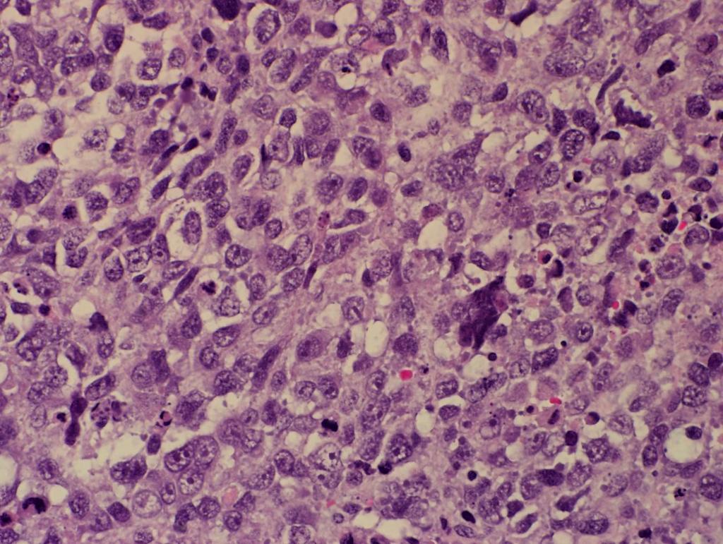
5 Case 17. Undifferentiated neoplasm 5

6 Case 17. Moderately pleomorphic primitive cells Lymphs, mits, karyo 6

7 Case 17. Anaplasia 7

8 Case 17. Anaplasia 8

9 Case 17. Anaplasia 9
10 10
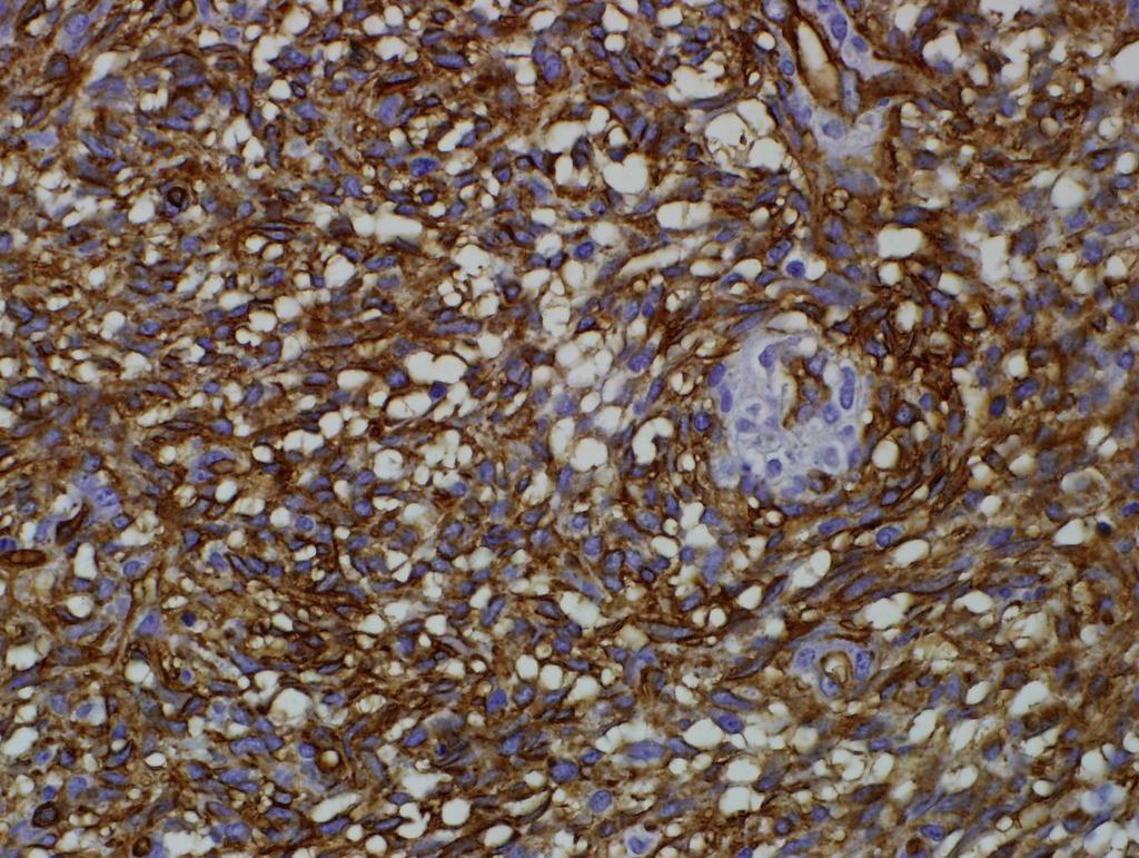
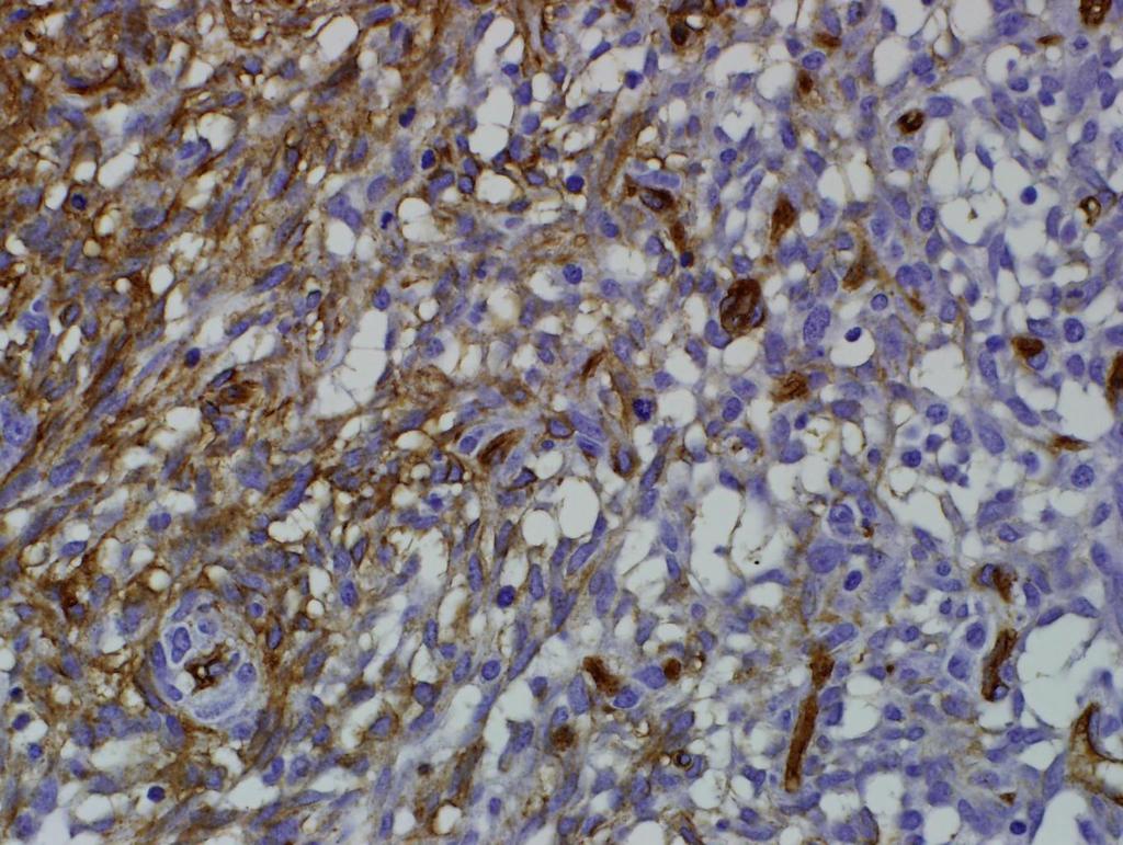
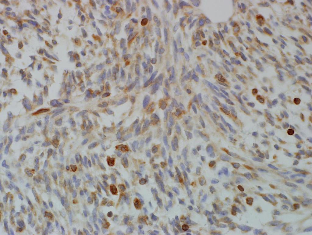
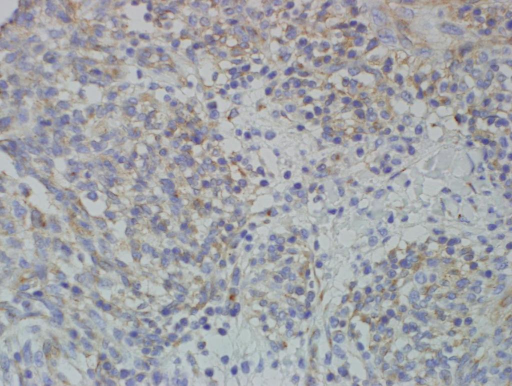
11 IHC Desmin: strongly positive MyoD1: strongly positive SOX-9: strongly positive [by report] Myogenin: focally positive 11
12 Cytogenetics FISH for FOXO1 (13q14.1) gene rearrangement: negative (performed at two institutions) 12
13 Case 17 Diagnosis: Treated embryonal rhabdomyosarcoma, with diffuse anaplasia, fusion-negative 13
14 Anaplastic rhabdomyosarcoma (RMS) Original concepts Seen in both forms of RMS, embryonal and alveolar Builds on concepts of anaplastic Wilms tumor Nucleomegaly (>3X size of adjacent cells) Hyperchromasia Atypical multipolar mitoses Diffuse and focal forms Diffuse = clonal Focal = occasional isolated cell 14
15 Anaplastic RMS: prognosis Persists with chemotherapy (marker of chemoresistance) Proven marker of bad outcome in embryonal RMS in prospective and retrospectic studies No effect on alveolar RMS outcome Did not make level of significance in multivariate analysis Has not been used to make protocol-based treatment decisions 15
16 Qualman et al: IRSG/COG studies Nine per cent of embryonal RMS (23/223 cases) Eight per cell of alveolar RMS (12/139 cases) Two of 17 spindle cell RMS cases One of 36 botryoid RMS cases Total 7% of RMS cases (39/463) 16
17 Qualman et al: Outcome analysis of embryonal RMS No difference between diffuse and focal anaplasia Univariate analysis: 5 year survivals With anaplasia: 68% Without anaplasia: 82% Only predictive with intermediate risk disease Multivariate analysis Demonstrated a statistical trend (p = 0.08) Was not a significant factor compared to proven factors like metastasis, age, stage, site, group, etc. 17
18 Anaplastic RMS: Genetics Qualman: Double minutes (gene amplification) found in 6 of 9 karyotypes Bridge: Genomic amplication frequent on comparative genomic hydridization All but one of 6 ERMS with amplicons showed anaplasia These 6 Involved 15q, 18q, 11q, and 12 q. 18
19 Case Presentations 18
20 Case month-old male infant with large buttock mass Pulmonary metastasis found on imaging. Wide excision of soft tissue mass was performed. Slides courtesy of Dr. Denise Malicki, Rady Children s Hospital, San Diego. 20

21 Case 18. Fibrosarcoma-like spindle cell tumor 21

22 Case 18: Permeation of adjacent fat 22

23 Case 18: Permeation of adjacent fat 23

24 Focal myxoid change 24

25 Focal myxoid change 25

26 Herring bone pattern 26
27 CD34 27
28 CD34 28
29 BCL2 29
30 CD99 30
31 Glut 1: negative CD31: negative D2-40: negative Factor XIIIa: negative Other IHC 31
32 ETV6-PDGFB RT-PCR: negative for fusion SS18 FISH: Cytogenetics negative for 18q11.2 rearrangement NTRKC FISH: negative for rearrangement 32
33 Diagnosis: CD34 positive fibrosarcoma, most consistent with fibrosarcoma ex dermatofibrosarcoma protuberans 33
34 Comment: Cannot absolutely exclude extrapleural solitary fibrous tumor. STAT6 testing may be helpful. 34
35 Differential diagnosis of pediatric fibrosarcoma Infantile fibrosarcoma: t(12;15), ETV6-NTRK3 fusion Low grade fibromyxoid sarcoma: MUC4, alternating pattern, large collagenous rosettes, FUS-CREB3L1/FUS-CREBL2 fusions Synovial sarcoma: EMA/CK/TLE1 positivity, SSX-SS18 fusions MPNST: involvement of nerve or neurofibroma, NF1 history, S100 positivity Fibroma-like epithelioid sarcoma: EMA/CK positivity, INI1 negativity 35
36 Differential diagnosis of pediatric fibrosarcoma Dermatofibrosarcoma protuberans Solitary fibrous tumor 36
37 Dermatofibrosarcoma protuberans (DFSP) in children Relatively uncommon in children but being increasingly recognized Congenital lesions may appear innocuous Slowly progressive lesion most often in trunk May be associated with immunodeficiency May be multifocal 37
38 DFSP: pathology Classic, giant cell, myxoid, fibrosarcomatous, and pigmented forms Permeation of fat CD34 positive, Factor XIIIa negative (beware intermediate lesions in spectrum of dermatofibroma) CD34 expression often lost in fibrosarcomatous areas Has t(17;22) translocation, COL1A1-PDGFB fusion Two of seven RT-PCR negative in one study 38
39 DFSP: Differential diagnosis Nodular fasciitis (positive for USP6 fusions) Infantile myofibroma (actin positive) Deep dermatofibroma/intermediate lesions (may be Factor XIIIa and CD34 positive) Spindle cell melanoma (check epidermis, melanocytic markers) 39
40 Solitary fibrous tumor in children Extremely rare, limited to isolated case reports Pleural or extrapleural Patternless and hemangiopericytomatous patterns CD34, CD99, BCL2 positive (may be actin positive) A new marker: STAT6 (has gene fusion and overexpression) 40
41 Case 19 41
42 Case year old girl with tumor involving metaphysis of distal radius. Periosteal elevation and soft tissue extension found on radiographs. The patient was treated with chemotherapy. An en bloc resection was performed afterwards. 42

43 Case 18. malignant cartilage 43

44 Case 19. Cartilage with osteoid 44

45 Case 19. Lacy osteoid, no cartilage 45

46 Case 19. Small cell pattern 46
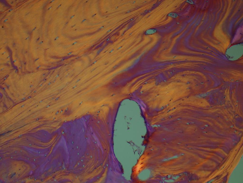
47 Diagnosis: Conventional osteosarcoma, high grade, chondroblastic variant 47
48 Differential diagnosis: malignant chondroblastic neoplasms in children Primary chondrosarcoma Rare but may occur in spine and head and neck Usually mesenchymal variant High grade chondroblastic osteosarcoma (more common in kids) Differentiate from osteoid producing mesenchymal chondrosarcoma Intermediate grade chondroblastic periosteal osteosarcoma Teratomatous lesions (look for other elements) Heterologous differentiation in dedifferentiated liposarcoma, Sertoli-Leydig tumor, PNET, RMS (other elements) Chordoma (location and cytokeratin/brachyury expression) 48
49 Primary pediatric chondrosarcoma Age range of 16 months-81 years in one large metaanalysis, with mean of years and peak between years Includes mesenchymal chondrosarcoma (covered in another lecture) Genetic predisposition Ollier's disease Maffucci's disease Hereditary exostosis (EXT1, EXT2, EXT3 mutations) Even rarer osteodysplasias (PTPN11, ACP5 mutations) Acquired mutations in IDH1, IDH2 frequent overall Isocitrate dehydrogenase genes in Kreb cycle 49
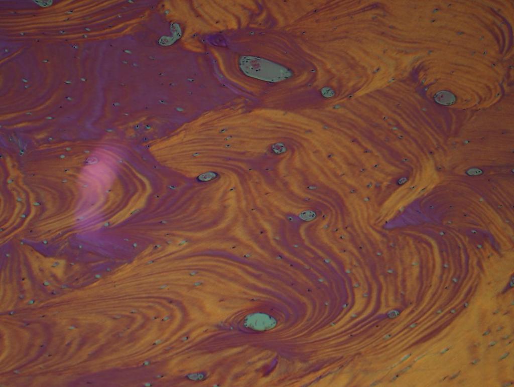
50 Chondroblastic osteosarcoma One of multiple other variants Osteoblastic/sclerosing Fibroblastic Anaplastic Fibrohisticytic or epithelioid (beware cytokeratin positivity) Telangiectatic Round cell (covered in another lecture) 50
51 51
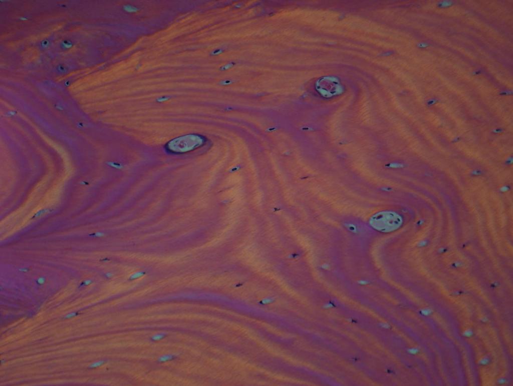
52 Case 20 52
53 Case year old male who develops swelling and increasing pain of lower jaw Follows wisdom teeth extraction. CT shows a destructive lytic and sclerotic lesion of mandible. A biopsy was performed. 53

54 Case 20: irregular bony trabeculae 54

55 Case 20. Collagenous intertrabecular region 55

56 Case 20: Sparse lymphocytes 56

57 Variable osteoid seam 57

58 Variable osteoid seam 58

59 Case 20. dense cortical bone 59
60 60
61 61
62 62

63 Case 20. Dense collagneous zones, with osteoid transition 63

64 Dense collagen 64
65 65
66 Diagnosis: Chronic sclerosing osteomyelitis 66
67 Differential diagnosis: non-neoplastic bony lesions Fracture callus (hemorrhage, organization, chondroid matrix) Osteomyelitis (inflammation, osteonecrosis, reactive bone) Fibrous dysplasia of bone (woven bone, non-osteoblastic) Osteofibrous dysplasia (tibia, cytokeratin) Osteitis fibrosa cystica (hyperparathyroidism, high serum Ca) In soft tissue, consider myositis ossificans, dermatomyositis, heterotopic bone, trauma, periostitis ossificans 67
68 Garré's sclerosing osteomyelitis First described by Carl Garré in 1893 Non-suppurative chronic osteomyelitis form of periostitis ossificans Proliferative periosteum Peripheral subperiosteal bone formation Mild but prolonged infection or irritation, without necrosis or exudate Particularly common in tibia, jaws 68
69 Garré's sclerosing osteomyelitis Clinical features Facial assymetry, bony assymetry Progressive swelling Variable pain, malaise, trismus Unilateral induration with normal overlying skin or mucosa 69
70 Garré's sclerosing osteomyelitis May or may not respond to antibiotics Resolution of pain, bony changes May require multiple CT scans, with relatively high dose of irradiation Rarely progresses to secondary osteosarcoma May occur with multifocal recurrent osteomyelitis, an autoimmune disease with a genetic basis 70
71 Garré's sclerosing osteomyelitis: histology Fibrous dysplasia-like pattern ("ground glass" appearance on imaging) Retiform pattern (lace-like) Paralled (lamellated) pattern - do not confuse with parosteal osteosarcoma Overlapping features with Caffey's disease (cortical hyperostosis), seen in infants 71
72 Other diseases to consider Low grade, osteoblastic-like osteosarcoma Parosteal osteosarcoma Hypertrophic osteoarthopathy History of heart or lung disease Congenital syphilis Cherubism (bilateral, extensive) 72
73 73
74 74
75 75
76 76
77 77
78 78
79 79
80 80
81 81
82 82
83 83
84 84
85 85
86 86
87 87
88 88
Disclosures. An update on ancillary techniques in the diagnosis of soft tissue tumors. Ancillary techniques. Introduction
 Disclosures An update on ancillary techniques in the diagnosis of soft tissue tumors. I have nothing to disclose. Andrew Horvai, MD, PhD Clinical Professor, Pathology Introduction Ancillary techniques
Klinisch belang van chromosomale translocatie detectie in sarcomen
 Translocations in sarcomas Klinisch belang van chromosomale translocatie detectie in sarcomen Judith V.M.G. Bovée, M.D., Ph.D. Department of Pathology Leiden University Medical Center RNA binding DNA binding
Disclosures. An update on ancillary techniques in the diagnosis of soft tissue tumors. Ancillary techniques. Introduction
 Disclosures An update on ancillary techniques in the diagnosis of soft tissue tumors. I have nothing to disclose. Andrew Horvai, MD, PhD Clinical Professor, Pathology Introduction Ancillary techniques
Introduction to Musculoskeletal Tumors. James C. Wittig, MD Orthopedic Oncologist Sarcoma Surgeon
 Introduction to Musculoskeletal Tumors James C. Wittig, MD Orthopedic Oncologist Sarcoma Surgeon www.tumorsurgery.org Definitions Primary Bone / Soft tissue tumors Mesenchymally derived tumors (Mesodermal)
Evening Specialty Conference Bone and Soft Tissue Pathology. Diagnostic pitfalls in bone and soft tissue pathology
 Evening Specialty Conference Bone and Soft Tissue Pathology. Case 1 Elizabeth G Demicco, MD, PhD Mount Sinai Hospital, New York Disclosure of Relevant Financial Relationships USCAP requires that all planners
5/10. Pathology Soft tissue tumors. Farah Bhani. Mohammed Alorjani
 5/10 Pathology Soft tissue tumors Mohammed Alorjani Farah Bhani Slides are included in this sheet. Objectives: Soft tissue tumors 1. Describe soft tissue tumors. 2. Understand the classification of soft
3/27/2017. Disclosure of Relevant Financial Relationships
 Ophthalmic Pathology Evening Specialty Conference USCAP 2017 5 th March, 2017 Mukul K. Divatia, MD Assistant Professor Department of Pathology & Genomic Medicine Weill Cornell Medical College Houston Methodist
Molecular pathology in soft tissue tumors. Sylvia Höller Pathologie
 Molecular pathology in soft tissue tumors Sylvia Höller Pathologie When do we perform molecular testing? Morphology and IHC are not clearly fitting with an entity some translocations are entity specific
LAC + USC.
 Jeff McDavit,, M.D. LAC + USC mcdavit@usc.edu Clinical History 55 year old male with large, deep, non- tender left thigh mass. Seen at LAC+USC Med Ctr FNA clinic No h/o trauma or radiation Vimentin
Update on Cutaneous Mesenchymal Tumors. Thomas Brenn
 Update on Cutaneous Mesenchymal Tumors Thomas Brenn Cutaneous Mesenchymal Tumours Wide morphological and biological spectrum Myofibroblastic, smooth muscle, neural, vascular, apidocytic, undifferentiated;
Grading of Bone Tumors
 Grading of Bone Tumors Joon Hyuk Choi, M.D. Department of Pathology College of Medicine, Yeungnam University Introduction to grading system of bone tumor used at Mayo Clinic WHO Histologic Classification
INDEX. in this web service Cambridge University Press
 actin 14 adamantinoma 202, 290 292, 297 adenocarcinoma 136 adipocytes in hibernoma 149, 150 in lipoblastoma 148 in lipoma 141, 142, 145 in liposarcoma 152 in myelolipoma 151 adrenal gland tumors see myelolipoma
Musculoskeletal Sarcomas
 Musculoskeletal Sarcomas Robert C. Orth, M.D., Ph.D. Edward B. Singleton Department of Pediatric Radiology Texas Children s Hospital Page 0 xxx00.#####.ppt 9/23/2012 9:01:18 AM No disclosures Page 1 xxx00.#####.ppt
Rhabdomyomas and Rhabdomyosarcomas (RMS) David M. Parham, MD Chief of Anatomic Pathology
 David M. Parham, MD Chief of Anatomic Pathology") Rhabdomyomas and Rhabdomyosarcomas (RMS) David M. Parham, MD Chief of Anatomic Pathology Tumors of skeletal muscle: Rhabdomyomas and rhabdomyosarcomas Embryonal muscle 2 3 4 5 6 7 8 Rhabdomyoma Benign
Rhabdomyomas and Rhabdomyosarcomas (RMS) David M. Parham, MD Chief of Anatomic Pathology Tumors of skeletal muscle: Rhabdomyomas and rhabdomyosarcomas Embryonal muscle 2 3 4 5 6 7 8 Rhabdomyoma Benign
* I have no disclosures or any
 Howard Rosenthal, M.D. Associate Professor of Orthopedic Surgery University of Kansas Sarcoma Center I have no disclosures or any conflicts related to the content of this presentation. Objectives 1. Describe
Contents Part I Introduction 1 General Description 2 Natural History: Importance of Size, Site, Histopathology
 Contents Part I Introduction 1 General Description... 3 1.1 Introduction... 3 1.2 Incidence and Prevalence... 5 1.3 Predisposing and Genetic Factors... 8 References... 16 2 Natural History: Importance
Financial disclosures
 An update on immunohistochemical markers in mesenchymal neoplasms By Konstantinos Linos MD, FCAP, FASDP Assistant Professor of Pathology Geisel School of Medicine at Dartmouth Dartmouth-Hitchcock Medical
Bone Tumours - a synopsis. Dr Zena Slim SpR in Histopathology QAH 2009
 Bone Tumours - a synopsis Dr Zena Slim SpR in Histopathology QAH 2009 Aims General approach to diagnosis Common entities.and not so common ones. Mini quiz Challenge of bone tumour diagnosis Bone tumours
Newer soft tissue entities
 Newer soft tissue entities Examples among fibroblastic tumors Turku, May 6, 2010 Markku Miettinen, M.D. AFIP, Washington, DC Fibroblastic neoplasms Solitary fibrous tumor /Hemangiopericytoma Low-grade
Primary bone tumors > metastases from other sites Primary bone tumors widely range -from benign to malignant. Classified according to the normal cell
 Primary bone tumors > metastases from other sites Primary bone tumors widely range -from benign to malignant. Classified according to the normal cell counterpart and line of differentiation. Among the
From Morphology to Molecular Pathology: A Practical Approach for Cytopathologists Part 1-Cytomorphology. Songlin Zhang, MD, PhD LSUHSC-Shreveport
 From Morphology to Molecular Pathology: A Practical Approach for Cytopathologists Part 1-Cytomorphology Songlin Zhang, MD, PhD LSUHSC-Shreveport I have no Conflict of Interest. FNA on Lymphoproliferative
MARK D. MURPHEY MD, FACR. Physician-in-Chief, AIRP. Chief, Musculoskeletal Imaging
 ALPHABET SOUP AND CYSTIC LESIONS OF THE BONE MARK D. MURPHEY MD, FACR Physician-in-Chief, AIRP Chief, Musculoskeletal Imaging ALPHABET SOUP AND CYSTIC LESIONS OF THE BONE Giant cell tumor (GCT) Unicameral
Spindle Cell Lesions Of The Breast. Emad Rakha Professor of Breast Pathology and Consultant Pathologist
 Spindle Cell Lesions Of The Breast Emad Rakha Professor of Breast Pathology and Consultant Pathologist * SCLs comprise a wide spectrum of diseases, ranging from reactive processes to aggressive malignant
Selected Pseudomalignant Soft Tissue Tumors of the Skin and Subcutis
 Selected Pseudomalignant Soft Tissue Tumors of the Skin and Subcutis Andrew L. Folpe, M.D. Professor of Laboratory Medicine and Pathology Mayo Clinic, Rochester, MN folpe.andrew@mayo.edu 2016 MFMER slide-1
Aspen conference on pediatric disease. July through August Bone and Soft Tissue Update. David M. Parham, MD. Rhabdomyoma and rhabdomyosarcoma
 Aspen conference on pediatric disease July through August 2014 Bone and Soft Tissue Update David M. Parham, MD Rhabdomyoma and rhabdomyosarcoma Embryonic rhabdomyogenesis is a highly conserved process
1/10/2018. Soft Tissue Tumors Showing Melanocytic Differentiation. Overview. Desmoplastic/ Spindle Cell Melanoma
 2016 MFMER slide-1 2016 MFMER slide-2 2016 MFMER slide-3 Soft Tissue Tumors Showing Melanocytic Differentiation Andrew L. Folpe, M.D. Professor of Laboratory Medicine and Pathology Mayo Clinic, Rochester,
Immunohistochemistry in Bone and Soft Tissue Tumors. Sahar Rassi Zankoul, MD
 Immunohistochemistry in Bone and Soft Tissue Tumors Sahar Rassi Zankoul, MD Introduction Bone tumors represent a wide variety of tumors of various origins and malignant potentials. These different tumor
57th Annual HSCP Spring Symposium 4/16/2016
 An Unusual Malignant Spindle Cell Lesion to Involve the Breast Erinn Downs-Kelly, D.O. Associate Professor of Pathology University of Utah & ARUP Laboratories No disclosures Case 39 y/o female with no
Lung Tumor Cases: Common Problems and Helpful Hints
 Lung Tumor Cases: Common Problems and Helpful Hints Brandon T. Larsen, MD, PhD Senior Associate Consultant Department of Laboratory Medicine and Pathology Mayo Clinic Arizona Arizona Society of Pathologists
Surgical Pathology Evening Specialty Conference USCAP 2015
 Surgical Pathology Evening Specialty Conference USCAP 2015 John R. Goldblum, M.D. Chairman, Department of Pathology, Cleveland Clinic Professor of Pathology, Cleveland Clinic Lerner College of Medicine
Part 1. Slides 1-38, Rita Alaggio Soft tissue tumors Trondheim 14. mars 2013
 Part 1 Slides 1-38, Rita Alaggio Soft tissue tumors Trondheim 14. mars 2013 Pediatric Pathology Soft Tissue Tumors AN UPDATE Rita Alaggio Azienda Ospedaliera Università di Padova Soft Tissue Tumors More
Enterprise Interest Nothing to declare
 Enterprise Interest Nothing to declare Diagnoses one would not like to miss in soft tissue pathology early in your career Marta Sbaraglia, MD Department of Pathology Hospital of Treviso University of Padua
Update on Sarcomas of the Head and Neck. Kevin Harrington
 Update on Sarcomas of the Head and Neck Kevin Harrington Overview Classification and incidence of sarcomas Clinical presentation Challenges to treatment Management approaches Prognostic factors Radiation-induced
Diplomate of the American Board of Pathology in Anatomic and Clinical Pathology
 A 33-year-old male with a left lower leg mass. Contributed by Shaoxiong Chen, MD, PhD Assistant Professor Indiana University School of Medicine/ IU Health Partners Department of Pathology and Laboratory
Essential Dermatopathology. Jinah Kim, MD, PhD Department of Pathology and Dermatology Stanford University Medical Center
 Essential Dermatopathology Jinah Kim, MD, PhD Department of Pathology and Dermatology Stanford University Medical Center OBJECTIVES Review clinical, pathologic and molecular aspects of bone and fat tumors
أملس عضلي غرن = Leiomyosarcoma. Leiomyosarcoma 1 / 5
 Leiomyosarcoma 1 / 5 EPIDEMIOLOGY Exact incidence is unknown, but older studies suggest that leiomyosarcomas comprise approximately 3 percent of soft-tissue sarcomas. Superficial leiomyosarcoma occurs
Bone/Osteoid Producing Lesions
 Chapter 2 Bone/Osteoid Producing Lesions Introduction There are many lesions that are associated with reactive new bone formation; this chapter predominantly covers those in which deposition of osteoid/bone
USCAP 2014 Common problems in bone and soft tissue pathology: Cartilage tumors
 USCAP 2014 Common problems in bone and soft tissue pathology: Cartilage tumors Andrew Horvai MD PhD Clinical Professor, Pathology UCSF, San Francisco, CA Outline Common intramedullary tumors Enchondroma
Malignant bone tumors. Incidence Myeloma 45% Osteosarcoma 24% Chondrosarcoma 12% Lyphoma 8% Ewing s Sarcoma 7%
 Malignant bone tumors Incidence Myeloma 45% Osteosarcoma 24% Chondrosarcoma 12% Lyphoma 8% Ewing s Sarcoma 7% Commonest primary bone sarcoma is osteosarcoma X ray Questions to ask 1. Solitary or Multiple
The Radiology Assistant : Bone tumor - ill defined osteolytic tumors and tumor-like lesions
 Bone tumor - ill defined osteolytic tumors and tumor-like lesions Henk Jan van der Woude and Robin Smithuis Radiology department of the Onze Lieve Vrouwe Gasthuis, Amsterdam and the Rijnland hospital,
Bone Tumors Clues and Cues
 William Herring, M.D. 2002 Bone Tumors Clues and Cues In Slide Show mode, advance the slides by pressing the spacebar All Photos Retain the Copyright of their Authors Clues by Appearance of Lesion Patterns
2018 ICD-O-3 Updates in Table Format with Annotation for Reference
 Status Histology Description (this may be preferred term or a synonym) Report Comments New term 8010 3 Urachal carcinoma (C65.9, C66.9, C67._, C68._) New term 8013 3 Combined large cell neuroendocrine
Case 8 Soft tissue swelling
 Case 8 Soft tissue swelling 26-year-old female presented with a swelling on the back of the left knee joint since the last 6 months and chronic pain in the calf and foot since the last 2 months. Pain in
USCAP Pediatrics Evening Subspecialty Conference 2015
 USCAP Pediatrics Evening Subspecialty Conference 2015 Sunday 22 March 2015 Alexander Lazar MD/PhD Department of Pathology S Section of Bone Soft TIssue Pathology Sarcoma Research Center The Case Patient
Disclosures. Giant Cell Rich Tumors of Bone. Outline. The osteoclast. Giant cell rich tumors 5/21/11
 Disclosures Giant Cell Rich Tumors of Bone Andrew Horvai, MD, PhD Associate Clinical Professor, Pathology This lecture discusses "off label" uses of a number of pharmaceutical agents. The speaker is describing
WHAT IS MDM2? (MDMTWOMICS) MDM2 IN SARCOMAS? (MDMTWOMAS) MDM2MICS? NO CONFLICT OF INTERESTS 5/07/2018 MDM2 IN SOFT TISSUE AND BONE SARCOMAS
 MDM2 IN SARCOMAS? (MDMTWOMAS) MDM2MICS? NO CONFLICT OF INTERESTS 5/07/2018 MDM2 IN SOFT TISSUE AND BONE SARCOMAS") IN SOFT TISSUE AND BONE SARCOMAS WHAT IS? (MDMTWOMICS) Raf Sciot, M.D., PhD. Department of Pathology, University Hospitals Katholieke Universiteit Leuven, LEUVEN, Belgium IN SARCOMAS? (MDMTWOMAS) MICS?
IN SOFT TISSUE AND BONE SARCOMAS WHAT IS? (MDMTWOMICS) Raf Sciot, M.D., PhD. Department of Pathology, University Hospitals Katholieke Universiteit Leuven, LEUVEN, Belgium IN SARCOMAS? (MDMTWOMAS) MICS?
Molecular Genetics of Paediatric Tumours. Gino Somers MBBS, BMedSci, PhD, FRCPA Pathologist-in-Chief Hospital for Sick Children, Toronto, ON, CANADA
 Molecular Genetics of Paediatric Tumours Gino Somers MBBS, BMedSci, PhD, FRCPA Pathologist-in-Chief Hospital for Sick Children, Toronto, ON, CANADA Financial Disclosure NanoString - conference costs for
Cutaneous Mesenchymal Neoplasms with EWSR1 Rearrangement
 Cutaneous Mesenchymal Neoplasms with EWSR1 Rearrangement By Konstantinos Linos MD, FCAP, FASDP Bone, Soft Tissue and Dermatopathology Assistant Professor of Pathology Dartmouth-Hitchcock Medical Center
Tumores de células pequeñas, redondas y azules: diagnóstico diferencial cuando el tiempo apremia
 Tumores de células pequeñas, redondas y azules: diagnóstico diferencial cuando el tiempo apremia Sílvia Bagué Servei de Patologia Hospital de Sant Pau Barcelona Soft tissue sarcomas Heterogeneous group
Update On Lipomatous Tumors: Old Standbys and New Concepts
 Update On Lipomatous Tumors: Old Standbys and New Concepts John R. Goldblum, M.D. Chairman, Department of Anatomic Pathology Cleveland Clinic Professor of Pathology Cleveland Clinic Lerner College of Medicine
Slide Seminar Spanish Society of Pathology
 Slide Seminar Spanish Society of Pathology John R. Goldblum, M.D. Chairman, Department of Anatomic Pathology Cleveland Clinic Professor of Pathology Cleveland Clinic Lerner College of Medicine 1921 Original
Malignant Bone Tumours. PathoBasic, Daniel Baumhoer
 Malignant Bone Tumours PathoBasic, 20.03.18 Daniel Baumhoer FNCLCC Grading The differentiation score is defined as the extent to which a tumor resembles adult mesenchymal tissue (score 1), the extent to
An Overview of Genital Stromal Tumors
 An Overview of Genital Stromal Tumors By Konstantinos Linos MD, FCAP, FASDP Bone, Soft Tissue and Dermatopathology Assistant Professor of Pathology Dartmouth-Hitchcock Medical Center Geisel School of Medicine
Keywords solitary fibrous tumor, dedifferentiation, dedifferentiated solitary fibrous tumor, STAT6, GRIA2, cytokeratin, rhabdomyosarcomatous
 758452IJSXXX10.1177/1066896918758452International Journal of Surgical PathologyCreytens et al research-article2018 Pitfalls in Pathology Multifocal Cytokeratin Expression in a Dedifferentiated Solitary
Classification (1) Classification (3) Classification (2) Spindle cell lesions. Spindle cell lesions of bladder (Mills et al.
 Classification (3) Classification (2) Spindle cell lesions. Spindle cell lesions of bladder (Mills et al.") Non-epithelial tumours and nonepithelial tumour-like lesions of the bladder Dr Jonathan H Shanks The Christie NHS Foundation Trust, Manchester, UK Classification (1) Myofibroblastic proliferations and
Non-epithelial tumours and nonepithelial tumour-like lesions of the bladder Dr Jonathan H Shanks The Christie NHS Foundation Trust, Manchester, UK Classification (1) Myofibroblastic proliferations and
Effective January 1, 2018 ICD O 3 codes, behaviors and terms are site specific
 Effective January 1, 2018 codes, behaviors and terms are site specific /N 8551/3 Acinar adenocarcinoma (C34. _) Lung primaries diagnosed prior to 1/1/2018 use code 8550/3 For prostate (all years) see 8140/3
Case 1. Clinical history
 Case 1 Case 1 Clinical history 17-month-old boy with a kidney tumor found during routine childhood care program. CT scan showed a solid mass. Chemotherapy was given for 4 weeks using actinomycin D and
Effective January 1, 2018 ICD O 3 codes, behaviors and terms are site specific
 Effective January 1, 2018 codes, behaviors and terms are site specific Status /N 8010/3 Urachal carcinoma (C65.9, C66.9, C67. _, C68._) 8013/3 Combined large cell neuroendocrine carcinoma (C34. _, C37.9)
A 25 year old female with a palpable mass in the right lower quadrant of her abdomen
 May 2016 A 25 year old female with a palpable mass in the right lower quadrant of her abdomen Contributed by: Paul Ndekwe, MD, Resident Physician, Indiana University School of Department of Pathology and
Case 1. Disclosure. Imaging. Clinical history 5/10/2016. USCAP 2016 Annual Meeting Evening Specialty Conference Bone and Soft tissue Pathology
 Disclosure Dr. Agaram has nothing to disclose Case 1 Narsi Agaram, MBBS USCAP 2016 Annual Meeting Evening Specialty Conference Bone and Soft tissue Pathology Clinical history Imaging 1998 A three month
BONES & JOINTS INFECTION BONE TUMOURS
 BONES & JOINTS INFECTION BONE TUMOURS IMPORTANT SERIOUS CONSEQUENCE PLEASE DON T MISS!! EARLY DIAGNOSIS & PROPER TREATMENT HOW?? AWARE of THEIR EXISTENCE (Knowledge) PREPARE for THEIR OCCURRENCE A HIGH
Pathology Mystery and Surprise
 Pathology Mystery and Surprise Tim Smith, MD Director Anatomic Pathology Medical University of South Carolina Disclosures No conflicts to declare Some problem cases Kidney tumor Scalp tumor Bladder tumor
Bone and Joint Part 2. Leslie G Dodd, MD
 Bone and Joint Part 2 Leslie G Dodd, MD Relative rates of cancer Sarcomas are relatively uncommon tumors New cancer cases 2007 All sites 1.4 million prostate 218,890 lung 213,380 breast 180,510 Soft tissue
Pediatric Soft-Tissue Sarcomas. Beth McCarville, MD St. Jude Children s Research Hospital Memphis, Tn
 Pediatric Soft-Tissue Sarcomas Beth McCarville, MD St. Jude Children s Research Hospital Memphis, Tn Overview Histologic classifications Characteristic imaging features Helpful clinical characteristics
Special slide seminar
 Special slide seminar Tomáš Rozkoš The Fingerland Department of Pathology Charles University Medical Faculty and Faculty Hospital in Hradec Králové Czech Republic Case history, 33 years old resistance
Financial disclosures
 Cutaneous Mesenchymal Neoplasms with EWSR1 Rearrangement By Konstantinos Linos MD, FCAP, FASDP Bone, Soft Tissue and Dermatopathology Assistant Professor of Pathology Dartmouth-Hitchc Geisel School of
USCAP COMPANION MEETING INTERNATIONAL SOCIETY OF BONE AND SOFT TISSUE PATHOLOGY DENVER, March 2 nd 2008
 1 USCAP COMPANION MEETING INTERNATIONAL SOCIETY OF BONE AND SOFT TISSUE PATHOLOGY DENVER, March 2 nd 2008 THE EVOLUTION OF SOFT TISSUE TUMOUR TAXONOMY: WHAT STILL NEEDS TO BE DONE? Christopher D.M. Fletcher,
HOW MAY THE CLASSIFICATION OF SOFT TISSUE TUMORS EVOLVE?
 Spanish Society of Pathology Zaragoza, May 2011 ARTHUR PURDY STOUT SYMPOSIUM HOW MAY THE CLASSIFICATION OF SOFT TISSUE TUMORS EVOLVE? Christopher D.M. Fletcher, M.D., FRCPath Brigham and Women s Hospital
COPYRIGHT 2004 BY THE JOURNAL OF BONE AND JOINT SURGERY, INCORPORATED
 84 COPYRIGHT 2004 BY THE JOURNAL BONE AND JOINT SURGERY, INCORPORATED Radiographic Evaluation of Pathological Bone Lesions: Current Spectrum of Disease and Approach to Diagnosis BY BENJAMIN G. DOMB, MD,
Épidémiologie des sarcomes en Belgique
 1 Épidémiologie des sarcomes en Belgique Dr. Liesbet Van Eycken 12 es journées annuelles du GSF-GETO Oud Sint-Jan Bruges, 22-24 juin 2016 Overview Introduction Descriptive epidemiology Incidence Age specific
University Journal of Surgery and Surgical Specialities
 University Journal of Surgery and Surgical Specialities Volume 1 Issue 1 2015 EXTRA SKELETAL MESENCHYMAL CHONDROSARCOMA :A CASE REPORT Rajaraman R Subbiah S Navin Naushad Kilpaulk Medical College Abstract:
Desmoplastic Melanoma R/O BCC. Clinical Information. 74 y.o. man with lesion on left side of neck r/o BCC
 R/O BCC Sabine Kohler, M.D. Professor of Pathology and Dermatology Dermatopathology Service Stanford University School of Medicine Clinical Information 74 y.o. man with lesion on left side of neck r/o
Diagnostic Approach to Soft Tissue Tumors
 SECTION 2 Diagnostic Approach to Soft Tissue Tumors Overview Biopsy and Resection of Soft Tissue Tumors 20 Clinical Approach Age- and Location-Based Approach to Diagnosis 24 Histologic Approach Pattern-Based
Cheryl M. Coffin, M.D. Goodpasture Professor of Pathology, Microbiology, and Immunology Vanderbilt University Nashville, TN, USA
 Cutaneous Mesenchymal Tumors in Childhood Cheryl M. Coffin, M.D. Goodpasture Professor of Pathology, Microbiology, and Immunology Vanderbilt University Nashville, TN, USA I. Introduction Cutaneous tumors
Mayo Medical Laboratories
 Mayo Medical Laboratories Virtual Lectures 2014 MFMER 2016 MFMER slide-1 Virtual Lectures Planning Committee Disclosure Summary As a provider accredited by ACCME, College of Medicine, Mayo Clinic (Mayo
The Impact of Advances in Molecular Genetic Pathology on the. Classification, Diagnosis and Treatment of Selected Soft Tissue
 The Impact of Advances in Molecular Genetic Pathology on the Classification, Diagnosis and Treatment of Selected Soft Tissue Tumors of the Head and Neck Joaquín J. García MD and Andrew L. Folpe MD Department
Osteosarcomas. Osteosarcoma. Epidemiology 4/12/2018. David R. Lucas, MD
 Osteosarcomas David R. Lucas, MD Osteosarcoma Most common primary malignant bone tumor Neoplastic cells produce osteoid Before chemotherapy almost always fatal Now long term survival 60 65% Urgent need
PITFALLS AND TRAPS IN THE DIAGNOSIS AND STAGING OF RENAL TUMOURS OF CHILDHOOD. Gordan M. Vujanić Cardiff, U.K.
 PITFALLS AND TRAPS IN THE DIAGNOSIS AND STAGING OF RENAL TUMOURS OF CHILDHOOD Gordan M. Vujanić Cardiff, U.K. RENAL TUMOURS OF CHILDHOOD - CLASSIFICATION (2016) Nephroblastic tumours Mesenchymal tumours
Review of the AP Part II Practical Examination. Dr David Clift Co Chief Examiner
 Review of the AP Part II Practical Examination Dr David Clift Co Chief Examiner General Remarks The part II practical examination involved 15 cases which were presented with sufficient clinical data to
Financial disclosures
 Mesenchymal Neoplasms with Melanocytic Differentiation By Konstantinos Linos MD, FCAP, FASDP Bone, Soft Tissue and Dermatopathology Assistant Professor of Pathology Dartmouth-Hitchcock Medical Center Geisel
Index. Note: Page numbers of article titles are in boldface type.
 Magn Reson Imaging Clin N Am 12 (2004) 185 189 Index Note: Page numbers of article titles are in boldface type. A Acromioclavicular joint, MR imaging findings concerning, 161 Acromion, types of, 77 79
2 Berkeley Street, Suite 403, Toronto, Ontario M5A 2W3 Visit us at: Tel: Fax:
 E-Path A.I. Engine Knowledge Base Enhancements Version 1.0.0.29 April 1, 2018 The major enhancements in the E-Path Knowledge Base from versions 1.0.0.28 through 1.0.0.29 are as follows: 1. Addition/modification
IN THE NAME OF GOD Dr. Kheirandish Oral and maxillofacial pathology
 IN THE NAME OF GOD Dr. Kheirandish Oral and maxillofacial pathology ORAL FOCAL MUCINOSIS Uncommon Tumorlike Cutaneous myxoid cyst Overproduction of hyaluronic acid by firoblasts Young adults Female Gingiva
Soft Tissue Sarcomas: Questions and Answers
 Soft Tissue Sarcomas: Questions and Answers 1. What is soft tissue? The term soft tissue refers to tissues that connect, support, or surround other structures and organs of the body. Soft tissue includes
Diagnostic Value of Immunohistochemistry in Soft Tissue Tumors
 Original Article DOI: 10.21276/APALM.1637 Diagnostic Value of Immunohistochemistry in Soft Tissue Tumors Sridevi. V*., Susruthan Muralitharan., and Thanka. J Dept of Pathology, SriMuthukumaran Medical
Bizarre parosteal osteochondromatous proliferation
 * * Bizarre Parosteal Osteochondromatous Proliferation A Case Report with Literature Review Chi-Fu Kao Yang-Chih Lin Yu-Hung Wu Be-Fong Chen* We report the case of a 12-year-old female with a slowly erythematous
Disclosure of Relevant Financial Relationships
 Neuropathology Evening Specialty Conference Disclosure of Relevant Financial Relationships The USCAP requires that anyone in a position to influence or control the content of all CME activities disclose
PROBLEMS OF PROGNOSTICATION IN SOFT TISSUE TUMOURS. Christopher D.M. Fletcher Brigham and Women s Hospital and Harvard Medical School Boston, MA
 PROBLEMS OF PROGNOSTICATION IN SOFT TISSUE TUMOURS Christopher D.M. Fletcher Brigham and Women s Hospital and Harvard Medical School Boston, MA Dr. Fletcher has no conflict of interest or disclosures to
Differential Diagnosis of Oral Masses. Palatal Lesions
 Differential Diagnosis of Oral Masses Palatal Lesions Palatal Masses Periapical Abscess Torus Palatinus Mucocele Lymphoid Hyperplasia Adenomatous Hyperplasia Benign Salivary Neoplasms Malignant Salivary
I sarcomi dei tessuti molli
 Novità e sequenze terapeutiche nelle neoplasie ginecologiche, melanoma e tumori rari: I sarcomi dei tessuti molli Giacomo G. Baldi Oncologia Medica Sandro Pitigliani Nuovo Ospedale S.Stefano Azienda USL
Case 2. Dr. Sathima Natarajan M.D. Kaiser Permanente Medical Center Sunset
 Case 2 Dr. Sathima Natarajan M.D. Kaiser Permanente Medical Center Sunset History 24 year old male presented with a 3 day history of right flank pain, sharp in nature Denies fever, chills, hematuria or
Pathology of Sarcoma ELEANOR CHEN, MD, PHD, ASSISTANT PROFESSOR DEPARTMENT OF PATHOLOGY UNIVERSITY OF WASHINGTON
 Pathology of Sarcoma ELEANOR CHEN, MD, PHD, ASSISTANT PROFESSOR DEPARTMENT OF PATHOLOGY UNIVERSITY OF WASHINGTON Presentation outline Background and epidemiology of sarcomas Sarcoma classification Sarcoma
No financial or other disclosures
 Case 2014-5 Esther N. Bit-Ivan, DO Northwestern University Jason Wang, MD Jason Park, MD Korgun Koral, MD Children s Medical Center Charles Timmons, MD Veena Rajaram, MD No financial or other disclosures
Clinical History. Pediatric Tumors with Involvement of the Head & Neck
 Pediatric Tumors with Involvement of the Head & Neck John Hicks Texas Children s Hospital Baylor College of Medicine Houston, TX NO DISCLOSURES Clinical History 10 Yr-Old Hispanic Male From Mexico with
APMA 2018 Radiology Track Bone Tumors When to say Gulp!
 APMA 2018 Radiology Track Bone Tumors When to say Gulp! DANIEL P. EVANS, DPM, FACFAOM Professor, Department of Podiatric Medicine and Radiology Dr. Wm. Scholl College of Podiatric Medicine Conflict of
The Completeness of Soft Tissue Sarcoma Data in the National Cancer Data Repository
 The Completeness of Soft Tissue Sarcoma Data in the National Cancer Data Repository Tumours diagnosed between 2006 and 2008 West Midlands Cancer Intelligence Unit Completeness of cancer data 2008 Soft
Adipocytic Tumours in children
 Università degli Studi di Padova Dipartimento di Medicina Sezione di Anatomia Patologica Generale e Citopatologia Adipocytic Tumours in children Rita Alaggio Basel Seminars in Pathology Paediatric Pathology
The Relevance of Cytologic Atypia in Cutaneous Neural Tumors
 The Relevance of Cytologic Atypia in Cutaneous Neural Tumors Recent Findings - New Developments New Problems Zsolt B. Argenyi, M.D. Professor of Pathology & Dermatology Director of Dermatopathology Department
S known to occur as a complication of therapeutic
 RADIATION-IND UCED EXTRASKELETAL OSTEOSARCOMA LAURENCE 1. ALPERT, MD," IBRAHIM F. ABACI, MD,+ AND SEYMOUR WERTHAMER, M D ~ A case history is presented of a 41-year-old woman who developed an extraskeletal
Conceptual Evolution of Soft Tissue Tumors Classification
 Conceptual Evolution of Soft Tissue Tumors Classification Angelo P. Dei Tos M.D. Departments of Pathology & Oncology Treviso, Italy How WHO classification was reshaped Pathologists and Cytogeneticists