Liver Tumors Selected Topics Romil Saxena, MD
|
|
|
- Madison Heath
- 6 years ago
- Views:
Transcription


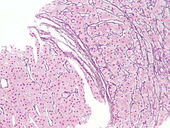
1 Liver Tumors Selected Topics Romil Saxena, MD Hepatocellular carcinoma 90% of all liver tumors Large cells with abundant eosinophilic cytoplasm that grow in a trabecular pattern 1
2 Case 1 55 male PET - multiple liver lesions, largest 8.2 cm No cirrhosis, no disease outside liver weight loss 15 lbs over 6 months, bad night sweats AFP


3 CD10 pcea AFP 3
 Negative: Hep Par1, HMB45, EMA,")

4 AFP IHC Positive: CD10 (cytoplasmic, membranous) AFP (cytoplasmic, patchy) Negative: Hep Par1, HMB45, EMA, CEA mono, CEA poly (membranous), CK20, CK7 Diagnosis: WD HCC vs metastatic hepatoid adenocarcinoma Chromogranin 4


5 Inhibin Hep Par1 5
6 Neuroendocrine tumor Positive: Chromogranin, synaptophysin Negative: Inhibin, PAX8 Case 2 71 F Biopsy of liver mass History of renal cell carcinoma 6


7 7

8 Positive: CK7, CD10, GATA3, GPC3 Negative: AFP, CDX2, vimentin, CK20, Hep Par1 Diagnosis: Neoplastic lesion, pending consultation Consultation report Malignant epithelial neoplasm with eosinophilic cytoplasm Additional stains done Positive: EMA, GPC3, ckit Negative: pcea, arginase, ER Diagnosis: Metastatic RCC, chromophobe type Remote history of RCC 15 years ago Arginase 1 8


9 Hep Par1 PAX8 Metastatic renal cell carcinoma, chromophobe type Positive: PAX8 Negative: Hep Par1, Arginase 1 9
10 Case 3 75 male 18 cm mass in non-cirrhotic liver AFP 45 Weight loss, fatigue, elevated LFTs for 2 months No h/o hepatitis Quit alcohol 10 years ago Quite smoking 10 years ago (never smoked heavily anyway) 10


11 Arginase 1 PAX8 (inhibin also negative) pcea 11

12 Reticulin Hepatocellular carcinoma Positive: Arginase 1, pcea Negative: Inhibin, PAX8 12
 Right hepatic mass 8 cm Pathology: FNA lung: non diagnostic Liver mass: HCC Referred to IU and second biopsy")
13 Case 4 41 female Left flank pain, myalgia, fatigue for 1 month CT left lower lung nodules (largest 1.5 cm) Right hepatic mass 8 cm Pathology: FNA lung: non diagnostic Liver mass: HCC Referred to IU and second biopsy performed HMB 45 13
 presentation Usually single, may be multifocal Variegated appearance grossly (hemorrhage, necrosis, yellow) Three components: Blood vessels, fat and myoid cells")
14 Hep Par1 Angiomyolipoma, epithelioid type Positive: HMB 45 Negative: Hep Par1 Angiomyolipoma PEComa Less common in liver than in kidney Middle aged individuals Incidental, localized (abdominal pain), or generalized (fever, malaise etc) presentation Usually single, may be multifocal Variegated appearance grossly (hemorrhage, necrosis, yellow) Three components: Blood vessels, fat and myoid cells Myoid cells may be spindled (SMA +) or epithelioid (HMB45 +) Epithelioid (pure/ predominant) variant common in liver 14


15 Lesion composed of large eosinophilic cells Hepatocellular Not hepatocellular Benign Evidence of hepatocellular differentiation Morphologic: Bile, Mallory Denk bodies, steatosis Immunohistochemical: Hep Par1, arginase 1, pcea, GPC3, BSEP Malignant Evidence of alternative differentiation Adrenal: Inhibin Renal: PAX8 Neuroendocrine: synaptophysin, chromogranin Melanoma: HMB45, S100, SOX10, Melan A Angiomyolipoma: HMB45 15


16 Immunohistochemical markers cytoplasmic +/- nuclear Hep Par 1 carbamoyl phosphate synthetase, urea cycle enzyme Arginase 1 binuclear manganese metalloenzyme, catalyzes hydrolysis of arginine to ornithine and urea Glypican 3 membrane anchored, heparin-sulfate proteoglycan normally expressed in fetal liver and placenta Alpha feto-protein expressed in fetal liver 16


17 IHC canalicular markers Polyclonal CEA cross reacts with a glycoprotein on bile canalicular membrane Bile salt export pump (BSEP) liver-specific ATP binding transporter involved in export of bile salts from hepatocytes Others CD10, villin IHC canalicular markers - Sensitivity Specificity HepPar1 PD HCC: 30% Stomach, lung, esophagus Arginase 1 PD HCC: 90-95%; all HCC: 85-90% Usually diffuse Works well in cytology material pancreas, cholangio rarely - breast, prostate GPC 3 PD HCC: 80-85%, WD HCC: 15% Diffuse -15% (not useful in biopsy) AFP All HCC: 30-50% Background staining a problem Negative: non-neoplastic liver Positive: cholangio, placental, germ cell Variety of tumors pcea PD HCC:<50% Membranous and luminal pattern in adeno BSEP PD HCC: <50% Diffuse staining: 10% Nguyen et al. Arch Pathol Lab Med 2015; 139: Only expressed in liver 17
18 Features of malignancy Characteristic histologic features 1/9/2018 Best markers Poorly differentiated HCC: Arginase 1 + GPC3 Well and moderately differentiated HCC: Hep Par1 + Arginase 1 Best overall: Arginase 1 Personal favorite: pcea Lesion composed of large eosinophilic cells Hepatocellular Not hepatocellular Benign Evidence of hepatocellular differentiation Morphologic: Bile, Mallory Denk bodies, steatosis Immunohistochemical: Hep Par1, arginase 1, pcea, GPC3, BSEP Malignant Evidence of alternative differentiation Adrenal: Inhibin Renal: PAX8 Neuroendocrine: synaptophysin, chromogranin Melanoma: HMB45, S100, SOX10, Melan A Angiomyolipoma: HMB45 Lesion composed of large eosinophilic cells Benign Hepatocellular Not hepatocellular Malignant 18

19 Reticulin stain CD34 stain Features of malignancy Characteristic histologic features 1/9/2018 Saxena et al. Diagnostic algorithms for tumors of the liver. In WHO Classification of Tumors of Digestive Diseases 4 th ed. 2010, p 255 Lesion composed of large eosinophilic cells Benign Hepatocellular Not hepatocellular Malignant Liver biopsy for diagnosis of liver mass in 50 year old patient with cirrhosis due to chronic hepatitis B 19


20 20


21 Benign Regenerative Nodule HCC HCC 21

22 Lesions composed of large eosinophilic cells Hepatocellular Not hepatocellular Benign Fibrotic Regenerative nodule Angiomyolipoma, epithelioid variant Non fibrotic Dysplastic nodule Focal nodular hyperplasia Hepatocellular adenoma Malignant Hepatocellular carcinoma Adrenal Renal Neuroendocrine Melanoma 71-year-old female with symptomatic cholelithiasis Incidental liver lesion discovered during cholecystectomy Biopsy of the lesion was performed Subsequent CT - solitary hepatic tumor within an otherwise normal appearing liver 22


23 Hep Par 1 PAX8 23


24 Case 2 71 year old female with HCC 24


25 Hep Par1 PAX8 Hepatocellular carcinoma clear cell predominant (mimics renal cell carcinoma) 25

 26")
26 Hepatocellular carcinoma prominent acinar / pseudoglandular structures (mimics adenocarcinoma, especially when tissue is scant, i.e. cytology, frozen section) 26


27 CD34 27
 Tumor cells creep along sinusoids and involve blood vessels Signet ring cells containing RBCs Positive for endothelial markers: Factor")
28 Metastatic lobular carcinoma Epithelioid hemangioendothelioma Commoner in women Most common in sixth decade May be discovered incidentally Occurs in non-fibrotic liver Usually multiple nodules Central hyalinization or ossification (firm, grey white nodules) Tumor cells creep along sinusoids and involve blood vessels Signet ring cells containing RBCs Positive for endothelial markers: Factor VIII, CD34, CD31 Epithelioid hemangioendothelioma Signet ring cells Mimics adenocarcinoma Look for intracytoplasmic red blood cells 28
 MCN Mucinous cystic neoplasm Hepatobiliary mucinous cystic neoplasm Biliary cystadenoma Pancreas Liver WHO 2010")
29 Pancreas Liver WHO 2010 terminology Liver former terminology PanIN Pancreatic intraepithelial neoplasia Biliary intraepithelial neoplasia (BilIN) Biliary dysplasia IPMN Intraductal papillary mucinous neoplasm Intraductal papillary neoplasm of bile duct Biliary papilloma(tosis) MCN Mucinous cystic neoplasm Hepatobiliary mucinous cystic neoplasm Biliary cystadenoma Pancreas Liver WHO 2010 terminology Liver former terminology PanIN Pancreatic intraepithelial neoplasia IPMN Intraductal papillary mucinous neoplasm Biliary intraepithelial neoplasia (BilIN) Intraductal papillary neoplasm of bile duct with invasion Biliary dysplasia Intraductal (papillary) cholangiocarcinoma, Biliary cystadenocarcinoma MCN Mucinous cystic neoplasm Hepatobiliary mucinous cystic neoplasm with invasion Biliary cystadenocarcinoma 29


30 30
 Atypical intraepithelial proliferation Multilayering of nuclei Flat or micropapillary projections into duct lumen Graded as")
31 Biliary Intraepithelial Neoplasia (BilIN) Flat, premalignant intraepithelial neoplasia of bile ducts Field change, multiple foci Precursor lesion of cholangiocarcinoma Risk factors: hepatolithiasis, PSC, chronic fluke infestation Biliary Intraepithelial Neoplasia (BilIN) Atypical intraepithelial proliferation Multilayering of nuclei Flat or micropapillary projections into duct lumen Graded as low, intermediate or high grade (BilIN 1,2,3) Foci of invasion may be seen BilIN may be seen associated with cholangiocarcinoma 31


32 32
")

33 Intraductal Papillary Neoplasm of Bile Duct Grossly visible lesion Fusiform or cystically dilated bile ducts containing soft, friable papillary, villous, or polypoid lesion(s) Unilocular or multilocular Single lesion, or continuous, or multiple with skip areas Communicate with biliary system, may be difficult to demonstrate Mucin hypersecretion in 1/3 of cases 33
")

34 Intraductal Papillary Neoplasm of Bile Duct Lining epithelium: pancreatobiliary, gastric, intestinal, oncocytic Dysplasia graded as low, intermediate, high Preinvasive neoplasm, may progress to invasive carcinoma Usually tubular adenocarcinoma, less frequently mucinous (colloid) adenocarcinoma Proposed terminology: Intraductal papillary biliary neoplasia with invasion or with invasive carcinoma has been variously called intraductal papillary cholangiocarcinoma, biliary papillomatosis, biliary papilloma, mucin-secreting biliary tumor, intraductal papillary mucinous tumor 34
35 Hepatobiliary Mucinous Cystic Neoplasm Occurs exclusively in women Grossly visible cyst Unilocular or multilocular No communication to biliary system Epithelium: single layer or stratified Ovarian stroma Positive for ER, PR and inhibin Benign Rare reports in literature of malignant lesions MCN with invasion or with invasive carcinoma Formerly, biliary cystadenocarcinoma CYST Epithelial-lined Without Epithelial Lining Cuboidal to low columnar Ciliate d Biliary / mucinous / oncocytic +/- papillary architecture Neoplastic Non-neoplastic Ovarian-like stroma No Epithelial epithelial invasion invasion into wall into wall w/o Ovarian-like stroma No Epithelial epithelial invasion invasion into wall into wall Laminate d wall Inflammation, necrosis Benign Malignant Solitary bile duct cyst Fibropolycy stic disease +/- associated renal disease Obstructive dilatation of bile duct Mucinous cystic neoplasm Ciliated foregut cyst Mucinous cystic neoplasm with associated invasive carcinoma Biliary intraductal papillary neoplasm Malignant biliary intraductal papillary neoplasm Intrahepatic cholangiocarcinoma with cystic change Mesench. hamartom a Cavernous hemangioma Cystic degeneration in any tumor Undif d embryonal sarcoma Cystic degeneration in any tumor Hydatid cyst Hydatid cyst Pyogenic abscess Amoebic abscess Necrotizing eosinophilic granuloma Hemorrhagic cyst Courtesy ND Theise, MD, NYU Adapted from Saxena et al. Diagnostic Algorithms for Tumors of the Liver. In: WHO Classification of Tumors of Digestive Diseases 4 th ed. 2010, p 259 Tumors that do not subscribe to a dichotomous paradigm of differentiation hepatocelular OR cholangiocytic Demonstrate biphenotypic differentiation, or stem/progenitor cell like features, or other variant patterns of differentiation? Trans-differentiation of malignant cells? Origin from stem/progenitor cells with divergent differentiation along variable pathways 35

36 Appropriate nomenclature, diagnostic criteria, prognostic significance and optimal therapeutic approach not completely defined due to lack of large series Not always identified correctly Lack of uniform terminology History 57 year old female, cirrhosis and chronic hepatitis C, genotype 1a Surveillance : Liver mass Dual phase CT with contrast: 3 hypervascular lesions, largest 1.8 cm nodule AFP 29.2ng/mL (normal 25 ng/ml) Transplanted 11 years folow-up, no recurrence of malignancy 36


37 37

38 Combined hepatocellularcholangiocarcinoma (chcc-cca) Case 2 74 year old female History of liver cirrhosis, right lobe lesion, r/o HCC 38


39 39
 phenotype")

40 pcea Combined hepatocellularcholangiocarcinoma (chcc-cca) Tumor of many names Combined HCC-CC Mixed HCC-CC HCC with dual (hepatocellular/biliary) phenotype Primary hepatic carcinoma of intermediate (hepatocyte/ cholangiocyte) phenotype Mixed hepatobiliary carcinoma Hepato-cholangiocarcinoma Biphenotypic primary liver carcinoma 40
41 Clinical Features Slightly more common in men, years of age Clinical features similar to other primary liver carcinomas Occur in background of chronic liver disease Particularly frequent after neo-adjuvant therapy Clinical Features Elevation of serum markers (AFP, CA19-9, CEA) variable Imaging: features of HCC or CC most commonly mimics CC or metastases Propensity to invade portal vein as well as regional lymph nodes Prognosis intermediate between HCC and CC Diagnosis Suspect combined hepatocellular-cholangiocarcinoma when simultaneous elevation of AFP and CA19-9 discordance between serum tumor markers and radiologic findings Treatment: complete surgical resection + regional lymph node dissection Frozen section recommended for all atypical HCC so that patient may benefit from LN dissection if tumor is combined chcc-cc 41

42 Case 3 23 year old woman, pain right lower thorax Imaging: multiple hypervascular nodules in liver Largest 7.5 cm, wash-out in portal phase. Serum AFP markedly elevated 42,720 IU/l Hepatitis B surface antigen +ve, most probably vertical transmission AFP CK19 Courtesy: Valerie Paradis, Beaujon Hospital Inserm, Clichy, France Hepatocellular carcinoma with K19 positivity 42
43 HCC with K19 expression One third of HCCs express biliary markers, K7 & K19 K19 expression in >5% of cells higher incidence of vascular invasion higher rate of recurrence after transplantation, TACE, resection HCCs that express CC-like and embryonic stem cell gene signatures show shorter recurrence free and overall survival* Enriched genes included K19 and EpCAM * Woo HG et al. Cancer Res 2010;70: References Seminars in Diagnostic Pathology 2017; Vol 34 Nakunama T, Sudo Y. Biliary tumors with pancreatic counterparts, pp Mann SA, Saxena R. Differential diagnosis of epithelioid and clear cell tumors in the liver, pp Hartke J, Johnson M, Ghabril. The diagnosis and treatment of hepatocellular carcinoma, pp Agni RM. Diagnostic histopathology of hepatocellular carcinoma: A case-based review, pp Sempoux C, Paradis V, Saxena R. Variant differentiation patterns in primary liver carcinoma, pp References Nguyen T, Phillips D, Jain D et al. Comparison of 5 immunohistochemical markers of hepatocellular differentiation for the diagnosis of hepatocellular carcinoma. Arch Pathol Lab Med 2015; 139: Saxena R, Albores-Saavedra J, Bioulac-Sage P et al. Diagnostic algorithms for tumors of the Liver. In: WHO classification of tumors of the Digestive System. 4 th edition, Lyon 2010, pp Woo HG, Lee JH, Yoon JH, et al. Identification of a cholangiocarcinoma-like gene expression trait in hepatocellular carcinoma. Cancer Res 2010;70:
Differential diagnosis of HCC
 Hepatocellular Carcinoma Quest for an Ideal Immunohistochemical Panel Sanjay Kakar, MD UCSF Differential diagnosis of HCC Hepatocellular lesions Adenoma, FNH, HG dysplasia Adenocarcinoma CholangioCA, metastasis
Biliary tract tumors
 Short Course 2010 Annual Fall Meeting of the Korean Society for Pathologists Biliary tract tumors Joon Hyuk Choi, M.D., Ph.D. Professor, Department of Pathology, Yeungnam Univ. College of Medicine, Daegu,
Liver Specialty Evening Conference. Matthew M. Yeh, MD, PhD Professor of Pathology Adjunct Professor of Medicine University of Washington, Seattle
 Liver Specialty Evening Conference Matthew M. Yeh, MD, PhD Professor of Pathology Adjunct Professor of Medicine University of Washington, Seattle Case History A 65 year-old man presents with abdominal
O Farrell Legacy UPDATE ON WHO NOMENCLATURE. World Health Organization, 2010 DISCLOSURES WITH EMPHASIS ON PROBLEM HEPATOCELLULAR TUMORS
 O Farrell Legacy UPDATE ON WHO NOMENCLATURE WITH EMPHASIS ON PROBLEM HEPATOCELLULAR TUMORS Linda Ferrell, MD University of California San Francisco Vice Chair, Director of Surgical Pathology World Health
Video Microscopy Tutorial 8
 Video Microscopy Tutorial 8 Common and Uncommon Lesions of the Liver Gladwyn Leiman, MD There are no disclosures necessary. Common and Uncommon Lesions in Liver FNA Gladwyn Leiman University of Vermont
8 years later! Next Generation Sequencing. Pathogenic Findings: HNF1A c.864delinscc, p.g292rfs*25 (NM_ ) (VAF: 59%) HNF1A Loss
 (VAF: 59%) HNF1A Loss") 8 years later! Next Generation Sequencing Pathogenic Findings: HNF1A c.864delinscc, p.g292rfs*25 (NM_000545.6) (VAF: 59%) HNF1A Loss Interpretation HNF1A c.864delinscc, p.g292rfs*25 (NM_000545.6) This
8 years later! Next Generation Sequencing Pathogenic Findings: HNF1A c.864delinscc, p.g292rfs*25 (NM_000545.6) (VAF: 59%) HNF1A Loss Interpretation HNF1A c.864delinscc, p.g292rfs*25 (NM_000545.6) This
Select problems in cystic pancreatic lesions
 Disclosure Select problems in cystic pancreatic lesions Five Prime Therapeutics shareholder Adicet Bio shareholder Bristol-Meyer Squibb advisory board grace.kim@ucsf.edu Pancreatic cystic lesions Intraductal
Neoplasms of the Canine, Feline and Lemur Liver:
 Neoplasms of the Canine, Feline and Lemur Liver: Classification and Prognosis Annual Seminar of the French Society of Veterinary Pathology John M. Cullen VMD PhD DACVP North Carolina State University Primary
Outline. Hepatocellular Carcinoma Histologic variants. HCC: Histologic variants
 2018 Park City AP Update Hepatocellular Carcinoma Histologic variants Sanjay Kakar, MD University of California, San Francisco Outline Histologic variants of HCC Morphologic and Immunohistochemical pitfalls
Pancreatic Cystic Lesions 원자력병원
 Pancreatic Cystic Lesions 원자력병원 박선 후 Lines of cellular differentiation Ductal Acinar Undetermined Ductal adenocarcinoma Serous/ mucinous tumor Intraductal papillary mucinous neoplasm Acinar cell carcinoma
Neoplasias Quisticas del Páncreas
 SEAP -Aproximación Práctica a la Patología Gastrointestinal- Madrid, 26 de mayo, 2006 Neoplasias Quisticas del Páncreas Gregory Y. Lauwers, M.D. Director, Service Massachusetts General Hospital Harvard
Intrahepatic cholangiocarcinoma Histologic spectrum, novel markers and molecular assays
 2018 Current Issues in Surgical Pathology Summary (not actual lecture) Intrahepatic cholangiocarcinoma Histologic spectrum, novel markers and molecular assays Sanjay Kakar, MD University of California,
Pathological Classification of Hepatocellular Carcinoma
 3 rd APASL Single Topic Conference: HCC in 3D Pathological Classification of Hepatocellular Carcinoma Glenda Lyn Y. Pua, M.D. HCC Primary liver cancer is the 2 nd most common cancer in Asia HCC is the
Liver Tumors. Prof. Dr. Ahmed El - Samongy
 Liver Tumors Prof. Dr. Ahmed El - Samongy Objective 1. Identify the most important features of common benign liver tumors 2. Know the risk factors, diagnosis, and management of hepatocellular carcinoma
Hepatobiliary and Pancreatic Malignancies
 Hepatobiliary and Pancreatic Malignancies Gareth Eeson MD MSc FRCSC Surgical Oncologist and General Surgeon Kelowna General Hospital Interior Health Consultant, Surgical Oncology BC Cancer Agency Centre
A 53 year-old woman with a lung mass, right hilar mass and mediastinal adenopathy.
 November 2015 Case of the Month A 53 year-old woman with a lung mass, right hilar mass and mediastinal adenopathy. Contributed by: Rasha Salama, M.D., IU Department of Pathology and Laboratory Medicine
NEOPLASMS AND TUMOR-LIKE CONDITIONS OF LIVER
 NEOPLASMS AND TUMOR-LIKE CONDITIONS OF LIVER Epithelial Tumors Focal nodular hyperplasia Focal nodular hyperplasia is a localized hyperplasic overgrowth of hepatocytes around a vascular anomaly, particularly
Synonyms. Nephrogenic metaplasia Mesonephric adenoma
 Nephrogenic Adenoma Synonyms Nephrogenic metaplasia Mesonephric adenoma Definition Benign epithelial lesion of urinary tract with tubular, glandular, papillary growth pattern Most frequently in the urinary
Pitfalls in the diagnosis of well-differentiated hepatocellular lesions
 2013 Colorado Society of Pathology Pitfalls in the diagnosis of well-differentiated hepatocellular lesions Sanjay Kakar, MD University of California, San Francisco Outline Hepatocellular adenoma: new WHO
X-Ray Corner. Imaging Approach to Cystic Liver Lesions. Pantongrag-Brown L. Solitary cystic liver lesions. Hepatic simple cyst (Figure 1)
") THAI J 136 Imaging Approach to Cystic Liver Lesions GASTROENTEROL 2013 X-Ray Corner Imaging Approach to Cystic Liver Lesions Pantongrag-Brown L Cystic liver lesions are common findings in daily practice
THAI J 136 Imaging Approach to Cystic Liver Lesions GASTROENTEROL 2013 X-Ray Corner Imaging Approach to Cystic Liver Lesions Pantongrag-Brown L Cystic liver lesions are common findings in daily practice
HEPATO-BILIARY IMAGING
 HEPATO-BILIARY IMAGING BY MAMDOUH MAHFOUZ MD PROF.OF RADIOLOGY CAIRO UNIVERSITY mamdouh.m5@gmail.com www.ssregypt.com CT ABDOMEN Indications Patient preparation Patient position Scanogram Fasting 4-6 hours
PATHOLOGY OF LIVER TUMORS
 PATHOLOGY OF LIVER TUMORS Pathobasic, 31.05.2016 WHO Classification Approach to a Liver Mass Lesion in a patient with chronic liver disease? Lesion in a patient without chronic liver disease? Malignant
40th European Congress of Cytology Liverpool, UK, 2-5 th October 2016
 40th European Congress of Cytology Liverpool, UK, 2-5 th October 2016 EUS FNA of abdominal organs: An approach to reporting and triage for ancillary testing Date and time: Sunday 2 nd October 2016 15.00-16.30
The Panel Approach to Diagnostics. Lauren Hopson International Product Specialist Cell Marque Corporation
 The Panel Approach to Diagnostics Lauren Hopson International Product Specialist Cell Marque Corporation Cell Marque Rocklin, California About Cell Marque: IVD primary antibody manufacturer Distributors
Hepatocellular carcinoma Cholangiocarcinoma. Jewels of hepatobiliary cancer imaging : what to look for? Imaging characteristics of HCC.
 Outline : Imaging Jewels Jewels of hepatobiliary cancer imaging : what to look for? Hepatocellular carcinoma Cholangiocarcinoma Surachate Siripongsakun, M.D. Chulabhorn Cancer Center Imaging characteristics
Cholangiocarcinoma. Judy Wyatt Dundee November 2010
 Cholangiocarcinoma Judy Wyatt Dundee November 2010 Making sense of cholangiocarcinoma Difficulties with diagnostic criteria How many entities within cholangiocarcinoma? Rapidly evolving Intrahepatic cholangiocarcinoma
Raga Ramachandran, MD, PhD Assistant Professor and Director of Medical Education, UCSF Pathology
 Variants of Hepatocellular Carcinoma: Practical Issues Raga Ramachandran, MD, PhD Assistant Professor and Director of Medical Education, UCSF Pathology raga.ramachandran@ucsf.edu A full copy of the presentation
Pancreatobiliary Frozen Section Nightmares
 Pancreatobiliary Frozen Section Nightmares Aatur D. Singhi, MD PhD Assistant Professor University of Pittsburgh Medical Center Department of Pathology singhiad@upmc.edu Objectives Briefly give an overview
Evaluation and Management of Cystic Lesions of the Pancreas: When to Resect, When to Follow and When to Forget
 Evaluation and Management of Cystic Lesions of the Pancreas: When to Resect, When to Follow and When to Forget Randall Brand, MD Professor of Medicine Division of Gastroenterology, Hepatology and Nutrition
Standardized Terminology in Pancreatobiliary Cytology: The Papanicolaou Society Guidelines
 Standardized Terminology in Pancreatobiliary Cytology: The Papanicolaou Society Guidelines Barbara Ann Centeno. M.D. Vice-Chair, Clinical Services, Anatomic Pathology Assistant Chief, Pathology Service
Intraductal papillary neoplasms in the bile ducts
 Intraductal papillary neoplasms in the bile ducts Seok Hwa Youn Myunghee Yoon Dong Hoon Shin Kosin University Gospel Hospital Department of general surgery Hepato-biliary-pancreatic division Introduction
Malignant Focal Liver Lesions
 Malignant Focal Liver Lesions Other Than HCC Pablo R. Ros, MD, MPH, PhD Departments of Radiology and Pathology University Hospitals Cleveland Medical Center Case Western Reserve University Pablo.Ros@UHhospitals.org
Mesenchymal Tumors MESENCHYMAL TUMORS OF THE LIVER: WHAT S NEW AND UNUSUAL (MY PERSPECTIVE)
") MESENCHYMAL TUMORS OF THE LIVER: WHAT S NEW AND UNUSUAL (MY PERSPECTIVE) CURRENT ISSUES IN ANATOMIC PATHOLOGY MAY 23, 2014 Linda Ferrell, MD, UCSF Mesenchymal Tumors Focus on Vascular Tumors Benign and
MESENCHYMAL TUMORS OF THE LIVER: WHAT S NEW AND UNUSUAL (MY PERSPECTIVE) CURRENT ISSUES IN ANATOMIC PATHOLOGY MAY 23, 2014 Linda Ferrell, MD, UCSF Mesenchymal Tumors Focus on Vascular Tumors Benign and
Evaluation of Liver Mass Lesions. American College of Gastroenterology 2013 Regional Postgraduate Course
 Evaluation of Liver Mass Lesions American College of Gastroenterology 2013 Regional Postgraduate Course Lewis R. Roberts, MB ChB, PhD Division of Gastroenterology and Hepatology Mayo Clinic College of
Mesenchymal Tumors. Cavernous Hemangioma (CH) VASCULAR TUMORS MESENCHYMAL TUMORS OF THE LIVER: WHAT S NEW AND UNUSUAL (MY PERSPECTIVE)
 VASCULAR TUMORS MESENCHYMAL TUMORS OF THE LIVER: WHAT S NEW AND UNUSUAL (MY PERSPECTIVE)") Mesenchymal Tumors MESENCHYMAL TUMORS OF THE LIVER: WHAT S NEW AND UNUSUAL (MY PERSPECTIVE) CURRENT ISSUES IN ANATOMIC PATHOLOGY MAY 23, 2014 Linda Ferrell, MD, UCSF Focus on Vascular Tumors Benign and
Mesenchymal Tumors MESENCHYMAL TUMORS OF THE LIVER: WHAT S NEW AND UNUSUAL (MY PERSPECTIVE) CURRENT ISSUES IN ANATOMIC PATHOLOGY MAY 23, 2014 Linda Ferrell, MD, UCSF Focus on Vascular Tumors Benign and
Interesting case. Vikas Kundra, M.D., Ph.D. October Vikas Kundra, M.D., Ph.D.
 Interesting case October 2012 Disclosure Information Vikas Kundra, M.D, Ph.D. I have no financial relationships to disclose. I WILL NOT include discussion of investigational or off-label use of a product
Gross appearance of nodular hyperplasia in material obtained from suprapubic prostatectomy. Note the multinodular appearance and the admixture of
 Tiền liệt tuyến Tiền liệt tuyến Gross appearance of nodular hyperplasia in material obtained from suprapubic prostatectomy. Note the multinodular appearance and the admixture of solid and microcystic areas.
04/10/2018. Intraductal Papillary Neoplasms Of Breast INTRADUCTAL PAPILLOMA
 Intraductal Papillary Neoplasms Of Breast Savitri Krishnamurthy MD Professor of Pathology Deputy Division Head The University of Texas MD Anderson Cancer Center 25 th Annual Seminar in Pathology Pittsburgh,
Appendix 4: WHO Classification of Tumours of the pancreas 17
 S3.01 The WHO histological tumour type must be recorded. CS3.01a The histological type of the tumour should be recorded based on the current WHO classification 17 (refer to Appendices 4-7). Appendix 4:
DIAGNOSTIC SLIDE SEMINAR: PART 1 RENAL TUMOUR BIOPSY CASES
 DIAGNOSTIC SLIDE SEMINAR: PART 1 RENAL TUMOUR BIOPSY CASES Dr. Andrew J. Evans MD, PhD, FACP, FRCPC Consultant in Genitourinary Pathology University Health Network, Toronto, ON Case 1 43 year-old female,
Radiology Pathology Conference
 Radiology Pathology Conference Nadia F. Yusaf, M.D. PGY-3 1/29/2010 Presentation material is for education purposes only. All rights reserved. 2010 URMC Radiology Page 1 of 90 Case 1 60 year- old man presents
Salivary Glands 3/7/2017
 Salivary Glands 3/7/2017 Goals and objectives Focus on the entities unique to H&N Common board type facts Information for your future practice Salivary Glands Salivary Glands Major gland. Paratid. Submandibular.
Intraductal Papillary Mucinous Neoplasms: We Still Have a Way to Go! Francesco M. Serafini, MD, FACS
 Intraductal Papillary Mucinous Neoplasms: We Still Have a Way to Go! Francesco M. Serafini, MD, FACS Brooklyn VAMC September 21 st GI Grand Rounds - What is it? - Clinical entity that has emerged from
MRI OF FOCAL LESIONS IN
 Introduction MRI OF FOCAL LESIONS IN THE NON-CIRRHOTIC LIVER Ivan Pedrosa M.D. Ph.D. Associate Professor of Radiology and Advanced Imaging Research Center University of Texas Southwestern. Dallas, TX Incidental
XIII. Tumours of the liver and biliary system
 XIII. Tumours of the liver and biliary system V. PONOMARKOV 1 & L. J. MACKEY 2 In this histological classification of liver and gall bladder tumours the tumour types largely correspond to those found in
Overview. Disclosure. PRE INVASIVE NEOPLASIA OF BILIARY TREE New Perspectives on Old Themes. N. Volkan Adsay, MD
 PRE INVASIVE NEOPLASIA OF BILIARY TREE New Perspectives on Old Themes N. Volkan Adsay, MD Professor and Vice-Chair Director of Anatomic Pathology Emory University and Emory Winship Cancer Institute Atlanta,
The clinically challenging entity of liver metastasis from tumors of unknown primary
 The clinically challenging entity of liver metastasis from tumors of unknown primary Xuchen Zhang, MD, PhD Associate Professor of Pathology Department of Pathology Yale University School of Medicine Liver
Papillary Lesions of the breast
 Papillary Lesions of the breast Emad Rakha Professor of Breast Pathology The University of Nottingham Papillary lesions of the breast are a heterogeneous group of disease, which are characterised by neoplastic
Mody. AIS vs. Invasive Adenocarcinoma of the Cervix
 Common Problems in Gynecologic Pathology Michael T. Deavers, M.D. Houston Methodist Hospital, Houston, Texas Common Problems in Gynecologic Pathology Adenocarcinoma in-situ (AIS) of the Cervix vs. Invasive
Pancreatic Cysts. Darius C. Desai, MD FACS St. Luke s University Health Network
 Pancreatic Cysts Darius C. Desai, MD FACS St. Luke s University Health Network None Disclosures Incidence Widespread use of cross sectional imaging Seen in over 2% of patients having abdominal imaging
Presentation material is for education purposes only. All rights reserved URMC Radiology Page 1 of 98
 Presentation material is for education purposes only. All rights reserved. 2011 URMC Radiology Page 1 of 98 Radiology / Pathology Conference February 2011 Brooke Koltz, Cytopathology Resident Presentation
Papillary Lesions of the Breast A Practical Approach to Diagnosis. (Arch Pathol Lab Med. 2016;140: ; doi: /arpa.
 Papillary Lesions of the Breast A Practical Approach to Diagnosis (Arch Pathol Lab Med. 2016;140:1052 1059; doi: 10.5858/arpa.2016-0219-RA) Papillary lesions of the breast Span the spectrum of benign,
Pancreatitis: A Potential Pitfall in Endoscopic Ultrasound Guided Pancreatic FNA
 Pancreatitis: A Potential Pitfall in Endoscopic Ultrasound Guided Pancreatic FNA Jack Yang, MD Department of Pathology, Medical University of South Carolina Objectives Understand the indication of EUS
Case Scenario 1: Thyroid
 Case Scenario 1: Thyroid History and Physical Patient is an otherwise healthy 80 year old female with the complaint of a neck mass first noticed two weeks ago. The mass has increased in size and is palpable.
CT & MRI of Benign Liver Neoplasms Srinivasa R Prasad
 CT & MRI of Benign Liver Neoplasms Srinivasa R Prasad No financial disclosures Acknowledgements Many thanks to Drs. Heiken, Narra & Menias (MIR) Dr. Sahani (MGH) for sharing images Benign Liver Tumors:
Management of Rare Liver Tumours
 Gian Luca Grazi Hepato-Biliary-Pancreatic Surgery National Cancer Institute Regina Elena Rome Fibrolamellar Carcinoma Mixed Hepato Cholangiocellular Carcinoma Hepatoblastoma Carcinosarcoma Primary Hepatic
List of Available TMAs in the PRN
 TMA RPCI_BrainCa01 RPCI_BrCa03 RPCI_BrCa04 RPCI_BrCa05 RPCI_BrCa0 RPCI_BrCa07 RPCI_BrCa08 RPCI_BrCa15 RPCI_BrCa1 RPCI_BrCa17 RPCI_BrCa18 RPCI_BrCa19 RPCI_BrCa20 RPCI_BrCa21 RPCI_BrCa24 RPCI_BrCa25 RPCI_BrCa2
A 42-year-old woman with a liver mass
 April 2016 Case of the Month A 42-year-old woman with a liver mass Contributed by: Natalia I. Rush, MD, Resident Physician, Indiana University School of Medicine, Department of Pathology and Laboratory
Enterprise Interest Nothing to declare
 Enterprise Interest Nothing to declare Update of mixed tumours of the GI tract, the pancreas and the liver Introduction to the concept of mixed tumours and clinical implication Jean-Yves SCOAZEC Surgical
I LOVE Immunostains. Two Types of Pitfalls 3/23/2017. Disclosure of Relevant Financial Relationships
 There Are No Magic Bullets: When Immunostains Can Get You into Trouble in Hepatic & Gastrointestinal Pathology John Hart, M.D. Sections of Surgical Pathology & Hepatology University of Chicago Medical
Renal tumours: use of immunohistochemistry & molecular pathology. Dr Lisa Browning John Radcliffe Hospital Oxford
 Renal tumours: use of immunohistochemistry & molecular pathology Dr Lisa Browning John Radcliffe Hospital Oxford Renal tumours: the use of immunohistochemistry & molecular pathology Classification of RCC
Matthew McCollough, M.D. April 9, 2009 University of Louisville
 Matthew McCollough, M.D. April 9, 2009 University of Louisville List the differential diagnosis for pancreatic cysts Review the epidemiology Illustrate the types of cysts through case discussions Discuss
Basement membrane in lobule.
 Bahram Memar, MD Basement membrane in lobule. Normal lobule-luteal phase Normal lobule-follicular phase Lactating breast Greater than 95% are adenocarcinomas in situ carcinomas and invasive carcinomas.
Urinary Bladder: WHO Classification and AJCC Staging Update 2017
 Urinary Bladder: WHO Classification and AJCC Staging Update 2017 Houston Society of Clinical Pathologists 58 th Annual Spring Symposium Houston, TX April 8, 2017 Jesse K. McKenney, MD Classification
GASTROINTESTINAL IMAGING STUDY GUIDE
 GASTROINTESTINAL IMAGING STUDY GUIDE Pharynx Diverticula Foreign bodies Trauma o Motility Disorders Esophagus Diverticula Trauma Esophagitis Barrett esophagus Rings, webs, and strictures Varices Benign
Outline. Intraductal Papillary Mucinous Neoplasm (IPMN) Guideline Review 4/6/2017. Case Example Background Classification Histology Guidelines
 Guideline Review 4/6/2017. Case Example Background Classification Histology Guidelines") Intraductal Papillary Mucinous Neoplasm (IPMN) Guideline Review The Nurse Practitioner Association New York State Capital Region Teaching Day Matthew Warndorf MD Case Example Background Classification
Intraductal Papillary Mucinous Neoplasm (IPMN) Guideline Review The Nurse Practitioner Association New York State Capital Region Teaching Day Matthew Warndorf MD Case Example Background Classification
Dr Claire Smith, Consultant Radiologist St James University Hospital Leeds
 Dr Claire Smith, Consultant Radiologist St James University Hospital Leeds Imaging in jaundice and 2ww pathway Image protocol Staging Limitations Pancreatic cancer 1.2.4 Refer people using a suspected
Kidney Case 1 SURGICAL PATHOLOGY REPORT
 Kidney Case 1 Surgical Pathology Report February 9, 2007 Clinical History: This 45 year old woman was found to have a left renal mass. CT urography with reconstruction revealed a 2 cm medial mass which
Workup of a Solid Liver Lesion
 Workup of a Solid Liver Lesion Joseph B. Cofer MD FACS Chief Quality Officer Erlanger Health System Affiliate Professor of Surgery UTHSC-Chattanooga I have no financial or other relationships with any
Pancreatico-biliary cytology: a practical approach to diagnosis. Corina Cotoi
 Pancreatico-biliary cytology: a practical approach to diagnosis Corina Cotoi Pancreatico-biliary lesions Solid: Ductal adenocarcinoma Cholangiocarcinoma Acinar cell carcinoma Neuroendocrine tumour / carcinoma
Histopathological diagnosis of CUP
 Histopathological diagnosis of CUP Dr Karin Oien karin.oien@glasgow.ac.uk Disclosure slide Dr Karin Oien has no financial interests in any company mentioned in this presentation. Dr Karin Oien is conducting
A LIVER TUMOR WITH AN IDENTITY CRISIS
 A LIVER TUMOR WITH AN IDENTITY CRISIS Stephen M. Lagana MD Assistant Professor of Pathology Columbia University Medical Center New York, NY USA SML2179@cumc.columbia.edu Disclosure of Relevant Financial
Disclosure. Relevant Financial Relationship(s) None. Off Label Usage None MFMER slide-1
 None. Off Label Usage None MFMER slide-1") Disclosure Relevant Financial Relationship(s) None Off Label Usage None 2013 MFMER slide-1 Case Presentation A 43 year old male, with partial nephrectomy for a right kidney mass 2013 MFMER slide-2 2013
Disclosure Relevant Financial Relationship(s) None Off Label Usage None 2013 MFMER slide-1 Case Presentation A 43 year old male, with partial nephrectomy for a right kidney mass 2013 MFMER slide-2 2013
Cutaneous metastases. Thaddeus Mully. University of California, San Francisco Professor, Departments of Pathology and Dermatology
 Cutaneous metastases Thaddeus Mully University of California, San Francisco Professor, Departments of Pathology and Dermatology DISCLOSURE OF RELATIONSHIPS WITH INDUSTRY Thaddeus Mully Course C005 Essential
performed to help sway the clinician in what the appropriate diagnosis is, which can substantially alter the treatment of management.
 Hello, I am Maura Polansky at the University of Texas MD Anderson Cancer Center. I am a Physician Assistant in the Department of Gastrointestinal Medical Oncology and the Program Director for Physician
Case Report Cystic and Papillary Neoplasm at the Hepatic Hilum Possibly Originating in the Peribiliary Glands
 Case Reports in Pathology Volume 2016, Article ID 9130754, 7 pages http://dx.doi.org/10.1155/2016/9130754 Case Report Cystic and Papillary Neoplasm at the Hepatic Hilum Possibly Originating in the Peribiliary
BREAST PATHOLOGY. Fibrocystic Changes
 BREAST PATHOLOGY Lesions of the breast are very common, and they present as palpable, sometimes painful, nodules or masses. Most of these lesions are benign. Breast cancer is the 2 nd most common cause
59 yo male with past medical history of prostate carcinoma, presented with upper abdominal pain
 December 2016 59 yo male with past medical history of prostate carcinoma, presented with upper abdominal pain Contributed by: Divya Sharma, MD. Fellow, Gastrointestinal Pathology, Department of Pathology
Diseases of the breast (1 of 2)
") Diseases of the breast (1 of 2) Introduction A histology introduction Normal ducts and lobules of the breast are lined by two layers of cells a layer of luminal cells overlying a second layer of myoepithelial
Diseases of the breast (1 of 2) Introduction A histology introduction Normal ducts and lobules of the breast are lined by two layers of cells a layer of luminal cells overlying a second layer of myoepithelial
Non Small Cell Lung Cancer Histopathology ד"ר יהודית זנדבנק
 Non Small Cell Lung Cancer Histopathology ד"ר יהודית זנדבנק 26.06.09 Lecture outlines WHO histological classification Macro/Micro assessment Early diagnosis Minimal pathology Main subtypes SCC, AdCa, LCLC
Biliary Tract Neoplasia: A Cyto-histologic Review. Michelle Reid, MD, MSc Professor of Pathology Director of Cytopathology Emory University Hospital
 Biliary Tract Neoplasia: A Cyto-histologic Review Michelle Reid, MD, MSc Professor of Pathology Director of Cytopathology Emory University Hospital Bile Duct Brushings (BDB) BDBs are the initial diagnostic
Approach to Liver Lesions. Anjana A. Pillai, MD Associate Professor of Medicine Director, Liver Tumor Clinic The University of Chicago Medical Center
 Approach to Liver Lesions Anjana A. Pillai, MD Associate Professor of Medicine Director, Liver Tumor Clinic The University of Chicago Medical Center Objectives Identify common clinical features and imaging
Spectrum of Preneoplastic and Neoplastic Cystic Lesions of the Kidney in Adult. by dr. Banan Burhan Mohammed Lecturer in Pathology Department
 Spectrum of Preneoplastic and Neoplastic Cystic Lesions of the Kidney in Adult by dr. Banan Burhan Mohammed Lecturer in Pathology Department Various hereditary, acquired, and neoplastic conditions can
Case 1. Case 1: EUS Report 5/1/2017. Interesting Cases of Pancreatic Masses
 Interesting Cases of Pancreatic Masses Martha Bishop Pitman, MD Professor of Pathology Harvard Medical School Director of Cytopathology Massachusetts General Hospital Boston, MA MASSACHUSETTS GENERAL PHYSICIANS
Carcinoma of Unknown Primary (CUP)
") Metasta c Carcinoma of Unknown Primary: Diagnos c Approach Using Immunohistochemistry James R. Conner, MD, PhD Mount Sinai Hospital Toronto, ON Carcinoma of Unknown Primary (CUP) 3-5% of all new malignant
Metasta c Carcinoma of Unknown Primary: Diagnos c Approach Using Immunohistochemistry James R. Conner, MD, PhD Mount Sinai Hospital Toronto, ON Carcinoma of Unknown Primary (CUP) 3-5% of all new malignant
Case: The patient is a 62 year old woman with a history of renal cell carcinoma that was removed years ago. A 2.4 cm liver mass was found on CT
 Case: The patient is a 62 year old woman with a history of renal cell carcinoma that was removed years ago. A 2.4 cm liver mass was found on CT during follow- up. ALT, AST, Alk Phos and bilirubin were
What is New in the 2015 WHO Lung Cancer Classification? Zhaolin Xu, MD, FRCPC, FCAP
 What is New in the 2015 WHO Lung Cancer Classification? Zhaolin Xu, MD, FRCPC, FCAP Professor, Dept of Pathology, Dalhousie University, Canada Pulmonary Pathologist and Cytopathologist, QEII HSC Senior
2 to 3% of All New Visceral Cancers Peak Incidence is 6th Decade M:F = 2:1 Grossly is a Bright Yellow, Necrotic Mass with a Pseudocapsule
 GENITOURINARY PATHOLOGY Kathleen M. O Toole, M.D. Renal Cell Carcinoma 2 to 3% of All New Visceral Cancers Peak Incidence is 6th Decade M:F = 2:1 Grossly is a Bright Yellow Necrotic Mass Grossly is a Bright
Hepato-Pancreatico-Biliary Surgery. Dr. Ankur J. Shah. MS, DNB, MNAMS, MRCSEd (UK), FRCS (UK)
, FRCS (UK)") Hepato-Pancreatico-Biliary Surgery Dr. Ankur J. Shah MS, DNB, MNAMS, MRCSEd (UK), FRCS (UK) Consultant Hepato-Pancreatico-Biliary and Liver Transplant Surgeon Ansh Liver Clinic Prevention to Cure Address
Hepato-Pancreatico-Biliary Surgery Dr. Ankur J. Shah MS, DNB, MNAMS, MRCSEd (UK), FRCS (UK) Consultant Hepato-Pancreatico-Biliary and Liver Transplant Surgeon Ansh Liver Clinic Prevention to Cure Address
Navigating the Biliary Tract with CT & MR: An Imaging Approach to Bile Duct Obstruction
 Navigating the Biliary Tract with CT & MR: An Imaging Approach to Bile Duct Obstruction Ann S. Fulcher, MD Medical College of Virginia Virginia Commonwealth University Richmond, Virginia Objectives To
Interesting Cases from Liver Tumor Board. Jeffrey C. Weinreb, M.D.,FACR Yale University School of Medicine
 Interesting Cases from Liver Tumor Board Jeffrey C. Weinreb, M.D.,FACR Yale University School of Medicine jeffrey.weinreb@yale.edu Common Liver Diseases Hemangioma Cyst FNH Focal Fat/Sparing THID Non-Cirrhotic
Objectives. Salivary Gland FNA: The Milan System. Role of Salivary Gland FNA 04/26/2018
 Salivary Gland FNA: The Milan System Dr. Jennifer Brainard Section Head Cytopathology Cleveland Clinic Objectives Introduce the Milan System for reporting salivary gland cytopathology Define cytologic
PSA. HMCK, p63, Racemase. HMCK, p63, Racemase
 Case 1 67 year old male presented with gross hematuria H/o acute prostatitis & BPH Urethroscopy: small, polypoid growth with a broad base emanating from the left side of the verumontanum Serum PSA :7 ng/ml
Updates in Urologic Pathology WHO Made Those Changes?! Peyman Tavassoli Pathology Department BC Cancer Agency
 Updates in Urologic Pathology WHO Made Those Changes?! Peyman Tavassoli Pathology Department BC Cancer Agency World Health Organization Available in Feb 2016 Frame work for reporting Major contributing
3/28/2017. Disclosure of Relevant Financial Relationships. GU Evening Subspecialty Case Conference. Differential Diagnosis:
 GU Evening Subspecialty Case Conference Rajal B. Shah, M.D. VP, Medical Director, Urologic Pathology Miraca Life Sciences, Irving, Texas Clinical Associate Professor of Pathology Baylor College of Medicine,
Cystic Disease of the Liver Work Up and Management. Louis Ferrari MD, PGY 3 6/9/16 SUNY Downstate Medical Center
 Cystic Disease of the Liver Work Up and Management Louis Ferrari MD, PGY 3 6/9/16 SUNY Downstate Medical Center The Case 73F presents to clinic after diagnostic laparoscopy at OSH. Known liver mass for
CTA/MRA of Pediatric Hepatic Masses Radiology-Pathology Correlation
 Acta Radiológica Portuguesa, Vol.XVIII, nº70, pág. 41-50, Abr.-Jun., 2006 CTA/MRA of Pediatric Hepatic Masses Radiology-Pathology Correlation Marilyn J. Siegel Mallinckrodt Institute of Radiology, Washington
Video Microscopy Tutorial 19
 Video Microscopy Tutorial 19 EUS FNA of Pancreatic Cysts Martha Pitman, MD There are no disclosures necessary. EUS-FNA of Pancreatic Cysts Martha Bishop Pitman, M.D. Massachusetts General Hospital Harvard
Financial Disclosure
 Benign Liver Masses Adil Abdalla, MBBS Creighton University-CHI Health August 25, 2018 Financial Disclosure Nothing to disclose Financial Disclosure 1 Objectives To assess patients with benign liver tumors
ARTHUR PURDY STOUT SOCIETY COMPANION MEETING: DIFFICULT NEW DIFFERENTIAL DIAGNOSES IN PROSTATE PATHOLOGY. Jonathan I. Epstein.
 1 ARTHUR PURDY STOUT SOCIETY COMPANION MEETING: DIFFICULT NEW DIFFERENTIAL DIAGNOSES IN PROSTATE PATHOLOGY Jonathan I. Epstein Professor Pathology, Urology, Oncology The Reinhard Professor of Urological