Brain and CNS Tumors
|
|
|
- Flora Martin
- 6 years ago
- Views:
Transcription
1 Brain and CNS Tumors FCDS 2011/2012 Educational Webcast Series January 19, 2012 Lynne Pearson, BHS, CTR, LHRM Steven Peace, BS, CTR Updated for 2012 Requirements and CSv
2 Presentation Outline Overview Anatomy of the Human Brain Multiple Primary and Histology Coding Rules Collaborative Stage Data Collection System (CSv2) C.S. Site Specific Factors 2 Treatment Options

3 Overview


4 Brain tumors are: Primary brain tumors - those that begin in the brain and tend to stay in the brain - occur in people of all ages, but they are statistically more frequent in children and older adults. Metastatic brain tumors those that begin as a cancer elsewhere in the body and spread to the brain are more common in adults than in children. 4 Source: American Brain Tumor Association Facts and Statistics
5 Brain tumors are: the second leading cause of cancer-related deaths in children (males and females) under age 20 (leukemia is the first) the second leading cause of cancer-related deaths in males ages the fifth leading cause of cancer-related deaths in females ages Source: American Brain Tumor Association Facts and Statistics
6 Brain tumors are: 6 Usually described as intracranial neoplasms with varying behaviors (benign, borderline, malignant) Are frequently grouped in discussions, statistics, training, treatment planning, and research to include pretty much any structure within the cranium (including hormone secreting ducts like the pineal and pituitary gland), the cranial nerves (optic nerve, olfactory nerve, acoustic nerve), the lining of the brain or meninges which also lines the rest of the central nervous system s critically important feature capable of distributing chemically charged nerve impulses with incredible speed and accuracy and a critical component of the function of the central nervous system, the spinal cord.

7 Intracranial vs. Extra-cranial Brain tumors are classified as either intra- or extra-cranial and both produce clinical/symptomatic effects that are similar in terms of mass effect, hemorrhage, seizure activity, and edema 7 SEER Training Module
8 ALL Brain Tumors are Reportable Public Law , the Benign Brain Tumor Cancer Registries Amendment Act, [PDF-185KB] requires programs participating in the National Program for Cancer Registries (NPCR) to collect data on benign and borderline tumors of the central nervous system in addition to the previously required data on malignant tumors. In addition to NPCR, the National Cancer Institute's (NCI) Surveillance, Epidemiology and End Results (SEER) program and the American College of Surgeons' (ACoS) Commission on Cancer began requiring that these tumors be reported, starting with cases diagnosed on January 1,


9 9 Brain tumors are ALL Reportable
10 Brain and CNS Tumors All Ages 2011 estimates in the United States 64,540 new cancer cases This includes: Malignant brain tumors (24,070) Non-malignant brain tumors (40,470) 10 Source: American Brain Tumor Association Facts and Statistics
11 Brain and CNS Tumors - Children Approximately 4,150 children younger than age 20 will be diagnosed with primary brain tumors in ,960 will be less than 15 years of age 1,190 will be between the ages of 15 and 19 Giomas represent a high percentage of childhood tumors 55% of all tumors and 71% of malignant tumors in children age % if all tumors and 74% of malignant tumors in children age Source: American Brain Tumor Association Facts and Statistics

12 Brain and CNS Tumors 12 Source: American Brain Tumor Association Facts and Statistics
13 13
 Remainder of tumors occur in spinal cord,")
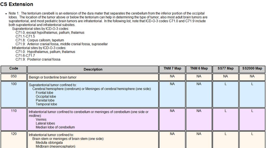
14 Childhood Brain Tumors Tentorium - extension of the dura mater separating the cerebellum from the occipital lobes 14 50% of childhood brain and CNS tumors are infratentorial, originating below the tentorium 20+% of childhood CNS tumors are located in the sellar or suprasellar region around the sella turcica (the bone that contains the pituitary gland) Remainder of tumors occur in spinal cord, brain stem, cranial nerves, etc.

15 Childhood Brain Tumors Supratentorial - childhood Craniopharyngiomas. Diencephalic and hypothalamic gliomas. Germ cell tumors. Low-grade astrocytomas. Anaplastic astrocytomas. Glioblastoma multiforme. Mixed gliomas. Oligodendrogliomas. Primitive neuroectodermal tumors. Low-grade or anaplastic ependymomas. Meningiomas. Choroid plexus tumors. Infratentorial - childhood Cerebellar astrocytomas (usually high-grade). Medulloblastomas (primitive neuroectodermal tumors). Ependymomas (low-grade or anaplastic). Brain stem gliomas (high-grade or low-grade). Atypical teratoid tumors 15
16 Pilocytic Astrocytoma Synonyms include: Juvenile pilocytic astrocytoma Cystic cerebellar astrocytoma Juvenile pilomyxoid astrocytoma 16 Characteristics: Usually slow growing, well-circumscribed neoplasm Associated with the formation of a single (or multiple) cyst(s) Arise in cerebellum near brainstem Other common sites include hypothalamic region and optic chiasm May occur in cerebral hemispheres and spinal cord Associated with neurofibromatosis Type 1 (NF1) 10 year survival greater than 90% with total removal Not associated with recurrence with total removal WHO Grade I - benign
17 Pilocytic Astrocytoma HOWEVER, when the ICD-O-3 was published, the behavior code for pilocytic astrocytoma downgraded from /3 (malignant behavior) to 1 (borderline behavior) as it still appears in the ICD-O-3 reference sitting on your desktop. Registrars in the United States were in 2000 and continue to be instructed by our national standard setting agencies to assign the behavior code /3 to these tumors despite the WHO downgrade. Rationale: To ensure complete reporting and data consistency, registrars should continue to assign the malignant behavior code (3) to pilocytic astrocytoma. This is the standard for all U.S. registries in all programs. 17 Confusing to researchers and public health studies since we reference ICD-O as our primary coding reference and ICD-O-3 has never published the U.S. change and does not assign a malignant behavior to this type of astrocytoma.

18 Causes and Risk Factors No single risk factor accounting for the majority of brain tumors has been identified even though many environmental and genetic factors are being studied 18
19 Causes and Risk Factors ENVIRONMENTAL Many studies have examined a wide spectrum of environmental factors as a cause for brain tumors. Of the long list of factors studied, only exposure to ionizing radiation has consistently been shown to put one at increased risk for developing a brain tumor. GENETIC There are a few rare genetic syndromes that involve brain tumors. NF1 (NF1 gene) NF2 (NF2 gene) Turcots (APC gene) Gorlins (PTCH gene) Tuberous sclerosis (TSC1 and TSC2 genes) Li-Fraumeni syndrome (TP53 gene) 19
20 Range of tumors and symptoms There are over 120 different types of brain/cns tumors. CNS tumors are associated with a range of symptoms and complications such as edema, seizures, endocrinopathy, fatigue, psychiatric disorder, venous thromboembolism that can seriously impact quality of life. Symptoms depend very much on the size and location of the tumor. General symptoms include persistent headaches which tend to be worse with activity, at night or early in the morning, convulsions, vomiting, subtle changes in personality, memory, mental ability, drowsiness, lethargy. 20 SEER Training Modules

21 21 Range of tumors and symptoms
22 Range of tumors and symptoms Symptoms are often location specific or provide clues Symptoms on the right side of the body may occur if the tumor is located on the left side of the brain and vice-versa. The speech center in most people is on the left side of the brain. Symptoms of a tumor located here may include difficulty saying correct words while still capable of understanding what is being said. If the tumor is located in the frontal lobe which controls intellectual function, thought process, behavior and memory, those activities may be affected. Similarity to closed head injury victims (motorcycle crash). 22 SEER Training Modules
23 Midline Shift and Mass Effect The bony cranium protects the brain from outside impacts to the head. When swelling occurs in the brain, there isn t much give. The swelling results in intracranial pressure and can cause a number of effects that begin to impact quality of life and comfort for the patient. 23 The easiest way to describe midline shift is to bring to mind siting in a movie theater. As soon as the person to one side of you puts his elbow onto the shared armrest between you, you tend to shift away. Source: Medscsape

24 Midline Shift and Mass Effect Midline is a central boundary separating the left and right hemispheres. Midline Shift Tumor crosses the brain to shift across the center line Mass Effect is Edema or swelling causes the brain to shift across center line 24 Both create new symptoms at cross-over Depends on the size and location of he tumor and level of spread Edema caused by many things Either cause pushes midline out of alignment Source: Medscsape

25 The Brain is Incapable of Feeling Pain Surgeons are able to cut living brains without fear of hurting their patients However, symptoms from tumors and their effect within the cranial cavity on various functions of the brain is a different story, altogether. Much is dependent upon tumor location and infiltration 25 Source: National Geographic, couretsy of Fred Hossler/Getty Images

26 Benign/Borderline/Malignant??? 26 Source: American Brain Tumor Association Facts and Statistics
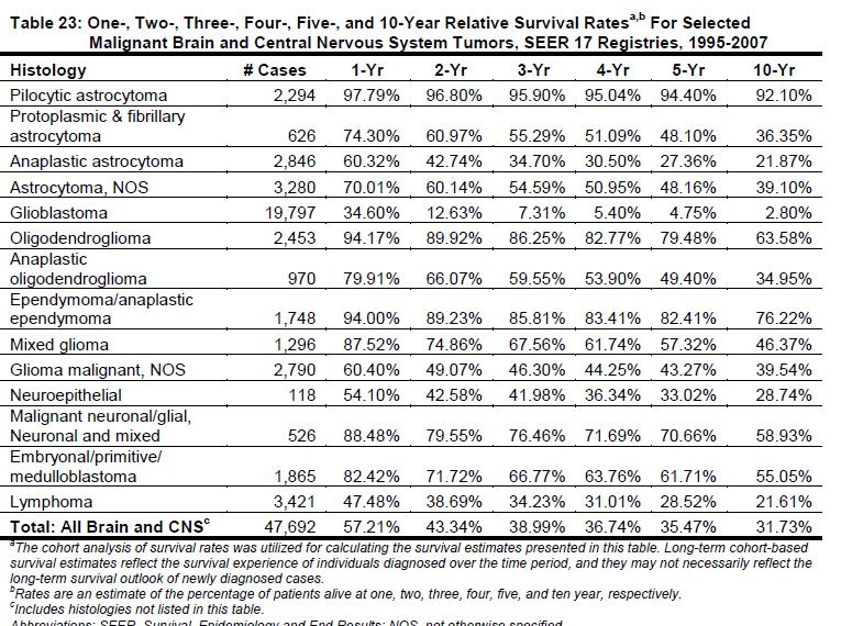
27 Survival Trends SEER data from year relative survival Males 34% Females 38% Children age 0-19 have the highest 5-year relative survival rate 72% The survival rate diminishes as age increases, down to 5% for persons age 75 and older 27
28 28
29 Tumor-Specific Statistics Meningioma Glioma (80% of all malignant brain tumors) 34% of all primary brain tumors 31% of all primary brain tumors Glioblastoma 17% of all primary brain tumors (54% of all gliomas) Astrocytoma 7% of all primary brain tumors Oligodendroglioma 2% of all primary brain tumors Ependymoma 1% of all primary brain tumors 29 Pituitary tumors Nerve sheath tumors Medulloblastoma/embryonal/and other tumors of primitive (developmental) nerve origin Lymphoma 13% of all primary brain tumors 9% of all primary brain tumors (ie: acoustic neuromas, schwannoma, malignant peripheral nerve sheath tumor) 3% of all primary brain tumors 2-3% of all primary brain tumors

30 30

31 31 Source: wikipedia.org and ccrcal.org
32 32
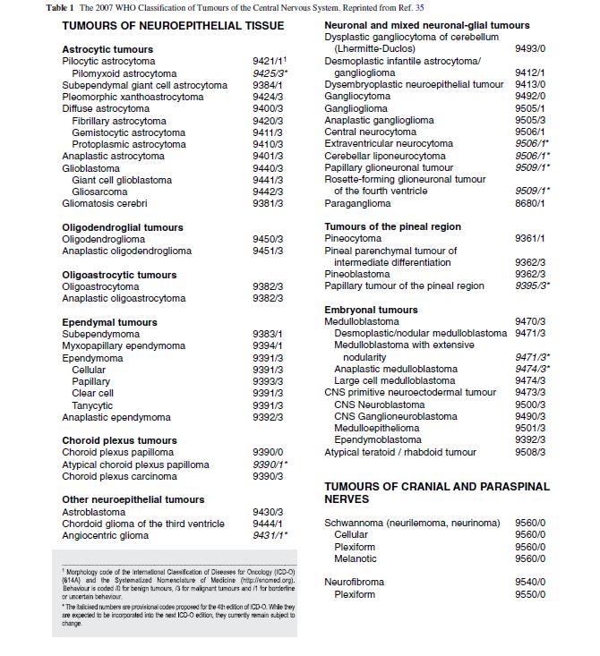
33 WHO Classification Groups Tumors of Neuroepithelial Tissue Tumors of Cranial and Paraspinal Nerves Tumors of Meninges Lymphomas and Hematopoietic Malignancies Germ Cell Tumors Tumors of the Sellar Region Metastatic Tumors 33
34 34
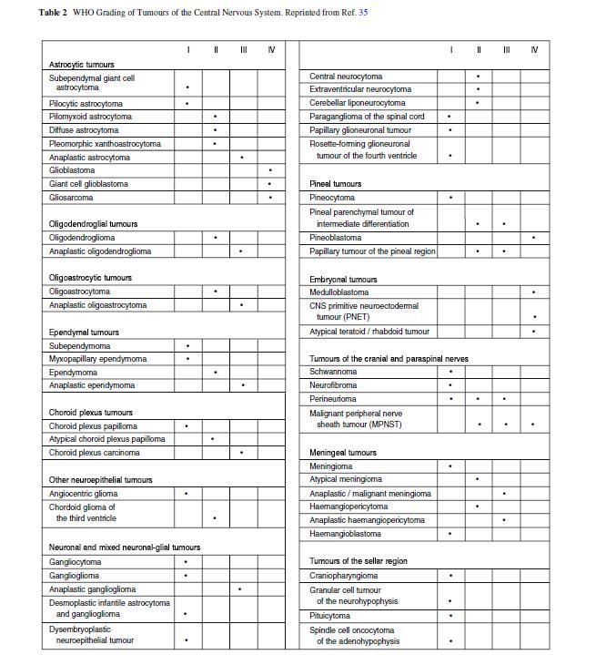
35 Four categories of tumor Grade I Grade II Grade III WHO Grade slow growing, non-malignant, associated with longterm survival benign tumors relatively slow-growing, sometimes recur as higher grade tumors, can be malignant or non-malignant (borderline malignant) malignant and often recur as higher grade tumors Grade IV reproduce rapidly and are very aggressive malignant tumors WHO grade is not recorded as part of the histology WHO grade is used by the clinician to plan treatment and predict prognosis 35
36 36

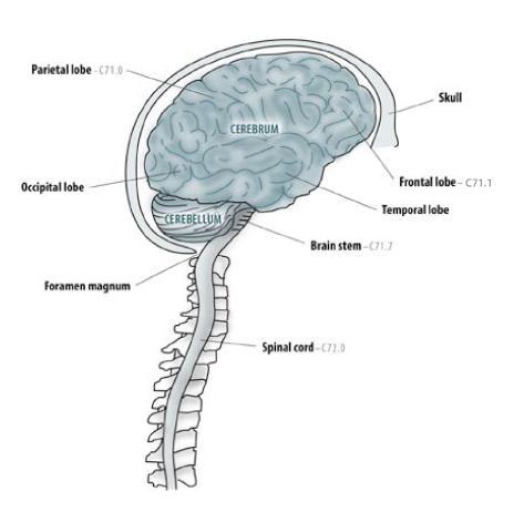
37 ANATOMY OF THE HUMAN BRAIN 37 Source: National Geographic, couretsy of Fred Hossler/Getty Images
38 THE HUMAN BRAIN The brain is the largest intracranial organ The brain is a 3-pound mass of jelly-like fats and tissues It is the most complex of all known living structures The skull or cranium is bone that covers the brain Up to one trillion nerve cells working together coordinate the physical actions and mental processes (voluntary and involuntary) that set humans apart from all other species 38 Source: CDC Data Collection of Primary CNS Tumors, NPCR Training Materials 2004
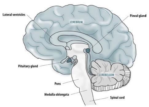
39 The CNS includes both intracranial sites (inside the cranium) and extra-cranial sites (outside the cranium). The pituitary gland, craniopharyngeal duct and pineal gland are found inside, alongside brain tissue Cranial nerves directly link to brain tissue The spinal cord is part of the CNS though not intracranial Any tumor that originates in the brain, spinal cord, the cranial nerves, one of the glands/ducts within the cranium (pineal, pituitary, craniopharyngeal), or the lining of the cranium (meninges) is reportable regardless of behavior (benign, borderline, or malignant). 39 Source: CDC Data Collection of Primary CNS Tumors, NPCR Training Materials 2004

40 40 Source: University of Illinois

41 41 ICD-O Topography Codes (Anatomic Site)
42 42
43 43

44 Ventricular System of the Brain Source: solarnavigator.net/human_brain 44


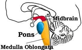
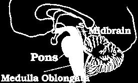
45 45 Meninges and Brain Stem

46 46 Cranial Nerves
47 Cranial Nerve Functions Cranial Nerve: I Olfactory II Optic III Oculomotor IV Trochlear V Trigeminal VI Abducens VII Facial VIII Vestibulocochlear IX Glossopharyngeal X Vagus XI Spinal Accessory XII Hypoglossal Major Functions: smell vision eyelid and eyeball movement turns eye downward and laterally, controls superior oblique muscles chewing, face & mouth touch & pain turns eye laterally facial expressions, taste, tears, saliva Also referred to as Auditory Nerve: hearing, equilibrium sensation Taste, senses carotid blood pressure aortic blood pressure, heart rate, stimulates digestive organs, taste controls trapezius & sternocleidomastoid muscles, controls swallowing controls tongue movements 47
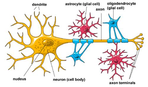
48 Characteristics of Brain Tumors Start in the brain and grow steadily there. Very rarely spread to other organs through the bloodstream. Are named for the cells from which they arise, each having a certain function essential to normal physiological functioning of the brain. For example: Gliomas arise from glial cells which support the CNS. Astrocytomas arise from astrocytes Ependymomas arise from ependymal cells which line the ventricles (fluid filled spaces within the brain) or central canal of the spinal cord. Oligodendrogliomas arise from oligodentdrocyte cells which make up the fatty substance called myolin that covers nerves like electrical insulation. Brain Stem Gliomas arise in the lowest part of the brain. 48

49 Characteristics of Brain Tumors 49 Source: medicalgeek.com/indian-post-graduate-exams
50 Histologic Type - Glioma Most common category of primary brain tumors. They begin in glial cells (supporting cells of the CNS) Often spread into surrounding brain tissue along nerve fibers invading the spaces between nearby normal brain cells. Some invade the surrounding brain more than others. Difficulty obtaining complete surgical removal. MRI scans show the largest part of the glioma, but cannot reliably show areas of the brain where tumor cells have invaded. Aggressive efforts to remove small numbers of tumor cells within the brain could cause loss of neurologic function. When it is not possible to remove the entire glioma, post-op radiation therapy and chemotherapy may be advised. Even with maximum safe resection followed by radiation and chemotherapy, gliomas can grow back. 50
51 Glioma 3 Main Histologic Types 1. Astrocytoma: In adults most often arise in the cerebrum. In children they occur in the brain stem, cerebrum and cerebellum. Rarely in brain stem in adults. Felt to be most aggressive of brain tumors. Grade I and II astrocytomas are low-grade astrocytomas. Grade III astrocytoma is an anaplastic astrocytoma. Grade IV astrocytoma is a glioblastoma multiforme. 51
52 Glioma 3 Main Histologic Types 2. Oligodroglioma: Rare tumor that usually occurs in the cerebrum, grows slowly and usually does not spread into surrounding brain tissue like astrocytoma does. Most common in middle-aged adults. 3. Ependymoma: Most commonly arise in children and young adults. They are also seen with neurofibromatosis Type II. (which we will discuss in a bit) 52


53 Glioma Other Subtypes There are other subtypes of gliomas, each with their own specific characteristics and modes of growth. Brain Stem Glioma Juvenile Pilocytic Astrocytoma Pleomorphic Xanthoastrocytoma Subependymoma Ganglioglioma 53
54 54 Glioma Tumor Markers
55 Non-Glial Tumors Medulloblastoma: Usually arises in the cerebrum, is the most common brain tumor in children, and is sometimes called a primitive neuroectodermal tumor or PNET. Meningioma: Arises from the meninges which are the outside coverings of the brain between the skull and the brain itself. It usually presses on the brain, but does not invade it and often grows slowly. 55
56 Non-Glial Tumors Schwannoma: Arises from Schwann cells present in certain nerves, including those that control balance and hearing. A common site is the vestibular nerve which carries signals from the inner ear to the brain stem. Tumors in this location are called acoustic neuromas (a.k.a. vestibular schwannoma), and occur most often in adults. 56
57 Non-Glial Tumors Craniopharyngioma: Grows at the base of the brain, arises from the tissue connecting the brain and the pituitary gland and occurs in both adults and children. 57 Pituitary Adenoma: Arises from the pituitary gland and may cause compression of the optic nerves causing vision problems. Some produce excessive amounts of hormones that can disrupt the body s metabolism. Roswell Park Cancer Insitute

58 Observing Migration of Glioma Cells 58 Source: Case Western Reserve University School of Medicine, public release 8/25/11
59 Neurofibromatosis The neurofibromatoses (NF) are a group of genetic disorders which cause tumors to grow along nerves and can also affect the development of non-nervous tissues such as bones and skin. Neurofibromatosis Type I (NF-I), also known as Peripheral NF and historically as von Recklinghausen Disease Occurs in 1:4,000 births Multiple cafe-au-lait spots (not reportable) Many, many neurofibromas on or under the skin (not reportable) Enlargement and deformation of bones and curvature of the spine Tumors may develop in brain, on cranial nerves, or the spinal cord 59 Neurofibromatosis Foundation

60 NF Type I: First documented photo Source Credit: Dr. Stanley B. Burns


61 Other Manifestions of NF Type I Lisch nodules on the eye Melanocytic hemartomas Café-au-lait spots on skin Discolored birth marks 61 Medscape Source: Dermnet.com; Dermatologic Manifestations of NF Type I
62 Neurofibromatosis Type II Neurofibromatosis Type II (NFII), also known as Multiple Inherited Schwannomas, Meningiomas and Ependymomas (MISME) or Bilateral Acoustic Neurofibromatosis (BAN ). Is a genetically inherited diseasecaused by mutations of the "Merlin" gene, which appears to influences the form and movement of cells Primary manifestation is a development of non-malignant brain tumors in the region of the cranial nerves, frequently bilaterally. The eighth cranial nerve is the auditory-vestibular nerve which transmits sensory information from the inner ear to the brain and is commonly affected. 62 Source: California Ear Institute

63 Acoustic Neuroma/Schwannoma 63 Source:


64 64 Multiple Primary Rules Histology Coding Rules

65 Different Rules for Benign and Malignant 65
66 66 Sequence Numbering for Brain Tumors Malignant primary brain and CNS tumors are assigned Sequence Codes in the range Sequence Chronologically Only count malignant tumors in the sequence If only one malignant tumor occurs, it is coded 00 If subsequent (multiple) primary malignant and/or in situ neoplasms, the sequence number for the first tumor begins at 01, the sequence number for the second primary tumor is 02, and so forth. Non-malignant primary brain and CNS tumors are assigned Sequence Codes in the range Sequence Chronologically Only count benign/borderline or reportable by agreement neoplasms in the sequence If only one non-malignant tumor occurs, it is coded 60. If subsequent (multiple) non-malignant neoplasms are diagnosed, the first tumor should be sequenced as 61, the second 62 and so forth.
67 67 Benign and Borderline Tumor Rules
68 Benign and Borderline Tumor Rules When multiple tumors are present registrars should identify and document specific characteristics for MPH Rules Text Date of Diagnosis (Timing is not used to determine number of abstracts or primary neoplasms to abstract) Method and Details of Diagnosis (some are never resected) Location of Tumor Laterality Histologic Type refer to Chart 1 Tumor Behavior Multiple Meningioma s (meningiomatosis) Neurofibromatosis Characteristics (when applicable) 68
69 69 Malignant Tumor Rules
70 70 Malignant Tumor Rules
71 Malignant Tumor Rules When multiple tumors are present registrars should identify and document specific characteristics for MPH Rules Text Date of Diagnosis (Timing is not used to determine number of abstracts or primary neoplasms to abstract) Method and Details of Diagnosis (most attempt resection) Location of Tumor (not spread or invasion but bulk of tumor) Histologic Type refer to Chart 1 and/or Chart 2 Tumor Behavior Variations or Combinations of One or More Glial Tumors Over Lifetime astrocytoma, glioblastoma, ependymoma, or oligodendroglioma Special rules for determining # abstracts Special rules for determining whether or not is mixed glioma Note: Recurrence, progression, or any reappearance of histologies on the same branch in Chart 1 or Chart 2 is always the same disease process. 71
72 Report /Sequence All Tumors Over Lifetime REMINDER: Sequence numbers for malignant neoplasms and for benign, borderline, and other reportable-by-agreement cases are assigned over a lifetime. Therefore, IF A PATIENT WAS DIAGNOSED WITH A NON-MALIGNANT CNS NEOPLASM BEFORE REPORTING WAS REQUIRED (January 1, 2004), THE NEW (SECOND) NEOPLASM SHOULD BE ASSIGNED SEQUENCE NUMBER 62 AND THE FIRST NEOPLASM (Seq 61) IS REPORTABLE AS A HISTORICAL CASE TO FCDS. (An abstract/accession is not be required by CoC or SEER but is by FCDS) Any benign and/or borderline brain or CNS tumor(s) diagnosed before January 1, 2004 ARE REPORTABLE TO FCDS as historical cases when accompanied by another reportable primary. 72 FCDS Data Acquisition Manual and CDC Data Collection of Primary Central Nervous System Tumors

73 2013 *2013* 73

74 74

75 75
76 76
77 77
78 78
79 Treatment
80 Surgical Option(s) Decisions regarding aggressiveness of surgery for primary brain lesions are complex and depend on the: Age and performance status of the patient Proximity to eloquent areas of the brain Feasibility of decreasing the mass effect with aggressive surgery Resectability of the tumor (including the number and location of lesions) In patients with recurrent disease, the time since the last surgery Surgical options include: Stereotactic biopsy Open biopsy or debulking procedure Subtotal resection Maximal safe resection 80

81 Craniotomy Any bony opening that is cut into the skull. 81 Source: Mayfield Clinic

82 Craniotomy Procedure A section of the skull, (called a bone flap) is removed to access the brain underneath. Typically the bone flap is replaced. If the flap is not replaced, the procedure is called a craniectomy 82 Source: MedlinePlus/US National Library of Medicine, NIH

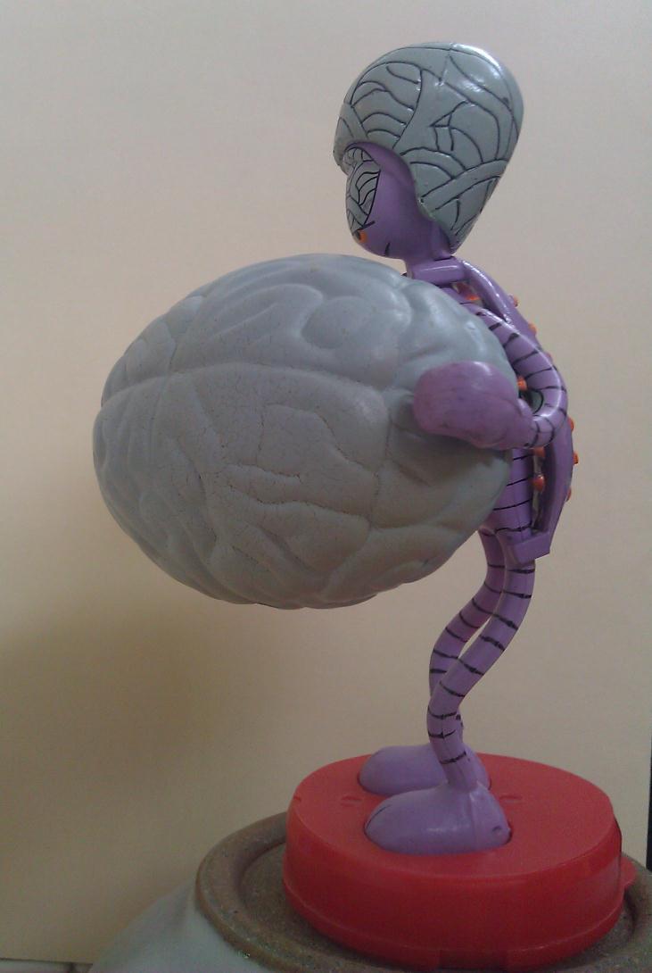
83 Surgeon has drawn the cutline circle around the tumor location 83 Source: The Alien-a set on Flickr

84 Surgeon has cut the scalp and pulled it back to expose the skull over the tumor 84 Source: The Alien-a set on Flickr

85 The skull is removed revealing the dura layer under which is the brain and tumor 85 Source: The Alien-a set on Flickr

86 Here you see the circular cut through the dura layer with the brain and tumor exposed. 86 Source: The Alien-a set on Flickr

87 Meningioma Resected 87 Source: The Alien-a set on Flickr

88 Pre- and Post-Operative Imaging Pre-op tumor is outlined in red Post-operative MRI shows complete resection of the tumor 88 Source: Desert Spine and Neurosurgical Institute
89 89 Surgery Codes
90 90 Surgery Codes
91 Radiation Therapies Primary XRT for brain tumors includes tumor volume/margins Tumor volume is defined by pre- and post-operative imaging Standard fractionated external beam radiation is most common Hypofractionation (daily dose given in smaller increments with 4 or 6 hours between treatments) is an emerging option Whole brain XRT and stereotactic radiosurgery for brain mets 91 Source: NCCN
92 Tumor Volume The larger the brain tumor, the more desirable fractionation (e.g. multiple smaller treatments, rather than one big one) Tumor size can determine schedule for fractionation and dose/tx Why: The "shell" of normal tissue outside the tumor volume will receive some part of the dose. For larger tumors, this "shell" volume increases rapidly as a function of tumor diameter Why: Fractionation spares this "shell" of normal tissue much more effectively than the single "shot" techniques 92 Source: Johns Hpkins Medicine
93 Stereotactic Radiosurgery (SRS) Despite name, SRS is an XRT treatment, not a surgical procedure Acoustic neuroma, pituitary tumors, spinal cord tumors and brain metastasis are candidates for this technique Special equipment focuses up to 200 beams of radiation on tumor Although each beam has very little effect on the brain tissue it passes through, a strong dose of radiation is delivered to the site where all the beams meet. Results in minimal damage to healthy tissues surrounding target. 93 Zdpirce Mayo Clinic

94 94 Source: San Diego Gamma Knife Center
95 Chemotherapy Chemotherapy is not an effective initial treatment for low-grade brain tumors. Why? Because most standard chemo agents have a hard time passing into the brain because of how the brain protects itself (the blood-brain barrier) Not all types of brain tumors respond to chemotherapy In general, chemotherapy for brain tumors is usually administered following surgery or radiation therapy Participation in clinical trials should be encouraged 95
96 Blood Brain Barrier Composed of special cells that make up brain s blood vessels Selectively prevents substances from entering the blood and brain, only allowing essential molecules such as amino acids, oxygen, glucose and water through Adenosine, a molecule produced by the body, seems to modulate the entry of large molecules into the brain When adenosine receptors are activated on cells that comprise the blood-brain barrier, a gateway into the barrier can be established 96 Science Daily Source: September 13, 2001
97 Approved Chemotherapy Agents Carmustine (BCNU) IV or dissolvable wafers placed surgically Temozolomide (Temodar) oral Lomustine (CCNU) oral Carboplatin Cisplatin Etoposide Irinotecan Vincristine Procarbazine (Matulane) oral Methotrexate - oral, by injection or intrathecally 97
98 NCCN Treatment Guidelines
99 Infiltrative Low-Grade Glioma Best management strategy has yet to be defined Small tumor samples can provide a lower histologic grade Rationale: Needle biopsies are often performed when lesions are in deep or critical regions of the brain, but can be misleading because gliomas often have varying degrees of cellularity, mitosis, or necrosis from one region to another 99 General recommendation is to first attempt as complete an excision of tumor as possible (based on postsurgical MRI verification) without compromising function No consensus exists regarding proper timing of postoperative external beam radiation Chemotherapy is not a traditional upfront treatment modality
100 Infiltrative Low-Grade Glioma When possible, maximal safe resection If gross total resection is achieved, some patients may be observed without adjuvant therapy. However, close follow-up is essential as over half of patients will eventually progress These tumors behave aggressively in patients over 40 years old Adjuvant radiation or chemotherapy is recommended 100 If stereotactic biopsy, open biopsy, or other subtotal excision was done, immediate fractionated external beam RT or chemotherapy should be given
101 Anaplastic Glioma and Glioblastoma Whenever possible, major tumor removal should be performed If glioblastoma is confirmed, options include radiation, chemotherapy, best supportive care, chemoradiation only if carmustine wafer was implanted If high-grade glioma is confirmed, BCNU wafer is an option In patients with good Karnofsky score (70 or above) options include fractionated external beam radiation therapy, chemotherapy or chemoradiation in the context of a clinical trial In patients with poor Karnofsky score (below 70) management may include radiation, chemotherapy or best supportive care 101
102 Intracranial Ependymoma Whenever possible, maximal safe resection should be attempted Adjuvant treatment depends on the extent of surgical resection, histology and staging by cranial spinal MRI and CSF cytology CSF dissemination occurs in up to 15% of intracranial ependymomas If MRI spine /CSF reveal disease, craniospinal radiation is mandatory If gross total resection with negative spinal MRI and CSF, adjuvant regional fractionated EBRT or observation may be considered 102
103 Medulloblastoma and PNET (supratentorial) MRI is the gold standard to assess PNET Maximal safe resection is recommended when possible Average Risk Patients: craniospinal radiation alone or concurrent chemoradiation followed by chemotherapy are both options High Risk Patients: patients with large cell or anaplastic medulloblastoma, supratentorial PNET, disease dissemination, unresectable tumors, or residual tumors over 1.5cm post-surgery are high risk and should undergo radiation followed by chemotherapy 103
104 Primary CNS Lymphoma Treatment to be initiated as immediately following diagnosis Treatment options depend on patient overall health and age For healthier patients a high-dose methotrexate regimen RT after systemic treatment depends on the responsiveness of the disease to the chemotherapy However, one or both may increase neurotoxicity, especially in patients older than 60 years of age 104
105 Primary Spinal Cord Tumors MRI is the gold standard for diagnosis of spinal cord lesions Asymptomatic patients may be observed or resected Symptomatic patients should undergo some form of surgery Maximal safe resection should be attempted Post-operative adjuvant radiation is not recommended However, if symptoms persist after incomplete resection or biopsy, radiation should be administered 105
106 Meningioma Meningiomas are typically diagnosed by CT or MRI imaging Biopsy may be considered for confirmation Options stratified by presence/absence of symptoms and tumor size Most asymptomatic patients with small tumors (<30mm) may just be observed. If neurological impairment is imminent, surgery (if accessible) or radiotherapy (EBRT OR SRS) is feasible Asymptomatic tumors >30mm can be either resected or observed 106
107 Meningioma Symptomatic disease requires active treatment by surgery if possible Non-surgical candidates should undergo radiation All patients with surgically resected grade III meningiomas (even after gross total resection) should receive adjuvant radiation for local control regardless of tumor size and symptom status 107
108 Additional Resources NCCN Evidence Based Treatment Guidelines, nccn.org, 2011 Collaborative Stage Data Collection System, AJCC, 2010 Multiple Primary and Histology Coding Rules, SEER 2007 The 2007 WHO Classification of Tumours of the Central Nervous System, David N. Louis, Hiroko Ohgaki, Otmar D. Wiestler, Webster K. Cavenee, Peter C. Burger, Anne Jouvet, Bernd W. Scheithauer and Paul Kleihues, World Health Organization, Lyon, France, 2007 Data collection of primary central nervous system tumors. National Program of Cancer Registries Training Materials. Department of Health and Human Services, Centers for Disease Control and Prevention. Atlanta, Georgia,
109 109
Brain and CNS Tumors
 Brain and CNS Tumors FCDS 2011/2012 Educational Webcast Series January 19, 2012 Lynne Pearson, BHS, CTR, LHRM Steven Peace, BS, CTR Updated for 2012 Requirements and CSv02.03.02 Presentation Outline Overview
1/17/2012. Brain and CNS Tumors. Presentation Outline. Overview. Overview. Anatomy of the Human Brain. Multiple Primary and Histology Coding Rules
 Brain and CNS Tumors FCDS 2011/2012 Educational Webcast Series January 19, 2012 Lynne Pearson, BHS, CTR, LHRM Steven Peace, BS, CTR Updated for 2012 Requirements and CSv02.03.02 Presentation Outline Overview
Neoplasms of the BRAIN and CNS
 Neoplasms of the BRAIN and CNS 2015-21016 FCDS Educational Webcast Series Steven Peace, BS, CTR October 15, 2015 2015 Focus Anatomy SSS 2000 MPH Rules AJCC TNM Presentation Outline Overview Reportable
2019 Neoplasms of the BRAIN/CNS. CDC & Florida DOH Attribution
 2019 Neoplasms of the BRAIN/CNS 2018-2019 FCDS Educational Webcast Series Steven Peace, BS, CTR March 21, 2019 CDC & Florida DOH Attribution Funding for this conference was made possible (in part) by the
Brain tumors: tumor types
 Brain tumors: tumor types Tumor types There are more than 120 types of brain tumors. Today, most medical institutions use the World Health Organization (WHO) classification system to identify brain tumors.
Bellringer: The central nervous system is comprised of: What is the name of the outermost layer of the brain? a. Brain. b.
 Bellringer: The central is comprised of: a. Brain b. Spinal cord c. Sensory receptors d. Both a and b What is the name of the outermost layer of the brain? a. Pia mater b. Dura mater c. Arachnoid d. Pons
Q&A. Fabulous Prizes. Collecting Cancer Data:CNS 2/7/12. NAACCR Webinar Series Collecting Cancer Data Central Nervous System
 Collecting Cancer Data Central Nervous System NAACCR 2012 2013 Webinar Series 2/7/2013 Q&A Please submit all questions concerning webinar content through the Q&A panel. Reminder: If you have participants
Tumors of the Nervous System
 Tumors of the Nervous System Peter Canoll MD. PhD. What I want to cover What are the most common types of brain tumors? Who gets them? How do they present? What do they look like? How do they behave? 1
CNS TUMORS. D r. Ali Eltayb ( U. of Omdurman. I ). M. Path (U. of Alexandria)
. M. Path (U. of Alexandria)") CNS TUMORS D r. Ali Eltayb ( U. of Omdurman. I ). M. Path (U. of Alexandria) CNS TUMORS The annual incidence of intracranial tumors of the CNS ISmore than intraspinal tumors May be Primary or Secondary
CNS TUMORS D r. Ali Eltayb ( U. of Omdurman. I ). M. Path (U. of Alexandria) CNS TUMORS The annual incidence of intracranial tumors of the CNS ISmore than intraspinal tumors May be Primary or Secondary
Peter Canoll MD. PhD.
 Tumors of the Nervous System Peter Canoll MD. PhD. What I want to cover What are the most common types of brain tumors? Who gets them? How do they ypresent? What do they look like? How do they behave?
The Nervous System: Central Nervous System
 The Nervous System: Central Nervous System I. Anatomy of the nervous system A. The CNS & the body by: 1. monitoring of the body 2. & information between parts of the body 3. acting as a to gather, store,
Site Specific Coding Rules MALIGNANT CENTRAL NERVOUS SYSTEM TUMORS
 Multiple Primary and Histology Site Specific Coding Rules MALIGNANT CENTRAL NERVOUS SYSTEM TUMORS 1 Prerequisites 2 Completion of Multiple Primary and Histology General Coding Rules 3 There are many ways
General: Brain tumors are lesions that have mass effect distorting the normal tissue and often result in increased intracranial pressure.
 1 Lecture Objectives Know the histologic features of the most common tumors of the CNS. Know the differences in behavior of the different tumor types. Be aware of the treatment modalities in the various
Meningioma tumor. Meningiomas are named according to their location (Fig. 1) and cause various symptoms: > 1
 and cause various symptoms: > 1") Meningioma tumor Overview A meningioma is a type of tumor that grows from the protective membranes, called meninges, which surround the brain and spinal cord. Most meningiomas are benign (not cancer) and
Meningioma tumor Overview A meningioma is a type of tumor that grows from the protective membranes, called meninges, which surround the brain and spinal cord. Most meningiomas are benign (not cancer) and
Principles of Anatomy and Physiology
 Principles of Anatomy and Physiology 14 th Edition CHAPTER 14 The Brain and Cranial Nerves Introduction The purpose of the chapter is to: 1. Understand how the brain is organized, protected, and supplied
Adult Brain and Spinal Cord Tumors
 Adult Brain and Spinal Cord Tumors An adult central nervous system (CNS) tumor is a disease in which abnormal cells form in the tissues of the brain and or the spinal cord. Major Parts of the Brain Anatomy
Tumors of the Central Nervous System
 Tumors of the Central Nervous System 1 Financial Disclosures I have NO SIGNIFICANT FINANCIAL, GENERAL, OR OBLIGATION INTERESTS TO REPORT Introduction General: Brain tumors are lesions that have mass effect
ACTIVITY 7: NERVOUS SYSTEM HISTOLOGY, BRAIN, CRANIAL NERVES
 ACTIVITY 7: NERVOUS SYSTEM HISTOLOGY, BRAIN, CRANIAL NERVES LABORATORY OBJECTIVES: 1. Histology: Identify structures indicated on three different slides or images of nervous system tissue. These images
CNS pathology Third year medical students. Dr Heyam Awad 2018 Lecture 12: CNS tumours 2/3
 CNS pathology Third year medical students Dr Heyam Awad 2018 Lecture 12: CNS tumours 2/3 Pilocytic astrocytoma Relatively benign ( WHO grade 1) Occurs in children and young adults Mostly: in the cerebellum
NON MALIGNANT BRAIN TUMOURS Facilitator. Ros Taylor Advanced Neurosurgical Nurse Practitioner Southmead Hospital Bristol
 NON MALIGNANT BRAIN TUMOURS Facilitator Ros Taylor Advanced Neurosurgical Nurse Practitioner Southmead Hospital Bristol Neurosurgery What will be covered? Meningioma Vestibular schwannoma (acoustic neuroma)
The Nervous System PART B
 7 The Nervous System PART B PowerPoint Lecture Slide Presentation by Jerry L. Cook, Sam Houston University ESSENTIALS OF HUMAN ANATOMY & PHYSIOLOGY EIGHTH EDITION ELAINE N. MARIEB Central Nervous System
Brain and Cranial Nerves (Ch. 15) Human Anatomy lecture. caudal = toward the spinal cord)
 Human Anatomy lecture. caudal = toward the spinal cord)") Insight: Some cranial nerve disorders Brain and Cranial Nerves (Ch. 15) Human Anatomy lecture I. Overview (Directional terms: rostral = toward the forehead caudal = toward the spinal cord) A. 3 Major parts
Insight: Some cranial nerve disorders Brain and Cranial Nerves (Ch. 15) Human Anatomy lecture I. Overview (Directional terms: rostral = toward the forehead caudal = toward the spinal cord) A. 3 Major parts
What is Brain Cancer? What is the brain?
 What is Brain Cancer? The brain and spinal column make up the central nervous system (CNS), where all vital functions of the body are controlled. When tumors arise in the central nervous system, they are
What are brain and spinal cord tumours? Contents
 13 11 20 Information and support What are brain and spinal cord tumours? Contents The brain and spinal cord Brain function What is a brain or spinal cord tumour? What types of tumours are there? How common
The Nervous System PART C. PowerPoint Lecture Slide Presentation by Patty Bostwick-Taylor, Florence-Darlington Technical College
 PowerPoint Lecture Slide Presentation by Patty Bostwick-Taylor, Florence-Darlington Technical College The Nervous System 7 PART C Protection of the Central Nervous System Scalp and skin Skull and vertebral
MALIGNANT GLIOMAS: TREATMENT AND CHALLENGES
 MALIGNANT GLIOMAS: TREATMENT AND CHALLENGES DISCLOSURE No conflicts of interest to disclose Patricia Bruns APRN, CNS Givens Brain Tumor Center Abbott Northwestern Hospital October 12, 2018 OBJECTIVES THEN
Oligodendrogliomas & Oligoastrocytomas
 Oligodendrogliomas & Oligoastrocytomas ABOUT THE AMERICAN BRAIN TUMOR ASSOCIATION Founded in 1973, the American Brain Tumor Association (ABTA) was the first national nonprofit organization dedicated solely
Nervous System: An Introduction. HAP Susan Chabot Lemon Bay High School
 Nervous System: An Introduction HAP Susan Chabot Lemon Bay High School Function of the Nervous System 3 overlapping functions SENSORY INPUT - Monitor changes inside and outside of the body; these changes
Site Specific Coding Rules Benign and Borderline Intracranial and CNS Tumors
 Multiple Primary and Histology Site Specific Coding Rules Benign and Borderline Intracranial and CNS Tumors 1 Prerequisites 2 Completion of Multiple Primary and Histology General Coding Rules 3 There are
Adult Central Nervous System Tumors Treatment (PDQ )
") 1 di 20 28/06/2016 11.18 NCBI Bookshelf. A service of the National Library of Medicine, National Institutes of Health. PDQ Cancer Information Summaries [Internet]. Bethesda (MD): National Cancer Institute
1 di 20 28/06/2016 11.18 NCBI Bookshelf. A service of the National Library of Medicine, National Institutes of Health. PDQ Cancer Information Summaries [Internet]. Bethesda (MD): National Cancer Institute
AMERICAN BRAIN TUMOR ASSOCIATION. Oligodendroglioma and Oligoastrocytoma
 AMERICAN BRAIN TUMOR ASSOCIATION Oligodendroglioma and Oligoastrocytoma ACKNOWLEDGEMENTS ABOUT THE AMERICAN BRAIN TUMOR ASSOCIATION Founded in 1973, the American Brain Tumor Association (ABTA) was the
AMERICAN BRAIN TUMOR ASSOCIATION. Oligodendroglioma and Oligoastrocytoma
 AMERICAN BRAIN TUMOR ASSOCIATION Oligodendroglioma and Oligoastrocytoma ACKNOWLEDGEMENTS ABOUT THE AMERICAN BRAIN TUMOR ASSOCIATION Founded in 1973, the American Brain Tumor Association (ABTA) was the
Year 2003 Paper two: Questions supplied by Tricia
 question 43 A 42-year-old man presents with a two-year history of increasing right facial numbness. He has a history of intermittent unsteadiness, mild hearing loss and vertigo but has otherwise been well.
C h a p t e r PowerPoint Lecture Slides prepared by Jason LaPres North Harris College Houston, Texas
 C h a p t e r 15 The Nervous System: The Brain and Cranial Nerves PowerPoint Lecture Slides prepared by Jason LaPres North Harris College Houston, Texas Copyright 2009 Pearson Education, Inc., publishing
Brain Tumors: an Introduction basic level
 Brain Tumors: an Introduction basic level Overview A tumor (also called a neoplasm or lesion) is abnormal tissue that grows by uncontrolled cell division. Normal cells grow in a controlled manner as new
Histopathological Study and Categorisation of Brain Tumors
 Histopathological Study and Categorisation of Brain Tumors Ruchira Wadhwa 1*, Purvi Patel 2, Hansa Goswami 3 1 Third Year Resident, 2 Assistant Professor, 3 Professor and Head, Department of Pathology,
The Brain 3 Main Areas: Cerebrum, Cerebellum, Brain Stem. Scope. Cerebrum 3/22/2017. Disclaimer
 Disclaimer Metro-Detroit Oncology Nursing Society PRACTICE PEARLS UPDATE IN CNS MALIGNANCY Gayle Groshko RN BSN OCN Beaumont Health Radiation Oncology Nurse Case Manager I have no conflicts of interest.
Pediatric Brain Tumors: Updates in Treatment and Care
 Pediatric Brain Tumors: Updates in Treatment and Care Writer Classroom Rishi R. Lulla, MD MS Objectives Introduce the common pediatric brain tumors Discuss current treatment strategies for pediatric brain
Brain Tumors: an Introduction
 1 2 Brain Tumors: an Introduction Overview A tumor is an abnormal growth of cells. Brain tumors are named after the cell type from which they grow some are benign, others malignant.. They may be primary
Supra- and infratentorial brain tumors from childhood to maternity
 Supra- and infratentorial brain tumors from childhood to maternity What to expect? I am going to show you the characteristic imaging findings of following tumors: Thierry A.G.M. Huisman, MD, FICIS, EQNR
The Brain and Cranial Nerves Pg. 129
 The Brain and Cranial Nerves Pg. 129 Three Main Regions of the Brain Forebrain Cerbral hemispheres Diencephalon Midbrain Brain stem Hindbrain Pons Cerebellum Medulla oblongata Forebrain Interprets sensory
Cranial Nerve: eyelid and eyeball movement innervates superior oblique turns eye downward and laterally chewing face & mouth touch & pain
 Cranial Nerves Cranial Nerve: I Olfactory II Optic III Oculomotor IV Trochlear V Trigeminal VI Abducens VII Facial VIII Vestibulocochlear (auditory) IX Glossopharyngeal X Vagus XI Spinal Accessory XII
Please submit all questions concerning webinar content through the Q&A panel. Reminder:
 NAACCR 2015-2016 Webinar Collecting Cancer Data: Series Central Nervous System NAACCR Webinar Series 2016 2017 Carol Hahn Johnson Jim Hofferkamp jhofferkamp@naaccr.org Q&A Please submit all questions concerning
The dura is sensitive to stretching, which produces the sensation of headache.
 Dural Nerve Supply Branches of the trigeminal, vagus, and first three cervical nerves and branches from the sympathetic system pass to the dura. Numerous sensory endings are in the dura. The dura is sensitive
A&P 1 Brain & Cranial Nerves Guide #1 - Pre-Lab Exercises
 A&P 1 Brain & Cranial Nerves Guide #1 - Pre-Lab Exercises In this "Pre-lab Guide", we will be looking at the brain & cranial nerves. This should be done before lab, so we don't waste time in lab! This
Brain and Spine Tumors
 Brain and Spine Tumors Andrew J. Fabiano, MD FAANS Associate Professor of Neurosurgery Roswell Park Cancer Institute SUNY at Buffalo School of Medicine Brain Tumors Brain Tumor Basics Types of Tumors Cases
Collecting Cancer Data: Central Nervous System Prizes! Question of the Month! Tip of the Month! Q&A
 Collecting Cancer Data: Central Nervous NAACCR 2008 2009 Webinar Series April 2, 2009 Prizes! Question of the Month! The participant that submits the best question of the session will receive a fbl fabulous
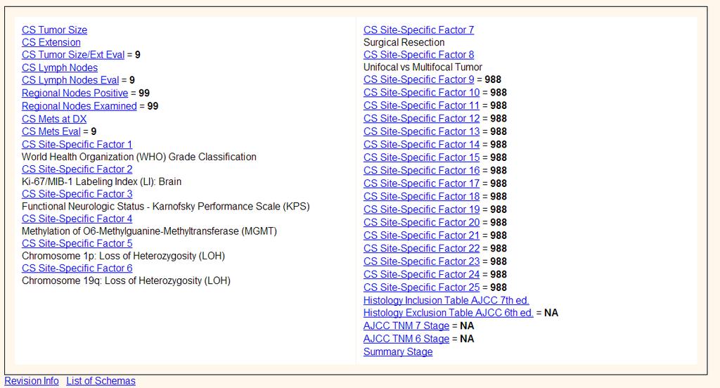
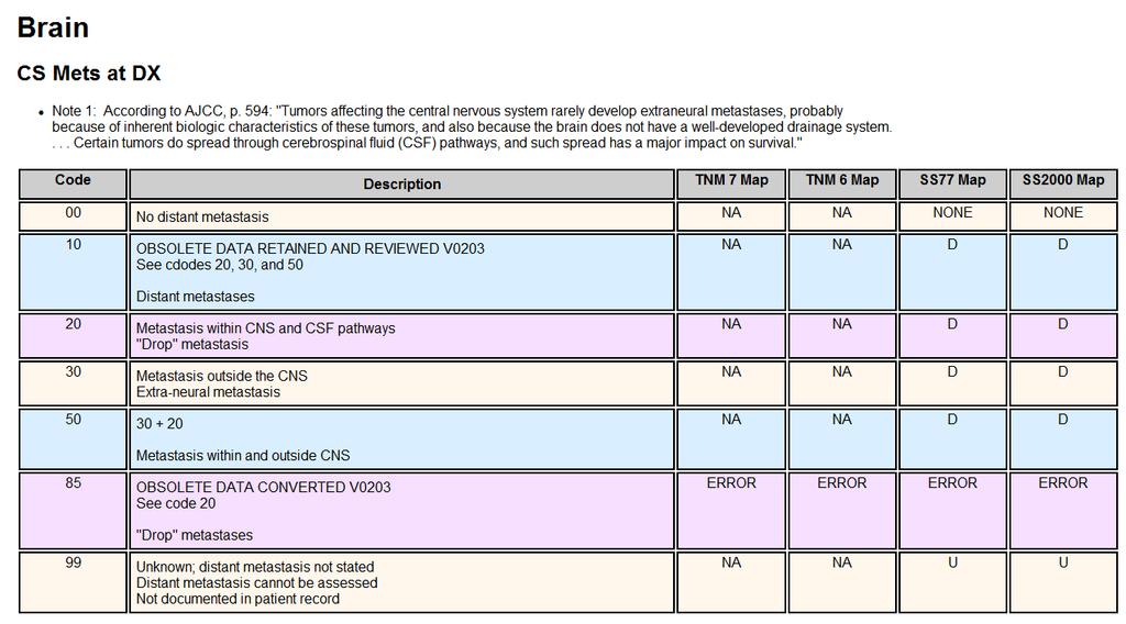
CS Tumor Size CS Extension CS Tumor Size/Ext Eval CS Lymph Nodes CS Lymph Nodes Eval Reg LN Pos Reg LN Exam CS Mets at DX CS Mets Eval
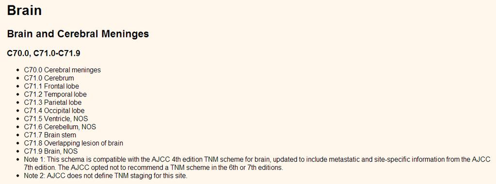
 C70.0, C71.0-C71.9 C70.0 Cerebral meninges C71.0 Cerebrum C71.1 Frontal lobe C71.2 Temporal lobe C71.3 Parietal lobe C71.4 Occipital lobe C71.5 Ventricle, NOS C71.6 Cerebellum, NOS C71.7 Brain stem C71.8
Brain Tumors. Andrew J. Fabiano, MD FAANS. Associate Professor of Neurosurgery Roswell Park Cancer Institute SUNY at Buffalo School of Medicine
 Brain Tumors Andrew J. Fabiano, MD FAANS Associate Professor of Neurosurgery Roswell Park Cancer Institute SUNY at Buffalo School of Medicine Brain Tumors Brain Tumor Basics Types of Tumors Cases Brain
2/20/2019 BRAIN DISSECTION CODING AND DOCUMENTATION OBJECTIVES INTRODUCTION
 BRAIN DISSECTION CODING AND DOCUMENTATION Diana R. Phelps, CPC, CPC-I, CEMC OBJECTIVES Identify general structure of the human brain Describe how the different parts work Recognized the two hemispheres
Nervous System. Student Learning Objectives:
 Nervous System Student Learning Objectives: Identify the primary parts of the neuron Identify the major structures of the central nervous system Identify the major structures of the peripheral nervous
The Brain and Cranial Nerves Pg Three Main Regions of the Brain. Forebrain
 The Brain and Cranial Nerves Pg. 129 Three Main Regions of the Brain Forebrain Cerbral hemispheres Diencephalon Midbrain Brain stem Hindbrain Pons Cerebellum Medulla oblongata Interprets sensory inputs
Nervous System The Brain and Spinal Cord Unit 7b
 Nervous System The Brain and Spinal Cord Unit 7b Chetek High School Mrs. Michaelsen 9.12 Meninges A. Meninges 1. The organs of the CNS are covered by membranes a. The meninges are divided into 3 layers:
Brain and spinal nerve. By: shirin Kashfi
 Brain and spinal nerve By: shirin Kashfi Nervous system: central nervous system (CNS) peripheral nervous system (PNS) Brain (cranial) nerves Spinal nerves Ganglions (dorsal root ganglions, sympathetic
LOW GRADE ASTROCYTOMAS
 LOW GRADE ASTROCYTOMAS This article was provided to us by David Schiff, MD, Associate Professor of Neurology, Neurosurgery, and Medicine at University of Virginia, Charlottesville. We appreciate his generous
Update on Pediatric Brain Tumors
 Update on Pediatric Brain Tumors David I. Sandberg, M.D. Director of Pediatric Neurosurgery & Associate Professor Dr. Marnie Rose Professorship in Pediatric Neurosurgery Pre-talk Questions for Audience
Brain Tumors: an Introduction
 Brain Tumors: an Introduction Overview A tumor (also called a neoplasm or lesion) is abnormal tissue that grows by uncontrolled cell division. Normal cells grow in a controlled manner as new cells replace
Neuro-oncology Update Andrew Kokkino, MD Medical Director, The Neurosciences Institute at Sacred Heart at Riverbend May 20, 2013
 Neuro-oncology Update 2013 Andrew Kokkino, MD Medical Director, The Neurosciences Institute at Sacred Heart at Riverbend May 20, 2013 Case 1 58 year old man with recent facial droop and HA s Thin, cachectic
Dosimetry, see MAGIC; Polymer gel dosimetry. Fiducial tracking, see CyberKnife radiosurgery
 Subject Index Acoustic neuroma, neurofibromatosis type 2 complications 103, 105 hearing outcomes 103, 105 outcome measures 101 patient selection 105 study design 101 tumor control 101 105 treatment options
Pediatric Brain Tumors Pre, Intra & Post Op Evaluation and Management. Timothy M. George, MD, FACS, FAAP
 Pediatric Brain Tumors Pre, Intra & Post Op Evaluation and Management Timothy M. George, MD, FACS, FAAP PEDIATRIC BRAIN TUMORS BACKGROUND: Incidence: Third most common pediatric tumor type (leukemia, neuroblastoma,
Neurosurgery Review. Mudit Sharma, MD May 16 th, 2008
 Neurosurgery Review Mudit Sharma, MD May 16 th, 2008 Dr. Mudit Sharma, Neurosurgeon Manassas, Fredericksburg, Virginia http://www.virginiaspinespecialists.com Phone: 1-855-SPINE FIX (774-6334) Fundamentals
DelMarVa-DC Regional Cancer Registrar s Educational Meeting. Doordan Conference Center Anne Arundel Medical Center Annapolis, MD
 DelMarVa-DC Regional Cancer Registrar s Educational Meeting Doordan Conference Center Anne Arundel Medical Center Annapolis, MD TNM Transition Updates & News from SEER Peggy Adamo, RHIT, CTR NCI SEER adamom@mail.nih.gov
b. The groove between the two crests is called 2. The neural folds move toward each other & the fuse to create a
 Chapter 13: Brain and Cranial Nerves I. Development of the CNS A. The CNS begins as a flat plate called the B. The process proceeds as: 1. The lateral sides of the become elevated as waves called a. The
Chapter 14: Nervous System Guided Notes (A-day)
") Chapter 14: Nervous System Guided Notes (A-day) Nervous System Overview Major Function: Control the body's and. Divided into the Nervous System (CNS=Brain and Spinal Cord) and the Nervous System (PNS=Cranial
Chapter 14: Nervous System Guided Notes (A-day) Nervous System Overview Major Function: Control the body's and. Divided into the Nervous System (CNS=Brain and Spinal Cord) and the Nervous System (PNS=Cranial
STEREOTACTIC RADIATION THERAPY. Monique Blanchard ANUM Radiation Oncology Epworth HealthCare
 STEREOTACTIC RADIATION THERAPY Monique Blanchard ANUM Radiation Oncology Epworth HealthCare Overview Stereotactic radiation therapy at Epworth Healthcare What is stereotactic radiation therapy? Delivery
SURGICAL MANAGEMENT OF BRAIN TUMORS
 SURGICAL MANAGEMENT OF BRAIN TUMORS LIGIA TATARANU, MD, Ph D NEUROSURGICAL CLINIC, BAGDASAR ARSENI CLINICAL HOSPITAL BUCHAREST, ROMANIA SURGICAL INDICATIONS CONFIRMING HISTOLOGIC DIAGNOSIS REDUCING TUMOR
Chapter 8 Nervous System
 Chapter 8 Nervous System Two message centers: Functions of these systems: 1. * 2. * Overview of the Nervous System Parts: General Functions: Functions Sensory input: Sensation via nerves Integration: interpretation
Pathologic Analysis of CNS Surgical Specimens
 2015 Kenneth M. Earle Memorial Neuropathology Review Pathologic Analysis of CNS Surgical Specimens Peter C. Burger, MD Interdisciplinary Quality Control Familiarity with entities Use of diagnostic algorithm
Neuro-Oncology. CBTRUS Statistical Report: Primary Brain and Central Nervous System Tumors Diagnosed in the United States in
 Neuro-Oncology Neuro-Oncology 17:iv1 iv62, 2015. doi:10.1093/neuonc/nov189 CBTRUS Statistical Report: Primary Brain and Central Nervous System Tumors Diagnosed in the United States in 2008-2012 Quinn T.
CHAPTER 11 Tumors Originating in the Brain Medulloblastomas, PNETs and Ependymomas
 Tumors Originating in the Brain Medulloblastomas, PNETs and Ependymomas Foolishly, I waited 7 months before I joined this (or any) group. By that time, my son had radiation, chemo, and a recurrence of
Childhood brain tumours
 Childhood brain tumours Our bodies are made up of billions of cells. Normally, these cells reproduce and repair themselves in a controlled way and do not cause us any problems. If for some reason this
Small and Big Operations: New Tools of the Trade for Brain Tumors. Disclosure. Incidence of Childhood Cancer
 Small and Big Operations: New Tools of the Trade for Brain Tumors Nalin Gupta MD PhD Chief, Division of Pediatric Neurosurgery Departments of Neurosurgery and Pediatrics University of California San Francisco
Collecting Cancer Data: CNS 1/6/2011. Collecting Cancer Data: Brain and Central Nervous System. NAACCR Webinar Series 1.
 Collecting Cancer Data: Brain and Central Nervous System January 6, 2011 NAACCR 2010-2011 Webinar Series Agenda Coding Moment Overview Multiple Primary/Histology Rules Collaborative Stage Treatment Fabulous
Chapter 10 The Nervous System: The Brain and Cranial Nerves
 Chapter 10 The Nervous System: The Brain and Cranial Nerves Copyright 2015 Wolters Kluwer Health Lippincott Williams & Wilkins Overview Key Terms aphasia corpus callosum meninges basal nuclei diencephalon
Childhood Brain and Spinal Cord Tumors Treatment Overview (PDQ )
") 1 di 14 27/11/2016 17.42 NCBI Bookshelf. A service of the National Library of Medicine, National Institutes of Health. PDQ Cancer Information Summaries [Internet]. Bethesda (MD): National Cancer Institute
1 di 14 27/11/2016 17.42 NCBI Bookshelf. A service of the National Library of Medicine, National Institutes of Health. PDQ Cancer Information Summaries [Internet]. Bethesda (MD): National Cancer Institute
FOR PUBLIC CONSULTATION ONLY STEREOTACTIC RADIOSURGERY/ STEROTACTIC RADIOTHERAPY FOR EPENDYMOMA
 1 EVIDENCE SUMMARY REPORT FOR PUBLIC CONSULTATION ONLY STEREOTACTIC RADIOSURGERY/ STEROTACTIC RADIOTHERAPY FOR EPENDYMOMA QUESTIONS TO BE ADDRESSED: 1. What is the evidence for the clinical effectiveness
Brain Tumor Treatment
 Scan for mobile link. Brain Tumor Treatment Brain Tumors Overview A brain tumor is a group of abnormal cells that grows in or around the brain. Tumors can directly destroy healthy brain cells. They can
X-Plain Brain Cancer Reference Summary
 X-Plain Brain Cancer Reference Summary Introduction Brain tumors are not rare. About 20,000 Americans are diagnosed with brain cancer or related cancer of the nervous system. This reference summary will
Data Collection of Primary Central Nervous System Tumors
 Slide 1 Data Collection of Primary Central Nervous System Tumors 1 In this presentation we are going to review many of the principles for abstracting CNS tumors. Many of the slides apply to non-malignant
CT & MRI Evaluation of Brain Tumour & Tumour like Conditions
 CT & MRI Evaluation of Brain Tumour & Tumour like Conditions Dr. Anjana Trivedi 1, Dr. Jay Thakkar 2, Dr. Maulik Jethva 3, Dr. Ishita Virda 4 1 M.D. Radiology, Professor and Head, P.D.U. Medical College
Brain Tumors. What is a brain tumor?
 Scan for mobile link. Brain Tumors A brain tumor is a collection of abnormal cells that grows in or around the brain. It poses a risk to the healthy brain by either invading or destroying normal brain
BRAIN TUMORS IN INFANTS
 BRAIN TUMORS IN INFANTS Dr Sergio Valenzuela M.D-( ISPN-ESPN-FLANC)&cols. Head Pediatric Neurosurgery Unit I Instituto de NeurocirugiaAsenjo Santiago CHILE RATE OF MENINGEAL,BRAIN AND OTHER CNS MALIGNANT
about brain tumors a primer for patients and caregivers
 about brain tumors a primer for patients and caregivers brain about tumors a primer for patients and caregivers 8550 W. Bryn Mawr Avenue, Suite 550 Chicago, IL 60631 CareLine: - -ABTA ( ) Email: info@abta.org
Unit Six The Nervous System
 Unit Six The Nervous System I. Introduction A. Definition a coordinating system of the body, composed of highly specialized cells that conduct nerve impulses to a center so responses can be made. The nervous
Unit VIII Problem 3 Neuroanatomy: Brain Stem, Cranial Nerves and Scalp
 Unit VIII Problem 3 Neuroanatomy: Brain Stem, Cranial Nerves and Scalp - Brain stem: It is connected to the cerebellum and cerebral hemispheres. Rostral end of brain stem: diencephalon is the area which
Neurological Assessment
 Neurological Assessment Name: Age: Gender: Date: History Review of history related to neurological system YES/NO If YES, provide details: General Neurological Mental Illness Neurological disease Severe
Selected radiosurgery cases from the Rotating Gamma Institute Debrecen, Hungary
 Selected radiosurgery cases from the Rotating Gamma Institute Debrecen, Hungary László Bognár M.D., Ph.D., József G. Dobai M.D., Gábor Csiky and Imre Fedorcsák M.D., Ph.D. Department of Neurosurgery, Medical
Imaging for suspected glioma
 Imaging for suspected glioma 1.1.1 Offer standard structural MRI (defined as T2 weighted, FLAIR, DWI series and T1 pre- and post-contrast volume) as the initial diagnostic test for suspected glioma, unless
Blood supply to the brain Blood brain barrier isolates neural tissue from general circulation
 The Brain and Cranial Nerves Objectives Name the major regions of the brain and describe their functions. Discuss the formation, circulation, and functions of the CSF. List the main components of the medulla
Dr Eddie Mee. Neurosurgeon Auckland City Hospital, Ascot Integrated Hospital, MercyAscot Hospitals, Auckland
 Dr Eddie Mee Neurosurgeon Auckland City Hospital, Ascot Integrated Hospital, MercyAscot Hospitals, Auckland 16:30-17:25 WS #48: Current Management of Brain Bleeds and Tumours 17:35-18:30 WS #58: Current
ACTIVITY 7: NERVOUS SYSTEM HISTOLOGY, BRAIN, CRANIAL NERVES NERVOUS SYSTEM TISSUES: HISTOLOGY SLIDES
 ACTIVITY 7: NERVOUS SYSTEM HISTOLOGY, BRAIN, CRANIAL NERVES OBJECTIVES: 1) How to get ready: Read Chapter 14 & 15 McKinley et al., Human Anatomy, 4e. All text references are for this textbook. Read dissection
Otolaryngologist s Perspective of Stereotactic Radiosurgery
 Otolaryngologist s Perspective of Stereotactic Radiosurgery Douglas E. Mattox, M.D. 25 th Alexandria International Combined ORL Conference April 18-20, 2007 Acoustic Neuroma Benign tumor of the schwann
Accuracy of intra-operative rapid diagnosis by Squash smear in CNS lesions An early institutional experience. KK Bansal,
 Accuracy of intra-operative rapid diagnosis by Squash smear in CNS lesions An early institutional experience. KK Bansal, Monika Bansal, Sanjeev Kishore, Anuradha K, Meena H, Dushyant G. Department of Neurosurgery
Human Nervous System:
 OLLI Brain: Making Sense of Our World: Lecture 3 Human Nervous System: The Motor & Sensory Divisions Copyright 2004 Pearson Education, Inc., publishing as Benjamin Cummings Organization of the Nervous
The Human Brain: Anatomy, Functions, and Injury
 The Human Brain: Anatomy, Functions, and Injury Main Menu Brain Anatomy Brain Functions Injury Mechanisms Brain Anatomy Menu Skull Anatomy Interior Skull Surface Blood Vessels of the Brain Arteries of
PRINCESS MARGARET CANCER CENTRE CLINICAL PRACTICE GUIDELINES
 PRINCESS MARGARET CANCER CENTRE CLINICAL PRACTICE GUIDELINES CENTRAL NERVOUS SYSTEM EPENDYMOMA Last Revision Date July 2015 1 CNS Site Group Ependymoma Author: Dr. Norm Laperriere 1. INTRODUCTION 3 2.
Stereotactic Radiosurgery/Fractionated Stereotactic Radiotherapy for Acoustic Neuroma (Vestibular Schwannomas)
") Strategic Commissioning Group West Midlands Commissioning Policy (WM/38) Stereotactic Radiosurgery/Fractionated Stereotactic Radiotherapy for Acoustic Neuroma (Vestibular Schwannomas) Version 1 July 2010
Strategic Commissioning Group West Midlands Commissioning Policy (WM/38) Stereotactic Radiosurgery/Fractionated Stereotactic Radiotherapy for Acoustic Neuroma (Vestibular Schwannomas) Version 1 July 2010
Chapter 10. The Nervous System
 Chapter 10 The Nervous System Objectives List the organs and divisions of the nervous system and describe the generalized functions Identify the major types of cells in the nervous system and discuss the
PRINCESS MARGARET CANCER CENTRE CLINICAL PRACTICE GUIDELINES
 PRINCESS MARGARET CANCER CENTRE CLINICAL PRACTICE GUIDELINES CENTRAL NERVOUS SYSTEM MENINGIOMA CNS Site Group Meningioma Author: Dr. Norm Laperriere Date: February 20, 2018 1. INTRODUCTION 3 2. PREVENTION
FCDS TENTATIVE V11 TO V11.1 IMPLEMENTATION SCHEDULE
 NAACCR V11.1 UPDATES ZIP CODE, FIPS COUNTY, FLORIDA CITY NAME VERIFICATION FILE (COMMA SEPARATED TEXT FILE) CANCER TRAINING: PRINCIPLES AND PRACTICE OF CANCER REGISTRATION, SURVEILLANCE AND CONTROL, JULY