Neoplasms of the BRAIN and CNS
|
|
|
- Alexina Rogers
- 6 years ago
- Views:
Transcription


1 Neoplasms of the BRAIN and CNS FCDS Educational Webcast Series Steven Peace, BS, CTR October 15, Focus Anatomy SSS 2000 MPH Rules AJCC TNM Presentation Outline Overview Reportable CNS Neoplasms Anatomy of the Human Brain & CNS WHO Grade for Brain and CNS Neoplasms Multiple Primary and Histology Coding Rules AJCC TNM Stage and SEER Summary Stage 2000 Image Source: news.discovery.com 2 Site Specific Factors 1

2 Overview Brain tumors are: Primary brain tumors - those that begin in the brain or central nervous system (or its supporting tissues) and tend to stay in the brain - occur in people of all ages, but they are statistically more frequent in children and older adults. Metastatic brain tumors those that begin as a cancer elsewhere in the body and spread to the brain are more common in adults than in children. 4 Source: American Brain Tumor Association Facts and Statistics 2
3 Brain tumors are: Usually described as intracranial neoplasms with varying behaviors (benign, borderline, malignant ref. ICD-O-3) Include most identifiable structures within the cranium including the brain itself, small hormone-secreting ducts like the pineal and pituitary glands, the cranial nerves (primarily the optic nerve, olfactory nerve, acoustic nerve), the outer protective lining of the brain (meninges), and the spinal cord. Metastatic neoplasms are excluded Certain benign tumors are excluded No malignant neoplasms are excluded Benign bone tumors are excluded 5 Brain Tumor Characteristics 6 Start in the brain and grow steadily there. Very rarely spread to other organs through the bloodstream. Are named for the anatomic location of tumor and/or the cells from which they arise, each having a certain function essential to normal physiological functioning of the brain. For example: Brain Stem Gliomas arise in the lowest part of the brain. Meningiomas arise in the meninges. Gliomas arise from glial cells that support the CNS. Astrocytomas arise from astrocytes Ependymomas arise from ependymal cells which line the ventricles Oligodendrogliomas arise from oligodentdrocyte cells which make up the fatty substance called myelin that covers nerves like electrical insulation. 3

4 Characteristics of Brain Tumors 7 Source: medicalgeek.com/indian-post-graduate-exams Range of Tumors and Symptoms There are over 120 different types of brain/cns tumors. CNS tumors are associated with a range of symptoms and complications such as edema, seizures, endocrinopathy, fatigue, psychiatric disorder, venous thromboembolism that can seriously impact quality of life. Symptoms depend very much on the size and location of the tumor. General symptoms include persistent headaches which tend to be worse with activity, at night or early in the morning, convulsions, vomiting, subtle changes in personality, memory, mental ability, drowsiness, lethargy. 8 SEER Training Modules 4

5 Brain Anatomy and Function 9 Tumor Location and Symptoms Symptoms are often tumor location specific or provide clues Symptoms on the right side of the body may occur if the tumor is located on the left side of the brain and vice-versa. The speech center in most people is on the left side of the brain. Symptoms of a tumor located here may include difficulty saying correct words while still capable of understanding what is being said. If the tumor is located in the frontal lobe which controls intellectual function, thought process, behavior and memory, those activities may be affected. Similarity to closed head injury victims (motorcycle crash). 10 SEER Training Modules 5
6 ALL Brain Tumors are Reportable Public Law , the Benign Brain Tumor Cancer Registries Amendment Act, [PDF-185KB] requires programs participating in the National Program for Cancer Registries (NPCR) to collect data on benign and borderline tumors of the central nervous system in addition to the previously required data on malignant tumors. In addition to NPCR, the National Cancer Institute's (NCI) Surveillance, Epidemiology and End Results (SEER) program and the American College of Surgeons' (ACoS) Commission on Cancer began requiring that these tumors be reported, starting with cases diagnosed on January 1, ALL Brain Tumors are Reportable BUT Do Not Report Benign/Borderline Tumors that were Diagnosed BEFORE 1/1/2004 NOT EVEN HISTORICAL This creates an ERROR for FCDS at Call for Data FCDS then has to Delete the Case and Adjust Sequences 12 6


7 ICD-O Topography Codes (Anatomic Site) 13 Brain Tumor Characteristics Benign Borderline Malignant Patient Age Tumor Location Tumor Histologic Type WHO Grade of Primary Tumor 14 American Brain Tumor Association 7
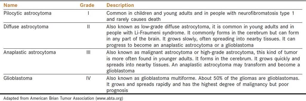
 Astrocytoma, Grade II (Low Grade Pilocytic Astrocytoma) Astrocytoma,")
8 Tumor Behavior Benign Tumors Slow growing Distinct borders Rarely spread Malignant Tumors Usually rapid growing Invasive Life threatening Borderline Malignant Tumors Rare Likely to Recur following Surgical Resection May become Life threatening 15 Common Histologic Types Astrocytoma, Grade I (Juvenile Pilocytic Astrocytoma) Astrocytoma, Grade II (Low Grade Pilocytic Astrocytoma) Astrocytoma, Grade III (Anaplastic Astrocytoma) Glioblastoma (Glioblastoma Multiforme/Astrocytoma, Grade IV) Ependymoma Medulloblastoma Meningioma Oligodendroglioma Oligoastrocytoma Pituitary Adenoma 16 8

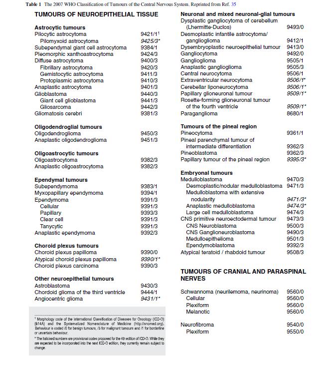
9 17 18 WHO Classification Groups Tumors of NeuroepithelialTissue Tumors of Cranial and Paraspinal Nerves Tumors of Meninges Tumors of Uncertain histogenesis Lymphomas and Hematopoietic Malignancies Germ Cell Tumors Cysts and Tumor-Like Lesions Tumors of the Sellar Region Metastatic Tumors 9


10


11 Distribution Primary Brain & CNS Tumors Behavior Malignant Nonmalignant 21 ALL Tumors Distribution Primary Brain & CNS Tumors Primary Site and Behavior Malignant Only


12 ALL Tumors Distribution Primary Brain & CNS Tumors Primary Site and Behavior Non-Malignant Only 23 Distribution Primary Brain & CNS Tumors Histologic Type All Behavior ALL Tumors


13 Distribution Primary Brain & CNS Tumors Behavior and Histologic Type Malignant Only 25 Distribution Primary Brain & CNS Tumors Behavior and Histologic Type Non- Malignant Only
 Remainder of tumors occur in spinal cord,")

14 Childhood Brain Tumors Tentorium - extension of the dura mater separating the cerebellum from the occipital lobes 27 50% of childhood brain and CNS tumors are infratentorial, originating below the tentorium 20+% of childhood CNS tumors are located in the sellar or suprasellar region around the sella turcica (the bone that contains the pituitary gland) Remainder of tumors occur in spinal cord, brain stem, cranial nerves, etc. Childhood Brain Tumors Supratentorial - childhood Craniopharyngiomas. Diencephalic and hypothalamic gliomas. Germ cell tumors. Low-grade astrocytomas. Anaplastic astrocytomas. Glioblastoma multiforme. Mixed gliomas. Oligodendrogliomas. Primitive neuroectodermal tumors. Low-grade or anaplastic ependymomas. Meningiomas. Choroid plexus tumors. Infratentorial - childhood Cerebellar astrocytomas (usually high-grade). Medulloblastomas (primitive neuroectodermal tumors). Ependymomas (low-grade or anaplastic). Brain stem gliomas (high-grade or low-grade). Atypical teratoid tumors 28 14


15 Distribution Primary Brain & CNS Tumors Children and Adolescents by Site 29 Distribution Primary Brain & CNS Tumors Children and Adolescents by Histologic Type
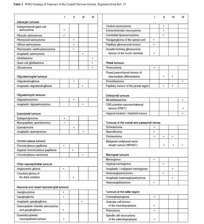
16 WHO Grade for Brain/CNS Tumors WHO Tumor Grades Grade I, II, III, and IV Higher the grade the more malignant the tumor A tumor can contain more than one grade of cell Always record and code the highest tumor grade noted Record WHO Grade in SSF1 NOT in Grade/Differentiation 31 WHO Grade for Brain/CNS Tumors LOW GRADE Neoplasms Grade I: least malignant tumors associated with long-term survival. They grow slowly and have an almost normal appearance when viewed through a microscope. Surgery alone may be an effective treatment for this grade tumor. Grade II: tumors are slow-growing and look slightly abnormal under a microscope. Some can spread into nearby normal tissue and recur, sometimes as a higher grade tumor

17 WHO Grade for Brain/CNS Tumors Grade III:These tumors are, by definition, malignant although there is not always a big difference between grade II and grade III tumors. The cells of a grade III tumor are actively reproducing abnormal cells, which grow into nearby normal brain tissue. These tumors tend to recur, often as a grade IV. Grade IV:The most malignant tumors. Tumors reproduce rapidly, can have a bizarre appearance when viewed under the microscope, and easily grow into nearby normal brain tissue. These tumors form new blood vessels so they can maintain their rapid growth. They also have areas of dead cells in their centers WHO Grade for Brain/CNS Tumors
18
19 37 Survival


20 ANATOMY OF THE HUMAN BRAIN 39 Source: National Geographic, couretsy of Fred Hossler/Getty Images 40 Source: University of Illinois 20


21 Brain Lobes and Fissures
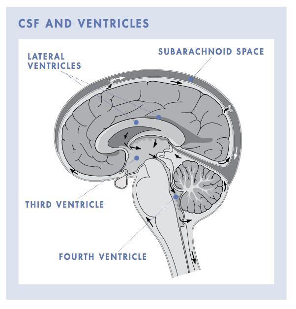

22 Ventricular System of the Brain 43 Ventricular System of the Brain 44 Source: solarnavigator.net/human_brain 22
23 Midline Shift and Mass Effect The bony cranium protects the brain from outside impacts to the head. When swelling occurs in the brain, there isn t much give. The swelling results in intracranial pressure and can cause a number of effects that begin to impact quality of life and comfort for the patient. 45 The easiest way to describe midline shift is to bring to mind siting in a movie theater. As soon as the person to one side of you puts his elbow onto the shared armrest between you, you tend to shift away. Source: Medscsape Midline Shift and Mass Effect Midline is a central boundary separating the left and right hemispheres. Midline Shift Tumor crosses the brain to shift across the center line Mass Effect is Edema or swelling causes the brain to shift across center line 46 Both create new symptoms at cross-over Depends on the size and location of he tumor and level of spread Edema caused by many things Either cause pushes midline out of alignment Source: Medscsape 23


24 Meninges and Brain Stem tentorium 47 Cranial Nerves Optic Acoustic 48 24
25 Cranial Nerve Functions Cranial Nerve: I Olfactory II Optic III Oculomotor IV Trochlear V Trigeminal VI Abducens VII Facial VIII Vestibulocochlear IX Glossopharyngeal X Vagus XI Spinal Accessory XII Hypoglossal smell vision eyelid and eyeball movement Major Functions: turns eye downward and laterally, controls superior oblique muscles chewing, face & mouth touch & pain turns eye laterally facial expressions, taste, tears, saliva Also referred to as Auditory Nerve: hearing, equilibrium sensation Taste, senses carotid blood pressure aortic blood pressure, heart rate, stimulates digestive organs, taste controls trapezius & sternocleidomastoid muscles, controls swallowing controls tongue movements 49 Sinus, Olfactory, Base of Skull Tumors Cancer Registries treat many of these as Head & Neck Neoplasms Some are intra-cranial but many are not intra-cranial or CNS Primary Site of Tumor Critical for Stage, Treatment, and Prognosis Histology is used to identify which are abstracted as Brain/CNS Highly specialized surgical procedures location of tumor Some Prognostic Factors Overlap 50 25
 can be Grade I-IV not just III-IV.")
26 Sinus, Olfactory, Base of Skull Tumors 51 Histologic Type - Glioma Most common category of primary brain tumors. They begin in glial cells (supporting cells of the CNS) can be Grade I-IV not just III-IV. Often spread into surrounding brain tissue along nerve fibers invading the spaces between nearby normal brain cells. Some invade the surrounding brain more than others. Difficulty obtaining complete surgical removal. MRI scans show the largest part of the glioma, but cannot reliably show areas of the brain where tumor cells have invaded. Aggressive efforts to remove small numbers of tumor cells within the brain could cause loss of neurologic function. When it is not possible to remove the entire glioma, post-op radiation therapy and chemotherapy may be advised. Even with maximum safe resection followed by radiation and chemotherapy, gliomas can grow back
27 Glioma 3 Main Histologic SubTypes 1. Astrocytoma: In adults most often arise in the cerebrum. In children they occur in the brain stem, cerebrum and cerebellum. Rarely in brain stem in adults. Felt to be most aggressive of brain tumors. Grade I and II astrocytomas are low-grade astrocytomas. Grade III astrocytoma is an anaplastic astrocytoma. Grade IV astrocytoma is a glioblastoma multiforme. 53 Glioma 3 Main Histologic SubTypes 2. Oligodendroglioma: Rare tumor that usually occurs in the cerebrum, grows slowly and usually does not spread into surrounding brain tissue like astrocytoma does. Most common in middle-aged adults. 3. Ependymoma: Most commonly arise in children and young adults. They are also seen with neurofibromatosis Type II. (which we will discuss in a bit) 54 27


28 Glioma Other Subtypes There are other subtypes of gliomas, each with their own specific characteristics and modes of growth. Brain Stem Glioma Juvenile Pilocytic Astrocytoma Pleomorphic Xanthoastrocytoma Subependymoma Ganglioglioma 55 Glioma Tumor Markers 56 28

29 Glioma Chromosome Alterations 57 Non-Glial Tumors Medulloblastoma: Usually arises in the cerebrum, is the most common brain tumor in children, and is sometimes called a primitive neuroectodermal tumor or PNET. Meningioma: Arises from the meninges which are the outside coverings of the brain between the skull and the brain itself. It usually presses on the brain, but does not invade it and often grows slowly
30 Meningioma Meningiomas are typically diagnosed by CT or MRI imaging Biopsy may be considered for confirmation Options stratified by presence/absence of symptoms and tumor size Most asymptomatic patients with small tumors (<30mm) may just be observed. If neurological impairment is imminent, surgery (if accessible) or radiotherapy (EBRT OR SRS) is feasible Asymptomatic tumors >30mm can be either resected or observed 59 Non-Glial Tumors Schwannoma: Arises from Schwann cells present in certain nerves, including those that control balance and hearing. May be called neuroma. A common site is the vestibular nerve which carries signals from the inner ear to the brain stem. Tumors in this location are called acoustic neuromas (a.k.a. vestibular schwannoma), and occur most often in adults
31 Non-Glial Tumors Craniopharyngioma: Grows at the base of the brain, arises from the tissue connecting the brain and the pituitary gland and occurs in both adults and children. Pituitary Adenoma: Arises from the pituitary gland and may cause compression of the optic nerves causing vision problems. Some produce excessive amounts of hormones that can disrupt the body s metabolism. 61 Roswell Park Cancer Insitute Neurofibromatosis The neurofibromatoses (NF) are a group of genetic disorders which cause tumors to grow along nerves and can also affect the development of non-nervous tissues such as bones and skin. Neurofibromatosis Type I (NF-I), also known as Peripheral NF and historically as von Recklinghausen Disease Occurs in 1:4,000 births Multiple cafe-au-lait spots (not reportable) Many, many neurofibromas on or under the skin (not reportable) Enlargement and deformation of bones and curvature of the spine Tumors may develop in brain, on cranial nerves, or the spinal cord 62 Neurofibromatosis Foundation 31


32 NF Type I: First documented photo Source Credit: Dr. Stanley B. Burns Other Manifestions of NF Type I Lisch nodules on the eye Melanocytic hemartomas Café-au-lait spots on skin Discolored birth marks 64 Medscape Source: Dermnet.com; Dermatologic Manifestations of NF Type I 32

33 Neurofibromatosis Type II Neurofibromatosis Type II (NFII), also known as Multiple Inherited Schwannomas, Meningiomas and Ependymomas (MISME) or Bilateral Acoustic Neurofibromatosis (BAN ). Is a genetically inherited diseasecaused by mutations of the "Merlin" gene, which appears to influences the form and movement of cells Primary manifestation is a development of non-malignant brain tumors in the region of the cranial nerves, frequently bilaterally. The eighth cranial nerve is the auditory-vestibular nerve which transmits sensory information from the inner ear to the brain and is commonly affected. 65 Source: California Ear Institute Acoustic Neuroma/Schwannoma 66 Source: 33


34 Multiple Primary Rules Histology Coding Rules 67 Different Rules for Benign and Malignant 68 34
35 69 Sequence Numbering for Brain Tumors Malignant primary brain and CNS tumors are assigned Sequence Codes in the range Sequence Chronologically Only count malignant tumors in the sequence If only one malignant tumor occurs, it is coded 00 If subsequent (multiple) primary malignant and/or in situ neoplasms, the sequence number for the first tumor begins at 01, the sequence number for the second primary tumor is 02, and so forth. Non-malignant primary brain and CNS tumors are assigned Sequence Codes in the range Sequence Chronologically Only count benign/borderline or reportable by agreement neoplasms in the sequence If only one non-malignant tumor occurs, it is coded 60. If subsequent (multiple) non-malignant neoplasms are diagnosed, the first tumor should be sequenced as 61, the second 62 and so forth. Benign and Borderline Tumor Rules 70 35
36 Benign and Borderline Tumor Rules When multiple tumors are present registrars should identify and document specific characteristics for MPH Rules Text Date of Diagnosis (Timing is not used to determine number of abstracts or primary neoplasms to abstract) Method and Details of Diagnosis (some are never resected) Location of Tumor Laterality Histologic Type refer to Chart 1 Tumor Behavior Multiple Meningioma s (meningiomatosis) Neurofibromatosis Characteristics (when applicable) 71 Malignant Tumor Rules 72 36
37 Malignant Tumor Rules 73 Malignant Tumor Rules When multiple tumors are present registrars should identify and document specific characteristics for MPH Rules Text Date of Diagnosis (Timing is not used to determine number of abstracts or primary neoplasms to abstract) Method and Details of Diagnosis (most attempt resection) Location of Tumor (not spread or invasion but bulk of tumor) Histologic Type refer to Chart 1 and/or Chart 2 Tumor Behavior Variations or Combinations of One or More Glial Tumors Over Lifetime astrocytoma, glioblastoma, ependymoma, or oligodendroglioma Special rules for determining # abstracts Special rules for determining whether or not is mixed glioma Note: Recurrence, progression, or any reappearance of histologies on the same branch in Chart 1 or Chart 2 is always the same disease process
, THE FIRST NEOPLASM IS NOT REPORTABLE TO FCDS EVENAS A HISTORICAL CASE THE NEW")
38 Do NOT Report Benign Tumors Dx d < 2004 REMINDER: Sequence numbers for malignant neoplasms and for benign, borderline, and other reportable-by-agreement cases are usually assigned over a lifetime. HOWEVER, WHEN A PATIENT WAS DIAGNOSED WITH A NON-MALIGNANT CNS NEOPLASM BEFORE REPORTING WAS REQUIRED (January 1, 2004), THE FIRST NEOPLASM IS NOT REPORTABLE TO FCDS EVENAS A HISTORICAL CASE THE NEW (SECOND) NEOPLASM SHOULD BE ASSIGNED SEQUENCE NUMBER 60 DO NOT REPORT THE BENIGN NEOPLASM IF DIAGNOSED BEFORE 1/1/2004 THIS HAS BEEN A SOURCE OF CONFUSION PLEASE REVIEW AND SAVE 75 FCDS Data Acquisition Manual and CDC Data Collection of Primary Central Nervous System Tumors Staging Brain and CNS Neoplasms 76 38
39 AJCC TNM, 7 th edition Attempts at developing a TNM-based classification and staging system for tumors of the central nervous system have not been successful. Basic TNM Concepts are not applicable to brain and CNS sites. It continues to be the recommendation of the ASJCC CNS Tumor Task Force that a formal classification and staging system not be attempted. AJCC Stage = 88 (Not Applicable) Includes: ANY Benign or ANY Malignant Neoplasm 77 AJCC TNM, 7 th edition Factors Felt to be of Prognostic and/or Clinical Significant Include: Tumor Histology Location of Tumor Unifocal or Multifocal WHO Grade of Tumor Patient Age at Diagnosis Functional Neurologic Status *KPS) Primary or Recurrent Tumor Extent of Resection Metastatic Spread Proliferative Fraction (Ki-67, M1 B-1) Gene Deletions (1p, 19q) MGMT Methylation 78 39
40 SEER Summary Stage 2000 To obtain a FREE electronic copy of the SS2000 Manual: 79 SS


41 Steps to Assign SS2000 Three summary stage groups can be ruled out quickly: in situ, distant, and localized FIRST In Situ Stage is Not Applicable for Brain/CNS Tumors 81 In Situ Stage is Not the Same as Benign or Borderline Behavior SS2000 for Benign/Borderline Tumors =


42 Steps to Assign SS2000 Three summary stage groups can be ruled out quickly: in situ, distant, and localized SECOND 83 Brain and CNS these are usually CSF Involvement cells in fluid On rare occasion you may see drop metastasis code as Distant Stage Steps to Assign SS2000 Three summary stage groups can be ruled out quickly: in situ, distant, and localized THIRD 84 Most common Summary Stage unless tumor crosses midline 42

43 Steps to Assign SS2000 Three summary stage groups can be ruled out quickly: in situ, distant, and localized FINAL (if needed) 85 Additional Resources The 2007 WHO Classification of Tumours of the Central Nervous System, David N. Louis, Hiroko Ohgaki, Otmar D. Wiestler, Webster K. Cavenee, Peter C. Burger, Anne Jouvet, Bernd W. Scheithauer and Paul Kleihues, World Health Organization, Lyon, France, 2007 Central Brain Tumor Registry of the United States (CBTRUS), 2015 NCCN Evidence Based Treatment Guidelines, nccn.org, 2015 Data collection of primary central nervous system tumors. National Program of Cancer Registries Training Materials. Department of Health and Human Services, Centers for Disease Control and Prevention. Atlanta, Georgia, Multiple Primary and Histology Coding Rules, SEER 2007 AJCC Cancer Staging Manual, 7 th ed., AJCC, 2010 SEER Summary Staging Manual
44 87 44
2019 Neoplasms of the BRAIN/CNS. CDC & Florida DOH Attribution
 2019 Neoplasms of the BRAIN/CNS 2018-2019 FCDS Educational Webcast Series Steven Peace, BS, CTR March 21, 2019 CDC & Florida DOH Attribution Funding for this conference was made possible (in part) by the
Brain and CNS Tumors
 Brain and CNS Tumors FCDS 2011/2012 Educational Webcast Series January 19, 2012 Lynne Pearson, BHS, CTR, LHRM Steven Peace, BS, CTR Updated for 2012 Requirements and CSv02.03.02 Presentation Outline Overview
Brain and CNS Tumors
 Brain and CNS Tumors FCDS 2011/2012 Educational Webcast Series January 19, 2012 Lynne Pearson, BHS, CTR, LHRM Steven Peace, BS, CTR Updated for 2012 Requirements and CSv02.03.02 Presentation Outline Overview
1/17/2012. Brain and CNS Tumors. Presentation Outline. Overview. Overview. Anatomy of the Human Brain. Multiple Primary and Histology Coding Rules
 Brain and CNS Tumors FCDS 2011/2012 Educational Webcast Series January 19, 2012 Lynne Pearson, BHS, CTR, LHRM Steven Peace, BS, CTR Updated for 2012 Requirements and CSv02.03.02 Presentation Outline Overview
Brain tumors: tumor types
 Brain tumors: tumor types Tumor types There are more than 120 types of brain tumors. Today, most medical institutions use the World Health Organization (WHO) classification system to identify brain tumors.
Bellringer: The central nervous system is comprised of: What is the name of the outermost layer of the brain? a. Brain. b.
 Bellringer: The central is comprised of: a. Brain b. Spinal cord c. Sensory receptors d. Both a and b What is the name of the outermost layer of the brain? a. Pia mater b. Dura mater c. Arachnoid d. Pons
Q&A. Fabulous Prizes. Collecting Cancer Data:CNS 2/7/12. NAACCR Webinar Series Collecting Cancer Data Central Nervous System
 Collecting Cancer Data Central Nervous System NAACCR 2012 2013 Webinar Series 2/7/2013 Q&A Please submit all questions concerning webinar content through the Q&A panel. Reminder: If you have participants
CNS TUMORS. D r. Ali Eltayb ( U. of Omdurman. I ). M. Path (U. of Alexandria)
. M. Path (U. of Alexandria)") CNS TUMORS D r. Ali Eltayb ( U. of Omdurman. I ). M. Path (U. of Alexandria) CNS TUMORS The annual incidence of intracranial tumors of the CNS ISmore than intraspinal tumors May be Primary or Secondary
CNS TUMORS D r. Ali Eltayb ( U. of Omdurman. I ). M. Path (U. of Alexandria) CNS TUMORS The annual incidence of intracranial tumors of the CNS ISmore than intraspinal tumors May be Primary or Secondary
Site Specific Coding Rules MALIGNANT CENTRAL NERVOUS SYSTEM TUMORS
 Multiple Primary and Histology Site Specific Coding Rules MALIGNANT CENTRAL NERVOUS SYSTEM TUMORS 1 Prerequisites 2 Completion of Multiple Primary and Histology General Coding Rules 3 There are many ways
Tumors of the Nervous System
 Tumors of the Nervous System Peter Canoll MD. PhD. What I want to cover What are the most common types of brain tumors? Who gets them? How do they present? What do they look like? How do they behave? 1
Brain and Cranial Nerves (Ch. 15) Human Anatomy lecture. caudal = toward the spinal cord)
 Human Anatomy lecture. caudal = toward the spinal cord)") Insight: Some cranial nerve disorders Brain and Cranial Nerves (Ch. 15) Human Anatomy lecture I. Overview (Directional terms: rostral = toward the forehead caudal = toward the spinal cord) A. 3 Major parts
Insight: Some cranial nerve disorders Brain and Cranial Nerves (Ch. 15) Human Anatomy lecture I. Overview (Directional terms: rostral = toward the forehead caudal = toward the spinal cord) A. 3 Major parts
General: Brain tumors are lesions that have mass effect distorting the normal tissue and often result in increased intracranial pressure.
 1 Lecture Objectives Know the histologic features of the most common tumors of the CNS. Know the differences in behavior of the different tumor types. Be aware of the treatment modalities in the various
Please submit all questions concerning webinar content through the Q&A panel. Reminder:
 NAACCR 2015-2016 Webinar Collecting Cancer Data: Series Central Nervous System NAACCR Webinar Series 2016 2017 Carol Hahn Johnson Jim Hofferkamp jhofferkamp@naaccr.org Q&A Please submit all questions concerning
NON MALIGNANT BRAIN TUMOURS Facilitator. Ros Taylor Advanced Neurosurgical Nurse Practitioner Southmead Hospital Bristol
 NON MALIGNANT BRAIN TUMOURS Facilitator Ros Taylor Advanced Neurosurgical Nurse Practitioner Southmead Hospital Bristol Neurosurgery What will be covered? Meningioma Vestibular schwannoma (acoustic neuroma)
ACTIVITY 7: NERVOUS SYSTEM HISTOLOGY, BRAIN, CRANIAL NERVES
 ACTIVITY 7: NERVOUS SYSTEM HISTOLOGY, BRAIN, CRANIAL NERVES LABORATORY OBJECTIVES: 1. Histology: Identify structures indicated on three different slides or images of nervous system tissue. These images
What is Brain Cancer? What is the brain?
 What is Brain Cancer? The brain and spinal column make up the central nervous system (CNS), where all vital functions of the body are controlled. When tumors arise in the central nervous system, they are
CS Tumor Size CS Extension CS Tumor Size/Ext Eval CS Lymph Nodes CS Lymph Nodes Eval Reg LN Pos Reg LN Exam CS Mets at DX CS Mets Eval
 C70.0, C71.0-C71.9 C70.0 Cerebral meninges C71.0 Cerebrum C71.1 Frontal lobe C71.2 Temporal lobe C71.3 Parietal lobe C71.4 Occipital lobe C71.5 Ventricle, NOS C71.6 Cerebellum, NOS C71.7 Brain stem C71.8
What are brain and spinal cord tumours? Contents
 13 11 20 Information and support What are brain and spinal cord tumours? Contents The brain and spinal cord Brain function What is a brain or spinal cord tumour? What types of tumours are there? How common
Peter Canoll MD. PhD.
 Tumors of the Nervous System Peter Canoll MD. PhD. What I want to cover What are the most common types of brain tumors? Who gets them? How do they ypresent? What do they look like? How do they behave?
Meningioma tumor. Meningiomas are named according to their location (Fig. 1) and cause various symptoms: > 1
 and cause various symptoms: > 1") Meningioma tumor Overview A meningioma is a type of tumor that grows from the protective membranes, called meninges, which surround the brain and spinal cord. Most meningiomas are benign (not cancer) and
Meningioma tumor Overview A meningioma is a type of tumor that grows from the protective membranes, called meninges, which surround the brain and spinal cord. Most meningiomas are benign (not cancer) and
The Nervous System: Central Nervous System
 The Nervous System: Central Nervous System I. Anatomy of the nervous system A. The CNS & the body by: 1. monitoring of the body 2. & information between parts of the body 3. acting as a to gather, store,
Tumors of the Central Nervous System
 Tumors of the Central Nervous System 1 Financial Disclosures I have NO SIGNIFICANT FINANCIAL, GENERAL, OR OBLIGATION INTERESTS TO REPORT Introduction General: Brain tumors are lesions that have mass effect
Principles of Anatomy and Physiology
 Principles of Anatomy and Physiology 14 th Edition CHAPTER 14 The Brain and Cranial Nerves Introduction The purpose of the chapter is to: 1. Understand how the brain is organized, protected, and supplied
The Nervous System PART B
 7 The Nervous System PART B PowerPoint Lecture Slide Presentation by Jerry L. Cook, Sam Houston University ESSENTIALS OF HUMAN ANATOMY & PHYSIOLOGY EIGHTH EDITION ELAINE N. MARIEB Central Nervous System
Histopathological Study and Categorisation of Brain Tumors
 Histopathological Study and Categorisation of Brain Tumors Ruchira Wadhwa 1*, Purvi Patel 2, Hansa Goswami 3 1 Third Year Resident, 2 Assistant Professor, 3 Professor and Head, Department of Pathology,
Site Specific Coding Rules Benign and Borderline Intracranial and CNS Tumors
 Multiple Primary and Histology Site Specific Coding Rules Benign and Borderline Intracranial and CNS Tumors 1 Prerequisites 2 Completion of Multiple Primary and Histology General Coding Rules 3 There are
Nervous System: An Introduction. HAP Susan Chabot Lemon Bay High School
 Nervous System: An Introduction HAP Susan Chabot Lemon Bay High School Function of the Nervous System 3 overlapping functions SENSORY INPUT - Monitor changes inside and outside of the body; these changes
Cranial Nerve: eyelid and eyeball movement innervates superior oblique turns eye downward and laterally chewing face & mouth touch & pain
 Cranial Nerves Cranial Nerve: I Olfactory II Optic III Oculomotor IV Trochlear V Trigeminal VI Abducens VII Facial VIII Vestibulocochlear (auditory) IX Glossopharyngeal X Vagus XI Spinal Accessory XII
Adult Brain and Spinal Cord Tumors
 Adult Brain and Spinal Cord Tumors An adult central nervous system (CNS) tumor is a disease in which abnormal cells form in the tissues of the brain and or the spinal cord. Major Parts of the Brain Anatomy
CNS pathology Third year medical students. Dr Heyam Awad 2018 Lecture 12: CNS tumours 2/3
 CNS pathology Third year medical students Dr Heyam Awad 2018 Lecture 12: CNS tumours 2/3 Pilocytic astrocytoma Relatively benign ( WHO grade 1) Occurs in children and young adults Mostly: in the cerebellum
Brain and spinal nerve. By: shirin Kashfi
 Brain and spinal nerve By: shirin Kashfi Nervous system: central nervous system (CNS) peripheral nervous system (PNS) Brain (cranial) nerves Spinal nerves Ganglions (dorsal root ganglions, sympathetic
DelMarVa-DC Regional Cancer Registrar s Educational Meeting. Doordan Conference Center Anne Arundel Medical Center Annapolis, MD
 DelMarVa-DC Regional Cancer Registrar s Educational Meeting Doordan Conference Center Anne Arundel Medical Center Annapolis, MD TNM Transition Updates & News from SEER Peggy Adamo, RHIT, CTR NCI SEER adamom@mail.nih.gov
The Nervous System PART C. PowerPoint Lecture Slide Presentation by Patty Bostwick-Taylor, Florence-Darlington Technical College
 PowerPoint Lecture Slide Presentation by Patty Bostwick-Taylor, Florence-Darlington Technical College The Nervous System 7 PART C Protection of the Central Nervous System Scalp and skin Skull and vertebral
Oligodendrogliomas & Oligoastrocytomas
 Oligodendrogliomas & Oligoastrocytomas ABOUT THE AMERICAN BRAIN TUMOR ASSOCIATION Founded in 1973, the American Brain Tumor Association (ABTA) was the first national nonprofit organization dedicated solely
C h a p t e r PowerPoint Lecture Slides prepared by Jason LaPres North Harris College Houston, Texas
 C h a p t e r 15 The Nervous System: The Brain and Cranial Nerves PowerPoint Lecture Slides prepared by Jason LaPres North Harris College Houston, Texas Copyright 2009 Pearson Education, Inc., publishing
Adult Central Nervous System Tumors Treatment (PDQ )
") 1 di 20 28/06/2016 11.18 NCBI Bookshelf. A service of the National Library of Medicine, National Institutes of Health. PDQ Cancer Information Summaries [Internet]. Bethesda (MD): National Cancer Institute
1 di 20 28/06/2016 11.18 NCBI Bookshelf. A service of the National Library of Medicine, National Institutes of Health. PDQ Cancer Information Summaries [Internet]. Bethesda (MD): National Cancer Institute
AMERICAN BRAIN TUMOR ASSOCIATION. Oligodendroglioma and Oligoastrocytoma
 AMERICAN BRAIN TUMOR ASSOCIATION Oligodendroglioma and Oligoastrocytoma ACKNOWLEDGEMENTS ABOUT THE AMERICAN BRAIN TUMOR ASSOCIATION Founded in 1973, the American Brain Tumor Association (ABTA) was the
Childhood Brain and Spinal Cord Tumors Treatment Overview (PDQ )
") 1 di 14 27/11/2016 17.42 NCBI Bookshelf. A service of the National Library of Medicine, National Institutes of Health. PDQ Cancer Information Summaries [Internet]. Bethesda (MD): National Cancer Institute
1 di 14 27/11/2016 17.42 NCBI Bookshelf. A service of the National Library of Medicine, National Institutes of Health. PDQ Cancer Information Summaries [Internet]. Bethesda (MD): National Cancer Institute
Neuro-Oncology. CBTRUS Statistical Report: Primary Brain and Central Nervous System Tumors Diagnosed in the United States in
 Neuro-Oncology Neuro-Oncology 17:iv1 iv62, 2015. doi:10.1093/neuonc/nov189 CBTRUS Statistical Report: Primary Brain and Central Nervous System Tumors Diagnosed in the United States in 2008-2012 Quinn T.
The Brain and Cranial Nerves Pg. 129
 The Brain and Cranial Nerves Pg. 129 Three Main Regions of the Brain Forebrain Cerbral hemispheres Diencephalon Midbrain Brain stem Hindbrain Pons Cerebellum Medulla oblongata Forebrain Interprets sensory
A&P 1 Brain & Cranial Nerves Guide #1 - Pre-Lab Exercises
 A&P 1 Brain & Cranial Nerves Guide #1 - Pre-Lab Exercises In this "Pre-lab Guide", we will be looking at the brain & cranial nerves. This should be done before lab, so we don't waste time in lab! This
Nervous System The Brain and Spinal Cord Unit 7b
 Nervous System The Brain and Spinal Cord Unit 7b Chetek High School Mrs. Michaelsen 9.12 Meninges A. Meninges 1. The organs of the CNS are covered by membranes a. The meninges are divided into 3 layers:
The Brain and Cranial Nerves Pg Three Main Regions of the Brain. Forebrain
 The Brain and Cranial Nerves Pg. 129 Three Main Regions of the Brain Forebrain Cerbral hemispheres Diencephalon Midbrain Brain stem Hindbrain Pons Cerebellum Medulla oblongata Interprets sensory inputs
AMERICAN BRAIN TUMOR ASSOCIATION. Oligodendroglioma and Oligoastrocytoma
 AMERICAN BRAIN TUMOR ASSOCIATION Oligodendroglioma and Oligoastrocytoma ACKNOWLEDGEMENTS ABOUT THE AMERICAN BRAIN TUMOR ASSOCIATION Founded in 1973, the American Brain Tumor Association (ABTA) was the
Supra- and infratentorial brain tumors from childhood to maternity
 Supra- and infratentorial brain tumors from childhood to maternity What to expect? I am going to show you the characteristic imaging findings of following tumors: Thierry A.G.M. Huisman, MD, FICIS, EQNR
Nervous System. Student Learning Objectives:
 Nervous System Student Learning Objectives: Identify the primary parts of the neuron Identify the major structures of the central nervous system Identify the major structures of the peripheral nervous
LOW GRADE ASTROCYTOMAS
 LOW GRADE ASTROCYTOMAS This article was provided to us by David Schiff, MD, Associate Professor of Neurology, Neurosurgery, and Medicine at University of Virginia, Charlottesville. We appreciate his generous
Collecting Cancer Data: CNS 1/6/2011. Collecting Cancer Data: Brain and Central Nervous System. NAACCR Webinar Series 1.
 Collecting Cancer Data: Brain and Central Nervous System January 6, 2011 NAACCR 2010-2011 Webinar Series Agenda Coding Moment Overview Multiple Primary/Histology Rules Collaborative Stage Treatment Fabulous
The dura is sensitive to stretching, which produces the sensation of headache.
 Dural Nerve Supply Branches of the trigeminal, vagus, and first three cervical nerves and branches from the sympathetic system pass to the dura. Numerous sensory endings are in the dura. The dura is sensitive
Neurological Assessment
 Neurological Assessment Name: Age: Gender: Date: History Review of history related to neurological system YES/NO If YES, provide details: General Neurological Mental Illness Neurological disease Severe
Dosimetry, see MAGIC; Polymer gel dosimetry. Fiducial tracking, see CyberKnife radiosurgery
 Subject Index Acoustic neuroma, neurofibromatosis type 2 complications 103, 105 hearing outcomes 103, 105 outcome measures 101 patient selection 105 study design 101 tumor control 101 105 treatment options
b. The groove between the two crests is called 2. The neural folds move toward each other & the fuse to create a
 Chapter 13: Brain and Cranial Nerves I. Development of the CNS A. The CNS begins as a flat plate called the B. The process proceeds as: 1. The lateral sides of the become elevated as waves called a. The
Brain Tumors: an Introduction
 1 2 Brain Tumors: an Introduction Overview A tumor is an abnormal growth of cells. Brain tumors are named after the cell type from which they grow some are benign, others malignant.. They may be primary
Course: Physical Assessment II Date: October 17, 2008 Doc: Practice Quiz 1
 Course: Physical Assessment II Date: October 17, 2008 Doc: Practice Quiz 1 This is the practice quiz we did in Class 4. The answers are at the end of the quiz should you wish to test yourself. Complete
Brain Tumors: an Introduction
 Brain Tumors: an Introduction Overview A tumor (also called a neoplasm or lesion) is abnormal tissue that grows by uncontrolled cell division. Normal cells grow in a controlled manner as new cells replace
Chapter 10 The Nervous System: The Brain and Cranial Nerves
 Chapter 10 The Nervous System: The Brain and Cranial Nerves Copyright 2015 Wolters Kluwer Health Lippincott Williams & Wilkins Overview Key Terms aphasia corpus callosum meninges basal nuclei diencephalon
Collecting Cancer Data: Central Nervous System Prizes! Question of the Month! Tip of the Month! Q&A
 Collecting Cancer Data: Central Nervous NAACCR 2008 2009 Webinar Series April 2, 2009 Prizes! Question of the Month! The participant that submits the best question of the session will receive a fbl fabulous
Human Nervous System:
 OLLI Brain: Making Sense of Our World: Lecture 3 Human Nervous System: The Motor & Sensory Divisions Copyright 2004 Pearson Education, Inc., publishing as Benjamin Cummings Organization of the Nervous
Chapter 14: Nervous System Guided Notes (A-day)
") Chapter 14: Nervous System Guided Notes (A-day) Nervous System Overview Major Function: Control the body's and. Divided into the Nervous System (CNS=Brain and Spinal Cord) and the Nervous System (PNS=Cranial
Chapter 14: Nervous System Guided Notes (A-day) Nervous System Overview Major Function: Control the body's and. Divided into the Nervous System (CNS=Brain and Spinal Cord) and the Nervous System (PNS=Cranial
Unit VIII Problem 3 Neuroanatomy: Brain Stem, Cranial Nerves and Scalp
 Unit VIII Problem 3 Neuroanatomy: Brain Stem, Cranial Nerves and Scalp - Brain stem: It is connected to the cerebellum and cerebral hemispheres. Rostral end of brain stem: diencephalon is the area which
X-Plain Brain Cancer Reference Summary
 X-Plain Brain Cancer Reference Summary Introduction Brain tumors are not rare. About 20,000 Americans are diagnosed with brain cancer or related cancer of the nervous system. This reference summary will
Year 2003 Paper two: Questions supplied by Tricia
 question 43 A 42-year-old man presents with a two-year history of increasing right facial numbness. He has a history of intermittent unsteadiness, mild hearing loss and vertigo but has otherwise been well.
Chapter 8 Nervous System
 Chapter 8 Nervous System Two message centers: Functions of these systems: 1. * 2. * Overview of the Nervous System Parts: General Functions: Functions Sensory input: Sensation via nerves Integration: interpretation
Data Collection of Primary Central Nervous System Tumors
 Slide 1 Data Collection of Primary Central Nervous System Tumors 1 In this presentation we are going to review many of the principles for abstracting CNS tumors. Many of the slides apply to non-malignant
Blood supply to the brain Blood brain barrier isolates neural tissue from general circulation
 The Brain and Cranial Nerves Objectives Name the major regions of the brain and describe their functions. Discuss the formation, circulation, and functions of the CSF. List the main components of the medulla
Unit 18: Cranial Cavity and Contents
 Unit 18: Cranial Cavity and Contents Dissection Instructions: The calvaria is to be removed without damage to the dura mater which is attached to the inner surface of the calvaria. Cut through the outer
Unit Six The Nervous System
 Unit Six The Nervous System I. Introduction A. Definition a coordinating system of the body, composed of highly specialized cells that conduct nerve impulses to a center so responses can be made. The nervous
Childhood brain tumours
 Childhood brain tumours Our bodies are made up of billions of cells. Normally, these cells reproduce and repair themselves in a controlled way and do not cause us any problems. If for some reason this
MALIGNANT GLIOMAS: TREATMENT AND CHALLENGES
 MALIGNANT GLIOMAS: TREATMENT AND CHALLENGES DISCLOSURE No conflicts of interest to disclose Patricia Bruns APRN, CNS Givens Brain Tumor Center Abbott Northwestern Hospital October 12, 2018 OBJECTIVES THEN
ACTIVITY 7: NERVOUS SYSTEM HISTOLOGY, BRAIN, CRANIAL NERVES NERVOUS SYSTEM TISSUES: HISTOLOGY SLIDES
 ACTIVITY 7: NERVOUS SYSTEM HISTOLOGY, BRAIN, CRANIAL NERVES OBJECTIVES: 1) How to get ready: Read Chapter 14 & 15 McKinley et al., Human Anatomy, 4e. All text references are for this textbook. Read dissection
PERIPHERAL NERVOUS SYSTEM
 CHAPTER 13 PERIPHERAL NERVOUS SYSTEM Functional division of nervous system = afferent info to the CNS ascending spinal cord = efferent info from CNS descending spinal cord somatic skin, muscles visceral
about brain tumors a primer for patients and caregivers
 about brain tumors a primer for patients and caregivers brain about tumors a primer for patients and caregivers 8550 W. Bryn Mawr Avenue, Suite 550 Chicago, IL 60631 CareLine: - -ABTA ( ) Email: info@abta.org
Lab 16: PNS: Nerves and Autonomic NS Hamilton Answers to Pre- Lab Assignments
 Lab 16: PNS: Nerves and Autonomic NS Hamilton Answers to Pre- Lab Assignments Pre-Lab Activity 1: 1. a. olfactory nerve b. optic nerve c. oculomotor nerve d. abducens nerve e. trochlear nerve f. trigeminal
Chapter 14: The Brain and Cranial Nerves. Copyright 2009, John Wiley & Sons, Inc.
 Chapter 14: The Brain and Cranial Nerves Development of the Brain Three to four-week embryo: prosencephalon, mesencephalon and rhombencephalon. Five-week embryo: telencephalon (cerebrum), diencephalon
2/20/2019 BRAIN DISSECTION CODING AND DOCUMENTATION OBJECTIVES INTRODUCTION
 BRAIN DISSECTION CODING AND DOCUMENTATION Diana R. Phelps, CPC, CPC-I, CEMC OBJECTIVES Identify general structure of the human brain Describe how the different parts work Recognized the two hemispheres
Brain Tumors: an Introduction basic level
 Brain Tumors: an Introduction basic level Overview A tumor (also called a neoplasm or lesion) is abnormal tissue that grows by uncontrolled cell division. Normal cells grow in a controlled manner as new
CT & MRI Evaluation of Brain Tumour & Tumour like Conditions
 CT & MRI Evaluation of Brain Tumour & Tumour like Conditions Dr. Anjana Trivedi 1, Dr. Jay Thakkar 2, Dr. Maulik Jethva 3, Dr. Ishita Virda 4 1 M.D. Radiology, Professor and Head, P.D.U. Medical College
4 main parts 1) Cerebrum 2) Diencephalon 3) Brain stem 4) Cerebellum
 Cerebrum 2) Diencephalon 3) Brain stem 4) Cerebellum") 4 main parts 1) Cerebrum 2) Diencephalon 3) Brain stem 4) Cerebellum White Matter = myelinated tracts or nerves Gray Matter = unmyelinated tracts or nerves Brain: gray matter on outside, white matter inside
4 main parts 1) Cerebrum 2) Diencephalon 3) Brain stem 4) Cerebellum White Matter = myelinated tracts or nerves Gray Matter = unmyelinated tracts or nerves Brain: gray matter on outside, white matter inside
STEREOTACTIC RADIATION THERAPY. Monique Blanchard ANUM Radiation Oncology Epworth HealthCare
 STEREOTACTIC RADIATION THERAPY Monique Blanchard ANUM Radiation Oncology Epworth HealthCare Overview Stereotactic radiation therapy at Epworth Healthcare What is stereotactic radiation therapy? Delivery
NERVOUS SYSTEM MODULE. Academic Year Study Guide
 NERVOUS SYSTEM MODULE Academic Year 2004-2005 Study Guide CNS Objectives At the end of this course, students should recognize the followings: 1. Histological structure of the brain meninges and the supporting
This lab activity is aligned with Visible Body s Human Anatomy Atlas app.
 1 This lab activity is aligned with Visible Body s Human Anatomy Atlas app. Learn more at visiblebody.com/professors We've split our Cranial Nerves lab activity into two parts. Part 1 is pre-lab exercises
CHAPTER 11 Tumors Originating in the Brain Medulloblastomas, PNETs and Ependymomas
 Tumors Originating in the Brain Medulloblastomas, PNETs and Ependymomas Foolishly, I waited 7 months before I joined this (or any) group. By that time, my son had radiation, chemo, and a recurrence of
CANCER REPORTING IN CALIFORNIA: ABSTRACTING AND CODING PROCEDURES California Cancer Reporting System Standards, Volume I
 CANCER REPORTING IN CALIFORNIA: ABSTRACTING AND CODING PROCEDURES California Cancer Reporting System Standards, Volume I Changes and Clarifications 16 th Edition April 15, 2016 Quick Look- Updates to Volume
Neuro-Oncology. Introduction. Background. Neuro-Oncology 18:i1 i50, doi: /neuonc/nov297
 Neuro-Oncology Neuro-Oncology 18:i1 i50, 2015. doi:10.1093/neuonc/nov297 American Brain Tumor Association Adolescent and Young Adult Primary Brain and Central Nervous System Tumors Diagnosed in the United
The Brain 3 Main Areas: Cerebrum, Cerebellum, Brain Stem. Scope. Cerebrum 3/22/2017. Disclaimer
 Disclaimer Metro-Detroit Oncology Nursing Society PRACTICE PEARLS UPDATE IN CNS MALIGNANCY Gayle Groshko RN BSN OCN Beaumont Health Radiation Oncology Nurse Case Manager I have no conflicts of interest.
M555 Medical Neuroscience Lab 1: Gross Anatomy of Brain, Crainal Nerves and Cerebral Blood Vessels
 M555 Medical Neuroscience Lab 1: Gross Anatomy of Brain, Crainal Nerves and Cerebral Blood Vessels Anatomical Directions Terms like dorsal, ventral, and posterior provide a means of locating structures
Cranial Nerves. Steven McLoon Department of Neuroscience University of Minnesota
 Cranial Nerves Steven McLoon Department of Neuroscience University of Minnesota 1 Course News Change in Lab Sequence Week of Oct 2 Lab 5 Week of Oct 9 Lab 4 2 Sensory and Motor Systems Sensory Systems:
CRANIAL NERVES. Dr. Amani A. Elfaki Associate Professor Department of Anatomy
 CRANIAL NERVES Dr. Amani A. Elfaki Associate Professor Department of Anatomy LEARNING OBJECTIVES Named the cranial nerves Identify the funcunal component of each cranial nerve Identify the effect of each
Chapter 10. The Nervous System
 Chapter 10 The Nervous System Objectives List the organs and divisions of the nervous system and describe the generalized functions Identify the major types of cells in the nervous system and discuss the
Update on Pediatric Brain Tumors
 Update on Pediatric Brain Tumors David I. Sandberg, M.D. Director of Pediatric Neurosurgery & Associate Professor Dr. Marnie Rose Professorship in Pediatric Neurosurgery Pre-talk Questions for Audience
Peripheral Nervous System Dr. Gary Mumaugh
 Peripheral Nervous System Dr. Gary Mumaugh Spinal Nerves Overview Thirty-one pairs of spinal nerves are connected to the spinal cord No special names; numbered by level of vertebral column at which they
Epidemiology and Outcome of Brain Disorders & Damage Across the Life Span: Some Notes. Vincent W. Hevern, SJ, Ph.D. 2016
 Epidemiology and Outcome of Brain Disorders & Damage Across the Life Span: Some Notes Cerebrovascular Disease or Stroke Vincent W. Hevern, SJ, Ph.D. 2016 A. Epidemiology of Brain Disorders & Damage Number
Accuracy of intra-operative rapid diagnosis by Squash smear in CNS lesions An early institutional experience. KK Bansal,
 Accuracy of intra-operative rapid diagnosis by Squash smear in CNS lesions An early institutional experience. KK Bansal, Monika Bansal, Sanjeev Kishore, Anuradha K, Meena H, Dushyant G. Department of Neurosurgery
Chapter 7 Nervous System
 Chapter 7 Nervous System Two message centers: Functions of these systems: 1. * 2. * Overview of the Nervous System Parts: General Functions: Functions Sensory input: Sensation via nerves Integration: interpretation
Pediatric Brain Tumors: Updates in Treatment and Care
 Pediatric Brain Tumors: Updates in Treatment and Care Writer Classroom Rishi R. Lulla, MD MS Objectives Introduce the common pediatric brain tumors Discuss current treatment strategies for pediatric brain
Pathologic Analysis of CNS Surgical Specimens
 2015 Kenneth M. Earle Memorial Neuropathology Review Pathologic Analysis of CNS Surgical Specimens Peter C. Burger, MD Interdisciplinary Quality Control Familiarity with entities Use of diagnostic algorithm
Ms. K. GOWRI. M.Pharm., Lecturer.
 Ms. K. GOWRI. M.Pharm., Lecturer. CENTRAL NERVOUS SYSTEM (CNS) central nervous system consists of brain and spinal cord membrane covering the brain and spinal cord are surrounded by three membrane Meninges
Examination and Diseases of Cranial Nerves
 Cranial nerve evaluation is an important part of a neurologic exam. There are some differences in the assessment of cranial nerves with different species but the general principles are the same. Going
FCDS TENTATIVE V11 TO V11.1 IMPLEMENTATION SCHEDULE
 NAACCR V11.1 UPDATES ZIP CODE, FIPS COUNTY, FLORIDA CITY NAME VERIFICATION FILE (COMMA SEPARATED TEXT FILE) CANCER TRAINING: PRINCIPLES AND PRACTICE OF CANCER REGISTRATION, SURVEILLANCE AND CONTROL, JULY
Epidemiology and Outcome of Brain Disorders & Damage Across the Life Span: Some Notes. Vincent W. Hevern, SJ, Ph.D. 2018
 Epidemiology and Outcome of Brain Disorders & Damage Across the Life Span: Some Notes Cerebrovascular Disease or Stroke Vincent W. Hevern, SJ, Ph.D. 2018 A. Epidemiology of Brain Disorders & Damage Number