Pediatric Orbital Tumors and Lacrimal Drainage System. Peter MacIntosh, MD University of Illinois
|
|
|
- Loraine Patrick
- 6 years ago
- Views:
Transcription


1 Pediatric Orbital Tumors and Lacrimal Drainage System Peter MacIntosh, MD University of Illinois
2 No financial disclosures

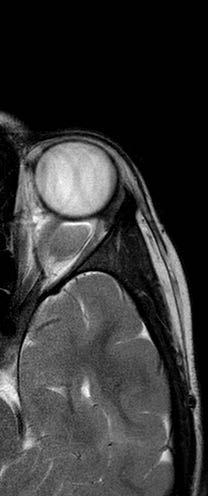
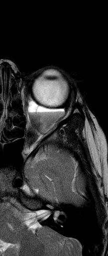
3 Dermoid Cyst Congenital Keratinized epidermis Dermal appendage Trapped during embryogenesis 6% of lesions 40-50% of orbital pediatric orbital lesion Usually discovered in the first year of life Painless/firm/subQ mass Rarely presents as an acute inflammatory lesion (Rupture?) Frontozygomatic (70%) Maxillofrontal (20%) suture


4 Imaging - CT Erosion/remodeling of bone Adjacent bony changes: smooth fossa (85%) Dumbell dermoid: extraorbital and intraorbital components through bony defect


5 Imaging - MRI Encapsulated Enhancement of wall but not lumen

6


7 Treatment Options Observation Risk of anesthesia Surgical Removal Changes to bone Rupture of cyst can lead to acute inflammation Irrigation Abx Steroids


8 Dermoid
 Clinical findings Proptosis Strabismus Astigmatism")
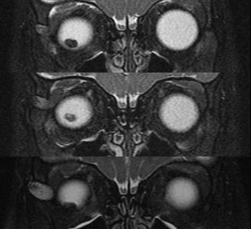
9 INFANTILE/Capillary Hemangioma Common BENIGN orbital lesion of children F>M Prematurity Appears in 1 st or 2 nd week of life Soft, bluish mass deep to the eyelid Superonasal orbit Rapidly expands over 6-12 months Increases with valsalva (crying) Clinical findings Proptosis Strabismus Astigmatism Amblyopia


10 INFANTILE/Capillary Hemangioma May enlarge for 1-2 years then regress 70-80% resolve before age 7 HIGH flow on doppler Kasabach-Merritt Syndrome Multiple large visceral capillary hemangiomas Sequestration of platelets into tumor Consumptive thrombocytopenia Supportive therapy and treat underlying tumor Complications DIC death

11 Homogenous soft tissue mass that infiltrates throughout the orbit MRI: Serpiginous Signal Voids because of high flow rate (stimulated vs. unstimulated blood) Isointense to brain on pre-contrast

12
13 INFANTILE/Capillary HemangiomaManagement Small lesions without visual compromise Observation 75% of lesions spontaneously resolve over 4-5 years Large lesions with risk of amblyopia Treat Intralesional steroids Complications: skin depigmentation, fat atrophy, eyelid necrosis, CRAO Interferon-alpha Laser oral steroids Propranolol Excision
14 Lymphatic malformation Benign Unencapsulated Hamartoma Composed of thinned-walled vascular channels Not well defined: diffuse, infiltrating Typically persist for life 10% may have involvement of other head and neck structures Palatal mucosa
15 Lymphatic malformation Signs/Symptoms Sudden proptosis Spontaneous hemorrhage chocolate cyst Enlarge with URI Enhances with contrast Difficult to debulk Defer surgery unless absolutely necessary Drainage/Sclerotherapy/Sildenafil

16 Lymphatic malformation


17

18


19 10/17/ /4/2008


20

21

22 Rhabdomyosarcoma Most common MALIGNANT orbital tumor in children 1% of all biopsied masses 4-5% of pediatric orbital masses 40% of pediatric malignant orbital masses Average Age: 8 75% in first decade Up to 78 yo Females > Males Arises from pluriopotential mesenchymal cells NOT from striated EOM
23 Rhabdomyosarcoma Can originate primarily in orbit or surrounding sinus Acute Rapid proptosis (80-100%) Unilateral Superonasal location Globe displacement (80%) Ptosis (30-50%) Eyelid swelling (60%) 20% lid signs dominate Pain (10%) Palpable mass (25%)


24 Imaging CT: moderately well circumscribed but irregular, homogenous mass. +/- adjacent bony destruction Enhances with contrast T1 MRI Isointense to EOM Heterogeneous enhancement T2 MRI Hyperintense to fat and muscle
25 Rhabdomyosarcoma Four Types Embryonal Most Common (80%) Superonasal Good survival rate Alveolar Most Aggressive Inferior Orbit Pleomorphic Most Benign Most rare Typically in adults, extremeties not orbit Botryoid Grapelike appearance Extends to orbit from adjacent sinus or conjunctiva
26 Rhabdomyosarcoma Management Biopsy/Surgical debulking Systemic evaluation to r/o metastatic dz (i.e.lung, brain, lymph nodes) Staging (Intergroup Rhabdomyosarcoma Study Group) I Localized disease, completely resected II Microscopic disease remaining after biopsy III Gross residual disease remaining after biopsy IV Distant metastasis present at onset Chemotherapy and radiotherapy Prognosis Improved greatly since early 70 s: 74% (Alveolar) 94% (Embryonal) Favorable location Tumor morphology Age of presentation Infants under 1 have worse prognosis

27

28 Metastatic Neuroblastoma Most common met. orbital tumor in children 90% before age of 10 Mean Age: % of neuroblastomas met. to orbit 8% present with ophthalmic features Usually from ADRENALS Also: retroperitoneal, mediastinum, neck 60-72% in abdomen Proptosis, ptosis, ecchymosis Bilateral 60% Paraneoplastic opsoclonus
29 Metastatic Neuroblastoma Work up Palpate abdomen CT: orbit, neck, chest, abdomen and pelvis Serology for catecholamines Bone Scan Bone marrow biopsy Radiation Chemotherapy Treatment
30 Optic Nerve Glioma Usually benign First decade F>M 25-50% associated with neurofibromatosis 1 Clinical Presentation Gradual growth Painless Unilateral axial proptosis Vision loss APD
31 OPTIC NERVE GLIOMA Malignant ONG (glioblastoma) Rare Adult males Clinical Presentation Massive swelling and hemorrhage of optic nerve head Pain Treatment Chemotherapy High-dose radiationtherapy Prognosis Poor

32 OPTIC NERVE GLIOMA Fusiform enlargement of ON with kinking

33 Optic Nerve Glioma Other ocular findings Swollen optic disc Retinochoroidal shunt vessels Strabismus Nystagmus Because of NF1 association, check: Café-au-lait spots Iris Lisch nodules
34 Optic Nerve Glioma Observation Good vision Follow closely Surgical excision Goal is to isolate from chiasm Lesions that involve chiasm and brain can be fatal When hypothalamus is involved mortality increases from 5% to 50% Radiation If tumor cannot be resected (in chiasm) Chemotherapy May delay need for radiation
35 Optic nerve sheath Meningioma Benign neoplasms that originate from arachnoid layer of meninges Women (80%) in 3 rd -4 th decade Present Gradual, unilateral painless vision loss Optic atrophy, optociliary shunts Clinical association Neurofibromatosis 2 People with NF-2 have a higher incidence but only a minority of meningiomas have NF-2

36 Optociliary shunt vessels DDx: Optic nerve sheath meningioma CRVO Chronic Glaucoma Chronic papilledema

37 Optic nerve sheath Meningioma Tram tracking on CT
38 Optic nerve sheath Meningioma Observation Radiotherapy Chemotherapy Surgery reserved for patients with intracranial extension or severe visual loss

39 Sphenoid wing meningioma Begins in arachnoid that lines the sphenoid Invades orbit from intracranial space Assoc with NF2 CT: Hyperostotic sphenoid bone

40

41

42

43

44
45 Sphenoid wing meningioma Treatment Close observation Surgical resection Radiation Hormone therapy (future)


46 Lacrimal Drainage System 12-15mm Valve Lacrimal of Rosenmüller Sac 2mm vertical canaliculi Posterior medial Canthal ligament 8-10 mm horizontal canaliculi with common canaliculus 12-18mm nasolacrimal duct Anterior medial Canthal ligament Anterior and posterior Lacrimal crests form the Lacrimal sac fossa Valve of Hasner in inferior meatus Fay & Dolman 46

47 Schirmer Testing Schirmer Testing Schirmer I No topical anesthetic Schirmer strip for 5 min Tests basal and reflex tearing Normal >10 Schirmer II No topical anesthetic Schirmer strip for 5 min Nasal stimulation for reflex tearing Normal >15 Basal Tear Secreation Topical anesthetic Schirmer strip for 5 min Normal = 10-15mm 47 Fay & Dolman

48 Canalicular Probing BCSC 48

49 Dye Disappearance Test Fluorescein instilled into fornices Persistence of dye or asymmetric persistence at 5 min suggest obstruction BCSC 49
50 Jones Testing Dye instilled into fornix Jones 1 (physiologic) Cotton tipped applicator at inferior meatus 2-5 min later If negative test, proceed to Jones 2 Jones 2 (non-physiologic) Wipe dye from fornix Irrigate with clear saline No fluid on Q-tip = anatomic blockage Clear fluid on Q-tip = lacrimal pump failure Dye on Q-tip = functional obstruction 50 Fay & Dolman

51 Lacrimal Irrigation A. Same canaliculus clear fluid return: Canalicular obstruction B. Opposite canaliculus clear fluid return: Common canalicular obstruction C. Opposite canaliculus mucoid fluid return: NLDO D. Opposite canaliculus and nose clear fluid return: Partial NLDO E. No fluid return: Patent NLD 51 BCSC
 Fay &")
52 Lacrimal Drainage Abnormalities Congenital lacrimal-cutaneous fistula Intranasal to medial canthus Tearing from the skin 1/3 may have NLDO If simple without NLDO direct surgical excision If NLDO Add DCR Aplasia/Hypoplasia/Stenosis Punctal aplasia/stenosis Manage with probing and intubation Complete absence of canalicular system CDCR (Jones tube) Fay & Dolman 52
53 Lacrimal Drainage Abnormalities Congenital NLDO Membrane blocking valve of Hasner Canalization of NLD completed late in pregnancy or shortly after birth 20% of babies may have watery eyes 2-6% of babies will have clinically evident epiphora 95% resolve spontaneously in first year of life Management Conservative management Observation Crigler lacrimal massage Antibiotics for infections Surgical Management >1year Probe Probe and intubate 53
54 Lacrimal Drainage Abnormalities Dacryocystocele Secondary to congenital NLDO with amniotic fluid collection Clinically swelling below the MCT Swelling above MCT Meningoencephalocele Dermoid cyst Management Crigler lacrimal massage Probing If airway bilateral with obstruction Urgent marsupialization 54

55

56

57

58
59 Conclusion Dermoid cyst is the most common orbital tumor of children Rhabdomyosarcoma is the most common malignant tumor of children Infantile hemangioms are effectively treated with propranolol Dacryocystocele below MCT consider imaging if in doubt Rule out meningoencephalocele Clinical Finding Sphenoid wing hypoplasia Optic nerve glioma Sphenoid wing meningioma Optic nerve sheath meningioma Systemic Association NF1 NF1 NF2 NF2 59
Orbital Tumors - A Clinico Pathological Study
 Orbital Tumors - A Clinico Pathological Study Radha. J. DO, Ani Sreedhar. MS. Little Flower Hospital, Angamaly, Kerala ORIGINAL ARTICLES Abstract: Aim. To study the clinical and histopathological profiles
NASOLACRIMAL DUCT OBSTRUCTION (BLOCKED TEAR DUCT) AND TEARY EYE - PATIENT INFORMATION
 AND TEARY EYE - PATIENT INFORMATION") NASOLACRIMAL DUCT OBSTRUCTION (BLOCKED TEAR DUCT) AND TEARY EYE - PATIENT INFORMATION What is lacrimal sac and nasolacrimal duct? The lacrimal apparatus is composed of a lacrimal gland (tear producing
NASOLACRIMAL DUCT OBSTRUCTION (BLOCKED TEAR DUCT) AND TEARY EYE - PATIENT INFORMATION What is lacrimal sac and nasolacrimal duct? The lacrimal apparatus is composed of a lacrimal gland (tear producing
Advances In Orbital Neuropathology
 Advances In Orbital Neuropathology Charles G. Eberhart, MD PhD Associate Professor of Pathology, Ophthalmology and Oncology Johns Hopkins University School of Medicine Overview Non-neoplastic lesions Microphthalmos/pseudoglioma
Clinical ophthalmology: a systematic approach. Jack Kanski Chapters 1, 2 and 17. Between lesser and greater wings of sphenoid.
 1 Orbital Disease Dr Sarah Osborne Learning objectives Knowledge of the anatomy of the normal nasolacrimal system Understand the causes of epiphora Identify common skin tumours Recognise the signs of thyroid
Ocular warning signs in GP practice: Paediatric Eye Pointers
 Ocular warning signs in GP practice: Paediatric Eye Pointers Dr Benjamin Chang MB, BCh, BAO, MMedSci, FRCS(Irel), FRCS(Edin), FRCOphth(Lond) Senior Consultant Ophthalmology and Visual Sciences Khoo Teck
CLINICAL PEARLS IN OCULAR ONCOLOGY
 CLINICAL PEARLS IN OCULAR ONCOLOGY IRIS NEVUS - Two kinds circumscribed and diffuse - Photodocumentation important to monitor growth - Risk Factors for iris nevus growth to melanoma (ABCDEF) A Age (young),
Pediatric Ocular Sonography
 Pediatric Ocular Sonography Cicero J Torres A Silva, MD Associate Professor of Radiology 2016 SPR Pediatric Ultrasound Course Yale University School of Medicine None Disclosures Objectives of Presentation
Year 2003 Paper two: Questions supplied by Tricia
 question 43 A 42-year-old man presents with a two-year history of increasing right facial numbness. He has a history of intermittent unsteadiness, mild hearing loss and vertigo but has otherwise been well.
13/02/1440 بسم ا هلل ا لرحمن ا لر حيم
 بسم ا هلل ا لرحمن ا لر حيم 1 Slowly progressive versus rapidly progressive proptosis by Ali M ISMAIL professor of ophthalmology @SOHAG U H Occuloplastic fellow @NNUH Occuloplastic fellow @Cambridge UH
Vision Health: Conditions, Disorders & Treatments EYELID DISORDERS
 Vision Health: Conditions, Disorders & Treatments EYELID DISORDERS There are a number of disorders that can affect the eyelid. Entropion Entropion is an inward turning of the eyelid and lashes toward the
C. Douglas Phillips MD FACR Director of Head and Neck Imaging Weill Cornell Medical College/NewYork-Presbyterian Hospital
 C. Douglas Phillips MD FACR Director of Head and Neck Imaging Weill Cornell Medical College/NewYork-Presbyterian Hospital Disclosures Neither I nor any family members have any pertinent financial relations
MRI masterfile Part 5 WM Heme Strokes.ppt 1
 Ocular and Orbital Trauma Eye Trauma: Incidence 1.3 million eye injuries in the US per year. 40,000 of these injuries lead to blindness in the US. Patrick Sibony, MD March 23, 2013 Ophthalmic Emergencies
A Case of Carotid-Cavernous Fistula
 A Case of Carotid-Cavernous Fistula By : Mohamed Elkhawaga 2 nd Year Resident of Ophthalmology Alexandria University A 19 year old male patient came to our outpatient clinic, complaining of : -Severe conjunctival
Orbital facia. Periororbital facia Orbital septum Bulbar facia Muscular facia
 Anatomy Orbital facia Periororbital facia Orbital septum Bulbar facia Muscular facia Physiology of symptoms 1) Proptosis ( exophthalmos) Pseudoproptosis Axial Non axial Pulsating Positional Intermittent
Mom, There s Something Wrong With My Eye
 Mom, There s Something Wrong With My Eye Veeral Shah MD, PHD Texas Children's Hospital Most Common Issues Seen by the Pediatrician Emergent Ocular Issues Seen by the Pediatrician 1 What does this baby
Endoscopic Assisted resection for congenital Midline Nasal Mass
 Endoscopic Assisted resection for congenital Midline Nasal Mass Ahmed Aly Ibrahim A.prof ORL Department Alexandria University Emad. A Magdy prof ORL Department Alexandria University Haytham Morsi,MD Mohammad
Assessment and Management of Ocular Trauma. Disclosure I have no direct financial interests in today s subject matter. 3/25/2019. Normal Eye Anatomy
 Assessment and Management of Ocular Trauma Samiksha Fouzdar Jain, MD,FRCS Department of Ophthalmology & Visual Sciences Truhlsen Eye Institute Disclosure I have no direct financial interests in today s
Bony orbit Roof The orbital plate of the frontal bone Lateral wall: the zygomatic bone and the greater wing of the sphenoid
 Bony orbit Roof: Formed by: The orbital plate of the frontal bone, which separates the orbital cavity from the anterior cranial fossa and the frontal lobe of the cerebral hemisphere Lateral wall: Formed
Wilms Tumor and Neuroblastoma
 Wilms Tumor and Neuroblastoma Wilm s Tumor AKA: Nephroblastoma the most common intra-abdominal cancer in children. peak incidence is 2 to 3 years of age Biology somatic mutations restricted to tumor tissue
Dr. Najah طب بغداد. Lecture: 14
 2015 2016 The lacrimal apparatus The lacrimal system consists of: Lecture: 14 Dr. Najah طب بغداد 1- Secretory portion: Comprises the following: a- The lacrimal gland: Which is divided into palpebral (Aqueoussecretion)
PEDIATRICS WK 3 HEAD AND NECK ALISON WALLACE MD, PHD
 PEDIATRICS WK 3 HEAD AND NECK ALISON WALLACE MD, PHD Topics 1. Cervical lymphadenopathy 2. Lymphatic malformation 3. Thyroglossal duct cysts 4. Branchial cleft cysts 5. Thyroid masses CASE 1 Case 1 A 2
Optic Pathway Gliomas, Germinomas, Spinal Cord Tumours. Colin Kennedy March 2015
 Optic Pathway Gliomas, Germinomas, Spinal Cord Tumours Colin Kennedy March 2015 Glioma of the optic chiasm. T1-weighted MRI with gadolinium enhancement, showing intense irregular uptake of contrast. The
Juvenile Angiofibroma
 Juvenile Angiofibroma Disclaimer The pictures used in this presentation have been obtained from a number of sources. Their use is purely for academic and teaching purposes. The contents of this presentation
Pediatric Epiphora. Lacrimal drainage system begins to develop at the. Pediatric Ophthalmology. Saurbhi Khurana MD, FICO
 Pediatric Ophthalmology Pediatric Epiphora Saurbhi Khurana MD, FICO Saurbhi Khurana MD, FICO, A.K Grover MD, MNAMS, FRCS (Glasgow) FIMSA, FICO Vision Eye Centre, Siri Fort Road, New Delhi Lacrimal drainage
Optic Nerve Disorders: Structure and Function and Causes
 Optic Nerve Disorders: Structure and Function and Causes Using Visual Fields, OCT and B-scan Ultrasound to Diagnose and Follow Optic Nerve Visual Losses Ohio Ophthalmological Society and Ophthalmic Tech
Dr.Dafalla Ahmed Babiker Jazan University
 Dr.Dafalla Ahmed Babiker Jazan University Brain tumors are the second commonest malignancy in children Infratentorial tumors are more common As a general rule they do not metastasize out of the CNS, but
Technicians & Nurses Program
 ASCRS ASOA Symposium & Congress Technicians & Nurses Program May 6-10, 2016 New Orleans Evaluation and Treatment of Eyelid Malignancies Richard C. Allen MD PhD FACS Professor Section of Ophthalmology Dept.
BILATERAL OPTIC MALIGNANT ASTROCYTOMA IN A 3 YEAR OLD CHILD WITH NFI CASE PRESENTATION
 BILATERAL OPTIC MALIGNANT ASTROCYTOMA IN A 3 YEAR OLD CHILD WITH NFI CASE PRESENTATION BOGDAN ILIESCU 1, M. VUKIC 2, ZIYAD FAIYAD 1, RAMONA FILIPESCU*, ION POEATA 1 1 3rd Neurosurgery Department, Prof.
The Investigation of Proptosis in Paediatric Practice.
 The Investigation of Proptosis in Paediatric Practice. Ms Sayantani Ghosh 1, Mr Saugat Dey 1 1 Bankura Sammilani Medical College and Hospital, 136, Dr. Meghnad Saha Road, PRATYASHA APARTMENT Flat-4C, KOLKATA-
Ocular and Periocular Trauma. Tina Rutar, MD. Assistant Professor of Ophthalmology and Pediatrics. Director, Visual Center for the Child
 Ocular and Periocular Trauma Tina Rutar, MD Assistant Professor of Ophthalmology and Pediatrics Director, Visual Center for the Child University of California, San Francisco Phone: 415-353-2560 Fax: 415-353-2468
Orbital and Ocular Adnexal Disorders with Red Eyes
 Orbital and Ocular Adnexal Disorders with Red Eyes Jason Lee Associate Consultant Department of Ophthalmology and Visual Sciences Practical Ophthalmology for the Family Physician 21 Jan 2017 No financial
Paediatric acute ophthalmology. Harry Bradshaw
 Paediatric acute ophthalmology Harry Bradshaw Approach Red eye Leukocoria Neurological Trauma Visual loss Red eye Orbital Eyelid Conjunctiva Cornea Uvea Orbital Orbit fixed volume Contiguous with sinuses,
Lower Eyelid Malposition
 Oculoplastic Surgeon s DDX for the Red Eye Geeta Belsare Been,MD The Center for Facial Plastic Surgery Barrington, IL Lower Eyelid Malposition Ectropion Involutional Cicatricial Paralytic Entropion Involutional
Diplomate of the American Board of Pathology in Anatomic and Clinical Pathology
 A 33-year-old male with a left lower leg mass. Contributed by Shaoxiong Chen, MD, PhD Assistant Professor Indiana University School of Medicine/ IU Health Partners Department of Pathology and Laboratory
The many faces of extranodal lymphoma
 The many faces of extranodal lymphoma Frank Pameijer Departments of Radiology and Radiation Oncology University Medical Center Utrecht Special thanks to Ilona M Schmalfuss, MD University of Florida Gainesville,
The orbit-2. Dr. Heba Kalbouneh Assistant Professor of Anatomy and Histology
 The orbit-2 Dr. Heba Kalbouneh Assistant Professor of Anatomy and Histology Eyelids The eyelids (act like the curtains) protect the eye from injury and excessive light by their closure The upper eyelid
The sebaceous glands (glands of Zeis) open directly into the eyelash follicles, ciliary glands (glands of Moll) are modified sweat glands that open
 open directly into the eyelash follicles, ciliary glands (glands of Moll) are modified sweat glands that open") The Orbital Region The orbits are a pair of bony cavities that contain the eyeballs; their associated muscles, nerves, vessels, and fat; and most of the lacrimal apparatus upper eyelid is larger and more
The Orbital Region The orbits are a pair of bony cavities that contain the eyeballs; their associated muscles, nerves, vessels, and fat; and most of the lacrimal apparatus upper eyelid is larger and more
Chapter 2 Nonmalignant Tumors of the Orbit
 Chapter 2 Nonmalignant Tumors of the Orbit Eric M. Hink and Vikram Durairaj Abstract Most orbital tumors are nonmalignant. Nonmalignant orbital tumors can arise from any of the structures within the orbit,
Examining Children s Eyes
 Paediatric Ophthalmology What to refer & when? Aims Tips for assessing a child s eyes in general practice Common paediatric ophthalmology symptoms and signs What needs to be referred and when? MISS FARIHA
MRI masterfile Part 5 WM Heme Strokes.ppt 2
 Imaging of Orbital Trauma Corneal Abrasion CT scan is preferable to MRI Bone, Rapid, Easy to monitor patient Foreign bodies, air, hemorrhage Fractures Cost Needed for an MRI MRI Globe and intraocular injuries
Pediatric Orbital Tumours in Upper Egypt: A 3-year Retrospective Analysis at a University Hospital.
 Commentary http://www.alliedacademies.org/clinical-ophthalmology-and-vision-science/ Pediatric Orbital Tumours in Upper Egypt: A 3-year Retrospective Analysis at a University Hospital. Ahmad Mostafa Abdallah
Neuro-Ocular Grand Rounds
 Neuro-Ocular Grand Rounds Anthony B. Litwak,OD, FAAO VA Medical Center Baltimore, Maryland Dr. Litwak is on the speaker and advisory boards for Alcon and Zeiss Meditek COMMON OPTIC NEUROPATHIES THAT CAN
1 Eyelids. Lacrimal Apparatus. Orbital Region. 3 The Orbit. The Eye
 1 1 Eyelids Orbital Region 2 Lacrimal Apparatus 3 The Orbit 4 The Eye 2 Eyelids The eyelids protect the eye from injury and excessive light by their closure. The upper eyelid is larger and more mobile
Kidney Case 1 SURGICAL PATHOLOGY REPORT
 Kidney Case 1 Surgical Pathology Report February 9, 2007 Clinical History: This 45 year old woman was found to have a left renal mass. CT urography with reconstruction revealed a 2 cm medial mass which
Nasolacrimal Duct Blockage
 The eyelids play a key role in protecting the eyes. They help spread moisture (tears) over the surface of the eyes when they close (for example, while blinking); thus, they help prevent the eyes from becoming
DISORDERS OF THE SALIVARY GLANDS Neoplasms Dr.M.Baskaran Selvapathy S IV
 DISORDERS OF THE SALIVARY GLANDS Neoplasms Dr.M.Baskaran Selvapathy S IV NEOPLASMS A) Epithelial I. Benign Pleomorphic adenoma( Mixed tumour) Adenolymphoma (Warthin s tumour) Oxyphil adenoma (Oncocytoma)
Ocular and periocular trauma
 Ocular and periocular trauma No financial disclosures. Tina Rutar M.D. Assistant Professor of Clinical Ophthalmology and Pediatrics Director, Visual Center for the Child University of California San Francisco
Objectives. 1. Recognizing benign skin lesions. 2.Know which patients will likely need surgical intervention.
 The Joy of Pediatric Skin Dr. Claire Sanger University of Kentucky Plastic & Reconstructive Surgery Objectives 1. Recognizing benign skin lesions 2.Know which patients will likely need surgical intervention.
Hemangiomas and Other Vascular Tumors
 facebook.com/cincykidsrad Hemangiomas and Other Vascular Tumors Disclosures No relevant financial disclosures Bernadette L. Koch, M.D. Departments of Radiology and Pediatrics Cincinnati Children s Hospital
J of Evolution of Med and Dent Sci/ eissn , pissn / Vol. 4/ Issue 80/ Oct. 05, 2015 Page 14022
 STUDY ON MANAGEMENT OF EPIPHORA IN PATIENTS WITH OBSTRUCTION AT VARIOUS LEVELS OF NASOLACRIMAL APPARATUS S. Shivranjani 1, Jaishree Dwivedi 2, Neha Mithal 3, Sandeep Mithal 4 HOW TO CITE THIS ARTICLE:
Meningioma tumor. Meningiomas are named according to their location (Fig. 1) and cause various symptoms: > 1
 and cause various symptoms: > 1") Meningioma tumor Overview A meningioma is a type of tumor that grows from the protective membranes, called meninges, which surround the brain and spinal cord. Most meningiomas are benign (not cancer) and
Meningioma tumor Overview A meningioma is a type of tumor that grows from the protective membranes, called meninges, which surround the brain and spinal cord. Most meningiomas are benign (not cancer) and
Imaging the Spinal Cord & Intradural Disease
 Department of Radiology University of California San Diego Imaging the Spinal Cord & Intradural Disease John R. Hesselink, M.D. Spinal Cord Diseases Tumors Syringohydromyelia Trauma Ischemia / Infarction
monitored anesthesia care (MAC)
") Entropion Entropion Entropion is an inward turning of the eyelid and lashes toward the eye, usually caused by relaxation of the eye muscles and tissue due to aging. Entropion usually affects the lower
Entropion Entropion Entropion is an inward turning of the eyelid and lashes toward the eye, usually caused by relaxation of the eye muscles and tissue due to aging. Entropion usually affects the lower
2. The clinician will know how to manage common pediatric ocular diseases
 Ida Chung, OD, MSHE, FCOVD, FAAO Western University College of Optometry Associate Professor/Assistant Dean of Learning 309 E. Second Street, Pomona, CA 91766 Office: 909 938 4140 Email: ichung@westernu.edu
Learn Connect Succeed. JCAHPO Regional Meetings 2017
 Learn Connect Succeed JCAHPO Regional Meetings 2017 Financial Disclosure Evaluation and Treatment of Orbital Cellulitis Thomas E. Johnson, M.D. Bascom Palmer Eye Institute University of Miami School of
Disclosures. Visual Pathways. Visual Pathways. Visual Loss Understanding the Patterns. I have no financial disclosures. Tabby A.
 Visual oss Understanding the Patterns Tabby A. Kennedy, MD University of Wisconsin Department of adiology I have no financial disclosures Acknowledgements: indell Gentry Greg Avey JP Yu Judy Chen Disclosures
Neuro-Ocular Grand Rounds Anthony B. Litwak,OD, FAAO VA Medical Center Baltimore, Maryland
 Neuro-Ocular Grand Rounds Anthony B. Litwak,OD, FAAO VA Medical Center Baltimore, Maryland Dr. Litwak is on the speaker and advisory boards for Alcon and Zeiss Meditek COMMON OPTIC NEUROPATHIES THAT CAN
EYE TRAUMA: INCIDENCE
 Introduction EYE TRAUMA: INCIDENCE 2.5 million eye injuries per year in U.S. 40,000 60,000 of eye injuries lead to visual loss Introduction Final visual outcome of many ocular emergencies depends on prompt,
Orbital Tumors October 2001
 TITLE: Orbital Tumors SOURCE: Grand Rounds Presentation, UTMB, Dept. of Otolaryngology DATE: October 31, 2001 RESIDENT PHYSICIAN: Michael Underbrink, MD FACULTY ADVISOR: Shawn Newlands, MD SERIES EDITORS:
INFECTION. HIV Infection DWI
 HIV Infection INFECTION DWI Fig Axial CT and MRI images show multiple enlarged lymph nodes in the neck as well as in the parotid gland bilaterally. These nodes were suppurative with positive diffusion.
Imaging Work-Up of a Neck Mass - Adults & Children
 Disclosures Imaging Work-Up of a Neck Mass - Adults & Children I have nothing to disclose Christine M Glastonbury MBBS Professor of Radiology & Biomedical Imaging Otolaryngology-Head & Neck Surgery and
Imaging features of orbital neoplasm developed in pediatrics
 Imaging features of orbital neoplasm developed in pediatrics Poster No.: C-1119 Congress: ECR 2015 Type: Educational Exhibit Authors: J. H. Yoo; Seoul/KR Keywords: Eyes, Head and neck, Paediatric, CT,
Malignant Cardiac Tumors Rad-Path Correlation
 Malignant Cardiac Tumors Rad-Path Correlation Vincent B. Ho, M.D., M.B.A. 1 Jean Jeudy, M.D. 2 Aletta Ann Frazier, M.D. 2 1 Uniformed Services University of the Health Sciences 2 University of Maryland
Plexiform Tumor of the Orbit
 Plexiform Tumor of the Orbit Anat Stemmer-Rachamimov, MD Department of Pathology Massachusetts General Hospital Harvard Medical School Disclosure of Relevant Financial Relationships USCAP requires that
DISCLOSURES. PEDIATRIC RED EYES Rachel M. Smith, OD, FCOVD HISTORY, HISTORY, HISTORY WHY RED EYES? EXAMINE THE EYE RED FLAGS TO REFER 3/25/2019
 DISCLOSURES Consultant/Speakers bureaus Research funding PEDIATRIC RED EYES Rachel M. Smith, OD, FCOVD Pediatric Optometrist Children s Hospital & Medical Center Stock ownership/corporate boards employment
Chapter 16: Proptosis. W. Bruce Wilson. Plan of Evaluation
 Chapter 16: Proptosis W. Bruce Wilson The term proptosis as generally used is synonymous with exophthalmos. The term signifies that one or both eyes are displaced in a direction anterior to their normal
Pediatric Soft-Tissue Sarcomas. Beth McCarville, MD St. Jude Children s Research Hospital Memphis, Tn
 Pediatric Soft-Tissue Sarcomas Beth McCarville, MD St. Jude Children s Research Hospital Memphis, Tn Overview Histologic classifications Characteristic imaging features Helpful clinical characteristics
A rare cause of intermittent respiratory distress and epiphora in the newborn: congenital dacryocystocele
 Case Report A rare cause of intermittent respiratory distress and epiphora in the newborn: congenital dacryocystocele Onur Ismi 1, Fatma Merve Bozkurt 2, Gokhan Icme 2, Can Eti 3, Ayca Sari 4 1 Department
Update on RECIST and Staging of Common Pediatric Tumors Ethan A. Smith, MD
 Update on RECIST and Staging of Common Pediatric Tumors Ethan A. Smith, MD Section of Pediatric Radiology C.S. Mott Children s Hospital University of Michigan ethans@med.umich.edu Disclosures No relevant
Case Scenario 1 Worksheet. Primary Site C44.4 Morphology 8743/3 Laterality 0 Stage/ Prognostic Factors
 CASE SCENARIO 1 9/10/13 HISTORY: Patient is a 67-year-old white male and presents with lesion located 4-5cm above his right ear. The lesion has been present for years. No lymphadenopathy. 9/10/13 anterior
Evaluation of Neck Mass. Disclosure. Learning Objectives 3/24/2014. Karen T. Pitman MD, FACS Banner MDACC, Gilbert AZ. Nothing to disclose
 Evaluation of Neck Mass Karen T. Pitman MD, FACS Banner MDACC, Gilbert AZ Nothing to disclose Disclosure Learning Objectives 1. Describe a systematic method to evaluate a patient with a neck mass 2. Select
Sepideh Tara Rousta, MD FAAO Robert Wood Johnson University Hospital Saint Peter s University Hospital Wills Eye Hospital
 Sepideh Tara Rousta, MD FAAO Robert Wood Johnson University Hospital Saint Peter s University Hospital Wills Eye Hospital 14 mo old w R eye cross (parents) 9 mo old R eye crossing getting worse for past
PUBLISHED VERSION.
 PUBLISHED VERSION Shay Keren, Gad Dotan, Leah Leibovitch, Dinesh Selva, and Igal Leibovitch Indocyanine green assisted removal of orbital lacrimal duct cysts in children Ophthalmology, 2015; 2015:130215-1-130215-5
Congenital lacrimal fistula
 Ugurbas 6-03-2000 14:59 Pagina 22 European Journal of Ophthalmology / Vol. 10 no. 1, 2000 / pp. 22-26 S.H. UǦURBAŞ, G. ZILELIOǦLU Department of Ophthalmology, Ankara University Faculty of Medicine, Ankara
Study of success rates in endoscopic dacryocystorhinostomy with and without stenting. dacryocystorhinostomy with and
 Original Research Article Study of success rates in endoscopic dacryocystorhinostomy with and without stenting Kirti Ambani 1, Niraj Suri 2, Hiren Parmar 3* 1 Assistant Professor, ENT Department, GMERS
Remember from the first year embryology Trilaminar disc has 3 layers: ectoderm, mesoderm, and endoderm
 Development of face Remember from the first year embryology Trilaminar disc has 3 layers: ectoderm, mesoderm, and endoderm The ectoderm forms the neural groove, then tube The neural tube lies in the mesoderm
The Child With An Abdominal Mass
 The Child With An Abdominal Mass Today we are going to talk about pediatric surgery, the abdominal masses in children. Firstly we have to take a full history and make a general, local and rectal examination
Pathology of Sarcoma ELEANOR CHEN, MD, PHD, ASSISTANT PROFESSOR DEPARTMENT OF PATHOLOGY UNIVERSITY OF WASHINGTON
 Pathology of Sarcoma ELEANOR CHEN, MD, PHD, ASSISTANT PROFESSOR DEPARTMENT OF PATHOLOGY UNIVERSITY OF WASHINGTON Presentation outline Background and epidemiology of sarcomas Sarcoma classification Sarcoma
PITUITARY PARASELLAR LESIONS. Kim Learned, MD
 PITUITARY PARASELLAR LESIONS Kim Learned, MD DIFFERENTIALS Pituitary Sella Clivus, Sphenoid Sinus Suprasellar Optic chiasm, Hypothalamus, Circle of Willis Parasellar Cavernous Sinus Case 1 17 YEAR-OLD
Core Curriculum Syllabus Emergencies in Otolaryngology-Head and Neck Surgery FACIAL FRACTURES
 Core Curriculum Syllabus Emergencies in Otolaryngology-Head and Neck Surgery A. General Considerations FACIAL FRACTURES Look for other fractures like skull and/or cervical spine fractures Test function
Congenital Neck Masses C. Stefan Kénel-Pierre, MD
 Congenital Neck Masses C. Stefan Kénel-Pierre, MD SUNY-LICH Medical Center Department of Surgery Case Presentation xx year old male presents with sudden onset left lower neck swelling x 1 week Denies pain,
Musculoskeletal Sarcomas
 Musculoskeletal Sarcomas Robert C. Orth, M.D., Ph.D. Edward B. Singleton Department of Pediatric Radiology Texas Children s Hospital Page 0 xxx00.#####.ppt 9/23/2012 9:01:18 AM No disclosures Page 1 xxx00.#####.ppt
Around The Globe in 60 Minutes
 Around The Globe in 60 Minutes Around the GLOBE in Sixty Minutes Basic Ocular Anatomy, Examination, and Diagnostic Techniques Introduction Focusing on canine and feline ocular anatomy and basic examination
CT of Maxillofacial Fracture Patterns. CT of Maxillofacial Fracture Patterns
 CT of Maxillofacial Fracture Patterns CT of Maxillofacial Fracture Patterns Stuart E. Mirvis, M.D., FACR Department of Radiology University of Maryland School of Medicine Viking 1 1976 MGS 2001 Technology
Sonography of soft-tissue vascular lesions
 Sonography of soft-tissue vascular lesions Oscar M. Navarro Associate Professor, University of Toronto Dept. of Diagnostic Imaging, The Hospital for Sick Children Toronto, Canada Declaration of Disclosure
Soft Tissue Sarcomas: Questions and Answers
 Soft Tissue Sarcomas: Questions and Answers 1. What is soft tissue? The term soft tissue refers to tissues that connect, support, or surround other structures and organs of the body. Soft tissue includes
ASSESSING THE EYES. Structures. Eyelids Extraocularmuscles Eyelashes Lacrimal glands: Lacrimal ducts Cornea Conjunctiva Sclera Pupils Iris.
 ASSESSING THE EYES Structures External Eyelids Extraocularmuscles Eyelashes Lacrimal glands: Lacrimal ducts Cornea Conjunctiva Sclera Pupils Iris 1 2 Structures Internal Optic disc Physiological cup Retinal
Minimally Invasive Technique of Dacryocystorhinostomy
 Cronicon OPEN ACCESS EC OPHTHALMOLOGY Research Protocol Mohammad Idrees* Consultant Ophthalmology, REDO Eye Hospital, Rawalpindi, Pakistan *Corresponding Author: Mohammad Idrees, Consultant Ophthalmology,
Neckmasses in infancy and childhood: Clinical and radiological classification and imaging approaches M. Mearadji
 Neckmasses in infancy and childhood: Clinical and radiological classification and imaging approaches M. Mearadji International Foundation for Pediatric Imaging Aid Introduction Neck masses are a frequent
3/27/2017. Disclosure of Relevant Financial Relationships
 Ophthalmic Pathology Evening Specialty Conference USCAP 2017 5 th March, 2017 Mukul K. Divatia, MD Assistant Professor Department of Pathology & Genomic Medicine Weill Cornell Medical College Houston Methodist
1/25/13 Right partial nephrectomy followed by completion right radical nephrectomy.
 History and Physical Case Scenario 1 45 year old white male presents with complaints of nausea, weight loss, and back pain. A CT of the chest, abdomen and pelvis was done on 12/8/12 that revealed a 12
Paper #A The efficacy of a video-based online teaching tool for medical students in oculoplastic surgery
 Paper #A-00051 The efficacy of a video-based online teaching tool for medical students in oculoplastic surgery Christina Leung, Hussein Hollands, Vladimir Kratky Purpose: To determine the effectiveness
AJCC Staging of Head & Neck Cancer (7 th edition, 2010) -LIP & ORAL CAVITY-
 -LIP & ORAL CAVITY-") TX: primary tumor cannot be assessed T0: no evidence of primary tumor Tis: carcinoma in situ. T1: tumor is 2 cm or smaller AJCC Staging of Head & Neck Cancer (7 th edition, 2010) -LIP & ORAL CAVITY- T2:
TX: primary tumor cannot be assessed T0: no evidence of primary tumor Tis: carcinoma in situ. T1: tumor is 2 cm or smaller AJCC Staging of Head & Neck Cancer (7 th edition, 2010) -LIP & ORAL CAVITY- T2:
Mucocele of paranasal sinuses
 From the SelectedWorks of Balasubramanian Thiagarajan March 7, 2012 Mucocele of paranasal sinuses Balasubramanian Thiagarajan Available at: https://works.bepress.com/drtbalu/57/ Mucoceles of paranasal
THE PROTECTIVE SYSTEM OF THE EYE
 THE PROTECTIVE SYSTEM OF THE EYE Introduction The eye is offered the same type of protection as the brain being enclosed in a cavity in the skull called the bony orbit (figs1, 2). Fig 1: the bony orbit.
Melanoma Case Scenario 1
 Melanoma Case Scenario 1 History and physical 11/5/16 Patient is a single, 48-year-old male in good health who presented to his primary physician for a yearly physical exam during which a 3.4 x 2.8 x 1.5
SESSION 1: GENERAL (BASIC) PATHOLOGY CONCEPTS Thursday, October 16, :30am - 11:30am FACULTY COPY
 PATHOLOGY CONCEPTS Thursday, October 16, :30am - 11:30am FACULTY COPY") SESSION 1: GENERAL (BASIC) PATHOLOGY CONCEPTS Thursday, October 16, 2008 9:30am - 11:30am FACULTY COPY GOAL: Describe the basic morphologic (structural) changes which occur in various pathologic conditions.
SESSION 1: GENERAL (BASIC) PATHOLOGY CONCEPTS Thursday, October 16, 2008 9:30am - 11:30am FACULTY COPY GOAL: Describe the basic morphologic (structural) changes which occur in various pathologic conditions.
A CASE OF A Huge Submandibular Pleomorphic Adenoma
 ISPUB.COM The Internet Journal of Head and Neck Surgery Volume 4 Number 2 S VERMA Citation S VERMA.. The Internet Journal of Head and Neck Surgery. 2009 Volume 4 Number 2. Abstract Pleomorphic adenoma
COPE Library Sample
 Breast Anatomy LOBULE LOBE ACINI (MILK PRODUCING UNITS) NIPPLE AREOLA COMPLEX ENLARGEMENT OF DUCT AND LOBE LOBULE SUPRACLAVICULAR NODES INFRACLAVICULAR NODES DUCT DUCT ACINI (MILK PRODUCING UNITS) 8420
Case Presentation: Indications for orbital decompression in TED: Modern surgical techniques for orbital decompression in TED: Inferomedial
 Case Presentation: Jonathan W. Kim, MD Director, Oculoplastic Surgery Stanford Medical Center 61 year old man with active Graves orbitopathy Visual acuity 20/30 OD 20/50 OS Left RAPD Bilateral optic disc