Glioma. Glioma : Outline Kazi Manir. MD,DNB,ECMO R.G.Kar Medical College,Kolkata
|
|
|
- Sherman Nicholson
- 5 years ago
- Views:
Transcription

1 Glioma Glioma : Outline Kazi Manir MD,DNB,ECMO R.G.Kar Medical College,Kolkata

2
")
3 Non-neuronal cells having capacities to divide. Astrocytes control chemical environments of neurons & blood flow (fmri detection) Oligo-dendrocytes produces myelin sheath in CNS. Glial cells

4 Devita,Cancer 10 th edition page 1404

5


6 WHO classification 2016 Summery of Major changes: Restructuring of diffuse gliomas, incorporating genetically defined entities IDH-wild type and IDH-mutant GBMs

7 Grading

8 Molecular outline
9

10
11

12 Devita 10 th edition Page 1406


13


14 1p/19q deletion Brandner et al 2015
15 Prognostic and predictive Oligo-dendroglial > Oligo-astrocytic histology Response to PCV and TMZ strongly correlates with co-del. Longer PFS and better response to Chemotherapy
16 Diagnostic role: Astrocytoma vs Oligo-astrocytoma Oligo-dendroglioma

17 ESMO Master class 2016 Zurich
18 Isocitrate Dehydrogenase (IDH): IDH 1 R132H Mutation (MC) and codon 172 mutation in mitochondrial IDH2 gene (rare) Grade II & Grade III astrocytic Grade II % Grade III Oligodendroglial Secondary GBM (from astrocytoma) Favorable prognosis in Grade III>II Low grade prognostic significance doubtfull High diagnostic value: IDH IHC +ve in low grade/anaplasitc oligodendroglioma -ve in primary GBM clear cell ependymoma Pilocytic astrocytoma GBM with oligo-dendroglial differentiation

19 ESMO Master class 2016 Zurich
20 MGMT Promoter methylation O6-Methylguanin-methyltransferase (MGMT, AGAT), a DNA repair protein, counteracts the effect of alkylating agents. MGMT Promoter methylation makes MGMT inactive. Prognostic and predictive in Malignant Glioma. Methylation specific PCR based detection.


21 MGMT Promoter methylation Brander et al 2015 ESMO Master Class Zurich 2016

22 ESMO Master class 2016 Zurich
23 ATRX loss (Alfa Thalassemia/Mentally Retarded X linked 52 /RAD 52) Can be detected by IHC/Gene Sequencing Found in exclusively in IDH mutant astrocytoma 67% Grade II and 73% in Grade III astrocytoma Rarely seen in GBM Diagnostic: to differentiate from Oligo astro/dendro Mutually exclusive with 1p/19q co-deletion Survival benefit adding TMZ/PCV in ATRX mutant AA Wistler B. et al. Acta Neuropathol 2013
24 Telomerase Reverse Trascriptase (TERT) mutation 75% in GBM and 18% in Grade II & III TERT promoter mutation presents with 1p/19q codeletion Mutually exclusive with IDH1 in GBM/astro Rarely presents with IDH1 in Oligodendroglioma Prognostic role still unclear Survival of GBM: IDH mutation only (42m) > IDH + TERT both wild type (16m) > TERT mutation only (11m) Nonoguchi N et al Acta Neuropathol 2013

25 B RAF V600E mutation In Refractory /recurrent BRAF mutant Pediatric Low grade Glioma Debrefeninb improves outcome with manageable side effects Kieren et al ESMO congress Copenhagen 2016
26 Other Mutations Histone H3F3A mutation: Paediatric Brainstem Glioma>adult GBM Coexists with ATRX mutation EGFR amplification: 28% primary IDH wild type GBM CIC mutation (19q) & FUBP1 mutation (1p): Additional diagnostic tool (IHC) to classify oligo vs astro tumors ahead of 1p 19 q test

27 Integrated diagnostic approach S. Brandner and A. von Deimling 2015


28 Integrated Diagnostic approach Diagnostic Panel: Non GBM Diagnostic Panel : GBM

29 Franceschi E. ESMO Congress September 2016
30 Radiological evaluation CT MRI DWI ADC MRI MR Perfusion: Dynamic Susceptibility Contrast Imaging (DSCI) relative cerebral blood flow volume (rcbv) relative cerebral blood flow volume (rcbf) Arterial Spin Labeling Perfusion Imaging (ASL MRI) MR Spectroscopy PET
31 Radiological evaluation MRI gold standard Most useful T1-weighted sagittal images Gd enhanced and unenhanced T1 axial images T2-weighted axial images CEMR helps :benign/malignant & low/high grade Commonly : Low grade Glioma do not enhance (except PCA and PXA) Presence of calcification is unique for Oligodendroglioma Negative prognostic factors include: the degree of necrosis the degree of enhancement deep location (e.g. thalamus)
32 CT: GBM irregular thick margins: iso to slightly hyper attenuating (high cellularity) irregular hypodense centre representing necrosis marked mass effect surrounding vasogenic oedema hemorrhage occasionally seen calcification is uncommon intense irregular, heterogeneous enhancement of the margins is almost always present
33 MRI: GBM T1 hypo to isointense mass within white matter central heterogeneous signal (necrosis, intratumoural haemorrhage) T1 C+ (Gd) enhancement is variable but is almost always present typically peripheral and irregular with nodular components usually surrounds necrosis T2/FLAIR hyperintense surrounded by vasogenic oedema flow voids occasionally seen


34 GBM : T1 and T1 contrast


35 GBM : T1 and T1 contrast


36 GBM: T2 and FLAIR
37 Diffusion weighted MRI Apparent Diffusion Coefficient (ADC) solid component elevated signal on DWI is common in solid/enhancing component ADC values correlate with grade WHO IV (GBM) = 745 ± 135 x 10-6 mm 2 /s WHO III (anaplastic) = 1067 ± 276 x 10-6 mm 2 /s WHO II (low grade) = 1273 ± 293 x 10-6 mm 2 /s
38 MR spectroscopy Typical spectroscopic characteristics include choline: increased lactate: increased lipids: increased NAA: decreased myoinositol: decreased

39 MRS Lesion site

40 MRS Normal Brain site
41 MR perfusion DSCI ASL MRI Helps to differentiate : Glioma/TB Low grade/high grade Radiation Necrosis/Recurrence rcbv (also? rcbf) elevated compared to lower grade tumours and normal brain Thomsen H et al. Acta Radiologica 2012


42 GBM T1 Axial T1 C+ Axial


43 GBM T2 Axial T2 FLAIR


44 GBM ADC DWI


45 GBM DSCI : CBV CBV
46 D/D with GBM cerebral metastasis may look identical both may appear multifocal metastases usually are centred on grey-white matter junction and spare the overlying cortex rcbv in the 'oedema' will be reduced primary CNS lymphoma should be considered especially in patients with AIDS, as in this setting central necrosis is more common otherwise usually homogeneously enhancing


47 Low grade Glioma T1 and T1 contrast


48 Low Grade Glioma: T2 and FLAIR


49
50 Glioma: Diffuse Brainstem Glioma Site: Mesencephalic Pontine (70%) (Diffuse Intrinsic Pontine Glioma): DIPG Medullary May present with obstructive hydrocephalus CT: Typically hypodense with little enhancement. MRI: T1: decreased intensity T2: heterogeneously increased T1 C+ (Gd): usually minimal (can enhance post RT) DWI: usually normal, occasionally mildly restricted


51 DIPG T2 T1 + C
52 Focal Brain stem Glioma Children /NF1 associated Tectal Plate more common May be associated with Hydrocephalus CT: Homogenous expansion of tectal plate No enhancement MRI: T1: iso to slightly hypointense to grey matter T2: hyperintense to grey matter T1 C+ (Gd): usually no enhancement


53 Axial T2 FLAIR T1 + C
: solid components of large tumours often contrast enhance T1 +C Fat")
54 Opto chiastmatic Glioma Children/NF1 association H/P: JPA Most common Indolent course Heterogenous with mixed solid & Cystic components MRI: T1: almost always hypointense T2: hyperintense FLAIR: hyperintense T1 C+ (Gd): solid components of large tumours often contrast enhance T1 +C Fat Saturation
55
56
57
58
59 Post surgical Treatment Personalised appraoch

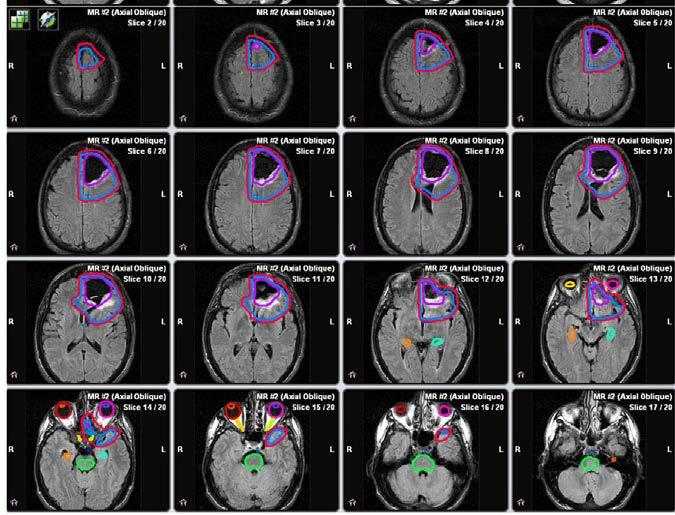
60 Post surgical Treatment outline

61 ESMO Master class Zurich 2016

62


63 ASTRO guideline 2016 Benefit of Adj RT after Biopsy/Sx Benefit of CCT/Adj TMZ


64 ASTRO guideline 2016 Dose & # in patients <70 years with good PS


65 Elderly patients (>70) ASTRO guideline 2016
66 ASTRO guideline 2016 Poor PS: Re RT in recurrence:

67 Target Volume: ASTRO guideline 2016
 Focal RT 3DCRT IMRT VMAT")
68 Whole Brain RT 2phase Partial Brain RT (3D RT) Focal RT 3DCRT IMRT VMAT
69 GBM contouring guideline 2016 Preparatory phase: Immobilization mask: head neutral 1-3mm CT : Flat couch: vertex to C3 Up to date ( 2weeks) CEMRI to be fused If MRI contraindicated Use CECT T1 + T2/FLAIR sequence dwmri, MRS, 18F-FET PET or [11C]methionine PET : investigational
70 General facts T2/FLAIR signals fluctuate depending on tumor mass-effect and postop edema. Contouring only based on T2/FLAIR sequence overestimates CTV volume. GBM is not a focal but infiltrative disease. Tumor cell may found varying density in whole Brain. Recurrence may occur due to sub-therapeutic doses (OAR limiting doses) 80% recurrences occur <2cm area of contrast enhanced lesions on CT or MRI.
71 MRI scan : Principle Post op MRI fused with planning CT: MUST Post op MRI (<48 hrs of Sx) : underestimates (shifting of brain/potential rec) Ideal timing: 2weeks of simulation Contrast enhanced T1 + T2/FLAIR Thin slice 3mm 1mm isotropic MPRAGE image: better white vs grey matter difference. Investigational: PWI/DWI MRI.
72 GTV: Resection cavity (if present) + all T1 CE areas with out T2/FLAIR peri-tumoral edema. Caveats: Post op gliosis mimics CE residual tumor. Secondary GBM (background of Non CE glioma) needs inclusion of T2 hyper intensity with T1 CE areas.
73 CTV: GTV + 2cm margin all around along white matter tracts Edit: Skull (0 mm, using bone window) Ventricles (5 mm) Falx (5 mm) Tentorium cerebelli (5 mm) visual pathway/ chiasm & brainstem (each 0 mm)
74 CTV issues FLAIR derived volumes > T2 derived volumes Some schools : don t edit CTV : include all T2 hyper intense areas No outcome difference Edema vs residual tumor is difficult to distinguish

75 Niyazi M et al. Radiother Oncol 118(2016)

76

77

78

79
80

81 OAR Niyazi M et al. Radiother Oncol 118(2016)
82 PTV margin : Uncertainties of CT MRI fusion & set up. Ideally departmental fixation protocol and QA protocol specific. Modern Linac with on/offline correction 3-5mm PTV margin suffice.
83 Technique Small spherical frontal/parietal tumor: 3DCRT IMRT/VMAT: Temporal /Insular Close proximity to orbit or brainstem Irregular shape VMAT similar conformity with 3DCRT with faster planning and delivery time Dose prescription: (ICRU 50 & 62) 100% at the isocentre, ensuring that the 95% isodose surface covers at least 95% of the PTV.

84 Immunotherapy: current approaches ESMO Congress 2016 Copenhagen
85 Major ongoing changes in Immunotherapy Rindopepimut for EGFRvIII-positive GBM? ICT-107 for HLA2-positive glioblastoma? Checkpoint inhibitors


86

87 ReACT trial
88 Immune checkpoint inhibitors Immune checkpoint inhibitors may exert strong anti-tumor activity: Melanoma: anti-ctla-4 alone vs. anti-pd-1 alone vs. combined treatment Pembrolizumab and nivolumab have been approved for advanced melanoma and other tumor entities May these drugs also mount anti-tumor immune responses against neoplasms in the CNS?
89 Current status PD1/PDL1 axis in GBM PD-L1 is expressed in human glioblastoma in vivo. Major prognostic role for PD-L1 expression in glioblastoma not defined. The significance as a biomarker of tumoral versus non-tumoral PD-L1 expression remains to be determined in glioblastoma, like in many other cancers. Preclinical studies demonstrate activity of PD- 1 inhibition in rodent glioma models.

90 CheckMate 143 : Phase II
91

92 CheckMate 143: Phase II to Phase III
93
94 SPECIAL SITUATION
95 Paediatric Glioma Gliomas are the most common primary brain tumors in children Histologically, they can be either low grade or high grade tumors Molecular biology now better defines subgroups of pediatric gliomas Surgery remains the mainstay of treatment for PLGG with exceptions PLGG is associated with good long-term outcomes (20-year OS~90%) Therapeutic regimen needs to take into account long-term effects Chemotherapy preferred as initial therapy in PLGG (after surgery) Pediatric HGG is fortunately much less common than PLGG Pediatric GBM is molecularly very distinct from its adult counterpart Unfortunately it is associated with universally dismal outcomes (as in adults) Targeted therapy likely to be the future in most pediatric gliomas
96 Management of Pediatric Low Grade Glioma Surgical resection: treatment of choice for resectable tumors Maximal safe resection: paradigm of choice during surgery Gross total resection/near total resection: curative treatment Surgical decompression/biopsy: still needed in vast majority Eloquent sites (optic pathway, brainstem, thalamus): difficult to biopsy Achieves immediate symptom relief by decompressing the brain Enables accurate histomorphological diagnosis (tumor type and grade) Provides tumor tissue for appropriate molecular testing Facilitates further adjuvant therapy (as and when necessary)
 Diffuse expansion of pons Triad of")
97 Some important exceptions to surgical resection 1: Focal Tectal Plate Glioma Clinically indolent tumor Presents with hydrocephalus Only CSF diversion (ETV) recommended 2: Optic Pathway Glioma (OPG) in NF1 One of the diagnostic criteria for NF1 Relatively slow growing/indolent course May regress/resolve spontaneously 3: Diffuse Intrinsic Pontine Glioma (DIPG) Diffuse expansion of pons Triad of signs & symptoms Aggressive biological behaviour
98 What after surgery - When to treat Further Adjuvant Therapy vs Observation Severe tumor-related symptoms (deficits, diencephalic syndrome) Unequivocal clinico-radiological evidence of tumor progression Recurrent tumor after achieving gross total resection previously COG Recommendations for Therapy

99 General Approach to Management of PLGG Stucklin et al, Neuropaediatrics 2016

100 BRAF signalling pathway: implicated in significant proportion of PLGG

101 BRAF alterations in PLGG: diagnostic, prognostic, or predictive? Useful aid for confirming diagnosis

102 Targets and opportunities for targeted therapy Penman et al, Front Oncol, 2015

103 Pediatric Glioblastoma
104 Management of Pediatric HGG including GBM Maximal safe resection remains the first-line therapy for pediatric HGG Post-operative adjuvant radiochemotherapy improves outcomes Focal Conformal RT (55.8Gy-59.4Gy in fx) is the current standard Concurrent and adjuvant temozolomide (Stupp regimen) adopted Despite multimodality treatment, recurrence/progression is the rule Universally dismal survival (median OS rarely exceeds months) Benefit of adding bevacizumab in upfront setting unknown Molecularly targeted therapy being tested and developed
105 Pediatric Glioblastoma: Molecularly Different Disease Schwatrzentruber et al, Nature 2012

106 Molecular subgroups of pediatric high grade glioma Gajjar et al, JCO 2015
107
Precision medicine for gliomas
 Precision medicine for YAZMIN ODIA, MD MS LEAD PHYSICIAN OF MEDICAL NEURO-ONCOLOGY DISCLOSURES Novocure: Advisory Board for Optune in No other financial conflicts of interest Glioma OVERVIEW INFILTRATIVE,
CNS SESSION 3/8/ th Multidisciplinary Management of Cancers: A Case based Approach
 CNS SESSION Chair: Ruben Fragoso, MD/PhD UC Davis Fellow: Michael Cardenas, MD UC Davis Panel: Gordon Li, MD Stanford Seema Nagpal, MD Stanford Jennie Taylor, MD UCSF HPI: 46 yo right handed woman who
Gliomas in the 2016 WHO Classification of CNS Tumors
 Gliomas in the 2016 WHO Classification of CNS Tumors Hindi N Al-Hindi, MD, FCAP Consultant Neuropathologist and Head Section of Anatomic Pathology Department of Pathology and Laboratory Medicine King Faisal
PRINCESS MARGARET CANCER CENTRE CLINICAL PRACTICE GUIDELINES
 PRINCESS MARGARET CANCER CENTRE CLINICAL PRACTICE GUIDELINES CENTRAL NERVOUS SYSTEM LOW GRADE GLIOMAS CNS Site Group Low Grade Gliomas Author: Dr. Norm Laperriere 1. INTRODUCTION 3 2. PREVENTION 3 3. SCREENING
PRINCESS MARGARET CANCER CENTRE CLINICAL PRACTICE GUIDELINES
 PRINCESS MARGARET CANCER CENTRE CLINICAL PRACTICE GUIDELINES CENTRAL NERVOUS SYSTEM ANAPLASTIC GLIOMAS CNS Site Group Anaplastic Gliomas Author: Dr. Norm Laperriere Date: February 20, 2018 1. INTRODUCTION
Oncological Management of Brain Tumours. Anna Maria Shiarli SpR in Clinical Oncology 15 th July 2013
 Oncological Management of Brain Tumours Anna Maria Shiarli SpR in Clinical Oncology 15 th July 2013 Outline General considerations of Primary Brain Tumours: epidemiology, pathology, presentation. Diagnosis
Radioterapia no Tratamento dos Gliomas de Baixo Grau
 Radioterapia no Tratamento dos Gliomas de Baixo Grau Dr. Luis Souhami University Montreal - Canada Low Grade Gliomas Relatively rare Heterogeneous, slow growing tumors WHO Classification Grade I Pilocytic
Clinical Trials for Adult Brain Tumors - the Imaging Perspective
 Clinical Trials for Adult Brain Tumors - the Imaging Perspective Whitney B. Pope, M.D., Ph.D. Department of Radiology David Geffen School of Medicine at UCLA August 22, 2015 1 Disclosure of Financial Relationships
MALIGNANT GLIOMAS: TREATMENT AND CHALLENGES
 MALIGNANT GLIOMAS: TREATMENT AND CHALLENGES DISCLOSURE No conflicts of interest to disclose Patricia Bruns APRN, CNS Givens Brain Tumor Center Abbott Northwestern Hospital October 12, 2018 OBJECTIVES THEN
A clinical perspective on neuropathology and molecular genetics in brain tumors
 A clinical perspective on neuropathology and molecular genetics in brain tumors M.J. van den Bent Erasmus MC Cancer Institute Rotterdam, the Netherlands Disclosures Member speakersbureau: MSD Consultancy:
Imaging for suspected glioma
 Imaging for suspected glioma 1.1.1 Offer standard structural MRI (defined as T2 weighted, FLAIR, DWI series and T1 pre- and post-contrast volume) as the initial diagnostic test for suspected glioma, unless
Systemic Treatment. Third International Neuro-Oncology Course. 23 May 2014
 Low-Grade Astrocytoma of the CNS: Systemic Treatment Third International Neuro-Oncology Course São Paulo, Brazil 23 May 2014 John de Groot, MD Associate Professor, Neuro-Oncology UT MD Anderson Cancer
Pediatric Brain Tumors: Updates in Treatment and Care
 Pediatric Brain Tumors: Updates in Treatment and Care Writer Classroom Rishi R. Lulla, MD MS Objectives Introduce the common pediatric brain tumors Discuss current treatment strategies for pediatric brain
Clinical significance of genetic analysis in glioblastoma treatment
 Clinical significance of genetic analysis in glioblastoma treatment Department of Neurosurgery, Graduate School of Medical Sciences, Kyushu University, Fukuoka, Japan Koji Yoshimoto Can we get prognostic
ARRO Case: Diffuse Intrinsic Pontine Glioma
 ARRO Case: Diffuse Intrinsic Pontine Glioma Nicholas DeNunzio, MD, PhD (PGY-3) Faculty Advisor: Louis S. Constine, MD, FASTRO Department of Radiation Oncology University of Rochester Case Presentation
DOES RADIOTHERAPY TECHNIQUE / DOSE / FRACTIONATION REALLY MATTER? YES
 DOES RADIOTHERAPY TECHNIQUE / DOSE / FRACTIONATION REALLY MATTER? YES Marco Krengli Radiotherapy, Department of Translational Medicine, University of Piemonte Orientale A. Avogadro THE STANDARD OF CARE
Case Presentation: USCAP Jason T. Huse, MD, PhD Assistant Member Department of Pathology Memorial Sloan Kettering Cancer Center
 Case Presentation: USCAP 2016 Jason T. Huse, MD, PhD Assistant Member Department of Pathology Memorial Sloan Kettering Cancer Center Case History 53 year old female with a long standing history of migraines
성균관대학교삼성창원병원신경외과학교실신경종양학 김영준. KNS-MT-03 (April 15, 2015)
") 성균관대학교삼성창원병원신경외과학교실신경종양학 김영준 INTRODUCTIONS Low grade gliomas (LGG) - heterogeneous group of tumors with astrocytic, oligodendroglial, ependymal, or mixed cellular histology - In adults diffuse, infiltrating
성균관대학교삼성창원병원신경외과학교실신경종양학 김영준 INTRODUCTIONS Low grade gliomas (LGG) - heterogeneous group of tumors with astrocytic, oligodendroglial, ependymal, or mixed cellular histology - In adults diffuse, infiltrating
UPDATES ON CHEMOTHERAPY FOR LOW GRADE GLIOMAS
 UPDATES ON CHEMOTHERAPY FOR LOW GRADE GLIOMAS Antonio M. Omuro Department of Neurology Memorial Sloan-Kettering Cancer Center II International Neuro-Oncology Congress Sao Paulo, 08/17/12 CHALLENGES IN
General: Brain tumors are lesions that have mass effect distorting the normal tissue and often result in increased intracranial pressure.
 1 Lecture Objectives Know the histologic features of the most common tumors of the CNS. Know the differences in behavior of the different tumor types. Be aware of the treatment modalities in the various
Neuropathology Evening Session: Case 3
 Neuropathology Evening Session: Case 3 Christine E. Fuller, MD Cincinnati Children s Hospital Medical Center Disclosure of Relevant Financial Relationships USCAP requires that all faculty in a position
CNS pathology Third year medical students. Dr Heyam Awad 2018 Lecture 12: CNS tumours 2/3
 CNS pathology Third year medical students Dr Heyam Awad 2018 Lecture 12: CNS tumours 2/3 Pilocytic astrocytoma Relatively benign ( WHO grade 1) Occurs in children and young adults Mostly: in the cerebellum
Classification of Diffuse Gliomas: Progress, Pearls and Pitfalls. Rob Macaulay Neuropathologist, MCC October 21, 2017
 Classification of Diffuse Gliomas: Progress, Pearls and Pitfalls Rob Macaulay Neuropathologist, MCC October 21, 2017 Objectives Explain why the designation high grade glioma is preferable to GBM for intraoperative
MOLECULAR DIAGNOSTICS OF GLIOMAS
 MOLECULAR DIAGNOSTICS OF GLIOMAS Arie Perry, M.D. Director, Neuropathology Division DIFFUSE GLIOMAS Cell types Astrocytomas (A) Oligodendrogliomas (O) Mixed oligoastrocytoma (MOA) Three WHO grades: II,
Corporate Medical Policy
 Corporate Medical Policy Analysis of MGMT Promoter Methylation in Malignant Gliomas File Name: Origination: Last CAP Review: Next CAP Review: Last Review: analysis_of_mgmt_promoter_methylation_in_malignant_gliomas
Glioblastoma and CNS tumors
 Glioblastoma and CNS tumors PRECEPTORSHIP PROGRAMME IMMUNO-ONCOLOGY Amsterdam, 27 May 2017 Patrick Roth Department of Neurology and Brain Tumor Center University Hospital Zurich Challenges in immunooncology
General Identification. Name: 江 X X Age: 29 y/o Gender: Male Height:172cm, Weight: 65kg Date of admission:95/09/27
 General Identification Name: 江 X X Age: 29 y/o Gender: Male Height:172cm, Weight: 65kg Date of admission:95/09/27 Chief Complaint Sudden onset of seizure for several minutes Present illness This 29-year
Posterior fossa tumors: clues to differential diagnosis with case-based review
 Posterior fossa tumors: clues to differential diagnosis with case-based review Poster No.: C-0323 Congress: ECR 2017 Type: Educational Exhibit Authors: H. A. Aboughalia, M. Abdelhady; Doha/QA Keywords:
Collection of Recorded Radiotherapy Seminars
 IAEA Human Health Campus Collection of Recorded Radiotherapy Seminars http://humanhealth.iaea.org The Role of Radiosurgery in the Treatment of Gliomas Luis Souhami, MD Professor Department of Radiation
The New WHO Classification and the Role of Integrated Molecular Profiling in the Diagnosis of Malignant Gliomas
 The New WHO Classification and the Role of Integrated Molecular Profiling in the Diagnosis of Malignant Gliomas Stefan Prokop, MD Neuropathology Fellow Hospital of the University of Pennsylvania Background
21/03/2017. Disclosure. Practice Changing Articles in Neuro Oncology for 2016/17. Gliomas. Objectives. Gliomas. No conflicts to declare
 Practice Changing Articles in Neuro Oncology for 2016/17 Disclosure No conflicts to declare Frances Cusano, BScPharm, ACPR April 21, 2017 Objectives Gliomas To describe the patient selection, methodology
Examining large groups of cancer patients to identify ways of predicting which therapies cancers might respond to.
 Stratified Medicine Examining large groups of cancer patients to identify ways of predicting which therapies cancers might respond to. Looking in detail at cancer cells and their genetic make up. Permit
Brain tumours (primary) and brain metastases in adults
 and brain metastases in adults") Brain tumours (primary) and brain metastases in adults NICE guideline Draft for consultation, January 0 This guideline covers diagnosing, monitoring and managing any type of primary brain tumour or brain
Brain tumours (primary) and brain metastases in adults NICE guideline Draft for consultation, January 0 This guideline covers diagnosing, monitoring and managing any type of primary brain tumour or brain
Pediatric CNS Tumors. Disclosures. Acknowledgements. Introduction. Introduction. Posterior Fossa Tumors. Whitney Finke, MD
 Pediatric CNS Tumors Disclosures Whitney Finke, MD Neuroradiology Fellow PGY-6 University of Utah Health Sciences Center Salt Lake City, Utah None Acknowledgements Introduction Nicholas A. Koontz, MD Luke
NICE guideline Published: 11 July 2018 nice.org.uk/guidance/ng99
 Brain tumours (primary) and brain metastases in adults NICE guideline Published: 11 July 2018 nice.org.uk/guidance/ng99 NICE 2018. All rights reserved. Subject to Notice of rights (https://www.nice.org.uk/terms-and-conditions#notice-ofrights).
Concepts for a personalized neurosurgical oncology. XXIV Annual Conference Pietro Paoletti 27. November 2015
 Concepts for a personalized neurosurgical oncology Jörg-Christian Tonn Dept. of Neurosurgery Ludwig-Maximilian University München Großhadern Germany XXIV Annual Conference Pietro Paoletti 27. November
Morphological features and genetic alterations
 Morphological features and genetic alterations Tutor : Audrey Rousseau Caget Lise: Université d Angers Iorio Vittoria: Seconda Università degli studi di Napoli Manaila Roxana: Iuliu Hatieganu University
The Radiologic Evaluation of Glioblastoma (GBM) and Differentiation from Pseudoprogression
 and Differentiation from Pseudoprogression") The Radiologic Evaluation of Glioblastoma (GBM) and Differentiation from Pseudoprogression Alexis Roy, Harvard Medical School, Year III Our Patient AB: Clinical Presentation 53 year old female with a past
The Radiologic Evaluation of Glioblastoma (GBM) and Differentiation from Pseudoprogression Alexis Roy, Harvard Medical School, Year III Our Patient AB: Clinical Presentation 53 year old female with a past
Case 7391 Intraventricular Lesion
 Case 7391 Intraventricular Lesion Bastos Lima P1, Marques C1, Cabrita F2, Barbosa M2, Rebelo O3, Rio F1. 1Neuroradiology, 2Neurosurgery, 3Neuropathology, Coimbra University Hospitals, Portugal. University
Enterprise Interest None
 Enterprise Interest None Heterogeneous chromosomal profiles in a unique series of DIPG in children and young adults European Congress of Pathology Amsterdam, 6 th September 2017 Charlotte Dufour, Romain
CT & MRI Evaluation of Brain Tumour & Tumour like Conditions
 CT & MRI Evaluation of Brain Tumour & Tumour like Conditions Dr. Anjana Trivedi 1, Dr. Jay Thakkar 2, Dr. Maulik Jethva 3, Dr. Ishita Virda 4 1 M.D. Radiology, Professor and Head, P.D.U. Medical College
Glioblastoma and CNS tumors
 Glioblastoma and CNS tumors PRECEPTORSHIP PROGRAMME IMMUNO-ONCOLOGY Amsterdam, 1 October 2016 Patrick Roth Department of Neurology and Brain Tumor Center University Hospital Zurich Immunology in the CNS
Pediatr Blood Cancer 2014
 Low grade Glioma! 40% of pediatric brain tumors Pathologically, anatomically, clinically and biologically heterogeneous Leptomeningeal metastases in 5% Frequently protracted clinical course Long-Term Outcome
Genomic analysis of childhood High grade glial (HGG) brain tumors
 brain tumors") Genomic analysis of childhood High grade glial (HGG) brain tumors Linda D Cooley Children s Mercy, Kansas City The Children s Mercy Hospital, 2017 Genomic analysis of childhood High grade glial (HGG) brain
Genomic analysis of childhood High grade glial (HGG) brain tumors Linda D Cooley Children s Mercy, Kansas City The Children s Mercy Hospital, 2017 Genomic analysis of childhood High grade glial (HGG) brain
RING ENCHANCING LESION BY M.S. HEMHNATH
 RING ENCHANCING LESION BY M.S. HEMHNATH A 21 YRS FEMALE CAME WITH H/O HEADACHE AND SEIZURE FOR THE PAST ONE MONTH. NO OTHER FOCAL NEUROLOGICAL DEFICIT. DIFFERENTIAL DIAGNOSIS For this case are Neurocysticerosis
Structural and functional imaging for the characterization of CNS lymphomas
 Structural and functional imaging for the characterization of CNS lymphomas Cristina Besada Introduction A few decades ago, Primary Central Nervous System Lymphoma (PCNSL) was considered as an extremely
Tumors of the Central Nervous System
 Tumors of the Central Nervous System 1 Financial Disclosures I have NO SIGNIFICANT FINANCIAL, GENERAL, OR OBLIGATION INTERESTS TO REPORT Introduction General: Brain tumors are lesions that have mass effect
Oligodendroglioma: imaging findings, radio-pathological correlation and evolution
 Oligodendroglioma: imaging findings, radio-pathological correlation and evolution Poster No.: C-2104 Congress: ECR 2013 Type: Authors: Keywords: DOI: Scientific Exhibit A. Hernandez Castro, M. D. Monedero
WHY BIOPSY? Diagnosis and Research
 WHY BIOPSY? Diagnosis and Research 9 2 4 1 3 1 1. Diagnosis only by Imaging (like no other tumor) The issue of Typical versus Atypical DIPG T1 FLAIR Gad. T2 Tractography Functional MRI Diffusion/Perfusion
Paolo Tini 1,3 M.D. :
 Correlazione tra espressione di Epidermal Growth Factor Receptor (EGFR) e Patterns di recidiva/progressione di malattia dopo trattamento radio-chemioterapico in pazienti affetti da Glioblastoma (GB). Paolo
Masses of the Corpus Callosum
 Masses of the Corpus Callosum Kesav Raghavan, HMS Year III Dr. Agenda Corpus Callosum Development and Anatomy Our Patient: Clinical Presentation Differential Diagnosis of Masses in the Corpus Callosum
Table 7: PBTC Protocols [ ] Protocol Title Strata Status Neuroimaging Objective/Test
![Table 7: PBTC Protocols [ ] Protocol Title Strata Status Neuroimaging Objective/Test](/thumbs/82/85455518.jpg "Table 7: PBTC Protocols [ ] Protocol Title Strata Status Neuroimaging Objective/Test") Table 7: PBTC Protocols [001 009] Protocol Title Strata Status Neuroimaging Objective/Test PBTC-001 PBTC-002 PBTC-003 PBTC-004 PBTC-005 PBTC-006 PBTC-007 PBTC-009 A Pilot of Systemic and Intrathecal Chemotherapy
Table 7: PBTC Protocols [001 009] Protocol Title Strata Status Neuroimaging Objective/Test PBTC-001 PBTC-002 PBTC-003 PBTC-004 PBTC-005 PBTC-006 PBTC-007 PBTC-009 A Pilot of Systemic and Intrathecal Chemotherapy
CNS Tumors: The Med Onc Perspective. Ronald J. Scheff, MD Associate Clinical Professor Weill Medical College of Cornell U.
 CNS Tumors: The Med Onc Perspective Ronald J. Scheff, MD Associate Clinical Professor Weill Medical College of Cornell U. Disclosure Speakers Bureau, Merck Basic Oncology Concepts Tissue Diagnosis Stage
Institute of Oncology & Radiobiology. Havana, Cuba. INOR
 Institute of Oncology & Radiobiology. Havana, Cuba. INOR 1 Transition from 2-D 2 D to 3-D 3 D conformal radiotherapy in high grade gliomas: : our experience in Cuba Chon. I, MD - Chi. D, MD - Alert.J,
CNS TUMORS. D r. Ali Eltayb ( U. of Omdurman. I ). M. Path (U. of Alexandria)
. M. Path (U. of Alexandria)") CNS TUMORS D r. Ali Eltayb ( U. of Omdurman. I ). M. Path (U. of Alexandria) CNS TUMORS The annual incidence of intracranial tumors of the CNS ISmore than intraspinal tumors May be Primary or Secondary
CNS TUMORS D r. Ali Eltayb ( U. of Omdurman. I ). M. Path (U. of Alexandria) CNS TUMORS The annual incidence of intracranial tumors of the CNS ISmore than intraspinal tumors May be Primary or Secondary
Joana Ramalho, MD C. Ryan Miller, MD, PhD
 Joana Ramalho, MD C. Ryan Miller, MD, PhD Case 1 3 month old baby girl Presented with new onset of seizures Newborn. Questionable blurring of the gray-white junction within the right occipital lobe. Findings
Brain Tumors: Radiologic Perspective
 Brain Tumors: Radiologic Perspective Alberto Bizzi, M.D. Neuroradiology Humanitas Research Hospital Milan, Italy The job of the neuroradiologist in the work-up of brain tumors has quite changed in the
Antiangiogenic drugs in unresectable glioblastoma. Dra. Carmen Balañá. /
 Antiangiogenic drugs in unresectable glioblastoma Dra. Carmen Balañá. / Outcome for unresectable GBM Overall survival for unresectable GBM without further treatment is: 3 months at most. Radiotherapy increases
Evaluation of Three-dimensional Conformal Radiotherapy and Intensity Modulated Radiotherapy Techniques in High-Grade Gliomas
 1 Carol Boyd Comprehensive Case Study July 11, 2013 Evaluation of Three-dimensional Conformal Radiotherapy and Intensity Modulated Radiotherapy Techniques in High-Grade Gliomas Abstract: Introduction:
Contemporary Management of Glioblastoma
 Contemporary Management of Glioblastoma Incidence Rates of Primary Brain Tumors Central Brain Tumor Registry of the United States, 1992-1997 100 Number of Cases per 100,000 Population 10 1 0.1 x I x I
RINDOPEPIMUT (CDX-110) IN GLIOBLASTOMA
 IN GLIOBLASTOMA") RINDOPEPIMUT (CDX-110) IN GLIOBLASTOMA MULTIFORM GEINO 2014 Dra Estela Pineda Madrid Hospital Clínic Barcelona EGFRvIII in glioblastoma multiform The most common mutation of EGFR in GBM Expressed in 30%
RINDOPEPIMUT (CDX-110) IN GLIOBLASTOMA MULTIFORM GEINO 2014 Dra Estela Pineda Madrid Hospital Clínic Barcelona EGFRvIII in glioblastoma multiform The most common mutation of EGFR in GBM Expressed in 30%
Treatment Planning Evaluation of Volumetric Modulated Arc Therapy (VMAT) for Craniospinal Irradiation (CSI)
 for Craniospinal Irradiation (CSI)") Treatment Planning Evaluation of Volumetric Modulated Arc Therapy (VMAT) for Craniospinal Irradiation (CSI) Tagreed AL-ALAWI Medical Physicist King Abdullah Medical City- Jeddah Aim 1. Simplify and standardize
Treatment Planning Evaluation of Volumetric Modulated Arc Therapy (VMAT) for Craniospinal Irradiation (CSI) Tagreed AL-ALAWI Medical Physicist King Abdullah Medical City- Jeddah Aim 1. Simplify and standardize
Disclaimers. Molecular pathology of brain tumors. Some aspects only. Some details are inevitably personal opinions
 Molecular pathology of brain tumors Disclaimers Some aspects only H.K. Ng The Chinese University of Hong Kong Some details are inevitably personal opinions Free ppt : http://www.acp.cuhk.edu.hk/hkng Why
Pr D.Figarella-Branger Service d Anatomie Pathologique et de Neuropathologie, La Timone, Marseille UMR 911 Inserm, Université d Aix-Marseille
 Novelties in the WHO 2016 classification of brain tumours Pr D.Figarella-Branger Service d Anatomie Pathologique et de Neuropathologie, La Timone, Marseille UMR 911 Inserm, Université d Aix-Marseille The
Advancements in Neuro- Oncology
 Advancements in Neuro- Oncology Ricky Chen, MD Director, Neuro-Oncology Providence St Vincent Medical Center November 30 th, 2018 No disclosures Recognizing a brain tumor New and *Persistent Headaches*
Astroblastoma: Radiologic-Pathologic Correlation and Distinction from Ependymoma
 AJNR Am J Neuroradiol 23:243 247, February 2002 Case Report Astroblastoma: Radiologic-Pathologic Correlation and Distinction from Ependymoma John D. Port, Daniel J. Brat, Peter C. Burger, and Martin G.
Goals for this Lecture. Case 1. Key Points MRI TECHNIQUES FOR DIFFERENTIAL DIAGNOSIS OF RECURRENT BRAIN LESIONS
 MRI TECHNIQUES FOR DIFFERENTIAL DIAGNOSIS OF RECURRENT BRAIN LESIONS Goals for this Lecture 1. Review common appearances for recurrent tumor and treatment effects on conventional MRI 2. Discuss current
WHO 2016 CNS Tumor Classification Update. DISCLOSURES (Arie Perry, MD) PATTERN RECOGNITION. Arie Perry, M.D. Director, Neuropathology
 PATTERN RECOGNITION. Arie Perry, M.D. Director, Neuropathology") WHO 2016 CNS Tumor Classification Update Arie Perry, M.D. Director, Neuropathology DISCLOSURES (Arie Perry, MD) I have no financial relationships to disclose. - and - I will not discuss off label use or
WHO 2016 CNS Tumor Classification Update Arie Perry, M.D. Director, Neuropathology DISCLOSURES (Arie Perry, MD) I have no financial relationships to disclose. - and - I will not discuss off label use or
Corporate Medical Policy
 Corporate Medical Policy Brachytherapy, Intracavitary Balloon Catheter for Brain Cancer File Name: Origination: Last CAP Review: Next CAP Review: Last Review: brachytherapy_intracavitary_balloon_catheter_for_brain_cancer
Review: Diagnostic, prognostic and predictive relevance of molecular markers in gliomas
 Review: Diagnostic, prognostic and predictive relevance of molecular markers in gliomas Sebastian Brandner MD FRCPath 1 and Andreas von Deimling 2 MD 1) Division of Neuropathology, The National Hospital
Target Delineation in Gliomas. Prof PK Julka Department of Radiotherapy and Oncology AIIMS, New Delhi
 Target Delineation in Gliomas Prof PK Julka Department of Radiotherapy and Oncology AIIMS, New Delhi 1 What is a glioma? A primary brain tumour that originated from a cell of the nervous system 2 Recommendations:
Diagnostic application of SNParrays to brain cancers
 Diagnostic application of SNParrays to brain cancers Adriana Olar 4/17/2018 No disclosures 55 yo M, focal motor seizure T2 T1-post C DIAGNOSIS BRAIN, LEFT FRONTAL LOBE, BIOPSY: - DIFFUSE GLIOMA, OLIGODENDROGLIAL
Innovative Multimodal Imaging Techniques in Brain Tumor Clinical Trials
 Innovative Multimodal Imaging Techniques in Brain Tumor Clinical Trials Benjamin M. Ellingson, Ph.D. Assistant Professor of Radiology, Biomedical Physics, and Bioengineering Brain Tumor Imaging Laboratory
Neurosurgical Management of Brain Tumours. Nicholas Little Neurosurgeon RNSH
 Neurosurgical Management of Brain Tumours Nicholas Little Neurosurgeon RNSH General Most common tumours are metastatic 10x more common than primary Incidence of primary neoplasms is 20 per 100000 per year
RINGS N THINGS: Imaging Patterns in Differential Diagnosis. Anne G. Osborn, M.D.
 RINGS N THINGS: Imaging Patterns in Differential Diagnosis Anne G. Osborn, M.D. ExpDDxs: Intra-axial (Parenchymal) Lesions Ring-enhancing lesions, solitary 1 Ring-enhancing lesion crossing corpus callosum
Defining Target Volumes and Organs at Risk: a common language
 Defining Target Volumes and Organs at Risk: a common language Eduardo Rosenblatt Section Head Applied Radiation Biology and Radiotherapy (ARBR) Section Division of Human Health IAEA Objective: To introduce
Thierry M. Muanza, MSc, MD, FRCPC,, McGill University Segal Cancer Centre, Jewish General Hospital Montreal, QC, Canada
 Thierry M. Muanza, MSc, MD, FRCPC,, McGill University Segal Cancer Centre, Jewish General Hospital Montreal, QC, Canada Déclarations Aucun conflit d intérêt Objectifs d apprentissage Évolution de la radiothérapie
Off-Label Treatments. Clinical Trials for Recurrent GBM UCSF Radiation Oncology Course: Management of Recurrent Disease. Outline
 Off-Label Treatments Clinical Trials for Recurrent GBM UCSF Radiation Oncology Course: Management of Recurrent Disease Jennifer Clarke, MD, MPH Assistant Professor Division of Neuro-Oncology Depts of Neurological
SYSTEMIC MANAGEMENT OF PEDIATRIC PRIMARY BRAIN TUMORS
 SYSTEMIC MANAGEMENT OF PEDIATRIC PRIMARY BRAIN TUMORS María E. Echevarría, MD Assistant Professor University of Puerto Rico Medical Sciences Campus DISCLOSURES No disclosures INTRODUCTION Pediatric CNS
PRINCESS MARGARET CANCER CENTRE CLINICAL PRACTICE GUIDELINES
 PRINCESS MARGARET CANCER CENTRE CLINICAL PRACTICE GUIDELINES CENTRAL NERVOUS SYSTEM MENINGIOMA CNS Site Group Meningioma Author: Dr. Norm Laperriere Date: February 20, 2018 1. INTRODUCTION 3 2. PREVENTION
CURRENT CONTROVERSIES IN THE MANAGEMENT OF HIGH GRADE GLIOMAS: AN INTERACTIVE CASE DISCUSSION *
 CURRENT CONTROVERSIES IN THE MANAGEMENT OF HIGH GRADE GLIOMAS: AN INTERACTIVE CASE DISCUSSION * Alessandro Olivi, MD, Jaishri Blakeley, MD, and Allen K. Sills, MD, FACS ABSTRACT The management of glioma
2017 Diagnostic Slide Session Case 3
 2017 Diagnostic Slide Session Case 3 Andrew Gao, MD Lili-Naz Hazrati, MD, PhD Cynthia Hawkins, MD, PhD Hospital for Sick Children and University of Toronto, Toronto, Canada Disclosures: none Clinical History
Prognostic value of ADC in glioblastoma multiforme and its correlation with survival and MGMT promoter methylation status.
 Prognostic value of ADC in glioblastoma multiforme and its correlation with survival and MGMT promoter methylation status. R. Zalazar, M.D. Hernández, M. Páramo, P. Slon, M. Millor Muruzabal, J. Solorzano
Ruolo dell imaging nella pianificazione del trattamento
 Simposio AIRO-SIRM: Diagnostica per immagini morfologica e funzionale nella stadiazione, terapia e follow-up dei sarcomi delle parti molli Ruolo dell imaging nella pianificazione del trattamento Marco
PRINCESS MARGARET CANCER CENTRE CLINICAL PRACTICE GUIDELINES
 PRINCESS MARGARET CANCER CENTRE CLINICAL PRACTICE GUIDELINES CENTRAL NERVOUS SYSTEM BRAIN METASTASES CNS Site Group Brain Metastases Author: Dr. Norm Laperriere Date: February 20, 2018 1. INTRODUCTION
Intensity modulated radiotherapy (IMRT) for treatment of post-operative high grade glioma in the right parietal region of brain
 for treatment of post-operative high grade glioma in the right parietal region of brain") 1 Carol Boyd March Case Study March 11, 2013 Intensity modulated radiotherapy (IMRT) for treatment of post-operative high grade glioma in the right parietal region of brain History of Present Illness:
1 Carol Boyd March Case Study March 11, 2013 Intensity modulated radiotherapy (IMRT) for treatment of post-operative high grade glioma in the right parietal region of brain History of Present Illness:
MRI Applications in Radiation Oncology:
 MRI Applications in Radiation Oncology: Physician s Perspective Jeff Olsen, MD Department of Radiation Oncology Washington University, St. Louis, MO Disclosures Washington University has research and service
Supra- and infratentorial brain tumors from childhood to maternity
 Supra- and infratentorial brain tumors from childhood to maternity What to expect? I am going to show you the characteristic imaging findings of following tumors: Thierry A.G.M. Huisman, MD, FICIS, EQNR
PRINCESS MARGARET CANCER CENTRE CLINICAL PRACTICE GUIDELINES
 PRINCESS MARGARET CANCER CENTRE CLINICAL PRACTICE GUIDELINES CENTRAL NERVOUS SYSTEM MEDULLOBLASTOMA AND PNET CNS Site Group Medulloblastoma and PNET Author: Dr. Norm Laperriere 1. INTRODUCTION 3 2. PREVENTION
Correlation of quantitative proton MR spectroscopy with local histology from stereotactic brain biopsy to evaluate heterogeneity of brain tumors
 Correlation of quantitative proton MR spectroscopy with local histology from stereotactic brain biopsy to evaluate heterogeneity of brain tumors Steve H. Fung, MD 1, Edward F. Jackson, PhD 2, Samuel J.
Precision medicine: How to exploit the growing knowledge on the evolving genomes of cells to improve cancer prevention and therapy.
 Precision medicine: How to exploit the growing knowledge on the evolving genomes of cells to improve cancer prevention and therapy Joe Costello, PhD Department of Neurological Surgery A more accurate and
MRS and Perfusion of Brain Tumors
 Department of Radiology University of California San Diego MRS and Perfusion of Brain Tumors John R. Hesselink, M.D. MRS & Perfusion of Brain Tumors Tumor histology Degree of malignancy Delineate tumor
Immuno-Oncology. Glioblastoma and CNS tumors 5 July 2016 Siena, Italy
 ESMO Preceptorship Programme Immuno-Oncology From the essentials of tumour immunology to clinical application Glioblastoma and CNS tumors 5 July 2016 Siena, Italy Michael Weller Department of Neurology
Management of Glioma: The Basics Glioma Update The clinical challenge. Glioma a malignant disease of the CNS
 Management of Glioma: The Basics Glioma Update 3 oger Stupp, MD Department of Oncology & Cancer Center University Hospital Zurich, Switzerland (roger.stupp@usz.ch) Bern, 3. August 3 The clinical challenge
What yield in the last decade about Molecular Diagnostics in Neuro
 What yield in the last decade about Molecular Diagnostics in Neuro Oncology? Raphael Salles S.Medeiros Neuropathologist at HC FMUSP Clinical Research Project Manager at Oncology department at Hospital
Protocol Abstract and Schema
 Protocol Abstract and Schema This is a phase I/II study to determine: 1) the maximum tolerated dose (MTD) or recommended phase II dose of ABT-888 in combination with radiation therapy, and 2) the efficacy
SURGICAL MANAGEMENT OF BRAIN TUMORS
 SURGICAL MANAGEMENT OF BRAIN TUMORS LIGIA TATARANU, MD, Ph D NEUROSURGICAL CLINIC, BAGDASAR ARSENI CLINICAL HOSPITAL BUCHAREST, ROMANIA SURGICAL INDICATIONS CONFIRMING HISTOLOGIC DIAGNOSIS REDUCING TUMOR
Challenging Paediatric Brain Tumours. ASP Belfast March 2017 Dr Jane Pears Consultant Paediatric Oncologist, Dublin
 Challenging Paediatric Brain Tumours ASP Belfast March 2017 Dr Jane Pears Consultant Paediatric Oncologist, Dublin Overview (i) Paediatric malignancy (ii) Central nervous system tumours (iii) Diffuse Intrinsic
Scottish Medicines Consortium
 Scottish Medicines Consortium temozolomide 5, 20, 100 and 250mg capsules (Temodal ) Schering Plough UK Ltd No. (244/06) New indication: for the treatment of newly diagnosed glioblastoma multiforme concomitantly
MRI OF BRAIN METASTASIS. Dr P. AGUETTAZ Hôpital Privé Clairval Marseille
 MRI OF BRAIN METASTASIS Dr P. AGUETTAZ Hôpital Privé Clairval Marseille Purpose To avoid a few common pitfalls when imaging patient suspected of brain metastasis 1. Does hyperintensity on post contrast