Cerebral Cortex Structure, Function, Dysfunction Reading Ch 10 Waxman Dental Neuroanatomy Lecture. Suzanne Stensaas, Ph.D.
|
|
|
- Barnard Roberts
- 5 years ago
- Views:
Transcription
1 Cerebral Cortex Structure, Function, Dysfunction Reading Ch 10 Waxman Dental Neuroanatomy Lecture Suzanne Stensaas, Ph.D. March 7, 2012
2 Anatomy Review Lobes and layers Brodmann s areas Vascular Supply Major Neurological Findings Frontal, Parietal, Temporal, Occipital, Limbic Quiz Questions?
3 Types of Cortex Sensory (Primary) Motor (Primary) Unimodal association Association and Multimodal necessary for language, reason, plan, imagine, create


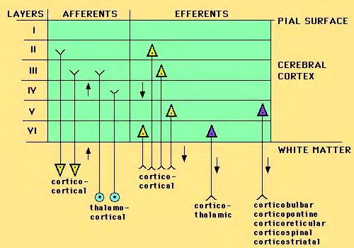
4 Structure of neocortex (6 layers)

5 The general pattern of primary, association and mulimodal association cortex (Mesulam)
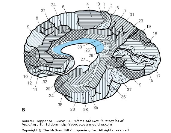
6 Brodmann, Lateral Left Hemisphere
7 MCA left hemisphere from D.Haines
8
9 ACA and PCA -Haines
10 Issues of Functional Localization Earliest studies -Signs, symptoms and note location at autopsy Electrical discharge (epilepsy) suggested function Ablation - deficit suggest function Reappearance of infant functions suggest loss of inhibition (disinhibition), i.e. grasp, suck, Babinski Linked networks of afferent and efferent neurons in several regions working to accomplish a task (attention) Functional imaging does not always equate with abnormal function associated with location of lesion fmri activation of several cortical regions Same sign from lesions in different areas i.e.paraphasias Notion of the right hemisphere as "emotional" in contrast to the left one as "logical" has no basis in fact.
11 Limbic System (not a true lobe) involves Cingulate gyrus - affect, pain, memory Hippocampus- short term memory Amygdala- fear, agression, mating Fornix pathway to hypothalamus Hypothalamus- ANS & endocr control Prefrontal Cortex- appropriate behavior

12 Schematic Diagram of principal limbic areas From College of DuPage Biology 1152 Syllabus

13 Amygdala and relationship to ventricle and hippocampus
14 Classic Hippocampal Circuit


15 Hippocampal Formation & Amygdala

16 Hippocampus


17 Hypothalamus Interbrain, Springer Verlag
18 Lateral view gross brain. Left hemisphere Frontal Lobe
19 Frontal Lobe Motor areas Contralateral weakness or paralysis (area 4) Premotor planning of action (area 6) Frontal eye fields for moving eyes to opposite side (area 8) e.g. Epileptic discharge Speech production (Broca s area 44, 45) Prefrontal areas
20 Apraxia (Error in execution of learned movements without coexisting weakness) Damage to dominant parietal, premotor, and supplementary motor areas Dominant hemisphere association areas Parietal - integrates motor sequences with vision and somatic sensory info Frontal lobe - execution of act
21 Frontal Lobe prefrontal association cortex Bilateral prefrontal damage distractible, apathetic lack foresight, abstract reasoning, initiative stubborn, perseverate, lack ambition, responsibility, judgment or social graces
22 Parietal Lobe Somatosensory Cortex-paresthesias Dominant Parietal lobe-reading, writing, naming L. Angular gyrus L. Supramarginal gyrus L. Multimodal cortex Agraphia can be frontal or parietal
23 Contralateral Neglect (asomatognosia) Right parietal Right side is dominant for attention - do not attend to opposite side, ie. Dressing apraxia Severe - failure to recognize one s opposite limb Impaired visuospatial ability (drawing, copying, 3D, manipulate objects in space Fail to appreciate humor
24 Temporal Lobe Association auditory cortex Speech comprehension Important in naming Memory - bilateral medial temporal lobe near hippocampus Superior part of contralateral visual field

25 Aphasias-Motor Full Broca s involves operculum, insula and subjacent white matter with contralateral hemiparesis of face, arm Telegraphic speech Agrammatism - syntax more affected than semantics Usually agraphia too Transcortical - interruption of inferred linkage paths inward to Broca s area
26 Global aphasia Dominant hemisphere Frontal Temporal Parietal Head of caudate associated with language disorders Internal carotid or proximal MCA, hemorrhage, or large tumor
27 Aphasias-Sensory Wernicke s Dominant (left usually) hemisphere Fluent, paraphasias, poor comprehension, Naming, repetition, reading and writing impaired Less aware and less frustrated than motor aphasias
28 Right hemisphere and aphasia Emotional tone modulation Propositional prosody Body language gestures
29 Temporal Lobe Functions Wernicke speech comprehension - dominant side Verbal learning- dominant Inferior temporal gyrus naming and faces - bilateral R or L Superior homonymous quadrantanopia - (Meyers loop) Hallucination incld gustatory, visual, auditory with emotion Lyrics in dominant lobe Harmony and melody is impaired by lesions of the nondominant, Visual learning- nondominant Visual agnosia dominant, auditory agnosia nondominant hemisphere Bilateral: cortical deafness. Otherwise subtle Bilateral: psychic blindness, Klüver-Bucy rarely full in man. Bilateral hippocampal formation : Amnesia
30 Agnosia-impaired perception or recognition with OK vision, hearing, sensation, attention, intelligence Visual: colors, faces, letters Auditory: tunes, spoken words, pure word deafness Somatosensory - stereognosis, graphesthesia May not have other signs: aphasia, apraxia Atrophy or metastatic disease Disconnections of specific sensory association areas Corpus callosum, deep white matter near main sensory areas

31 Occipital Lobe Brodmann, Lateral

32 Ventral and dorsal Stream, MT

33 Medial Gross Brain
34 Visual Path
35 Ventral, Gross Brain

36 PCA ventral view right hemisphere from D.Haines


37 Write the patient history Posterior view, angiogram R L R L FIRST ANGIO SECOND ANGIO next day
38 Doctors say a bystander can recognize a stroke by asking three simple questions: STROKE:Remember the 1st Four Letters...S.T.R.O. If everyone can remember something this simple, we could save some folks.
39 Ask four simple questions: S Ask the person to SMILE T Ask the person to TALK and SPEAK A SIMPLE SENTENCE (Coherently) (i.e. It is sunny out today) R Ask them to RAISE BOTH ARMS. O Ask them to open their mouth and STICK OUT your tongue. (Does it deviate to one side?) K Kall 911 E Every minute counts (180 mins)
40 End of Dental lecture
Higher Cortical Function
 Emilie O Neill, class of 2016 Higher Cortical Function Objectives Describe the association cortical areas processing sensory, motor, executive, language, and emotion/memory information (know general location
Learning Objectives.
 Emilie O Neill, class of 2016 Learning Objectives 1. Describe the types of deficits that occur with lesions in association areas including: prosopagnosia, neglect, aphasias, agnosia, apraxia 2. Discuss
The origins of localization
 Association Cortex, Asymmetries, and Cortical Localization of Affective and Cognitive Functions Michael E. Goldberg, M.D. The origins of localization The concept that different parts of the brain did different
Lecture 35 Association Cortices and Hemispheric Asymmetries -- M. Goldberg
 Lecture 35 Association Cortices and Hemispheric Asymmetries -- M. Goldberg The concept that different parts of the brain did different things started with Spurzheim and Gall, whose phrenology became quite
Association Cortex, Asymmetries, and Cortical Localization of Affective and Cognitive Functions. Michael E. Goldberg, M.D.
 Association Cortex, Asymmetries, and Cortical Localization of Affective and Cognitive Functions Michael E. Goldberg, M.D. The origins of localization The concept that different parts of the brain did different
CEREBRUM. Dr. Jamila EL Medany
 CEREBRUM Dr. Jamila EL Medany Objectives At the end of the lecture, the student should be able to: List the parts of the cerebral hemisphere (cortex, medulla, basal nuclei, lateral ventricle). Describe
Psy /16 Human Communication. By Joseline
 Psy-302 11/16 Human Communication By Joseline Lateralization Left Hemisphere dominance in speech production in 95% of right handed and 70% of left handed people Left -> Timing, Sequence of events Right
Exam 1 PSYC Fall 1998
 Exam 1 PSYC 2022 Fall 1998 (2 points) Briefly describe the difference between a dualistic and a materialistic explanation of brain-mind relationships. (1 point) True or False. George Berkely was a monist.
CEREBRUM Dr. Jamila Elmedany Dr. Essam Eldin Salama
 CEREBRUM Dr. Jamila Elmedany Dr. Essam Eldin Salama Objectives At the end of the lecture, the student should be able to: List the parts of the cerebral hemisphere (cortex, medulla, basal nuclei, lateral
Chapter 2 Test. 1. Evolutionary structures within the are the most primitive. *a. hindbrain b. thalamus c. forebrain d. midbrain e.
 Cognitive Psychology In and Out of the Laboratory 5th Edition Galotti TEST BANK Full clear download (no formatting errors) at: https://testbankreal.com/download/cognitive-psychology-laboratory-5thedition-galotti-test-bank/
Homework Week 2. PreLab 2 HW #2 Synapses (Page 1 in the HW Section)
") Homework Week 2 Due in Lab PreLab 2 HW #2 Synapses (Page 1 in the HW Section) Reminders No class next Monday Quiz 1 is @ 5:30pm on Tuesday, 1/22/13 Study guide posted under Study Aids section of website
Homework Week 2 Due in Lab PreLab 2 HW #2 Synapses (Page 1 in the HW Section) Reminders No class next Monday Quiz 1 is @ 5:30pm on Tuesday, 1/22/13 Study guide posted under Study Aids section of website
PSY 215 Lecture 17 (3/28/2010) (Lateralization in the Brain) Dr. Achtman PSY 215
 (Lateralization in the Brain) Dr. Achtman PSY 215") PSY 215 Lecture 17 Topic: Lateralization in the Brain Chapter 14.1, pages 403-414 Corrections: Lecture 16 (page 4) Broca s Area: trouble producing language, comprehension is okay. Announcements: Review
PSY 215 Lecture 17 Topic: Lateralization in the Brain Chapter 14.1, pages 403-414 Corrections: Lecture 16 (page 4) Broca s Area: trouble producing language, comprehension is okay. Announcements: Review
Excellent Network Courses. Department of Neurology Affiliated hospital of Jiangsu University
 Excellent Network Courses Department of Neurology Affiliated hospital of Jiangsu University Agnosia Visual Agnosia Lissauer (1890) described 2 types: a) Apperceptive Cannot see objects b) Associative Does
The Frontal Lobes. Anatomy of the Frontal Lobes. Anatomy of the Frontal Lobes 3/2/2011. Portrait: Losing Frontal-Lobe Functions. Readings: KW Ch.
 The Frontal Lobes Readings: KW Ch. 16 Portrait: Losing Frontal-Lobe Functions E.L. Highly organized college professor Became disorganized, showed little emotion, and began to miss deadlines Scores on intelligence
-Zeina Assaf. -Omar Odeh. - Maha Beltagy
 -3 -Zeina Assaf -Omar Odeh - Maha Beltagy 1 P a g e The Inferior Surface Of The Brain The inferior surface of the brain is divide by the stem of the lateral fissure into 2 parts : The orbital surface and
Sensorimotor Functioning. Sensory and Motor Systems. Functional Anatomy of Brain- Behavioral Relationships
 Sensorimotor Functioning Sensory and Motor Systems Understanding brain-behavior relationships requires knowledge of sensory and motor systems. Sensory System = Input Neural Processing Motor System = Output
Outline of the next three lectures
 Outline of the next three lectures Lecture 35 Anatomy of the human cerebral cortex gross and microscopic cell types connections Vascular supply of the cerebral cortex Disorders involving the cerebral cortex
P. Hitchcock, Ph.D. Department of Cell and Developmental Biology Kellogg Eye Center. Wednesday, 16 March 2009, 1:00p.m. 2:00p.m.
 Normal CNS, Special Senses, Head and Neck TOPIC: CEREBRAL HEMISPHERES FACULTY: LECTURE: READING: P. Hitchcock, Ph.D. Department of Cell and Developmental Biology Kellogg Eye Center Wednesday, 16 March
Chapter 14, Part 2! Chapter 14 Part 2 Brain/Cranial Nerves! The Cerebrum and Cranial Nerves! pp !
 Chapter 14, Part 2! The Cerebrum and Cranial pp. 482 505! SECTION 14-9! The cerebrum, the largest region of the brain, contains motor, sensory, and association areas! 2! White Matter of the Cerebrum! 1.
Chapter 14, Part 2! The Cerebrum and Cranial Nerves! pp !
 Chapter 14, Part 2! The Cerebrum and Cranial pp. 482 505! SECTION 14-9! The cerebrum, the largest region of the brain, contains motor, sensory, and association areas! 2! 1! ! Chapter 14 Part 2 Brain/Cranial
Brain. Cerebral white matter. Brain cortex. Frontal lobe. Frontal lobe Brain cortex
 Brain Brain cortex Layer (stratum) of grey matter which cover hemisphers Longitudinal fissure - 2 hemispheres Enlargement of neocortex folding the brain surface into convolutions (gyri) separated by groves
THE VISUAL PATHWAY FOR DENTAL STUDENTS
 Neuroanatomy Suzanne S. Stensaas, Ph.D. February 16, 2012 Objectives: THE VISUAL PATHWAY FOR DENTAL STUDENTS A. Draw the expected visual fields seen in classic lesions of the nerve, chiasm, thalamus, optic
Motor Functions of Cerebral Cortex
 Motor Functions of Cerebral Cortex I: To list the functions of different cortical laminae II: To describe the four motor areas of the cerebral cortex. III: To discuss the functions and dysfunctions of
Introduction to Physiological Psychology Review
 Introduction to Physiological Psychology Review ksweeney@cogsci.ucsd.edu www.cogsci.ucsd.edu/~ksweeney/psy260.html n Learning and Memory n Human Communication n Emotion 1 What is memory? n Working Memory:
Inside Your Patient s Brain Michelle Peterson, APRN, CNP Centracare Stroke and Vascular Neurology
 Inside Your Patient s Brain Michelle Peterson, APRN, CNP Centracare Stroke and Vascular Neurology Activity Everyone stand up, raise your right hand, tell your neighbors your name 1 What part of the brain
Layered organization of cortex: Paleocortex 3 layers hippocampal formation / ventral & medial cortex closest to brainstem
 Layered organization of cortex: Paleocortex 3 layers hippocampal formation / ventral & medial cortex closest to brainstem Archicortex 3-4 layers hippocampal formation / amygdala Neocortex 6 layers more
Cerebral Cortex: Association Areas and Memory Tutis Vilis
 97 Cerebral Cortex: Association Areas and Memory Tutis Vilis a) Name the 5 main subdivisions of the cerebral cortex. Frontal, temporal, occipital, parietal, and limbic (on the medial side) b) Locate the
Human Paleoneurology and the Evolution of the Parietal Cortex
 PARIETAL LOBE The Parietal Lobes develop at about the age of 5 years. They function to give the individual perspective and to help them understand space, touch, and volume. The location of the parietal
OBJECTIVES. At the end of the lecture, students should be able to: List the cerebral arteries.
 DR JAMILA EL MEDANY OBJECTIVES At the end of the lecture, students should be able to: List the cerebral arteries. Describe the cerebral arterial supply regarding the origin, distribution and branches.
CEREBRUM & CEREBRAL CORTEX
 CEREBRUM & CEREBRAL CORTEX Seonghan Kim Dept. of Anatomy Inje University, College of Medicine THE BRAIN ANATOMICAL REGIONS A. Cerebrum B. Diencephalon Thalamus Hypothalamus C. Brain Stem Midbrain Pons
Biocomputer Wired for Action MWABBYH CTBIR LOBES
 Biocomputer Wired for Action MWABBYH CTBIR LOBES 100 100 100 100 100 200 200 200 200 200 300 300 300 300 300 400 400 400 400 400 500 500 500 500 500 Biocomputer Wired for Action MWABBYH CTBIR LOBES 100
Disorders of language and speech. Samuel Komoly MD PhD DHAS Professor and Chairman Department of Neurology
 Disorders of language and speech Samuel Komoly MD PhD DHAS Professor and Chairman Department of Neurology http://neurology.pote.hu major categories disorders of language and speech cortical types aphasias
C:\Documents and Settings\sstensaas\Desktop\dental visual 2010\VisualPath dental 2010.docVisualPath dental 2010.doc
 Neuroanatomy Suzanne Stensaas April 8, 2010, 10:00-12:00 p.m. Reading: Waxman Ch. 15, Computer Resources: HyperBrain Ch 7 THE VISUAL PATHWAY Objectives: 1. Describe the pathway of visual information from
The motor regulator. 2) The cerebellum
 The cerebellum") The motor regulator 2) The cerebellum Motor control systems outside the cortex Cerebellum -controls neural programs for the executionl of skilled movements Cerebellar Peduncles Atlas Fig. 2-31 Atlas Fig.
The motor regulator 2) The cerebellum Motor control systems outside the cortex Cerebellum -controls neural programs for the executionl of skilled movements Cerebellar Peduncles Atlas Fig. 2-31 Atlas Fig.
Cortical Organization. Functionally, cortex is classically divided into 3 general types: 1. Primary cortex:. - receptive field:.
 Cortical Organization Functionally, cortex is classically divided into 3 general types: 1. Primary cortex:. - receptive field:. 2. Secondary cortex: located immediately adjacent to primary cortical areas,
correlates with social context behavioral adaptation.
 REVIEW OF FRONTAL LOBE STRUCTURES Main organization of frontal cortex: 1. Motor area (precentral gyrus). 2. Premotor & supplementary motor areas (immediately anterior to motor area). Includes premotor,
Note: Waxman is very sketchy on today s pathways and nonexistent on the Trigeminal.
 Dental Neuroanatomy Thursday, February 3, 2011 Suzanne Stensaas, PhD Note: Waxman is very sketchy on today s pathways and nonexistent on the Trigeminal. Resources: Pathway Quiz for HyperBrain Ch. 5 and
FRONTAL LOBE. Central Sulcus. Ascending ramus of the Cingulate Sulcus. Cingulate Sulcus. Lateral Sulcus
 FRONTAL LOBE Central Ascending ramus of the Cingulate Cingulate Lateral Lateral View Medial View Motor execution and higher cognitive functions (e.g., language production, impulse inhibition, reasoning
Stroke School for Internists Part 1
 Stroke School for Internists Part 1 November 4, 2017 Dr. Albert Jin Dr. Gurpreet Jaswal Disclosures I receive a stipend for my role as Medical Director of the Stroke Network of SEO I have no commercial
Cognitive Neuroscience Cortical Hemispheres Attention Language
 Cognitive Neuroscience Cortical Hemispheres Attention Language Based on: Chapter 18 and 19, Breedlove, Watson, Rosenzweig, 6e/7e. Cerebral Cortex Brain s most complex area with billions of neurons and
Telencephalon (Cerebral Hemisphere)
") Telencephalon (Cerebral Hemisphere) OUTLINE The Cortex - Lobes, Sulci & Gyri - Functional Subdivisions - Limbic Lobe & Limbic System The Subcortex - Basal Ganglia - White Matter (Internal Capsule) - Relations
Telencephalon (Cerebral Hemisphere) OUTLINE The Cortex - Lobes, Sulci & Gyri - Functional Subdivisions - Limbic Lobe & Limbic System The Subcortex - Basal Ganglia - White Matter (Internal Capsule) - Relations
Prof. Saeed Abuel Makarem & Dr.Sanaa Alshaarawy
 Prof. Saeed Abuel Makarem & Dr.Sanaa Alshaarawy 1 Objectives By the end of the lecture, you should be able to: Describe the anatomy and main functions of the thalamus. Name and identify different nuclei
shows syntax in his language. has a large neocortex, which explains his language abilities. shows remarkable cognitive abilities. all of the above.
 Section: Chapter 14: Multiple Choice 1. Alex the parrot: pp.529-530 shows syntax in his language. has a large neocortex, which explains his language abilities. shows remarkable cognitive abilities. all
Neocortex. Hemispheres 9/22/2010. Psychology 472 Pharmacology of Psychoactive Drugs. Structures are divided into several section or lobes.
 Neocortex Psychology 472 Pharmacology of Psychoactive Drugs 1 Is the most developed in Humans Has many folds and fissures The folds of tissue are called gyri or a gyrus (single) The fissures or valleys
Nervous System. 1. What N.S. division controls skeletal muscles? 3. What kind of neuroglia myelinates axons in the PNS?
 . What N.S. division controls skeletal muscles? Nervous System SRS Review %. Central nervous system %. Peripheral nervous system %. Afferent division %. Somatic division %. Autonomic division %. Sympathetic
XIXth Century: Localization of Functions to Different Parts of the Brain
 XIXth Century: Localization of Functions to Different Parts of the Brain Studies by Bell and Magendie initiated an extremely important scientific procedure,, where a specific part of the nervous system
Define functional MRI. Briefly describe fmri image acquisition. Discuss relative functional neuroanatomy. Review clinical applications.
 Dr. Peter J. Fiester November 14, 2012 Define functional MRI. Briefly describe fmri image acquisition. Discuss relative functional neuroanatomy. Review clinical applications. Briefly discuss a few examples
Gives few collaterals, it is mainly a single process surrounded by a myelin sheath
 Lecture 1 - Nerve fiber refers to both axons and dendrites, the dendrites are the afferent fibers (sensory); they receive impulses from neighbouring neurons, and the axon is the efferent fiber (motor);
Chapter 3: 2 visual systems
 Chapter 3: 2 visual systems Overview Explain the significance of the turn to the brain in cognitive science Explain Mishkin and Ungerleider s hypothesis that there are two distinct visual systems Outline
CISC 3250 Systems Neuroscience
 CISC 3250 Systems Neuroscience Levels of organization Central Nervous System 1m 10 11 neurons Neural systems and neuroanatomy Systems 10cm Networks 1mm Neurons 100μm 10 8 neurons Professor Daniel Leeds
The motor regulator. 2) The cerebellum
 The cerebellum") The motor regulator 2) The cerebellum Motor control systems outside the cortex Cerebellum -controls neural programs for the executionl of skilled movements Feed-back and feed-forward control circuits By
The motor regulator 2) The cerebellum Motor control systems outside the cortex Cerebellum -controls neural programs for the executionl of skilled movements Feed-back and feed-forward control circuits By
Test Bank. Multiple Choice
 Chapter 2: The Brain: An Overview of Structure and Function Test Bank Multiple Choice 1. Evolutionary structures within the are the most primitive. a. hindbrain b. thalamus c. forebrain d. midbrain Answer
Anatomy and Physiology (Bio 220) The Brain Chapter 14 and select portions of Chapter 16
 The Brain Chapter 14 and select portions of Chapter 16") Anatomy and Physiology (Bio 220) The Brain Chapter 14 and select portions of Chapter 16 I. Introduction A. Appearance 1. physical 2. weight 3. relative weight B. Major parts of the brain 1. cerebrum 2.
Anatomy and Physiology (Bio 220) The Brain Chapter 14 and select portions of Chapter 16 I. Introduction A. Appearance 1. physical 2. weight 3. relative weight B. Major parts of the brain 1. cerebrum 2.
LEC 1B ANATOMY OF THE NERVOUS SYSTEM. Cogs 17 * UCSD
 LEC 1B ANATOMY OF THE NERVOUS SYSTEM Cogs 17 * UCSD Cerebral Cortex A 6-layer sheet of cells, unfolded = < 1 m square X 3 mm thick Cortex 6 layers of cells Nissl Stain for Cell Bodies Info projected to
XIXth Century: Localization of Functions to Different Parts of the Brain
 XIXth Century: Localization of Functions to Different Parts of the Brain Studies by Bell and Magendie initiated an extremely important scientific procedure,, where a specific part of the nervous system
Brain-Behavior Network. Central Nervous System. Cerebral Cortex Gyrus and Sulcus. Nervous System
 Brain-Behavior Network Nervous System Sensory information comes into and decisions come out of the central nervous system (CNS) Central Nervous System The nerves outside the CNS are called the peripheral
PARIETAL LOBE. Vasilios A. Zerris MD, MPH, MSc, FAANS
 PARIETAL LOBE Vasilios A. Zerris MD, MPH, MSc, FAANS Diplomate of the American Board of Neurological Surgery Fellow of the American Association of Neurological Surgeons Professor of Neurosurgery, European
Functional Neuroanatomy and Traumatic Brain Injury The Frontal Lobes
 Functional Neuroanatomy and Traumatic Brain Injury The Frontal Lobes Jessica Matthes, Ph.D., ABN Barrow TBI Symposium March 23, 2019 jessica.matthes@dignityhealth.org Outline TBI Mechanisms of Injury Types
Perceptual Learning. Motor Learning. Stimulus-Response Learning. Relational Learning
 Introduction to Physiological Psychology Review ksweeney@cogsci.ucsd.edu www.cogsci.ucsd.edu/~ksweeney/psy260.html Learning and Memory Human Communication Emotion 1 Working Memory: What is memory? Limited
Notes: Organization. Anatomy of the Nervous System. Cerebral cortex. Cortical layers. PSYC 2: Biological Foundations - Fall Professor Claffey
 PSYC 2: Biological Foundations - Fall 2012 - Professor Claffey Notes: Organization Version: 10/30/12 - original version Anatomy of the Nervous System Content covered in Hans's lecture: CNS & PNS Directions/Planes
Lab 12 Nervous System II
 Lab 12 Nervous System II Laboratory Objectives Identify the structural components of the central nervous system Label the functional areas of human cerebral cortex. Given a deficit affecting one or more
THE COCHLEA AND AUDITORY PATHWAY
 Dental Neuroanatomy Suzanne S. Stensaas, PhD February 23, 2012 Reading: Waxman, Chapter 16, Review pictures in a Histology book Computer Resources: http://www.cochlea.org/ - Promenade around the Cochlea
Parts of the Brain. Hindbrain. Controls autonomic functions Breathing, Heartbeat, Blood pressure, Swallowing, Vomiting, etc. Upper part of hindbrain
 Parts of the Brain The human brain is made up of three main parts: 1) Hindbrain (or brainstem) Which is made up of: Myelencephalon Metencephalon 2) Midbrain Which is made up of: Mesencephalon 3) Forebrain
PSYC& 100: Biological Psychology (Lilienfeld Chap 3) 1
 1") PSYC& 100: Biological Psychology (Lilienfeld Chap 3) 1 1 What is a neuron? 2 Name and describe the functions of the three main parts of the neuron. 3 What do glial cells do? 4 Describe the three basic
PSYC& 100: Biological Psychology (Lilienfeld Chap 3) 1 1 What is a neuron? 2 Name and describe the functions of the three main parts of the neuron. 3 What do glial cells do? 4 Describe the three basic
fmri (functional MRI)
") Lesion fmri (functional MRI) Electroencephalogram (EEG) Brainstem CT (computed tomography) Scan Medulla PET (positron emission tomography) Scan Reticular Formation MRI (magnetic resonance imaging) Thalamus
Lesion fmri (functional MRI) Electroencephalogram (EEG) Brainstem CT (computed tomography) Scan Medulla PET (positron emission tomography) Scan Reticular Formation MRI (magnetic resonance imaging) Thalamus
Human Nervous System
 Human Nervous System A network of interconnected parts that controls behavior & connects us to the world Central Nervous System consists of the brain and spinal cord Peripheral Nervous System consists
Systems Neuroscience Dan Kiper. Today: Wolfger von der Behrens
 Systems Neuroscience Dan Kiper Today: Wolfger von der Behrens wolfger@ini.ethz.ch 18.9.2018 Neurons Pyramidal neuron by Santiago Ramón y Cajal (1852-1934, Nobel prize with Camillo Golgi in 1906) Neurons
The Brain and Behavior
 PNS Chapter 1 The Brain and Behavior 18-698 / 42-632 Neural Signal Processing Spring 2017 Prof. Byron Yu Roadmap Introduction to neuroscience Chapter 1 The brain and behavior Chapter 2 Nerve cells and
Multiple Choice Identify the letter of the choice that best completes the statement or answers the question.
 Name: The Brain Multiple Choice Identify the letter of the choice that best completes the statement or answers the question. 1. The most obvious difference between the human brain and the brain of a carp
MULTI-CHANNEL COMMUNICATION
 INTRODUCTION Research on the Deaf Brain is beginning to provide a new evidence base for policy and practice in relation to intervention with deaf children. This talk outlines the multi-channel nature of
Disorders affecting region: depression anxiety
 Amygdala Involved in learning, and the processing of emotional memories. Measures sensory input for potential threat level, then hypothalamus Regulates volatile emotions like fear and anger. Disorders
PsychoBrain. 31 st January Dr Christos Pliatsikas. Lecturer in Psycholinguistics in Bi-/Multilinguals University of Reading
 PsychoBrain 31 st January 2018 Dr Christos Pliatsikas Lecturer in Psycholinguistics in Bi-/Multilinguals University of Reading By the end of today s lecture you will understand Structure and function of
Modules 4 & 6. The Biology of Mind
 Modules 4 & 6 The Biology of Mind 1 Neuron - 100 Billion - Communication System Glial cells Cell body (nucleus) Dendrites Axon Axon Terminals (terminal buttons) Synaptic cleft 3 4 Communication Within
CNS composed of: Grey matter Unmyelinated axons Dendrites and cell bodies White matter Myelinated axon tracts
 CNS composed of: Grey matter Unmyelinated axons Dendrites and cell bodies White matter Myelinated axon tracts The Brain: A Quick Tour Frontal Lobe Control of skeletal muscles Personality Concentration
Cerebral Cortex 1. Sarah Heilbronner
 Cerebral Cortex 1 Sarah Heilbronner heilb028@umn.edu Want to meet? Coffee hour 10-11am Tuesday 11/27 Surdyk s Overview and organization of the cerebral cortex What is the cerebral cortex? Where is each
Functional Neuroanatomy. IBRO ISN African Neuroscience School 4-13 th Dec 2014 Nairobi, Kenya
 Functional Neuroanatomy IBRO ISN African Neuroscience School 4-13 th Dec 2014 Nairobi, Kenya What is/are the function(s) of the nervous system? Sensation Perception Visceral activities (Homeostasis) Behavior
Cognitive Neuroscience of Memory
 Cognitive Neuroscience of Memory Types and Structure of Memory Types of Memory Type of Memory Time Course Capacity Conscious Awareness Mechanism of Loss Sensory Short-Term and Working Long-Term Nondeclarative
Gnosia synthesis of sensory impulses resulting in perception, appreciation and recognition of stimuli. Agnosia is inability to recognize the meaning
 Gnosia synthesis of sensory impulses resulting in perception, appreciation and recognition of stimuli. Agnosia is inability to recognize the meaning of a sensory stimuli even though it has been perceived
The Nervous System. Divisions of the Nervous System. Branches of the Autonomic Nervous System. Central versus Peripheral
 The Nervous System Divisions of the Nervous System Central versus Peripheral Central Brain and spinal cord Peripheral Everything else Somatic versus Autonomic Somatic Nerves serving conscious sensations
Telencephalon part 2
 Telencephalon part 2 1. Olfactory system, rhinencephalon 2. Limbic system: hippocampal formation amygdala 3. Main cortical areas: sensory areas of the cortex motor areas of the cortex 4. Functional localization
WHAT ARE the COMPONENTS OF THE NERVOUS SYSTEM?
 The Nervous System WHAT ARE the COMPONENTS OF THE NERVOUS SYSTEM? The nervous system is made of: the brain & the spinal cord the nerves the senses There are lots of proteins and chemicals in your body
Overview of Brain Structures
 First Overview of Brain Structures Psychology 470 Introduction to Chemical Additions Steven E. Meier, Ph.D. All parts are interrelated. You need all parts to function normally. Neurons = Nerve cells Listen
MENTAL HOSPITAL PHONE MENU
 If you have low self-esteem, please hang up. Our operators are too busy to talk with you. MENTAL HOSPITAL PHONE MENU Hello and thank you for calling The State Mental Hospital. Please select from the following
The Central Nervous System I. Chapter 12
 The Central Nervous System I Chapter 12 The Central Nervous System The Brain and Spinal Cord Contained within the Axial Skeleton Brain Regions and Organization Medical Scheme (4 regions) 1. Cerebral Hemispheres
 BRAIN AND ITS VITAL FUNCTIONS 1 Brain and Its Vital Functions Student s Name Institution Name Professor s Name Course Title BRAIN AND ITS VITAL FUNCTIONS 2 The brain is the integral organism and all its
Motor Systems I Cortex. Reading: BCP Chapter 14
 Motor Systems I Cortex Reading: BCP Chapter 14 Principles of Sensorimotor Function Hierarchical Organization association cortex at the highest level, muscles at the lowest signals flow between levels over
Hemispheric Specialization (lateralization) Each lobe of the brain has specialized functions (Have to be careful with this one.)
 Each lobe of the brain has specialized functions (Have to be careful with this one.)") Cerebral Cortex Principles contralaterality the right half of your brain controls the left half of your body and vice versa. (contralateral control.) Localization of function Specific mental processes
Cerebral Cortex Principles contralaterality the right half of your brain controls the left half of your body and vice versa. (contralateral control.) Localization of function Specific mental processes
Name: Period: Test Review: Chapter 2
 Name: Period: Test Review: Chapter 2 1. The function of dendrites is to A) receive incoming signals from other neurons. B) release neurotransmitters into the spatial junctions between neurons. C) coordinate
Nervous system, integration: Overview, and peripheral nervous system:
 Nervous system, integration: Overview, and peripheral nervous system: Some review & misc. parts [Fig. 28.11B, p. 573]: - white matter --> looks white due to the myelinated sheaths, which are quite fatty.
Chapter 3. Structure and Function of the Nervous System. Copyright (c) Allyn and Bacon 2004
 Allyn and Bacon 2004") Chapter 3 Structure and Function of the Nervous System 1 Basic Features of the Nervous System Neuraxis: An imaginary line drawn through the center of the length of the central nervous system, from the
Chapter 3 Structure and Function of the Nervous System 1 Basic Features of the Nervous System Neuraxis: An imaginary line drawn through the center of the length of the central nervous system, from the
Pathologies of postchiasmatic visual pathways and visual cortex
 Pathologies of postchiasmatic visual pathways and visual cortex Optic radiation: anatomy Pathologies of the postchiamsatic visual pathways and visual cortex Characterized by homonymous hemianopsia. This
Lecture XI. Integrating Functions. Additional References! Principal References! Methods to Study Brain Activity in Awake Humans!
 Readings Lecture XI. Integrating Functions Bio 3411 Wednesday 1 NEUROSCIENCE 5 th ed Chapters 26 & 27, pp.587 624 Page Figure Feature 619 27.4 R & L Auditory cortices differ 621 27.6 Activity with language
Thalamus and Sensory Functions of Cerebral Cortex
 Thalamus and Sensory Functions of Cerebral Cortex I: To describe the functional divisions of thalamus. II: To state the functions of thalamus and the thalamic syndrome. III: To define the somatic sensory
Forebrain Brain Structures Limbic System. Brain Stem Midbrain Basil Ganglia. Cerebellum Reticular Formation Medulla oblongata
 Brain structures (1) Cut out the following cards (2) Identify the three major divisions of the brain (as defined by your book). Initially, try this without any form of aid such as your textbook. (3) Organize
Human Brain. Lateralization of Function. An extension of the spinal cord. Dr. Coulson Cognitive Science Department UCSD
 Lateralization of Function Human Brain An extension of the spinal cord Dr. Coulson Cognitive Science Department UCSD Cerebral Hemispheres Corpus Callosum Cerebral Lobes Neurons Brain composed of neurons
The CNS and PNS: How is our Nervous System Organized?
 Honors Biology Guided Notes Chapter 28 Nervous System Name 28.10 28.19 The CNS and PNS: How is our Nervous System Organized? ANIMAL NERVOUS SYSTEMS Define Cephalization and Centralization. What type of
Clinical Learning Exercise #1
 Clinical Learning Exercise #1 Exercise: We are going to assume nothing is wrong with the peripheral nervous system and attempt to identify the central nervous system anatomical location for the following
The Neuroscience of Music in Therapy
 Course Objectives The Neuroscience of Music in Therapy Unit I. Learn Basic Brain Information Unit II. Music in the Brain; Why Music Works Unit III. Considerations for Populations a. Rehabilitation b. Habilitation
THE CENTRAL NERVOUS SYSTEM. The Brain & Spinal Cord
 THE CENTRAL NERVOUS SYSTEM The Brain & Spinal Cord Review: Nervous System Parallel Distributed Processing Composition of the CNS Nuclei: Clusters of neurons in the CNS ( neighborhoods ) Fiber Tracts/Pathways:
Cerebrum-Cerebral Hemispheres. Cuneyt Mirzanli Istanbul Gelisim University
 Cerebrum-Cerebral Hemispheres Cuneyt Mirzanli Istanbul Gelisim University The largest part of the brain. Ovoid shape. Two incompletely separated cerebral hemispheres. The outer surface of the cerebral