(Received May 7, 1936)
|
|
|
- Whitney Nash
- 5 years ago
- Views:
Transcription

1 62 6I2.31I.I:6I2.392.OI3:6I2.8 DIETARY DEFICIENCY, NERVE LESIONS AND THE DENTAL TISSUES BY J. D. KING (From the Field Laboratories, University of Sheffield) (Received May 7, 1936) IT has been previously reported that, in experimental animals, deficiency of vitamin A or carotene in the diet results, not only in hyperplasia of the gingival and subgingival tissues, with the subsequent supervention of periodontal disease [M. Mellanby, 1930], but also in degeneration of the sensory nerves of the teeth and jaws [M. Mellanby & King, 1934]. It had been suggested by E. Mellanby [1933, 1934] that the epithelial and nervous lesions observed in vitamin A deficiency were closely related and that the former might be due to removal of, or interference with, the normal trophic impulses. On this basis it was considered [M. Mellanby & King, 1934] that such a loss of neurotrophic control, due to the effects of the vitamin deficiency upon the nerve or nerve cell, might be partly responsible for so-called pyorrhcea alveolaris and other diseases of the teeth and jaws. If this were the case interference with the nerve supply, including experimental resection of the afferent fibres of the dental nerves, might also be accompanied by hyperplastic and other degenerative conditions of the periodontal tissues. In 1934 the experiments here described were begun with the object primarily of testing this point. It was, however, borne in mind that such operations would not necessarily reproduce alterations in nerve-cell function and structure similar to those observed in the metabolic derangement associated with a dietary deficiency of vitamin A. In the following series of experiments upon dogs and rabbits, the effects of resection of certain nerves are discussed and compared with those due to vitamin A deficiency. In the course of the investigation it was found that the part played by the vaso-motor system in the growth of the teeth had also to be included.

2 VITAMIN AND DENTAL NERVES 63 INFERIOR DENTAL NERVE OPERATIONS After examination of the distribution of the sensory dental nerves in the animals to be used, it was decided that those supplying the lower jaw were the more suitable for operation. Eventually the inferior dental nerve was selected for experimental section, in spite of the fact that the periodontal tissues of the lower jaw are also supplied by the lingual and long buccal nerves. This additional nerve supply is in some respects a serious disadvantage, but by severing the inferior dental little or no damage is done to other tissues; moreover, this nerve appears to possess no motor fibres and so is not likely to affect mastication. The inferior aspect of the mandible, below the roots of the cheek teeth, was chosen as the site of operation in both dogs and rabbits (see radiographs, P1. I and Text-fig. 1); in puppies, below the second deciduous molar; in adult dogs, below the fourth premolar or first molar; and in rabbits, beneath the roots of the second premolar and first molar. In all of these regions the under side of the mandible is free from muscular and tendinous attachments. Small branches of the inferior labial and submental arteries and of the facial vein, and cutaneous nerve terminations are the only structures, other than subcutaneous tissue, which are likely to be encountered between the skin and the periosteum. Relatively close to the under surface of the bone, and within its substance, lies the inferior dental canal containing vessels and nerve. In this position, therefore, the inferior dental canal was opened and, with the aid of a blunt aneurism needle, carefully inserted between the nerve and artery, about three-quarters of an inch of the former was lifted and excised from one side of the lower jaw of a number of dogs and rabbits. In the dogs the effects of such lesions were also compared with those due to dietary deficiency. SERIES I (DoGs) With the exception of animals a and b (Table I), which were adults, the excisions were performed on all of the dogs at an early age and the effects of such treatment adjudged by observation of the condition of the permanent dentition. Some of the animals received diets adequate, and others deficient in vitamin A, as shown in Table I. Age at beginning of the dietary period: 6-8 weeks. Basal diet: white bread, 120 g.; lean meat, 20 g.; separated milk powder, 20 g.; bakers' yeast, 7-5 g.; sodium chloride, 1 g.; orange juice, 6 c.c.

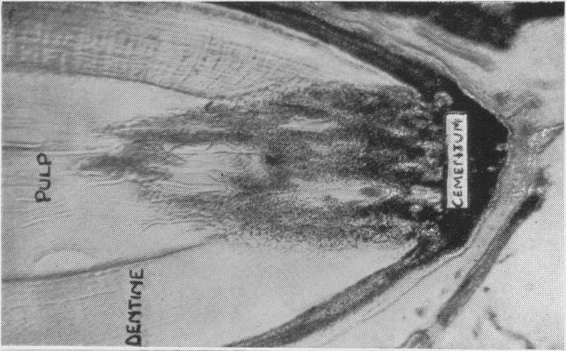
3 64 J. D. KING The above results, together with the radiographs and other illustrations shown in Pls. I and II, may be summarized as follows: (a) Vitamin A deficiency. The effects of deficiency of vitamin A upon the teeth and periodontal tissues of each dog were determined by the condition of the whole of the upper jaw and, as shown in Table I, by that of the left (unoperated) side of the mandible. In animals b, e, f and g (P1. I and II), receiving diets deficient in vitamin A, there was on the whole an increased deposition of tartar about the necks of the permanent teeth, accompanied by varying degrees of gingivitis. As seen in P1. II, figs. 2 and 4, dogs b and g, the alignment of the lower incisors was irregular, the latter perhaps being caused by crowding of the teeth due to deficient growth of the mandible. Radiographs of the lower jaws at death showed. the roots of the cheek teeth to be imperfectly developed and the laminat durae not well defined; the bone trabeculse in the immediate vicinity of the tooth roots appeared unusually opaque to X-rays in some regions (P1. I, figs. 2 and 4). Histologically, degeneration of sensory nerves and hyperplasia and downward proliferation of the subgingival epithelium were characteristic defects; in addition, the abnormal radiographic appearance of the tooth roots was seen to be due to excessive deposition of cementum, which in some cases had invaded the apical portion of the pulp chamber (P1. II, figs. 6 and 7). The addition of calcium carbonate to the ration of dog g (P1. I, fig. 4; P1. II, figs. 4 and 7) produced no obvious improvement in the periodontal tissues or in the alignment of the front teeth. In contrast with the above, animals a, c and d showed much more normal conditions. The amount of gingivitis and cervical tartar was considerably reduced; the incisor teeth were in proper alignment; abnormalities of cementum and bone were absent; and the nervous and epithelial tissues presented a normal appearance (P1. I, figs. 1 and 3; P1. II, figs. 1, 3 and 5). (b) Experimental nerve lesions. The effects due to resection of the right inferior dental nerve were assessed by comparing the right side of the lower jaw, peripheral to the site of operation, with the left (control) side. The most obvious result of the nerve lesion was the effect produced upon the alignment of the front teeth. Pronounced linguoversion of the second incisor, and to a lesser extent buccoversion of the third incisor, was observed even when the diet contained liberal amounts of vitamins A and D and other essentials (P1. II, figs. 1 and 3, dogs a and c). In those animals receiving A-deficient rations, since lack of this vitamin was itself associated with similar conditions in the lower
4 4) 0 4).-4)- C) 90 & * * & 44) ^ + ~. *,.> *- 1-1 H4, c C'* N. 1-- P> > 44'Go p p r$ + + ; *; P....P ~~~ O@@ MI+~ I+t" - E 4)4 + 4 ^ 4 *... s * *..*- fl C) 0.2 4)64 CH.4 *t bo 0-4a 4-4 *tl 0 64 Ca 40 Ca m w.a O al 44) W -0C o 0 0C )-44-5A ) O )i 06 CB 4) 0 C4) w4" 04) 344 B._ *z 64 4) CB-) C', 4) 4) 40* -~~~~~ 64,W4 o 4) Ca 4) Ca 4)~~~~~~~~~~4 4) o Q~~~~~~~~)46-44.~ 4 44 C), 4) 4) 4) C1 -~~~~~ ~ oo -4Q ~ ~ ~ ~~~ ~ ~ ~~ ~ *~~~~~~~ ~04)64 4),~~~~~~~~~~4 _ 4)44)44 4)44~~~~46 4 _ 4)o._. ~ 4) PH. LXXXVIII.
5 66 J. D. KING incisor region, the effects of the nerve operations cannot easily be judged (P1. II, figs. 2 and 4, dogs b and 9). In the experiments described here no marked differences, macroscopic, radiographic, or histological, in the condition of the periodontal tissues were observed between the operated and control sides of the lower jaw, even in the incisor region. When the diets were deficient, the changes associated with lack of vitamin A were present; when there was an adequate supply of this vitamin, the tissues appeared normal in structure. Although at first sight it might appear that the mechanism controlling the incisor misalignment, seen both in vitamin A deficiency and after experimental nerve excision, had a common origin, a consideration of the time of eruption of the permanent teeth of both sides of the lower jaw of two of the young operated dogs, receiving, respectively, diets adequate and deficient in vitamin A, indicated that the deformities arose from different causes, as shown in Table II. TABLE II. The time of eruption (piercing of the gum) of the permanent lower teeth. The effects of experimental excision of part of the right inferior dental nerve compared with those of deficiency of vitamin A in the diet Age at operation: 1j months. Postoperative period: 7 months. Diet: as in Table I. Dog d Dog f (Diet containing vitamin A) (Diet deficient in vitamin A) A Time of eruption Time of eruption (days after birth) Difference (days after birth) Difference C (- early, (-early, Perm. Unoperated Operated + late) Unoperated Operated + late) teeth side side days side side days II I I's C Pml Absent Absent * + 5 Pm * + 3 Pm * Pm * ml M M * It is probable that the tissues in the vicinity of these teeth may have been damaged during the operation. This table allows a comparison to be made between the effect of vitamin A deficiency and that of the experimental nerve lesion upon the rate of eruption of the mandibular permanent teeth of dogs d and f.

6 VITAMIN AND DENTAL NERVES In the first instance, on the unoperated side it is evident that the teeth of the vitamin A-deficient dog f erupted, on an average, about 13 days later than the corresponding ones of dog d, which received a liberal supply of vitamin A. The nerve operation, on the other hand, resulted in a slight acceleration of growth of the second or third incisor of the operated side, as compared with the corresponding teeth of the unoperated side of the same animal; it will be noted that the incisor teeth so affected were those which had previously been observed to be out of alignment (see Table I). The position of the canine of either dog, on account of the size of the tooth and the length of its root, would hardly have been expected to be much affected. The proximity to the site of operation of the premolars peripheral to the nerve lesion renders it inadvisable to draw any conclusions from the time of eruption of these teeth. It would seem, therefore, that vitamin A deficiency and excision of the inferior dental nerve tend: to have reverse effects upon the rate of eruption of the lower incisor teeth, although their end result is similar as regards tooth alignment. On this basis, however, it is difficult to account for the incisor irregularity produced in dog a, all of whose teeth were fully erupted prior to the nerve operation. SERIES 2 (RABBITS) Experimental removal of part of the right inferior dental nerve was also performed on eleven rabbits. Since the teeth of these animals grow from persistent pulps, by drilling small parallel holes in the cervical enamel of the incisor of each side of the lower jaw and observing the rate at which these holes approached the biting edge, it was 'possible to compare the rate of tooth growth on the operated and unoperated sides. This procedure was adopted in five of the operated rabbits and in seven unoperated controls. In all these animals the distance from the lower border of the drilled hole to the biting edge of the tooth was recorded at frequent intervals. In the other six operated animals, which were allowed to survive for 1-4 months after the nerve had been excised, the growth of the teeth was not estimated, the object in this instance being observation of any possible pathological changes in the teeth and periodontal tissues over a varying postoperative period. The diets of all the animals, experimental and control, included an adequate supply of fat-soluble vitamins, being composed of: "nibbed" oats, 40 g.; wheat bran, 10 g.; heated alfalfa, 10 g.; calcium carbonate, 075 g.; lemon juice, 1 c.c.; vitamin D, 500 i.u.; and cabbage, 30 g. or

 Text-fig. 1.")
, 17 days after holes had been drilled in the cervical enamel of the lower teeth, showing their normal rate of growth.")
. Text-fig. 4.")
7 68 J. D. KINGB mammalian liver oil (vitamin A), 250 "blue" units. The average age of the animals at the time of operation was 3 months, when they were receiving 70 g. of the above diet daily. Results. The effects of the operation upon the rate of growth of the teeth of rabbits can best be described in conjunction with the accompanying graphs (Text-figs. 5, 6 and 7), where AB represents the time Fig. 2. Fig. 1. Fig. 3. ~~~~~~Fig. 4. The effects of experimental resection of the inferior dental and cervical sympathetic nerves upon the rate of growth of the incisor teeth of rabbits. (See also p. 70, Text-figs ) Text-fig. 1. Radiograph of the lower jaw of a rabbit after excision of the right inferior dental nerve, showing the site of operation (0). Text-fig. 2. Photograph of the incisors of control animal 6 (series 2), 17 days after holes had been drilled in the cervical enamel of the lower teeth, showing their normal rate of growth. No nerve operation had been performed. Text-fig. 3. Photograph of the incisors of animal 1 (series 2), 17 days after resection of the right inferior dental nerve. Note the more rapid rate of growth of the lower tooth of the operated side (0). Text-fig. 4. Photograph of the incisors of animal 19 (series 3), 18 days after resection of the right cervical sympathetic trunk. Note the more rapid growth of the tooth of the operated side (0). after operation and BC the growth of the teeth during this period, the line passing horizontally through C being the level at which the drilled holes had grown out of the teeth. The following are typical of these experiments. Animal 1 (Text-fig. 5). On the eighth day after operation it is seen that the right incisor had grown distinctly faster than the left (control). This acceleration of growth increased steadily until, on the twenty-first

8 VITAMIN AND DENTAL NERVES day, the hole in its buccal surface was no longer visible. At this time the left incisor hole was still 2 mm. below the biting edge, which it passed on the thirtieth day after the operation. Text-fig. 3 illustrates the appearance of the lower incisors of this animal 17 days after resection of the right inferior dental nerve; the relative rate of growth of the teeth, as evidenced by the position of the holes in their buccal surface, is clearly indicated. Text-fig. 2 is a photograph of an unoperated control animal (No. 6) at a similar stage. In animals 2 and 3 (Text-figs. 6 and 7), following acceleration in growth of the right incisor during the first 8 days after operation, the teeth of both sides grew at relatively similar rates until the twenty-first and eighteenth day respectively. In each animal the growth of the right tooth then became retarded, and the holes in all the teeth disappeared on the twenty-seventh day. During the first 19 days after operation, the average rate of growth of the lower incisor teeth was approximately: on the operated side, 0-28 mm. per day; on the unoperated side, 021 mm. per day. Control animals (unoperated) 6-12 (Text-fig. 2). The incisor of each side grew at a rate corresponding to that of the control teeth in the operated animals. This shows that the resection has been followed by accelerated growth. In some cases the increase in growth took place in the first week only and it appears that subsequently a slight retardation may sometimes occur. The weights of animals 1-5 fell for the first day or two after operation, but soon recovered and increased more or less consistently throughout the experiments. Apart from the occasional presence of small, opaque, whitish patches on the enamel of both lower incisors, no other abnormal conditions were observed, clinically or radiographically, in the teeth or periodontal tissues of any of the animals in which the resection was done, even in those surviving for 4 months after operation. Histologically, increased vascularity of the tissues on the operated side of animals 1-5 was the only change to be noted in the teeth or surrounding structures peripheral to the nerve lesion. On the other hand, Astanin and Kauschansky [1931] claim to have produced "macroscopic caries" and gingivitis by severing the second or third divisions of the trigeminal nerve of rabbits; but since all the teeth of rabbits grow from persistent pulps, they can hardly be subject to typical dental caries. Reverting to the growth-rate experiments, the rabbit findings confirm those of series 1 (dogs). It appears, however, unlikely that destruction of the sensory fibres of the inferior dental nerve would accelerate 69


9 70 J. D. KING tooth growth. It is of course possible that in rabbits the nerve lesion in some way affected the composition of the tooth, rendering it more or less I OPIERAThD a CONTReL u6o WEMCHTIN ^RA Fig. 5. Fig. 6. Fig. 7. A DAY5 AFTER OR B A DAYS AFTER OP. B A DAYS AFTER OR B Fig. 8. Fig. 9. Fig. 10. The effect of experimental resection of the inferior dental and cervical sympathetic nerves upon the rate of growth of the incisor teeth of rabbits. Text-figs. 5-7 illustrate graphically the rate of growth of the lower incisors of animals 1-3 (series 2) after the inferior dental operation. Text-figs illustrate the growth of the lower incisors of animals (series 3) after the cervical sympathetic operation. The distance BC=6-5 mm. in Text-figs. 5 and 6, and 6*0 mm. in Text-figs. further description of graphs see text, pp. 67 and ; for brittle, but no such change was noted by the methods available. Certainly the incisal edge on the operated side must, if the tooth grew faster, have been more susceptible to attrition than that of the control tooth, otherwise it would have projected beyond the latter. That there was no

10 VITAMIN AND DENTAL NERVES obvious difference in length between the two teeth is shown by Textfig. 3. In any case such an argument has little or no bearing upon the dog experiments. It seemed more probable that some other nerves, such as those controlling the blood supply to the teeth, may have been in huch close proximity to the inferior dental nerve that they too were severed or injured during the operation. Vaso-constrictor fibres are said to accompany the peripheral nerves [Gilding, 1932], and if these had been destroyed, it might be expected that vaso-dilatation of the peripheral vessels, including those of the teeth distal to the lesion, by increasing the volume of blood available, would tend to accelerate the changes associated with tooth growth. It must be emphasized here that the inferior dental vessels themselves were not damaged by the operation in any of the dogs or rabbits. It was therefore decided to remove part of the cervical sympathetic cord, believed to supply vaso-constrictor fibres to the teeth and jaws, from a further series of rabbits. SERIES 3 (RABBITS) Operative procedure. About 1 in. cf the right cervical sympathetic cord, between the middle and superior cervical ganglia, was removed from each of five rabbits; holes for measuring growth rate were again drilled in the lower incisor teeth, as previously described. The animals received the same diets as those in series 2. Results. The effects of these operations are shown in Text-figs. 4, 8, 9 and 10. During the first 19 days after nerve resection, the average rate of growth of the lower incisor teeth of animals was approximately: on the operated side, 0-28 mm. per day; on the unoperated side, 0-23 mm. per day; thus demonstrating that an increased rate of growth may also follow excision of part of the cervical sympathetic. Probably, therefore, the effects attributed in series 1 and 2, to removal of the inferior dental nerve, were in reality due to injury to vaso-constrictor fibres. As in series 2, no periodontal changes were observed, but again opaque patches on both lower incisors were noted, and histological preparations demonstrated a larger blood content of the teeth of the operated side. DISCUSSION (a) Vitamin A deficiency (dogs). M. Mellanby [1930], from experiments on dogs, concluded that vitamin A was primarily concerned with the development of the soft periodontal structures, while vitamin D 71

11 72 J. D. KING controlled that of the calcified tissues. Retarded tooth eruption, incisor irregularity, and the supervention of periodontal disease were considered to result from a combined deficiency of both these fat-soluble vitamins. E. Mellanby [1926 a] found that in fat-soluble vitamin deficiency, susceptibility to infection was not related to the calcification of the bones, indicating that the former was due to deficiency of vitamin A rather than of D. Goldblatt and Benischek [1927] repeated the work of Mori [1922] and Wolbach and Howe [1925] on the dietary factor responsible for epithelial hyperplasia and metaplasia, but gave vitamin D in the form of irradiated cotton-seed oil, so that the diet was deficient only in vitamin A. The epithelial changes, as well as infective lesions also found, such as keratomalacia, broncho-pneumonia and enteritis, could now be definitely ascribed to vitamin A deficiency. As a result of further work on this problem, Green and E. Mellanby [1928, 1930] gave the name "anti-infective vitamin" to vitamin A, and later E. Mellanby [1933, 1934] showed that degeneration of sensory nerves, peripheral as well as central, resulted from lack of vitamin A. In the experiments described here all of the dogs received vitamin D, the variable factor being vitamin A. Deficiency of the latter resulted not only in epithelial hyperplasia, degeneration of sensory nerves, and a somewhat increased deposition of cervical tartar, but also in retarded tooth eruption, irregularities in position of the lower incisor teeth, imperfectly formed tooth roots with a tendency to apical hypercementosis, ill-defined laminee durae and changes in the alveolar bone in some regions (Pls. I and II). It is of interest here to note that in some cases of chronic periodontal disease in man it is not uncommon to find rounded and stunted tooth roots similar in radiographic appearance to those observed in experimental vitamin A deficiency. Although it is true that a combined deficiency of vitamin D and vitamin A accentuates some, but not all, of the abnormalities in the calcified structures and still further retards the eruption of the teeth, the results described here indicate that vitamin A, in addition to its influence upon epithelia and sensory nerves, may also play a part in the development of some of the calcified tissues of the body. In some unpublished experiments on rats the author has observed that vitamin A deficiency may be associated with degeneration of the ameloblasts and odontoblasts of their persistently growing incisors. In such A-deficient amimals the characteristic enamel pigmentation was lost, while the dentine was poorly calcified and contained many interglobular spaces, conditions reminiscent of vitamin D deficiency, although the animals

12 VITAMIN AND DENTAL NERVES were given ample amounts of vitamin D, calcium and phosphorus. The rat experiments confirm to some extent the findings of Wolbach and Howe [1933], who maintained that vitamin A played an extremely important part in the calcification of the teeth of rats. But the experiments of these investigators were not decisive, since their A-deficient rations were also deficient in vitamin D, while in their adequate diets vitamin A was given in the form of butter, a variable source of vitamin A which contains an appreciable amount of vitamin D. It is usually considered that vitamin D controls calcification by mobilizing the mineral salts of the blood. It seems possible that vitamin A may affect calcification by its influence upon the morphology and consequent activity of the lime-secreting cells, even when vitamin D and mineral salts are available. If this is so, its absence may contribute to the development of periodontal disease and even dental caries by causing defects of tooth structure as well as by increased susceptibility to infection. It is hoped that the experimental production of such diseases, preliminary reports of which have already been published [King, 1935 a, b], may shed further light on this problem. By means of a histological examination of human post-mortem material Wilkinson [1935] showed that degenerated cells of the subgingival epithelium may become calcified before the latter becomes separated from the tooth; later, when separation does occur, he claims that the calcified cells are left adhering to the tooth as calculus, the formation of the latter being similar to calcification in cartilage as described by Robison [1923]. In support of this theory Wilkinson cites the experiments of Smith [1930] and Adamson [1929], as demonstrating that desquamated cells of the gingival epithelia liberate a phosphatase, which, by setting phosphates free in the presence of calcium ions, allows calcium phosphate to be deposited in the dead cells. Wilkinson believes that calculus may thus be produced, resulting in the inflammatory complications so often encountered in human periodontal disease. Certainly epithelial degeneration, calculus formation, and gingivitis are common sequelae to experimental vitamin A deficiency, so that since these conditions are also found in the human subject, whatever be the exact role of vitamin A in this syndrome, it may be inferred that lack of this vitamin, especially during the developmental period, is an important predisposing factor in the aetiology of periodontal disease. (b) Experimental nerve lesions (dogs and rabbits). The relative uniformity in results of the experiments in series 2 and 3 (rabbits) indicates 73

13 74 J. D. KING that the effects produced by resection of the inferior dental nerve in both dogs and rabbits were caused by the unintentional severing of vasoconstrictor fibres in very close association with, and probably within the actual sheath of, the sensory nerve. It is probable, therefore, that in both species the temporarily increased rate of growth of the teeth peripheral to the lesion was due to an increase in the volume of blood available. Gilding [1932] showed that the distribution of the vaso-constrictor nerves is strictly unilateral, stopping abruptly at the mid-line, in spite of the fact that the blood vessels of one side anastomose freely with those of the other. The sensory distribution of the inferior dental nerve, on the other hand, frequently extends for some little distance across the mid-line. The growth changes here reported were confined to the operated side. The temporary nature of such changes suggests that the walls of the affected blood vessels soon recover the greater part, at least, of their normal tone, and this may perhaps be why no histological alterations in the dental tissues were found by the methods described. It is true that microscopic preparations of the rabbits' lower incisor teeth after death showed more blood in the tooth pulp of the operated side, but this may be merely because the normally innervated vessels on the other side were constricted in the asphyxia induced by the chloroform used for killing the animals. In this respect the animals in which the inferior dental nerve was excised were like those in which the sympathetic was cut, so that it is probable that the constrictor nerves had been severed in all three series of animals and that the growth changes observed were due to this. Leist [1927] described an increased rate of growth of the teeth of five out of nine guinea-pigs after cauterizing the sympathetic fibres around the carotid artery with 7 p.c. phenol; he also noted similar but less marked changes following such treatment of the internal maxillary and inferior dental arteries of a few dogs. In the cervical sympathetic of the dog there are dilator as well as constrictor fibres [Dastre and Morat, 1884], but not in the rabbit or cat [Feldberg and Schiff, 1926]. As the constrictors predominate over the dilators in the dog's splanchnic nerve [Dale, 1913], this may also be expected in its cervical sympathetic. Although resection of the inferior dental nerve, as previously described, has failed to cause lesions in the teeth or periodontal tissues, it is possible that, by destruction of the whole sensory supply to one side of one or both jaws and by preventing nerve regeneration for a sufficiently

14 VITAMIN AND DENTAL NERVES long period, pathological changes similar to those seen in vitamin A deficiency may be produced. Experiments along these lines are now in progress; in some, the sensory roots of the fifth nerve are resected, while in others the Gasserian ganglion from which they originate is itself removed. The results of the present nerve operations may have some bearing on the vexed problem of tooth eruption, for they show that, in addition to vitamins A and D, the blood supply and condition of the vascular nerves play a part in the development and growth of the teeth. G ottlieb [1927] claims that changes in the rate of growth of the teeth are of outstanding importance. He believes that eruption continues slowly after the teeth have come into occlusion, and that any conditions which disturb the synchronism between attrition of the tooth crowns and " postclinical" eruption accelerate the downgrowth of the subgingival epithelium and the development of periodontal disease. If the misalignment of the right lower incisors following the operation in the adult dog which received vitamin A was due to changes in the blood supply, it seems possible that such changes, resulting in abnormal movements of the teeth even after complete clinical eruption, may in turn give rise to changes in their supporting structures. The fact that in this dog the periodontal tissues appeared free from defects and there was no downgrowth of subgingival epithelium along the root or absorption of alveolar bone, does not seem to support Gottlieb's hypothesis, although more experiments on adult animals are necessary. Movements of the teeth after clinical eruption are, of course, quite commonly seen in the human mouth, associated with trauma, pressure from other teeth, or extraction of adjacent or opposing teeth. SUMMARY In dogs receiving vitamin A-deficient diets: (1) eruption of teeth was delayed; (2) hypercementosis occurred and the laminae durao and bone of the tooth sockets were malformed; (3) the alignment of the incisor teeth was irregular; in addition to the periodontal and nervous defects described in previous papers. In animals (dogs and rabbits) in which the inferior dental nerve was resected on one side: (1) the eruption in dogs, and in rabbits the growth of teeth was accelerated; (2) the alignment of the incisor teeth was irregular; but no defects in the dental or periodontal tissues were found. 75
15 76 J. D. KING As similar changes were observed on resection of the cervical sympathetic, the effects of severing the inferior dental nerve are regarded as due to damage to vasomotor fibres in it. The expenses of this investigation have been defrayed by the Medical Research Council, to whom my thanks are due. I am also indebted to Mrs M. Mellanby and Prof. G. A. Clark for their valuable advice and criticism, and to Miss I. Jopling for clerical assistance. REFERENCES Adamson, K. T. (1929). Aust. J. Dent. 33, 245. Astanin, P. P. & Kauschansky, L. J. (1931). Dtsch. Mschr. Zahnheilk. 49, 12. Dale, H. H. (1913). J. Physiol. 40, 291. Dastre & Morat (1884). Systeme neurveux va8omoteur. Paris. Feldberg & Schiff (1926). Pfuigers Arch. 212, 365. Gilding, H. P. (1932). J. Physiol. 74, 34. Goldblatt, H. & Benischek, M. (1927). J. exp. Med. 46, 699. Gottlieb, B. (1927). J. Amer. dent. A8s. 14, Green, H. N. & Mellanby, E. (1928). Brit. med. J. 2, 691. Green, H. N. & Mellanby, E. (1930). J. Exp. Path. 11, 81. King, J. D. (1935 a). Dent. Rec. 55, 522. King, J. D. (1935 b), Brit. dent. J. 59, 233 and 305. Leist, M. (1927). Z. Stomat. No. 8. Mellanby, E. (1926 a). Brit. med. J. 1, 515. Mellanby, E. (1933). Edin. med. J. 40, No. 4. Mellanby, E. (1934). J. Path. Bact., Lond., 38, 391. Mellanby, M. (1929). Sp. Rep. Ser. Med. Res. Coun. No Mellanby, M. (1930). Ibid. No Mellanby, M. & King, J. D. (1934). Brit. dent. J. 56, 538. Mori, S. (1922). J. Amer. med. Ass. 79, 197. Robison, R. (1923). Biochem. J. 17, 286. Smith, G. H. (1930). Aust. J. exp. Biol. Med. Sci. 7, 45. Wilkinson, F. C. (1935). Dent. Rec. 55, No. 3, p. i. Wolbach, S. B. & Howe, P. R. (1925). J. exp. Med. 42, 753. Wolbach, S. B. & Howe, P. R. (1933). Amer. J. Path. 9, 275.


16 THE JOURNAL OF PHYSIOLOGY, VOL. 88, No. 1 PLATE I Fig. 1. Doga. Fig. 2. Dogb. Fig. 3. Dog c. Fig. 4. Dogg. To face p. 76


17 PLATE 1I _~~~~~~~~~~~~~~~~ fd\ El THE JOURNAL OF PHYSIOLOGY, VOL. 88, No. 1 at~~~~~~~~~~~~~~~~~7 0PF`7' -0, '. 2?jiJ
18 VITAMIN AND DENTAL NERVES 77 EXPLANATION OF PLATES I AND 11 PLATE I Radiographs to illustrate the effects of vitamin A deficiency and of resection of the right inferior dental nerve upon the lower jaw and teeth of dogs. Fig. 1. Dog a. Age at operation, 22 months. Postoperative period, 12 months. Diet contained liberal supplies of vitamin A. The appearance of the teeth and bone of both sides is normal. Fig. 2. Dog b. Age at operation, 11 months. Postoperative period, 11 months. Diet was deficient in vitamin A. Note. Defective tooth roots, ill-defined lamine dure and, surrounding these, the bone trabeculae are more radio-opaque on both sides. Fig. 3. Dog c. Age at operation, 2 months. Postoperative period, 6 months. Diet included liberal amounts of vitamin A. The teeth and bone of both sides appear normal. Fig. 4. Dog g. Age at operation, 2 months. Postoperative period, 5 months. Diet was deficient in vitamin A but calcium carbonate was added. Note. On both operated and unoperated sides the tooth roots are defectively formed, the laminae dure are not well-defined and the surrounding bone trabeculse are more radio-opaque than normal; the lower margin of the jaw of this animal was injured after death. 0, site of operation. A, B, C, areas from which the photomicrographs shown in P1. II were prepared. PLATE II Photogrophs and photomicrographs to show the effects of vitamin A deficiency and of resection of the right inferior dental nerve of the dogs referred to in P1. I. Fig. 1. Dog a, operated when 22 months old. Diet contained liberal amount of vitamin A. Note. Irregular alignment of the lower front teeth of the operated side. Fig. 2. Dog b, operated when 11 months old. Diet was deficient in vitamin A. Note. Irregular alignment of lower incisors of both operated and unoperated sides; the canines were removed. Fig. 3. Dog c, operated when 2 months old. Diet included liberal supplies of vitamins A and D. Note. Irregularity of lower incisors of the operated side. Fig. 4. Dog g, operated when 2 months old. Diet was deficient in vitamin A but calcium carbonate was added. Note. Bilateral irregularity of lower incisors. 0, operated side. Fig. 5. Ground section of area A, P1. I, dog a. Note. Normal appearance of apical and peri-apical tissues; section is not quite central, but, when considered in conjunction with the radiograph (P1. I, fig. 1), the tissues are seen to be free from obvious defects. Fig. 6. Ground section of area B, P1. I, dog b. Note. Invasion and replacement of pulp and dentine by cementum. Fig. 7. Ground section of area C, P1. I, dog g. Note. Invasion of pulp and dentine by cementum.
Fundamental & Preventive Curvatures of Teeth and Tooth Development. Lecture Three Chapter 15 Continued; Chapter 6 (parts) Dr. Margaret L.
 Dr. Margaret L.") Fundamental & Preventive Curvatures of Teeth and Tooth Development Lecture Three Chapter 15 Continued; Chapter 6 (parts) Dr. Margaret L. Dennis Proximal contact areas Contact areas are on the mesial and
Fundamental & Preventive Curvatures of Teeth and Tooth Development Lecture Three Chapter 15 Continued; Chapter 6 (parts) Dr. Margaret L. Dennis Proximal contact areas Contact areas are on the mesial and
Dental Morphology and Vocabulary
 Dental Morphology and Vocabulary Palate Palate Palate 1 2 Hard Palate Rugae Hard Palate Palate Palate Soft Palate Palate Palate Soft Palate 4 Palate Hard Palate Soft Palate Maxillary Arch (Maxilla) (Uppers)
6610 NE 181st Street, Suite #1, Kenmore, WA
 660 NE 8st Street, Suite #, Kenmore, WA 9808 www.northshoredentalacademy.com.08.900 READ CHAPTER The Professional Dental Assistant (p.-9) No Key Terms Recall Questions:,,,, and 6 CLASS SYLLABUS DAY READ
Development of teeth. 5.DM - Pedo
 Development of teeth 5.DM - Pedo Tooth development process of continuous changes in predetermined order starts from dental lamina A band of ectodermal cells growing from the epithelium of the embryonic
Medical NBDE-II. Dental Board Exams Part I.
 Medical NBDE-II Dental Board Exams Part I http://killexams.com/exam-detail/nbde-ii Question: 149 Anatomically, the term "clinical root" can be defined as which of the following: A. The space in the tooth
Dental Anatomy and Occlusion
 CHAPTER 53 Dental Anatomy and Occlusion Ma Lou C. Sabino DDS, and Emily G. Smythe, DDS What numerical system is used most commonly in the United States for designating the adult dentition? Pediatric dentition?
SPACE MAINTAINER. Multimedia Health Education. Disclaimer
 Disclaimer This movie is an educational resource only and should not be used to manage your health. All decisions about the management of premature loss of primary teeth and use of space maintainers must
Advanced Probing Techniques
 Module 21 Advanced Probing Techniques MODULE OVERVIEW The clinical periodontal assessment is one of the most important functions performed by dental hygienists. This module begins with a review of the
1. What is the highest and sharpest cusp on the lower first deciduous molar? 2. Which of the following is NOT the correct location of an embrasure?
 1 1. What is the highest and sharpest cusp on the lower first deciduous molar? a. mesiobuccal b. distobuccal c. distolingual d.mesiolingual 2. Which of the following is NOT the correct location of an embrasure?
Applications in Dermatology, Dentistry and LASIK Eye Surgery using LASERs
 Applications in Dermatology, Dentistry and LASIK Eye Surgery using LASERs http://www.medispainstitute.com/menu_laser_tattoo.html http://www.life123.com/bm.pix/bigstockphoto_close_up_of_eye_surgery_catar_2264267.s600x600.jpg
Primary Teeth Chapter 18. Dental Anatomy 2016
 Primary Teeth Chapter 18 Dental Anatomy 2016 Primary Teeth - Introduction Synonyms deciduous teeth, baby teeth, temporary teeth, milk teeth. There are 20 primary teeth, designated as A thru T in the Universal
(Received February 6, 1935.)
") 272 6 I2.3II.1:577. i6c THE EFFECT OF VITAMIN D ON THE CALCIUM CONTENT OF THE DENTINE. BY E. WILFRED FISH. (From the Hale Research Laboratory, Royal Dental Hospital, London.) (Received February 6, 1935.)
272 6 I2.3II.1:577. i6c THE EFFECT OF VITAMIN D ON THE CALCIUM CONTENT OF THE DENTINE. BY E. WILFRED FISH. (From the Hale Research Laboratory, Royal Dental Hospital, London.) (Received February 6, 1935.)
[1920], in studies on the human pleural membrane, pointed out the
![[1920], in studies on the human pleural membrane, pointed out the](/thumbs/84/89127006.jpg "[1920], in studies on the human pleural membrane, pointed out the") 'ca -.101 6II.25:6II.OI8.86 NERVES AND NERVE ENDINGS IN THE VISCERAL PLEURA OF THE CAT. BY A. I. G. McLAUGHLIN. (From the Unit Laboratories, University College Hospital Medical School.) (Received September
'ca -.101 6II.25:6II.OI8.86 NERVES AND NERVE ENDINGS IN THE VISCERAL PLEURA OF THE CAT. BY A. I. G. McLAUGHLIN. (From the Unit Laboratories, University College Hospital Medical School.) (Received September
THE CANADA AND ALBERTA BSE SURVEILLANCE PROGRAM (CABSESP) GUIDELINES FOR AGE VERIFICATION IN CATTLE 1
 GUIDELINES FOR AGE VERIFICATION IN CATTLE 1") AGRICULTURE AND RURAL DEVELOPMENT Food Safety and Animal Health Division, Animal Health Branch 9 th Floor, O.S. Longman Building 6909-116 Street Edmonton, Alberta T6H 4P2 Telephone 780-644-2148 Fax 780-422-5734
AGRICULTURE AND RURAL DEVELOPMENT Food Safety and Animal Health Division, Animal Health Branch 9 th Floor, O.S. Longman Building 6909-116 Street Edmonton, Alberta T6H 4P2 Telephone 780-644-2148 Fax 780-422-5734
Structure of an Incisor
 MAMMALIAN TEETH Mammals have different types and shapes of teeth and they are thus termed Heterodonts. Those which have teeth of the same size and shapes are termed as Homodonts. In mammals teeth consist
Attachment G. Orthodontic Criteria Index Form Comprehensive D8080. ABBREVIATIONS CRITERIA for Permanent Dentition YES NO
 First Review IL HFS Dental Program Models Second Review Ortho cad Attachment G Orthodontic Criteria Index Form Comprehensive D8080 Ceph Film X-Rays Photos Narrative Patient Name: DOB: ABBREVIATIONS CRITERIA
PREMATURE PRIMARY TOOTH LOSS
 Disclaimer This movie is an educational resource only and should not be used to manage your dental health. All decisions about the management of premature primary tooth loss must be made in conjunction
Eruption and Shedding of Teeth
 Eruption and Shedding of Teeth Mixed Dentition: Presence of both dentitions Figure from Ten Cate s Oral Histology, Ed., Antonio Nanci, 6 th edition Tooth eruption is the process by which developing teeth
DENTAL TRAUMA IN DECIDUOUS TEETH
 Disclaimer This movie is an educational resource only and should not be used to manage your health. All decisions about the management of Dental Trauma in Deciduous Teeth must be made in conjunction with
Dental Anatomy High Yield Notes. **Atleast 35 questions comes from these areas of old lectures**
 Dental Anatomy High Yield Notes **Atleast 35 questions comes from these areas of old lectures** This review notes compiled and prepared by my sister for her own study, as a last day review session for
Archived SECTION 14 - SPECIAL DOCUMENTATION REQUIREMENTS
 SECTION 14 - SPECIAL DOCUMENTATION REQUIREMENTS 14.1 CERTIFICATE OF MEDICAL NECESSITY...2 14.2 OPERATIVE REPORT...2 14.2.A PROCEDURES REQUIRING A REPORT...2 14.3 PRIOR AUTHORIZATION REQUEST...2 14.3.A
IMPACTED CANINES. Unfortunately, this important tooth is the second most common tooth to be impacted after third molars
 IMPACTED CANINES After we talked about impacted third molars, today we ll discuss about maxillary impacted canines in upper dental arch, how to manage these cases as a dental surgeon. You will study about
Tooth eruption and movement
 Tooth eruption and movement Dr. Krisztián Nagy Diphydont dentition Deciduous dentition primary dentition Diphydont dentition Permanent dentition secondary dentition Mixed Dentition: Presence of both dentitions
Oral Embryology and Histology
 Oral Embryology and Histology Chapter 8 Copyright 2018, Elsevier Inc. All Rights Reserved. 1 Learning Objectives Lesson 8.1: Oral Embryology 1. Pronounce, define, and spell the key terms. 2. Define embryology
(Received 12 April 1938)
") 206 J. Physiol. (I938) 93, 206-2I4 6II.83:6II.314:6I2.392.OI3 A DEGENERATIVE CHANGES IN THE AXIS CYLINDERS OF THE DENTAL NERVES, DUE TO DIETS DEFICIENT IN VITAMIN A AND CAROTENE BY J. D. KING, W. LEWINSKY
206 J. Physiol. (I938) 93, 206-2I4 6II.83:6II.314:6I2.392.OI3 A DEGENERATIVE CHANGES IN THE AXIS CYLINDERS OF THE DENTAL NERVES, DUE TO DIETS DEFICIENT IN VITAMIN A AND CAROTENE BY J. D. KING, W. LEWINSKY
Lecture 2 Maxillary central incisor
 Lecture 2 Maxillary central incisor Generally The deciduous tooth appears in the mouth at 3 18 months of age, with 6 months being the average and is replaced by the permanent tooth around 7 8 years of
Morphology of an Anatomic Crown. By: Assistant Professor Dr. Baydaa Ali Al - Rawi
 Morphology of an Anatomic Crown By: Assistant Professor Dr. Baydaa Ali Al - Rawi October 4, 2009 Elevated landmarks Depressed landmarks A) Elevated landmarks : 1. Dental lobe : is one of the primary centers
Development of occlusion
 Development of occlusion The development of dentition is an important part of craniofacial growth as the formation, eruption, exfoliation and exchange of teeth take place during this period. Term occlusion
THE EFFECT OF FLUORINE UPON THE PHOSPHATASE CONTENT OF PLASMA, BONES, AND TEETH OF ALBINO RATS
 THE EFFECT OF FLUORINE UPON THE PHOSPHATASE CONTENT OF PLASMA, BONES, AND TEETH OF ALBINO RATS BY MARGARET CAMMACK SMITH AND EDITH M. LANTZ (From the Department oj Nutrition, Agricultural Experiment Station,
Lec. 11 & 12 Dr. Ali H. Murad Dental pulp 1- Coronal pulp
 Lec. 11 & 12 Dr. Ali H. Murad Dental pulp Is the soft connective tissue located in the central portion of each tooth. All pulps have similar morphologic characteristic, such as a soft, gelatinous consistency
Infratemporal fossa: Tikrit University college of Dentistry Dr.Ban I.S. head & neck Anatomy 2 nd y.
 Infratemporal fossa: This is a space lying beneath the base of the skull between the lateral wall of the pharynx and the ramus of the mandible. It is also referred to as the parapharyngeal or lateral pharyngeal
Educational Training Document
 Educational Training Document Table of Contents Part 1: Resource Document Disclaimer Page: 2 Part 2: Line Item Grade Sheets Page: 3 Release: 11/2016 Page 1 of 6 Part 1: Resource Document Disclaimer The
Lec [8]: Mandibular nerve:
![Lec [8]: Mandibular nerve:](/thumbs/94/121295776.jpg "Lec [8]: Mandibular nerve:") Lec [8]: Mandibular nerve: The mandibular branch from the trigeminal ganglion lies in the middle cranial fossa lateral to the cavernous sinus. With the motor root of the trigeminal nerve [motor roots lies
Lec [8]: Mandibular nerve: The mandibular branch from the trigeminal ganglion lies in the middle cranial fossa lateral to the cavernous sinus. With the motor root of the trigeminal nerve [motor roots lies
Periodontal Disease. Radiology of Periodontal Disease. Periodontal Disease. The Role of Radiology in Assessment of Periodontal Disease
 Radiology of Periodontal Disease Steven R. Singer, DDS srs2@columbia.edu 212.305.5674 Periodontal Disease! Includes several disorders of the periodontium! Gingivitis! Marginal Periodontitis! Localized
FRACTURES AND LUXATIONS OF PERMANENT TEETH
 FRACTURES AND LUXATIONS OF PERMANENT TEETH 1. Treatment guidelines and alveolar bone Followup Procedures INFRACTION Clinical findings Radiographic findings Treatment Follow-Up Favorable Outcome Unfavorable
The Course, Relations and Distribution of the Inferior Alveolar Nerve and Its Branches in the Cat
 The Course, Relations and Distribution of the Inferior Alveolar Nerve and Its Branches in the Cat P. P. ROBINSON Department of Physiology (Oral Biology), The Medical School, University Walk, Bristol BS8
#45 Ortho-Tain, Inc PREVENTIVE ERUPTION GUIDANCE -- PREVENTIVE OCCLUSAL DEVELOPMENT
 #45 Ortho-Tain, Inc. 1-800-541-6612 PREVENTIVE ERUPTION GUIDANCE -- PREVENTIVE OCCLUSAL DEVELOPMENT Analysis and Diagnosis of Occlusion: The ideal child of 5 y ears of age that probably has the best chance
Our Teeth. History Of Equine Dentistry EQUINE DENTISTRY. Who Should Do Equine Dentistry? Some Facts To Know About Teeth
 EQUINE DENTISTRY Mike Black, DVM Nebraska Equine Veterinary Clinic Omaha NE History Of Equine Dentistry Some evidence of equine dentistry dates back to 2,000 B.C. Bit-Seats: Information on bit-seats dates
Pet Dental Health. Tooth/Mouth Anatomy. The Tooth. The Tooth cont d. The Tooth cont d 8/22/2016
 General Session: Take a Bite out of the Competition by Getting to the Root of Pet Dental Health Presented on: September 14, 2016 Presenter: Image Placeholder Pet Dental Health 1. Anatomy/Terms 2. Signs
Franklin, 1933; Waterman, 1933]; indeed, the only negative findings, [Waterman, 1933]. Inasmuch, then, as Donegan was misled with
![Franklin, 1933; Waterman, 1933]; indeed, the only negative findings, [Waterman, 1933]. Inasmuch, then, as Donegan was misled with](/thumbs/91/106294335.jpg "Franklin, 1933; Waterman, 1933]; indeed, the only negative findings, [Waterman, 1933]. Inasmuch, then, as Donegan was misled with") 381 6I2.I34:6I2.893 THE CONSTRICTOR RESPONSE OF THE INFERIOR VENA CAVA TO STIMULATION OF THE SPLANCHNIC NERVE BY K. J. FRANKLIN AND A. D. McLACHLIN (From the University Department of Pharmacology, Oxford)
381 6I2.I34:6I2.893 THE CONSTRICTOR RESPONSE OF THE INFERIOR VENA CAVA TO STIMULATION OF THE SPLANCHNIC NERVE BY K. J. FRANKLIN AND A. D. McLACHLIN (From the University Department of Pharmacology, Oxford)
Dental Anatomy and Physiology for Clinical Dental Technicians. with Marnie Hayward
 Dental Anatomy and Physiology for Clinical Dental Technicians with Marnie Hayward Salivary glands Parotid Submandibular Sublingual Salivary glands position Parotid glands Lie below ear and behind angle
Tooth Variations. Suruedee Chinthakanan
 Tooth Variations Suruedee Chinthakanan Tooth variations Dental anomalies Cause : hereditary factor Developmental disturbances of teeth www.ectodermaldysplsia.org Tooth variations Enamel is formed from
Alveolar Growth in Japanese Infants: A Comparison between Now and 40 Years ago
 Bull Tokyo Dent Coll (2017) 58(1): 9 18 Original Article doi:10.2209/tdcpublication.2016-0500 Alveolar Growth in Japanese Infants: A Comparison between Now and 40 Years ago Hiroki Imai 1), Tetsuhide Makiguchi
Periodontal ligament
 Periodontal ligament The periodontium The periodontium includes: The gingiva Cementum Periodontal ligament Alveolar bone Def: The periodontal ligament is the dense fibrous connective tissue that occupies
RETENTION AND RELAPSE
 RETENTION AND RELAPSE DEFINITION Maintaining newly moved teeth long enough to aid in stabilizing their correction MOYERS loss of any correction achieved by any orthodontic treatment RELAPSE CAUSES OF RELAPSE
APPENDIX A. MEDICAID ORTHODONTIC INITIAL ASSESSMENT FORM (IAF) You will need this scoresheet and a disposable ruler (or a Boley Gauge)
 You will need this scoresheet and a disposable ruler (or a Boley Gauge)") APPENDIX A MEDICAID ORTHODONTIC INITIAL ASSESSMENT FORM (IAF) You will need this scoresheet and a disposable ruler (or a Boley Gauge) Name: _ I. D. Number: Conditions: 1. Cleft palate deformities 2. Deep
APPENDIX A MEDICAID ORTHODONTIC INITIAL ASSESSMENT FORM (IAF) You will need this scoresheet and a disposable ruler (or a Boley Gauge) Name: _ I. D. Number: Conditions: 1. Cleft palate deformities 2. Deep
Dr Robert Drummond. BChD, DipOdont Ortho, MChD(Ortho), FDC(SA) Ortho. Canad Inn Polo Park Winnipeg 2015
, FDC(SA) Ortho. Canad Inn Polo Park Winnipeg 2015") Dr Robert Drummond BChD, DipOdont Ortho, MChD(Ortho), FDC(SA) Ortho Canad Inn Polo Park Winnipeg 2015 Severely compromised FPM with poor prognosis Children often present with a developing dentition affected
Dr Robert Drummond BChD, DipOdont Ortho, MChD(Ortho), FDC(SA) Ortho Canad Inn Polo Park Winnipeg 2015 Severely compromised FPM with poor prognosis Children often present with a developing dentition affected
ORTHODONTIC INITIAL ASSESSMENT FORM (OIAF) w/ INSTRUCTIONS
 w/ INSTRUCTIONS") Use the accompanying Tip Sheet and How to Score the Orthodontic Initial Assessment Form for guidance in completion of the assessment form. You will need this score sheet and a disposable ruler (or a Boley
Use the accompanying Tip Sheet and How to Score the Orthodontic Initial Assessment Form for guidance in completion of the assessment form. You will need this score sheet and a disposable ruler (or a Boley
MAXILLARY INJECTION TECHNIQUE. Chinthamani Laser Dental Clinic
 MAXILLARY INJECTION TECHNIQUE Chinthamani Laser Dental Clinic Introduction A number of injection techniques are available to aid in providing clinically adequate anesthesia of the teeth and soft and hard
The Blood Supply of the Rat Mandible '
 The Blood Supply of the Rat Mandible ' DONALD F. HUELKE AND WALTER A. CASTELL12 Department of Anatomy, The University of Michigan, Ann Arbor, Michigan ABSTRACT The blood supply of the rat mandible was
Upper arch. 1Prosthodontics. Dr.Bassam Ali Al-Turaihi. Basic anatomy & & landmark of denture & mouth
 1Prosthodontics Lecture 2 Dr.Bassam Ali Al-Turaihi Basic anatomy & & landmark of denture & mouth Upper arch Palatine process of maxilla: it form the anterior three quarter of the hard palate. Horizontal
T O O T H A T L A S C O U R S E G U I D E A S S I S T A N T E D I T I O N
 T O O T H A T L A S C O U R S E G U I D E A S S I S T A N T E D I T I O N The information in this guide was prepared by ehuman with contributions from: Cara Miyasaki, RDHEF, MS, Foothill College Kay Murphy,
2018 Teeth. DENTITION refers to the makeup of a set of teeth, including their type, number, and arrangement.
 2018 Teeth DENTITION refers to the makeup of a set of teeth, including their type, number, and arrangement. A dog's mouth is constructed for the diet of a carnivore, being able to stab, catch, and hold
You know you would like to stop swearing at the computer after each shot. Troubleshooting oral radiography
 You know you would like to stop swearing at the computer after each shot Troubleshooting oral radiography Goals of oral radiology Achieve diagnostic images of the teeth and surrounding bone. Images should
NATIONAL EXAMINING BOARD FOR DENTAL NURSES
 NATIONAL EXAMINING BOARD FOR DENTAL NURSES NATIONAL DIPLOMA EXAMINATION DENTAL CHARTING NEBDN is a limited company registered in England & Wales No. 5580200 Registered with the Charity Commisioners No.
ANATOMY OF THE PERIODONTIUM. Dr. Fatin Awartani
 ANATOMY OF THE PERIODONTIUM Part II Cementum and Alveolar bone Associate Professor Periodontal division King Saud university Cementum Calcified mesenchymal tissue that forms the outer covering of the anatomic
Yokose, T; Sakamoto, T; Sueishi, K; Author(s) Tsujino, K; Kubo, S; Yakushiji, M; Journal Bulletin of Tokyo Dental College, 4
 Tsujino, K; Kubo, S; Yakushiji, M; Journal Bulletin of Tokyo Dental College, 4") Two cases with supernumerary teeth Title region Yokose, T; Sakamoto, T; Sueishi, K; Author(s) Tsujino, K; Kubo, S; Yakushiji, M; Journal Bulletin of Tokyo Dental College, 4 URL http://hdl.handle.net/10130/216
Two cases with supernumerary teeth Title region Yokose, T; Sakamoto, T; Sueishi, K; Author(s) Tsujino, K; Kubo, S; Yakushiji, M; Journal Bulletin of Tokyo Dental College, 4 URL http://hdl.handle.net/10130/216
A Case Report of Odontogenic Keratocyst in Anterior Mandibule Position
 A Case Report of Odontogenic Keratocyst in Anterior Mandibule Position Malihe Moeini 1, Seyed Ehsan Anvar 2, Rasool Barzegari Bafghi 3* 1.Resident of Oral and Maxillofacial Radiology, Faculty of Dentistry,
Unit 6L.4: Teeth and Eating
 Unit 6L.4: Teeth and Eating Types of teeth Preventing tooth decay Dentition of other animals Digestive system By the end of this unit you should: Know the structure, function and care of the human teeth.
following its stimulation. joined each superior thyroid artery and was found just cephalad to
 612.44: 612.817 THE THYROID NERVE IN THE DOG AND ITS FUNCTION. By W. DONALD Ross 1 and V. H. K. MOORHOUSE. From the Department of Physiology, Faculty of Medicine, University of Manitoba. (Received for
Diagnosis. overt Examination. Definitive Examination. History. atient interview. Personal History. Clinical Examination.
 Diagnosis overt Examination History Definitive Examination atient interview Personal History Mental Attitude Medical History Dental History Clinical Examination Extra Oral Oral Radiographic Evaluation
Corporate Medical Policy
 Corporate Medical Policy File Name: Origination: Last CAP Review: Next CAP Review: Last Review: orthodontics_for_pediatric_patients 2/2014 10/2017 10/2018 10/2017 Description of Procedure or Service Children
Plaque and Occlusion in Periodontal Disease Wednesday, February 25, :54 AM
 Plaque and Occlusion in Periodontal Disease Wednesday, February 25, 2015 9:54 AM 1. The definition of Trauma From Occlusion: Primary TFO, Secondary TFO, and Combined TFO 2. Clinical and Radiographic signs
The unerupted maxillary canine - a post-surgical review.
 The unerupted maxillary canine - a post-surgical review. Item Type Article Authors O'Dowling, Ian Citation The unerupted maxillary canine--a post-surgical review., 55 (5):232-6 J Ir Dent Assoc Publisher
06 Tooth Development and Eruption
 + 06 Tooth Development and Eruption Tooth development Root development PDL and alveolar bone development Primary tooth eruption and shedding Permanent tooth eruption Q. Where and how tooth starts to form?
OF THE LIP AND PALATE. By T. D. FOSTER, M.D.S., F.D.S., D.Orth.R.C.S. School of Dental Surgery, University of Birmingham
 MAXILLARY DEFORMITIES IN REPAIRED CLEFTS OF THE LIP AND PALATE By T. D. FOSTER, M.D.S., F.D.S., D.Orth.R.C.S. School of Dental Surgery, University of Birmingham IN patients with repaired clefts of the
THE INDICATIONS FOR THE TRANSPLANTATION OF MAXILLARY CANINES IN THE LIGHT OF 100 CASES. J. I. Moss, PhD.(Lond.), B.D.S., F.D.S., R.C.S.(Eng.
, B.D.S., F.D.S., R.C.S.(Eng.") British Journal of Oral surgery (1975), 12, 268-274 THE INDICATIONS FOR THE TRANSPLANTATION OF MAXILLARY CANINES IN THE LIGHT OF 100 CASES J. I. Moss, PhD.(Lond.), B.D.S., F.D.S., R.C.S.(Eng.) University
British Journal of Oral surgery (1975), 12, 268-274 THE INDICATIONS FOR THE TRANSPLANTATION OF MAXILLARY CANINES IN THE LIGHT OF 100 CASES J. I. Moss, PhD.(Lond.), B.D.S., F.D.S., R.C.S.(Eng.) University
Dr.Sepideh Falah-kooshki
 Dr.Sepideh Falah-kooshki MAXILLA Premaxillary/median palatal suture (radiolucent). Incisive fossa and foramen (radiolucent). Nasal passages (radiolucent). Nasal septum (radiopaque). Anterior nasal spine
Anatomy Sheet: Oral cavity Done by: rasha Rakan edited by: khansaa Mahmoud
 Anatomy Sheet: Oral cavity Done by: rasha Rakan edited by: khansaa Mahmoud The oral cavity has 2 parts: 1. Oral vestibule: outer part that consists of outside the teeth, between the teeth, the cheeks and
COMBINED PERIODONTAL-ENDODONTIC LESION. By Dr. P.K. Agrawal Sr. Prof and Head Dept. Of Periodontia Govt. Dental College, Jaipur
 COMBINED PERIODONTAL-ENDODONTIC LESION By Dr. P.K. Agrawal Sr. Prof and Head Dept. Of Periodontia Govt. Dental College, Jaipur Differential diagnosis For differential diagnostic purposed the endo-perio
Ankylosed primary teeth with no permanent successors: What do you do? -- Part 1
 Ankylosed primary teeth with no permanent successors: What do you do? -- Part 1 March 3, 2015 By David M. Sarver, DMD, MS The clinical problem You have a seven-year-old patient who comes to your office
DENTIN It a hard vital tissue, surrounds the pulp & underlies the enamel on the crown & the cementum on the roots of the teeth.
 Lec. 7 Dr. Ali H.Murad DENTIN It a hard vital tissue, surrounds the pulp & underlies the enamel on the crown & the cementum on the roots of the teeth. Physical properties: 1-Dentin is pale yellow in color,
ANTERIOR CERVICAL TRIANGLE (Fig. 2.1 )
") 2 Neck Anatomy ANTERIOR CERVICAL TRIANGLE (Fig. 2.1 ) The boundaries are: Lateral: sternocleidomastoid muscle Superior: inferior border of the mandible Medial: anterior midline of the neck This large triangle
2 Neck Anatomy ANTERIOR CERVICAL TRIANGLE (Fig. 2.1 ) The boundaries are: Lateral: sternocleidomastoid muscle Superior: inferior border of the mandible Medial: anterior midline of the neck This large triangle
Everything You Wanted to Know About Extractions but Were Afraid to Ask
 Everything You Wanted to Know About Extractions but Were Afraid to Ask Tooth extraction is a surgical procedure with serious potential complications and should only be performed by a trained veterinarian.
Oral Surgery. Basic Techniques of Dental Local Anesthesia. A variety of techniques used in administration and deposition of local anesthesia:
 Oral Surgery Lecture: 9 Dr. Saif Saadedeen Basic Techniques of Dental Local Anesthesia A variety of techniques used in administration and deposition of local anesthesia: 1. Topical anesthesia 2. Infiltration
Indications The selection of amalgam as a restorative material for class V cavity should involve the following considerations:
 1 Lec.7 د.عبد املنعم اخلفاجي CLASS V CAVITY PREPARATION FOR AMAGLAM Indications The selection of amalgam as a restorative material for class V cavity should involve the following considerations: 1- Caries:
B4 NUTRITION 4.3 Animal Nutrition
 B4 NUTRITION 4.3 Animal Nutrition 1. State the term balanced diet & describe how balanced diet is related to age, sex & activity of an individual. Balanced diet: A diet that contains all the main nutrients
Tooth and Surface Identification (TID and SID)
") Tooth and Surface Identification (TID and SID) Dental treatment documentation and billing require to properly identify teeth and tooth surfaces. Incorrect TID and SID are frequent reasons for claim denial
Tooth and Surface Identification (TID and SID) Dental treatment documentation and billing require to properly identify teeth and tooth surfaces. Incorrect TID and SID are frequent reasons for claim denial
swed dent j 2010; 34: hasselkvist, johansson, johansson
 swed dent j 2010; 34: 187-195 hasselkvist, johansson, johansson swedish dental journal vol. 34 issue 4 2010 187 swed dent j 2010; 34: 187-195 hasselkvist, johansson, johansson 188 swedish dental journal
Prosthetic Options in Implant Dentistry. Hakimeh Siadat, DDS, MSc Associate Professor
 Prosthetic Options in Dentistry Hakimeh Siadat, DDS, MSc Associate Professor Dental Research Center, Department of Prosthodontics & Dental s Faculty of Dentistry, Tehran University of Medical Sciences
Dowel restorations Treatment with a post and core
 Dowel restorations Treatment with a post and core A post and core is a dental restoration used to sufficiently buildup tooth structure for future restoration with a crown when there is not enough tooth
Unusual transmigration of canines report of two cases in a family
 ISSN: Electronic version: 1984-5685 RSBO. 2014 Jan-Mar;11(1):88-92 Case Report Article Unusual transmigration of canines report of two cases in a family Sulabha A. Narsapur 1 Sameer Choudhari 2 Shrishal
Furthermore, added choline may exert relatively little effect when. naturally occurring lipotropic factors are present in appreciable amounts
 343 6I2.352.2:547.922 THE EFFECTS OF CHOLESTEROL AND CHOLINE ON LIVER FAT BY C. H. BEST AND JESSIE H. RIDOUT (From the School of Hygiene, University of Toronto) (Received January 27, 1936) THE results
Treatment planning of nonskeletal problems. in preadolescent children
 In the name of GOD Treatment planning of nonskeletal problems in preadolescent children Presented by: Dr Somayeh Heidari Orthodontist Reference: Contemporary Orthodontics Chapter 7 William R. Proffit,
Clinical UM Guideline
 Clinical UM Guideline Subject: Clinical Crown Lengthening Guideline #: 04-206 Current Effective Date: 03/24/2017 Status: New Last Review Date: 02/08/2017 Description This document addresses the procedure
Techniques of local anesthesia in the mandible
 Techniques of local anesthesia in the mandible The technique of choice for anesthesia of the mandible is the block injection and this is attributed to the absence of the advantages which are present in
14/09/15. Assessment of Periodontal Disease. Outline. Why is Periodontal assessment needed? The Basics of Periodontal assessment
 Assessment of Periodontal Disease Dr Wendy Turner Outline Why is Periodontal assessment needed? The Basics of Periodontal assessment Probing: Basic Periodontal Examination for adults and children. Detailed
King's College Hospital Dental School, London, S.E. 5.
 OSTECTOMY AT THE MANDIBULAR SYMPHYSIS J. H. SOWRAY, B.D.S., F.D.S.R.C.S. (Eng.), L.R.C.P., M.R.C.S. and R. HASKELL, M.B., B.S., F.D.S.R.C.S. (Eng.). King's College Hospital Dental School, London, S.E.
MEDICAL ASSISTANCE BULLETIN COMMONWEALTH OF PENNSYLVANIA DEPARTMENT OF PUBLIC WELFARE
 MEDICAL ASSISTANCE BULLETIN COMMONWEALTH OF PENNSYLVANIA DEPARTMENT OF PUBLIC WELFARE ISSUE DATE EFFECTIVE DATE NUMBER October 21,1996 October 28,1996 03-96-06 SUBJECT BY Information on New Procedures
LOCAL ANESTHESIA IN PEDIATRIC DENTISTRY
 Disclaimer This movie is an educational resource only and should not be used to manage your health. All decisions about the management of local anesthesia in pediatric dentistry must be made in conjunction
part TWO Communication
 part TWO Communication chapter FOUR Dental Terminology OBJECTIVES After completing this chapter, you should be able to do the following: Spell and define key terms Discuss the purposes of teeth Identify
Surgical Procedure in Guided Tissue Regeneration with the. Inion GTR Biodegradable Membrane System
 Surgical Procedure in Guided Tissue Regeneration with the Inion GTR Biodegradable Membrane System 1 Introduction This presentation familiarizes you with the basic steps how to use the Inion GTR membrane
Development of occlusion:
 : Dr.Issam Aljorani (BDS, MSc. Ortho.) Postnatal development of the dentition When a child is born, mineralization of all the primary tooth crowns is well underway, with this process also beginning in
TRAUMA TO THE FACE AND MOUTH
 Dr.Yahya A. Ali 3/10/2012 F.I.C.M.S TRAUMA TO THE FACE AND MOUTH Bailey & Love s 25 th edition Injuries to the orofacial region are common, but the majority are relatively minor in nature. A few are major
Radiology. & supporting structures. Lec. 14 Common diseases of teeth Dr. Areej
 Radiology Lec. 14 Common diseases of teeth Dr. Areej & supporting structures A radiograph is only one part of the diagnostic process. Usually one does NOT make a diagnosis solely from a radiograph. A diagnosis
THE ACTION OF NICOTINE ON THE CILIARY GANGLION
 Brit. J. Pharmnacol. (1952), 7, 665. THE ACTION OF NICOTINE ON THE CILIARY GANGLION BY BRENDA M. SCHOFIELD From the Department of Pharmacology, University of Oxford (Received June 7, 1952) The existing
INTRODUCTION. IN a previous paper(l) we have been able to show that adrenaline may
 we have been able to show that adrenaline may") REVERSAL OF THE ACTION OF ADRENALINE. BY B. A. McSWINEY AND G. L. BROWN. (From the Department of Physiology, University of Manchester.) INTRODUCTION. IN a previous paper(l) we have been able to show that
REVERSAL OF THE ACTION OF ADRENALINE. BY B. A. McSWINEY AND G. L. BROWN. (From the Department of Physiology, University of Manchester.) INTRODUCTION. IN a previous paper(l) we have been able to show that
CIE Biology GCSE 7: Human nutrition
 CIE Biology GCSE 7: Human nutrition Notes Humans need many different nutrients to survive. To receive these nutrients in the correct quantities, a balanced diet must be eaten. A balanced diet includes