Role of brainstem in somatomotor (postural) functions
|
|
|
- Buddy Carroll
- 5 years ago
- Views:
Transcription
: Position of the head, Linear acceleration, Angular acceleration Function: Equilibrium Muscle tone regulation mainly in the antigravity muscles.")
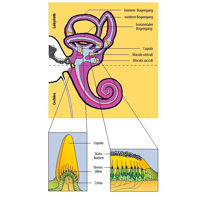
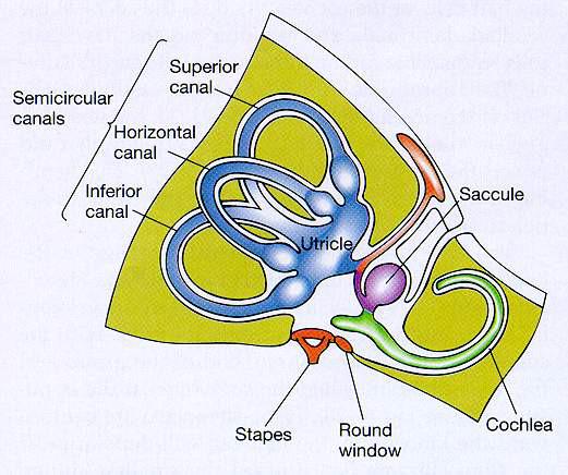
1 Role of brainstem in somatomotor (postural) functions (vestibular apparatus) The muscle tone and its regulation VESTIBULAR SYSTEM (Equilibrium) Receptors: Otolith organs Semicircular canals Sensation (information): Position of the head, Linear acceleration, Angular acceleration Function: Equilibrium Muscle tone regulation mainly in the antigravity muscles. Coordination of eye movements Coordination of head movements 1
2 2

3 utriculus 3


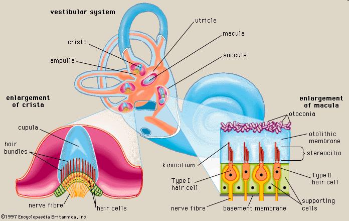
4 The structure of the hair cells 4

5 5


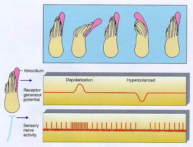
6 Function of hair cells Mechanosensitive cells Mechanoelectric transduction Basal surface: attach with perilymph (high Na + and low K + extracellular fluid) Apical surface: attach with endolymph (high K + and low Na + secretion IC) 6
 transmitter release - glutamate action potential on the afferent")
7 Apical surface: stereocilia + 1 kinocilium tip-link Mechanosensitive Kation (K + ) channels Resting situation: 10-15% opened K + channels partial depolarisation opening of some voltage gated Ca 2+ channels IC Ca 2+ (from perilymph) transmitter release - glutamate action potential on the afferent nerve 7
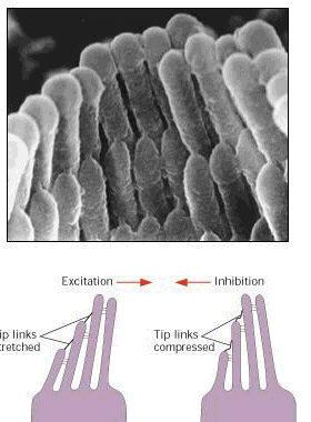
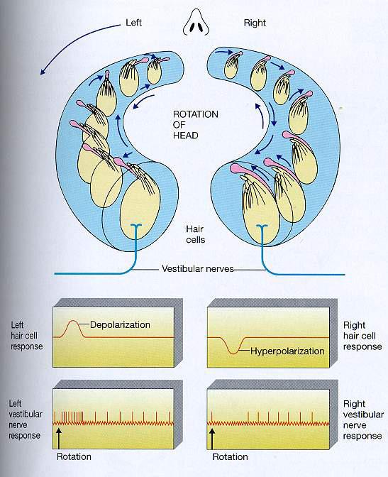
8 Activation (µs): Stereocilia move to the kinocilium opening of the mechanosensitive channels increased depolarisation opening of more voltage gated Ca 2+ channels IC Ca 2+ (from perilymph) Increased transmitter release - glutamate Increased action potential frequency in the afferent neurons Inhibition Stereocilia move away from the kinocilium closing of the mechanosensitive channels decreased depolarisation closing of voltage gated Ca 2+ channels IC Ca 2+ Decreased transmitter release Decreased action potential frequency in the afferent neurons 8
9 9

10 Cerebral cortex eyes Cerebellum Proprioceptors vestibular nuclei 10

11 NYSTAGMUS Nystagmus is a form of involuntary eye movement. It is characterized by alternating smooth pursuit in one direction and saccadic movement in the other direction. Vestibulo-ocular reflexes: Rotatory nystagmus Postrotatory nystagmus Caloric nystagmus Optokinetic nystagmus Optokinetic Nystagmus 11

12 Caloric nystagmus Sensori-motor system Limbic cortex Structure Subcortical Motivational sub areas Frontal cortex Task Motivation Sequence Plan Tim e Ascending system Basal ganglia Cerebellum (vermis) Brainstem Interneuron g.v. Association cortex Thalamus Mot. nuclei Motor cortex Motoneuron (spinal) Cerebellum (hemispheres) Descending system Voluntary Posture Spinal motoric (Reflexes) Program 800 ms 50 ms Execution Receptor Muscle (effektor) Length, tension, position, joint relation (posture) Light, sound, temperature (environmental stimuli) 12
 3. Pontine reticular formation (extensor facilitation) 4.")
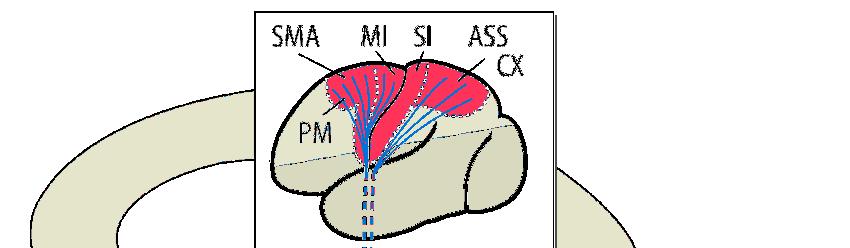
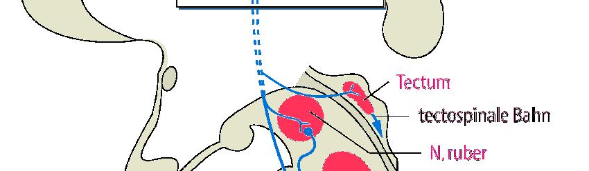
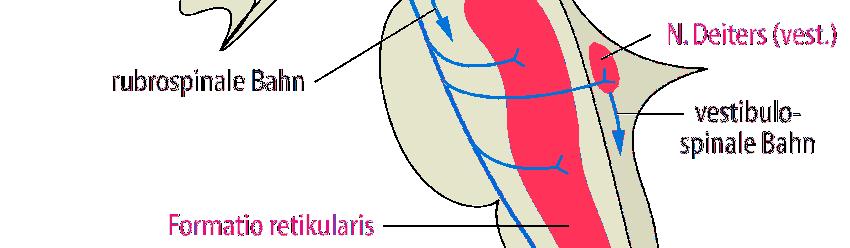
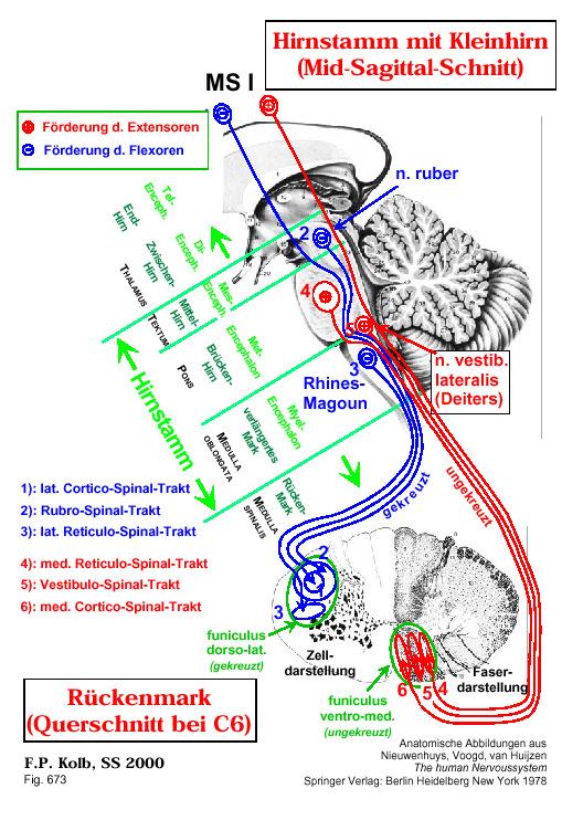
13 Brainstem structures in muscle tone regulation: (tonic control) 1. Red nucleus, nucleus ruber, rubrospinal pathway (extensor inhibition) 2. Deiters nucleus, vestibulospinal pathway (extensor facilitation) 3. Pontine reticular formation (extensor facilitation) 4. Medullary reticular formation (extensor inhibition) Midbrain: Tectum Red Nucleus Pons: Lateral (pontine) reticular formation Medulla: Medial (medullary) reticular formation Vestibular nuclei Centers 13


14 Descending pathways Vestibulospinal tracts: information from vestibular system for reflex-control and balance. Pontine reticulospinal tract: activation of extensor muscle for postural control Medullary reticulospinal tract: inhibition of extensor muscles Rubrospinal tract: activation of medullary reticular center; activation of motoneurons of distal muscles Tectospinal tract: control of eye and head movements 14
15 15
16 Decerebrated animal at low level Section below the midlevel of mesencephalon => the pontine and medullary reticular nuclei and the vestibulary system are intact. Increased tone in the antigravitiy muscles => decerebrate rigidity Tonic neck and labyrinthine reflexes 16
17 Causes: 1. Lack of inhibition of higher centers 2. Lack of activation of medullary reticular formation by higher centers 3. Pathways: Lateral vestibulospinal tract Medial (pontine) reticulospinalis tract Decerebrated animal at high level Section at the high level of mesencephalon, the pontine and medullary reticular nuclei and the vestibulary system and red nucleus are intact. Signs (in lower animals: cat, dog etc.): - Spontaneous righting - No rigidity - Righting reflexes: Receptors: vestibular app, neck muscle, neck joints, mechanical stimulus on the skin Labyrinthine righting reaction Neck righting reaction Body on head righting reflexes Body on body righting reflexes Vestibular placing reaction: extension of limbs and toes. 17
18 Causes: Inhibiton by red nucleus, Inhibition by cerebellum Inhibition by medullary reticular formation Signs in primates (monkey, human): Rigidity in the extensors in the lower limbs Increased tone in the flexors in the upper limbs Paralysis, no righting reflex Grasping reflex Pupil reflex, nystagmus Lift reaction: down: extension, up: flexion Decortication: Section in front of the subthalamic region Signs in lower animals (cat, dog): Spontaneous righting, walking, Normal behavior No learning Sings in primates: Rigidity in the extensors in the lower limbs Increased tone in the flexors in the upper limbs Paralysis, no righting reflex Grasping reflex 18


19 Decortication rigidity Decerebration rigidity extensor spasm in all four extremities 19
20 Brainstem postural reflexes (dynamic control) Principles 1. vestibular and neck reflexes stabilize the head and eyes 2. vestibular and neck afferents are synergetic on the neck but antagonistic on the limbs 3. vestibular and neck afferents are converging on vestibular nuclei and propriospinal neurons Brainstem control of motor function Postural reflexes Control of many stereotype movements of the body Control of equilibrium Control of eye, neck and axial (antigravity) muscle movements 20


21 Postural reflexes integrated in the brainstem: Tonic labirynthine reflexes Stimulus: gravity Receptor: otolithic organs Center: medulla Responses: contraction of limb extensor muscles (rigidity) Placed on its back: maximal extension in the four limbs Turned to left side: the rigidity level is higher at left side. Prone position: rigidity is minimal Head turned up: extension in the limbs Head turned down: flexion in the limbs Postural reflexes integrated in the brainstem: Tonic neck reflexes Stimulus: head turned Receptor: neck proprioceptors Center: medulla Responses: change in pattern of extensor contraction 1. to side: extension of limbs on side to which head is turned 2. up: hind legs flex 3. down: forelegs flex 21

22 Postural reflexes integrated in the brainstem Labyrinthine righting reflexes Stimulus: gravity Receptor: otolithic organs Center: midbrain Responses: head kept level Postural reflexes integrated in the brainstem: Neck righting reflexes Stimulus: stretch of neck muscles Receptor: muscle spindles Center: midbrain Responses: righting of thorax and shoulder, then pelvis 22
23 Postural reflexes integrated in the brainstem: Body on head righting reflexes Stimulus: pressure on side of body Receptor: exteroceptors Center: midbrain Responses: righting of head Postural reflexes integrated in the brainstem: Body on body righting reflexes Stimulus: pressure on side of body Receptor: exteroceptors Center: midbrain Responses: righting of body even when head held sideways 23
24 Postural reflexes integrated in the cortex: 1. Visual righting reflex 2. Visual placing reaction 3. Tactile placing reaction 4. Tactile grasping and avoiding reactions 5. Visual grasping and avoiding reactions 6. Babinsky test positive 7. Grasping reflex: flexion or clenching of the fingers or toes on stimulation of the palm or sole, normal only in infancy. Cerebral motor cortex Most "voluntary" movements initiated by the cerebral cortex are achieved when the cortex activates "patterns" of function stored in lower brain areas (spinal cord, brainstem). These lower centers, in turn, send specific control signals to the muscles. For a few types of movements, however, the cortex has almost a direct pathway to the anterior motor neurons of the cord, bypassing some motor centers on the way. This is especially true for control of the fine dexterous movements of the fingers and hands. Characteristics: complexity, flexibility, somototopy, contralateral, homunculus, feed-back 24


25 (1) the primary motor cortex, (2) the premotor area, (3) the supplementary motor area. Br8. control of eye movements Phases: Identification, localization, motivation: Cingular and dorsal part of parietal cortex Planning: premotor and supplementary areas Execution: primary motor cortex Homunculus, somatotopy 25
26 Execution: Primary motor cortex Precentral gyrus (Br 4): Contralateral Somatotopy Nerve signals generated the discrete patterns of muscle contractions Efferentation: corticospinal pathway Neurons are active before the muscle contraction The activity of neurons increases with the strength of the contraction Lesion: muscle weakness Supplementary and Premotor cortex (Br. 6): Nerve signals generated in the premotor area cause complex "patterns" of movement. Bilateral Planning, Motor image of the total muscle movement, Connection of the visual and auditory informations Regulation of posture for the correct movements Efferentation: Br4, basal ggl., spinal cord Lesion: apraxia (loss of the ability to execute or carry out learned purposeful movements, despite having the desire and the physical ability to perform the movements) especially for bilateral tasks, uncoordinated finger movements 26
27 Pyramid tract (Corticospinal pathway) 10 6 neuron Origin: 50% primary motor cortex Premotor area Somatosensory cortex Pathway: crossed, uncrossed End: alfa-, gamma-motoneurons, interneurons Lesion: decreased muscle tone, paralysis Collaterals: other pyramid cells, red nucl. thalamus, brainstem, striatum, cerebellum 27
28 Lateral cortico-spinal tract Ventral cortico-spinal tract % crossed to lateral ventral horn - monosynaptic to motoneurons of distal muscles -this enables fine independent finger movements - not fully developed at birth - most highly developed in primates - uncrossed - bilateral & polysynaptic to medial motoneurons of proximal/axial muscles - for posture 28
29 Corticomotoneuronal system 29
I: To describe the pyramidal and extrapyramidal tracts. II: To discuss the functions of the descending tracts.
 Descending Tracts I: To describe the pyramidal and extrapyramidal tracts. II: To discuss the functions of the descending tracts. III: To define the upper and the lower motor neurons. 1. The corticonuclear
Biological Bases of Behavior. 8: Control of Movement
 Biological Bases of Behavior 8: Control of Movement m d Skeletal Muscle Movements of our body are accomplished by contraction of the skeletal muscles Flexion: contraction of a flexor muscle draws in a
The Nervous System: Sensory and Motor Tracts of the Spinal Cord
 15 The Nervous System: Sensory and Motor Tracts of the Spinal Cord PowerPoint Lecture Presentations prepared by Steven Bassett Southeast Community College Lincoln, Nebraska Introduction Millions of sensory
Brain Stem and cortical control of motor function. Dr Z Akbari
 Brain Stem and cortical control of motor function Dr Z Akbari Brain stem control of movement BS nuclear groups give rise to descending motor tracts that influence motor neurons and their associated interneurons
The Motor Systems. What s the motor system? Plan
 The Motor Systems What s the motor system? Parts of CNS and PNS specialized for control of limb, trunk, and eye movements Also holds us together From simple reflexes (knee jerk) to voluntary movements
skilled pathways: distal somatic muscles (fingers, hands) (brainstem, cortex) are giving excitatory signals to the descending pathway
 (brainstem, cortex) are giving excitatory signals to the descending pathway") L15 - Motor Cortex General - descending pathways: how we control our body - motor = somatic muscles and movement (it is a descending motor output pathway) - two types of movement: goal-driven/voluntary
L15 - Motor Cortex General - descending pathways: how we control our body - motor = somatic muscles and movement (it is a descending motor output pathway) - two types of movement: goal-driven/voluntary
Motor tracts Both pyramidal tracts and extrapyramidal both starts from cortex: Area 4 Area 6 Area 312 Pyramidal: mainly from area 4 Extrapyramidal:
 Motor tracts Both pyramidal tracts and extrapyramidal both starts from cortex: Area 4 Area 6 Area 312 Pyramidal: mainly from area 4 Extrapyramidal: mainly from area 6 area 6 Premotorarea: uses external
Voluntary Movement. Ch. 14: Supplemental Images
 Voluntary Movement Ch. 14: Supplemental Images Skeletal Motor Unit: The basics Upper motor neuron: Neurons that supply input to lower motor neurons. Lower motor neuron: neuron that innervates muscles,
Chapter 8. Control of movement
 Chapter 8 Control of movement 1st Type: Skeletal Muscle Skeletal Muscle: Ones that moves us Muscles contract, limb flex Flexion: a movement of a limb that tends to bend its joints, contraction of a flexor
Unit VIII Problem 5 Physiology: Cerebellum
 Unit VIII Problem 5 Physiology: Cerebellum - The word cerebellum means: the small brain. Note that the cerebellum is not completely separated into 2 hemispheres (they are not clearly demarcated) the vermis
Spinal Interneurons. Control of Movement
 Control of Movement Spinal Interneurons Proprioceptive afferents have a variety of termination patterns in the spinal cord. This can be seen by filling physiologically-identified fibers with HRP, so their
cortical and brain stem control of motor function
 cortical and brain stem control of motor function cortical and brain stem control of motor function most voluntary movements initiated by the cerebral cortex are achieved when the cortex activates patterns
Spinal Cord Organization. January 12, 2011
 Spinal Cord Organization January 12, 2011 Spinal Cord 31 segments terminates at L1-L2 special components - conus medullaris - cauda equina no input from the face Spinal Cord, Roots & Nerves Dorsal root
 Spinal Cord Tracts DESCENDING SPINAL TRACTS: Are concerned with somatic motor function, modification of ms. tone, visceral innervation, segmental reflexes. Main tracts arise form cerebral cortex and others
CNS consists of brain and spinal cord PNS consists of nerves
 CNS consists of brain and spinal cord PNS consists of nerves 1 As with sensory input, motor output is organized in central nervous system Peripheral Nervous system divides efferent signals somatotopically
Biology 218 Human Anatomy
 Chapter 21 Adapted form Tortora 10 th ed. LECTURE OUTLINE A. Overview of Sensations (p. 652) 1. Sensation is the conscious or subconscious awareness of external or internal stimuli. 2. For a sensation
PETER PAZMANY CATHOLIC UNIVERSITY Consortium members SEMMELWEIS UNIVERSITY, DIALOG CAMPUS PUBLISHER
 PETER PAZMANY CATHOLIC UNIVERSITY SEMMELWEIS UNIVERSITY Development of Complex Curricula for Molecular Bionics and Infobionics Programs within a consortial* framework** Consortium leader PETER PAZMANY
Motor systems.... the only thing mankind can do is to move things... whether whispering or felling a forest. C. Sherrington
 Motor systems... the only thing mankind can do is to move things... whether whispering or felling a forest. C. Sherrington 1 Descending pathways: CS corticospinal; TS tectospinal; RS reticulospinal; VS
THE VESTIBULAR APPRATUS AND PATHWAY
 Dental Neuroanatomy February 23, 2012 Suzanne Stensaas, Ph.D. Reading: Waxman Chapter 17 Also pp 105-108 on control of eye movments Computer Resources: HyperBrain Ch. 8 Vestibulospinal Pathway Quiz http://library.med.utah.edu/kw/animations/hyperbrain/pathways/
Physiology of motor control (1)
") Physiology of motor control (1) Physiology of somatomotor system 1. Task: It controls the skeletal muscles 2. Content: Simple reflexes Muscle tone Posture Movement Sexual functions Motor component of emotions
Physiology of motor control (1) Physiology of somatomotor system 1. Task: It controls the skeletal muscles 2. Content: Simple reflexes Muscle tone Posture Movement Sexual functions Motor component of emotions
Neurophysiology of systems
 Neurophysiology of systems Motor cortex (voluntary movements) Dana Cohen, Room 410, tel: 7138 danacoh@gmail.com Voluntary movements vs. reflexes Same stimulus yields a different movement depending on context
Reflexes. Dr. Baizer
 Reflexes Dr. Baizer 1 Learning objectives: reflexes Students will be able to describe: 1. The clinical importance of testing reflexes. 2. The essential components of spinal reflexes. 3.The stretch reflex.
CNS MCQ 2 nd term. Select the best answer:
 Select the best answer: CNS MCQ 2 nd term 1) Vestibular apparatus: a) Represent the auditory part of the labyrinth. b) May help in initiating the voluntary movements. c) Contains receptors concerned with
Motor Functions of Cerebral Cortex
 Motor Functions of Cerebral Cortex I: To list the functions of different cortical laminae II: To describe the four motor areas of the cerebral cortex. III: To discuss the functions and dysfunctions of
Neural Integration I: Sensory Pathways and the Somatic Nervous System
 C h a p t e r 15 Neural Integration I: Sensory Pathways and the Somatic Nervous System PowerPoint Lecture Slides prepared by Jason LaPres Lone Star College - North Harris Copyright 2009 Pearson Education,
KINE 4500 Neural Control of Movement. Lecture #1:Introduction to the Neural Control of Movement. Neural control of movement
 KINE 4500 Neural Control of Movement Lecture #1:Introduction to the Neural Control of Movement Neural control of movement Kinesiology: study of movement Here we re looking at the control system, and what
Lecture X. Brain Pathways: Movement!
 Bio 3411 Readings (background only) Bio 3411 Monday Neuroscience 4 th ed Page(s) Feature 423-451Upper motor control of Brain Stem and Spinal Cord The Brain Atlas 3 rd ed Page(s) Feature 198-199 Vestibular
Lecture X. Brain Pathways: Movement!
 Bio 3411 Monday 1 Readings (background only) Neuroscience 5 th ed Page(s) Feature 353-398Upper motor control of Brain Stem and Spinal Cord Neuroscience 4 th ed Page(s) Feature 423-451Upper motor control
Cortical Control of Movement
 Strick Lecture 2 March 24, 2006 Page 1 Cortical Control of Movement Four parts of this lecture: I) Anatomical Framework, II) Physiological Framework, III) Primary Motor Cortex Function and IV) Premotor
Cervical reflex Giovanni Ralli. Dipartimento di Organi di Senso, Università di Roma La Sapienza
 Cervical reflex Giovanni Ralli Dipartimento di Organi di Senso, Università di Roma La Sapienza The development of the neck in vertebrates allows the individual to rotate the head independently of the trunk
1. Which part of the brain is responsible for planning and initiating movements?
 Section: Chapter 10: Multiple Choice 1. Which part of the brain is responsible for planning and initiating movements? p.358 frontal lobe hippocampus basal ganglia cerebellum 2. The prefrontal cortex is
A3.1.7 Motor Control. 10 November 2016 Institute of Psychiatry,Psychology and Neuroscience Marinela Vavla
 A3.1.7 Motor Control 10 November 2016 Institute of Psychiatry,Psychology and Neuroscience Marinela Vavla marinela.vavla@kcl.ac.uk Learning objectives Motor systems: components & organization Spinal cord
Introduction. HTML/image_m/fnhum g001.jpg
 13 Motor system II Introduction http://www.frontiersin.org/files/articles/42416/fnhum-07-00085- HTML/image_m/fnhum-07-00085-g001.jpg Introduction http://images.persianblog.ir/559630_ixfiuro0.jpg http://www.frontiersin.org/files/articles/42416/fnhum-07-00085-
Vestibular System Dr. Bill Yates Depts. Otolaryngology and Neuroscience 110 Eye and Ear Institute
 Vestibular System Dr. Bill Yates Depts. Otolaryngology and Neuroscience 110 Eye and Ear Institute 412-647-9614 byates@pitt.edu What is the Vestibular System? The vestibular system is the sensory system,
Degree of freedom problem
 KINE 4500 Neural Control of Movement Lecture #1:Introduction to the Neural Control of Movement Neural control of movement Kinesiology: study of movement Here we re looking at the control system, and what
The Somatosensory System
 The Somatosensory System Reading: BCP Chapter 12 cerebrovortex.com Divisions of the Somatosensory System Somatosensory System Exteroceptive External stimuli Proprioceptive Body position Interoceptive Body
Motor System Hierarchy
 Motor Pathways Lectures Objectives Define the terms upper and lower motor neurons with examples. Describe the corticospinal (pyramidal) tract and the direct motor pathways from the cortex to the trunk
HEAD AND NECK PART 2
 HEAD AND NECK PART 2 INTEGRATED CURRICULUM = Integrate Basic Science and Clinical Training 1- ENT PATIENT EXAM IN ICS COURSE - Today and next week - Review/Preview Anatomy underlying ENT exam 2- NEUROANATOMY/NEUROLOGY
THE BACK. Dr. Ali Mohsin. Spinal Cord
 Spinal Cord THE BACK Dr. Ali Mohsin The spinal cord is the elongated caudal part of the CNS. It starts as the inferior continuation of the medulla oblongata at the level of foramen magnum, & ends as an
OVERVIEW. Today. Sensory and Motor Neurons. Thursday. Parkinsons Disease. Administra7on. Exam One Bonus Points Slides Online
 OVERVIEW Today Sensory and Motor Neurons Thursday Parkinsons Disease Administra7on Exam One Bonus Points Slides Online 7 major descending motor control pathways from Cerebral Cortex or Brainstem
Chapter 13. The Nature of Muscle Spindles, Somatic Reflexes, and Posture
 Chapter 13 The Nature of Muscle Spindles, Somatic Reflexes, and Posture Nature of Reflexes A reflex is an involuntary responses initiated by a sensory input resulting in a change in the effecter tissue
Course: PG- Pathshala Paper number: 13 Physiological Biophysics Module number M23: Posture and Movement Regulation by Ear.
 Course: PG- Pathshala Paper number: 13 Physiological Biophysics Module number M23: Posture and Movement Regulation by Ear Principal Investigator: Co-Principal Investigator: Paper Coordinator: Content Writer:
Brainstem. Steven McLoon Department of Neuroscience University of Minnesota
 Brainstem Steven McLoon Department of Neuroscience University of Minnesota 1 Course News Change in Lab Sequence Week of Oct 2 Lab 5 Week of Oct 9 Lab 4 2 Goal Today Know the regions of the brainstem. Know
Department of Neurology/Division of Anatomical Sciences
 Spinal Cord I Lecture Outline and Objectives CNS/Head and Neck Sequence TOPIC: FACULTY: THE SPINAL CORD AND SPINAL NERVES, Part I Department of Neurology/Division of Anatomical Sciences LECTURE: Monday,
Somatic Nervous System: Motor Output *
 OpenStax-CNX module: m63208 1 Somatic Nervous System: Motor Output * Steven Telleen Based on Motor Responses by OpenStax This work is produced by OpenStax-CNX and licensed under the Creative Commons Attribution
Chapter 14: Integration of Nervous System Functions I. Sensation.
 Chapter 14: Integration of Nervous System Functions I. Sensation A. General Organization 1. General senses have receptors a. The somatic senses provide information about & 1. Somatic senses include: a.
CN V! touch! pain! Touch! P/T!
 CN V! touch! pain! Touch! P/T! Visual Pathways! L! R! B! A! C! D! LT! E! F! RT! G! hypothalamospinal! and! ALS! Vestibular Pathways! 1. Posture/Balance!!falling! 2. Head Position! 3. Eye-Head Movements
What is the effect on the hair cell if the stereocilia are bent away from the kinocilium?
 CASE 44 A 53-year-old man presents to his primary care physician with complaints of feeling like the room is spinning, dizziness, decreased hearing, ringing in the ears, and fullness in both ears. He states
Gross Anatomy of Lower Spinal Cord
 Chapter 13 Spinal Cord, Spinal Nerves and Somatic Reflexes Spinal cord Spinal nerves Somatic reflexes Gross Anatomy of Lower Spinal Cord Meninges of Vertebra & Spinal Cord Spina Bifida Congenital defect
At the highest levels of motor control, the brain represents actions as desired trajectories of end-effector
 At the highest levels of motor control, the brain represents actions as desired trajectories of end-effector Normal condition, using fingers and wrist Using elbow as folcrum Using shoulder as folcrum (outstretched
THE CENTRAL NERVOUS SYSTE M
 THE CENTRAL NERVOUS SYSTE M Structure and Functio n THIRD EDITIO N PER BRODAL A Brief Survey, x i Studying the Structures and Function of the Nervous System, xii i Animal Experiments Crucial for Progress,
Motor control. Proprioception and movement
 Motor control Proprioception and movement 2/24 in general we are not aware of information coming from proprioceptors though they belong to somatosensory receptors these receptors detect stretching of the
Neural Integration I: Sensory Pathways and the Somatic Nervous System
 15 Neural Integration I: Sensory Pathways and the Somatic Nervous System PowerPoint Lecture Presentations prepared by Jason LaPres Lone Star College North Harris An Introduction to Sensory Pathways and
Believe In Your Dreams. They Were Given To You For A Reason
 Text Important Formulas Numbers Doctor notes Notes and explanation Lecture No.16 Believe In Your Dreams. They Were Given To You For A Reason 1 Physiology of postural reflexes Objectives: 1. Postural reflexes
The Nervous System S P I N A L R E F L E X E S
 The Nervous System S P I N A L R E F L E X E S Reflexes Rapid, involuntary, predictable motor response to a stimulus Spinal Reflexes Spinal somatic reflexes Integration center is in the spinal cord Effectors
Biological Psych Frontal Lobes
 Biological Psych Frontal Lobes Frontal lobe What is it? Home to personality? Lesions: wide variety symptoms More than any part of brain Involved in: motor function problem solving spontaneity memory language
Chapter 16: Sensory, Motor, and Integrative Systems. Copyright 2009, John Wiley & Sons, Inc.
 Chapter 16: Sensory, Motor, and Integrative Systems Sensation n Conscious and subconscious awareness of changes in the external or internal environment. n Components of sensation: Stimulation of the sensory
Spinal cord. We have extension of the pia mater below L1-L2 called filum terminale
 Spinal cord Part of the CNS extend from foramen magnum to the level of L1-L2 (it is shorter than the vertebral column) it is covered by spinal meninges. It is cylindrical in shape. It s lower end become
Chapter 12b. Overview
 Chapter 12b Spinal Cord Overview Spinal cord gross anatomy Spinal meninges Sectional anatomy Sensory pathways Motor pathways Spinal cord pathologies 1 The Adult Spinal Cord About 18 inches (45 cm) long
Basal nuclei, cerebellum and movement
 Basal nuclei, cerebellum and movement MSTN121 - Neurophysiology Session 9 Department of Myotherapy Basal Nuclei (Ganglia) Basal Nuclei (Ganglia) Role: Predict the effects of various actions, then make
Arterial Blood Supply
 Arterial Blood Supply Brain is supplied by pairs of internal carotid artery and vertebral artery. The four arteries lie within the subarachnoid space Their branches anastomose on the inferior surface of
Cerebellum. Steven McLoon Department of Neuroscience University of Minnesota
 Cerebellum Steven McLoon Department of Neuroscience University of Minnesota 1 Anatomy of the Cerebellum The cerebellum has approximately half of all the neurons in the central nervous system. The cerebellum
Located below tentorium cerebelli within posterior cranial fossa. Formed of 2 hemispheres connected by the vermis in midline.
 The Cerebellum Cerebellum Located below tentorium cerebelli within posterior cranial fossa. Formed of 2 hemispheres connected by the vermis in midline. Gray matter is external. White matter is internal,
The Cerebellum. Outline. Overview Structure (external & internal) Micro-circuitry of the cerebellum Cerebellum and motor learning
 Micro-circuitry of the cerebellum Cerebellum and motor learning") The Cerebellum P.T Ji Jun Cheol Rehabilitation Center 1 HansarangAsan Hospital. Outline Overview Structure (external & internal) Micro-circuitry of the cerebellum Cerebellum and motor learning 2 1 Cerebellum
The Cerebellum P.T Ji Jun Cheol Rehabilitation Center 1 HansarangAsan Hospital. Outline Overview Structure (external & internal) Micro-circuitry of the cerebellum Cerebellum and motor learning 2 1 Cerebellum
Lecture VIII. The Spinal Cord, Reflexes and Brain Pathways!
 Reflexes and Brain Bio 3411! Monday!! 1! Readings! NEUROSCIENCE 5 th ed: Review Chapter 1 pp. 11-21;!!Read Chapter 9 pp. 189-194, 198! THE BRAIN ATLAS 3 rd ed:! Read pp. 4-17 on class web site! Look at
BIOH111. o Cell Module o Tissue Module o Integumentary system o Skeletal system o Muscle system o Nervous system o Endocrine system
 BIOH111 o Cell Module o Tissue Module o Integumentary system o Skeletal system o Muscle system o Nervous system o Endocrine system Endeavour College of Natural Health endeavour.edu.au 1 Textbook and required/recommended
The Spinal Cord. The Nervous System. The Spinal Cord. The Spinal Cord 1/2/2016. Continuation of CNS inferior to foramen magnum.
 The Nervous System Spinal Cord Continuation of CNS inferior to foramen magnum Simpler than the brain Conducts impulses to and from brain Two way conduction pathway Reflex actions Passes through vertebral
Neural Basis of Motor Control. Chapter 4
 Neural Basis of Motor Control Chapter 4 Neurological Perspective A basic understanding of the physiology underlying the control of voluntary movement establishes a more comprehensive appreciation and awareness
Motor Systems. Motor systems
 389 Motor systems The control of voluntary movements is complex. Many different systems across numerous brain areas need to work together to ensure proper motor control. We will start a journey through
Spinal Cord- Medulla Spinalis. Cuneyt Mirzanli Istanbul Gelisim University
 Spinal Cord- Medulla Spinalis Cuneyt Mirzanli Istanbul Gelisim University Spinal Column Supports the skull, pectoral girdle, upper limbs and thoracic cage by way of the pelvic girdle. Transmits body weight
THE CEREBELLUM SUDIVISIONS, STRUCTURE AND CONNECTIONS
 THE CEREBELLUM Damage to the cerebellum produces characteristic symptoms primarily with respect to the coordination of voluntary movements. The cerebellum receives information from the skin, joints, muscles,
The Vestibular System
 The Vestibular System Vestibular and Auditory Sensory Organs Bill Yates, Ph.D. Depts. Otolaryngology & Neuroscience University of Pittsburgh Organization of Sensory Epithelium Displacement of Stereocilia
By Dr. Saeed Vohra & Dr. Sanaa Alshaarawy
 By Dr. Saeed Vohra & Dr. Sanaa Alshaarawy 1 By the end of the lecture, students will be able to : Distinguish the internal structure of the components of the brain stem in different levels and the specific
NS201C Anatomy 1: Sensory and Motor Systems
 NS201C Anatomy 1: Sensory and Motor Systems 25th January 2017 Peter Ohara Department of Anatomy peter.ohara@ucsf.edu The Subdivisions and Components of the Central Nervous System Axes and Anatomical Planes
Auditory and Vestibular Systems
 Auditory and Vestibular Systems Objective To learn the functional organization of the auditory and vestibular systems To understand how one can use changes in auditory function following injury to localize
Fig Cervical spinal nerves. Cervical enlargement C7. Dural sheath. Subarachnoid space. Thoracic. Spinal cord Vertebra (cut) spinal nerves
 spinal nerves") Fig. 13.1 C1 Cervical enlargement C7 Cervical spinal nerves Dural sheath Subarachnoid space Thoracic spinal nerves Spinal cord Vertebra (cut) Lumbar enlargement Medullary cone T12 Spinal nerve Spinal nerve
Fig. 13.1 C1 Cervical enlargement C7 Cervical spinal nerves Dural sheath Subarachnoid space Thoracic spinal nerves Spinal cord Vertebra (cut) Lumbar enlargement Medullary cone T12 Spinal nerve Spinal nerve
Cranial Nerve VIII (The Vestibulo-Cochlear Nerve)
") Cranial Nerve VIII (The Vestibulo-Cochlear Nerve) Please view our Editing File before studying this lecture to check for any changes. Color Code Important Doctors Notes Notes/Extra explanation Objectives
Cranial Nerve VIII (The Vestibulo-Cochlear Nerve) Please view our Editing File before studying this lecture to check for any changes. Color Code Important Doctors Notes Notes/Extra explanation Objectives
Making Things Happen: Simple Motor Control
 Making Things Happen: Simple Motor Control How Your Brain Works - Week 10 Prof. Jan Schnupp wschnupp@cityu.edu.hk HowYourBrainWorks.net The Story So Far In the first few lectures we introduced you to some
Sensory Pathways & Somatic Nervous System. Chapter 15
 Sensory Pathways & Somatic Nervous System Chapter 15 How Does Brain Differentiate Sensations? Pain impulses make brain aware of injuries and infections. Impulses from eye, ear, nose and tongue make brain
Neural Basis of Motor Control
 Neural Basis of Motor Control Central Nervous System Skeletal muscles are controlled by the CNS which consists of the brain and spinal cord. Determines which muscles will contract When How fast To what
VESTIBULAR SYSTEM. Deficits cause: Vertigo. Falling Tilting Nystagmus Nausea, vomiting
 VESTIBULAR SYSTEM Objectives: Understand the functions of the vestibular system: What is it? How do you stimulate it? What are the consequences of stimulation? Describe the vestibular apparatus, the 2
The Motor System. The finger movements of a neurosurgeon manipulating. John C. Kincaid, M.D. THE SKELETON AS THE FRAMEWORK FOR MOVEMENT
 C H 5A P T E R The Motor System John C. Kincaid, M.D. CHAPTER OUTLINE THE SKELETON AS THE FRAMEWORK FOR MOVEMENT MUSCLE FUNCTION AND BODY MOVEMENT PERIPHERAL NERVOUS SYSTEM COMPONENTS FOR THE CONTROL OF
b. The groove between the two crests is called 2. The neural folds move toward each other & the fuse to create a
 Chapter 13: Brain and Cranial Nerves I. Development of the CNS A. The CNS begins as a flat plate called the B. The process proceeds as: 1. The lateral sides of the become elevated as waves called a. The
Vestibular Physiology Richard M. Costanzo, Ph.D.
 Vestibular Physiology Richard M. Costanzo, Ph.D. OBJECTIVES After studying the material of this lecture, the student should be able to: 1. Describe the structure and function of the vestibular organs.
Non-cranial nerve nuclei
 Brainstem Non-cranial nerve nuclei Nucleus Gracile nucleus Cuneate nucleus Infeiro olivary nucleus Pontine nucleus inferior colliculus superior colliculus Red nucleus Substantia nigra Pretectal area Site
CHAPTER 16 LECTURE OUTLINE
 CHAPTER 16 LECTURE OUTLINE I. INTRODUCTION A. The components of the brain interact to receive sensory input, integrate and store the information, and transmit motor responses. B. To accomplish the primary
PHYSIOLOHY OF BRAIN STEM
 PHYSIOLOHY OF BRAIN STEM Learning Objectives The brain stem is the lower part of the brain. It is adjoining and structurally continuous with the spinal cord. 1 Mid Brain 2 Pons 3 Medulla Oblongata The
Lecture - Chapter 13: Central Nervous System
 Lecture - Chapter 13: Central Nervous System 1. Describe the following structures of the brain, what is the general function of each: a. Cerebrum b. Diencephalon c. Brain Stem d. Cerebellum 2. What structures
Abdullah AlZibdeh. Dr. Maha ElBeltagy. Maha ElBeltagy
 19 Abdullah AlZibdeh Dr. Maha ElBeltagy Maha ElBeltagy Introduction In this sheet, we discuss the cerebellum; its lobes, fissures and deep nuclei. We also go into the tracts and connections in which the
Motor systems. Motor systems
 515 The control of voluntary movements is extremely complex. Many different systems across numerous brain areas need to work together to ensure proper motor control. We will start a journey through these
Neurology. Hollie Wilson
 Neurology Hollie Wilson Objectives Anatomy Physiology: Functional centres of brain UMN lesion vs. LMN lesion Spinal cord Main tracts ascending and descending Nerve roots and peripheral nerves action potentials
UNIVERSITY OF JORDAN FACULTY OF MEDICINE DEPARTMENT OF PHYSIOLOGY & BIOCHEMISTRY NEUROPHYSIOLOGY (MEDICAL) Spring, 2014
 Spring, 2014") UNIVERSITY OF JORDAN FACULTY OF MEDICINE DEPARTMENT OF PHYSIOLOGY & BIOCHEMISTRY NEUROPHYSIOLOGY (MEDICAL) Spring, 2014 Textbook of Medical Physiology by: Guyton & Hall, 12 th edition 2011 Eman Al-Khateeb,
UNIVERSITY OF JORDAN FACULTY OF MEDICINE DEPARTMENT OF PHYSIOLOGY & BIOCHEMISTRY NEUROPHYSIOLOGY (MEDICAL) Spring, 2014 Textbook of Medical Physiology by: Guyton & Hall, 12 th edition 2011 Eman Al-Khateeb,
Functional Distinctions
 Functional Distinctions FUNCTION COMPONENT DEFICITS Start Basal Ganglia Spontaneous Movements Move UMN/LMN Cerebral Cortex Brainstem, Spinal cord Roots/peripheral nerves Plan Cerebellum Ataxia Adjust Cerebellum
COGS 107B Week 2. Hyun Ji Friday 4:00-4:50pm
 COGS 107B Week 2 Hyun Ji Friday 4:00-4:50pm Lecture 3: Proprioception Principles: The Neuron Doctrine and The Law of Dynamic Polarization Proprioception Joint-protecting reflexes (ex. Knee jerk reflex)
Connection of the cerebellum
 CEREBELLUM Connection of the cerebellum The cerebellum has external layer of gray matter (cerebellar cortex ), & inner white matter In the white matter, there are 3 deep nuclei : (a) dentate nucleus laterally
1. The cerebellum coordinates fine movement through interactions with the following motor-associated areas:
 DENT/OBHS 131 2009 Take-home test 4 Week 6: Take-home test (2/11/09 close 2/18/09) 1. The cerebellum coordinates fine movement through interactions with the following motor-associated areas: Hypothalamus
The Cerebellum. The Little Brain. Neuroscience Lecture. PhD Candidate Dr. Laura Georgescu
 The Cerebellum The Little Brain Neuroscience Lecture PhD Candidate Dr. Laura Georgescu Learning Objectives 1. Describe functional anatomy of the cerebellum - its lobes, their input and output connections
CHAPTER 16: SENSORY, MOTOR, & INTEGRATIVE SYSTEM DR. WELCH
 BIOL 2401 DR. WELCH CHAPTER 16: SENSORY, MOTOR, & INTEGRATIVE SYSTEM I. Sensation conscious or subconscious awareness of internal & external changes. Define perception. Primary function of. Why isn t blood
Auditory and vestibular system
 Auditory and vestibular system Sensory organs on the inner ear inner ear: audition (exteroceptor) and vestibular apparatus (proprioceptor) bony and membranous labyrinths within the temporal bone (os temporale)
Stretch reflex and Golgi Tendon Reflex. Prof. Faten zakareia Physiology Department, College of Medicine, King Saud University 2016
 Stretch reflex and Golgi Tendon Reflex Prof. Faten zakareia Physiology Department, College of Medicine, King Saud University 2016 Objectives: Upon completion of this lecture, students should be able to