Motor System Hierarchy
|
|
|
- Sarah Fisher
- 5 years ago
- Views:
Transcription
1 Motor Pathways
2 Lectures Objectives Define the terms upper and lower motor neurons with examples. Describe the corticospinal (pyramidal) tract and the direct motor pathways from the cortex to the trunk and limbs. Briefly describe the indirect motor pathways from the cortex to the trunk and limbs through extrapyramidal tracts such as rubrospinal and reticulospinal tracts. Describe motor pathways to the face muscles. Compare the signs and symptoms of the upper and lower motor neuron lesions. Identify the centers that make the basal ganglia. Identify the different parts, regions and nuclei of the cerebellum. Summarize the motor system circuitry.
3 Motor System Hierarchy Control Systems Initiator Executers Lower motor neuron Muscle Basal Ganglia Cerebellum Cortex Upper motor neurons Lower motor neuron Muscle
4 Upper and Lower Motor Neurons
5 Lower Motor Neurons Neurons innervates muscles Alpha motoneurons Innervates normal fibers Gamma motor neurons Innervates fibers in muscle spindle Present in: Spinal cord Brainstem (in the cranial nerves nuclei) Muscle tone
6 Muscle Tone Determined by the level of activity in the lower motor neurons Tone refers to the resistance of a muscle to passive stretch Primary determinant of muscle tone is the level of activity in the stretch reflex
7 Upper Motor Neurons Project to LMN Innervate α and γ motor neurons and inhibitory interneurons Location of UMN Reticular formation: reticulospinal tract Vestibular nuclei: vestibulospinal tract Superior colliculus: tectospinal tract Red nucleus: rubrospinal tract Motor cortex: corticospinal & corticobulbar tracts (+ corticorubral & corticoreticular)
8 Descending Tracts Classification Classical classification: Pyramidal system Axons traverse the pyramids in medulla Corticospinal axons Extrapyramidal system Other descending tracts Basal ganglia and their connections with motor region Functional classification: Medial system Innervate medial motor nucleus Lateral system Innervate lateral motor nucleus
9 Descending Tracts Lateral pathways Terminate laterally in the ventral horn Crossed Involved in movements of the distal limbs (initiation & fine movement) Damage weakness or paralysis Contains Lateral corticospinal tract Ruprospinal tract

10 Ruprospinal tract From red nucleus Crossed immediately

11 The Pyramidal (Corticospinal & corticobulbar) Tract Origin cerebral cortex ⅓ from primary motor cortex ⅓ from premotor areas ⅓ from primary somatosensory cortex Terminate in dorsal horn and brainstem Modify processing in the somatosensory pathways

12 Corticospinal Tract Axons pass through: Corona radiata Internal capsule (posterior limb) Basis pedunculi midbrain Medullary pyramids Decussation Crossed Lateral corticospinal 90 95% Uncrossed Anterior corticospinal tract

13 Lateral Corticospinal Tract Somatotopic Organization
14 Corticobulbar Tract Same origin & course as corticospinals Leave tract in brainstem Terminate in the cranial nerve nuclei Bilateral in general Except to facial nucleus
15 Descending Tracts Medial pathways Terminate Medially in the ventral horn Trunk & proximal limb movements Important in maintaining balance & position Mostly happened unconsciously Damage Deficits in maintaining balance & posture Changes in muscle tone Contains: Reticulospinal tract Vestibuspinal tract Tectospinal tract Anterior corticospinal tract
 Contains")

16 Reticulospinal Tract From the reticular formation Important influence on muscle activity and reflexes Controlled by cortex (corticoreticular) Contains descending autonomic fibers controlled by thalamus Crossed and uncrossed Two tracts Lateral reticulospinal tract from Medulla Medial reticulospinal tract from Pons

17 Vestibulospinal Tract Importance in maintaining balance Influence axial muscles Uncrossed Medial & lateral vestibulospinal tracts

18 Tectospinal Tract Superior colliculus Terminate in the cervical regions Head movements in response to visual stimuli Mostly crossed


19 Anterior Corticospinal Tract Similar to other medial pathways Terminate in the medial motor n. Except it is voluntary Do not cross in pyramidal decussation May cross before termination
20 Lower Motor Neuron Lesion Flaccid paralysis or paresis (weakness) Hypo or areflexia Decreased muscle tone Atrophy muscle wasting Develops over time (weeks) Fasciculations small twitches that are visible to the eye
21 Upper Motor Neuron Lesion Paralysis or paresis Spasticity Hypertonia Hyperreflexia And maybe: Babinski sign Clonus Decreased superficial reflexes Abdominal reflex & Cremasteric reflex

22 Upper Motor Neuron Lesion Babinski Sign Abnormal response to stroking the lateral planter surface of the foot Not useful in babies Normal response: toes planter flex Abnormal: dorsiflexion of big toe
23 Upper Motor Neuron Lesion Clonus Repetitive flexion extension of a joint in response to single flexion or extension

; testes elevation with stroking inside of")
24 Superficial Reflexes Decrease With UMN lesions Abdominal reflex; abdominal muscles contract on stroking the abdomen Cremasteric reflex (useful in babies); testes elevation with stroking inside of the thigh

25 Lower-versus Upper-Motor- Neuron Lesions Variable Lower-Motor-Neuron Lesion Upper-Motor-Neuron Lesion Weakness Flaccid paralysis Spastic paralysis Deep tendon reflexes Decreased or absent Increased Babinski's reflex Absent Present Atrophy May be marked Absent or resulting from disuse Fasciculations and fibrillations May be present Absent
26 Spinal Shock Follows severe acute injury to the spinal cord For short period (days or weeks) Loss of all functions (motor & sensory) bellow level of injury Loss of reflexes Due to sudden loss of supraspinal inputs
27 Upper Motor Neurons Cerebral Cortex Lateral system Medial system Motor cortex Red nu. Spinal cord & Brainstem Retic. Form. Sup. Colliculus Vestibular Nu. γ α Intrafusal Muscle Extrafusal
28 Basal Nuclei (Ganglia)
29 Basal Ganglia The basal ganglia include the caudate, putamen, and globus pallidus and number of closely related nuclei They influence motor system primarily through projections to upper motor neurons Motor deficits depend on the specific nucleus damaged Understanding the neurochemistry of basal ganglia drives the development of clinical treatment
30 Basal Ganglia The basal ganglia act as Brake against involuntary movement Switch to turn on a fixed action pattern Their major output is to the VA of the thalamus Projects primarily to area 6 (premotor & supplementary motor areas)
31 Basal Ganglia Terminology Striatum (neostriatum) = caudate + putamen Lentiform nucleus = putamen + globus pallidus Corpus striatum = caudate + lentiform Basal ganglia = corpus striatum + amygdala Globus pallidus = pallidum = paleostriatum Claustrum is some times included with the basal ganglia Basal ganglia is included by the extrapyramidal system
32 Caudate nucleus Parts Location Relations Lateral ventricle Amygdaloid nucleus Basal Ganglia: Gross Anatomy

33 Lentiform nucleus Parts Putamen Globus pallidus Internal (GPi) External (GPe) Shape Location Relations External & internal capsules Claustrum Basal Ganglia: Gross Anatomy
 Pars compacta (SNc)")
34 Basal Ganglia: Gross Anatomy Amygdaloid nucleus Subthalamic nucleus Substantia nigra Pars reticulata (SNr) Pars compacta (SNc) Claustrum

35 Inputs Most inputs enter the striatum From cerebral cortex & thalamus These inputs are excitatory Outputs Most leave from Gpi & SNr Most go to VA nucleus of the thalamus, which projects to motor cortex The outputs are GABAergic and inhibitory VA excites motor cortex, leading to movements Increase basal ganglia output will inhibit the VA and reduce overall movements Basal Ganglia Circuitry
36 Basal Ganglia Circuitry Intrinsic Circuits Large number of connections between components of the basal ganglia Can be grouped into Direct pathway Indirect pathway These pathways affect the VA activity and thus the motor cortex activity
37 From striatum to Gpi Uses GABA, which inhibits another GABAergic projection (Gpi to VA) Disinhibition Cortical activity direct pathway Gpi activity VA activity Activity in the direct pathway leads to increased motor cortex activity and increased movements The Direct Pathway
38 Goes from striatum to GPe (GABA) to the subthalamic nucleus (GABA) Subthalamic nucleus to Gpi (Glu) activity in the cortex activity of subthalamic nu. GPi VA activity motor cortex activity The Indirect Pathway
39 Basal Ganglia Circuitry The direct pathway increase movements The indirect pathway decrease movements Normal behavior requires a balance between the direct and indirect pathways All pathways are uncrossed Right basal ganglia modulate right cortex and affect movements on the left side of the body Acetylcholine is used by the interneurons in the striatum It affect the output of the direct and indirect pathways It s a target for drug therapy
40 In the striatum different cell types give rise to the direct and indirect pathways Both cell types receive dopaminergic input from SN pars compacta These cells have different receptors for DA For direct pathway, DA excites the striatal cells For indirect pathway, DA inhibits the striatal cells Thus the nigrostriatal pathway the activity of the VA and motor cortex PD leads to direct pathway activity indirect pathway activity activity of VA and motor cortex Nigrostriatal Pathway
41 Cerebellum
42 Cerebellum The cerebellum is essential for normal movements It affects motor behavior by affecting UMNs The cerebellum acts as a comparator Compares intended movements (data from cerebral cortex) to the actual movements (sensory data) Sends corrective signals into the descending motor pathways
43 Cerebellar Function It affects all movements, it is important in: Balance Locomotion Simple & complex movements Eye movements, etc. Site of motor learning Important for learning new motor skills and adjusting movements to changing sensory inputs
44 Location. Relations.. The cerebellum consists of two hemispheres The hemispheres are connected by vermis Cerebellar Anatomy Gross Anatomy
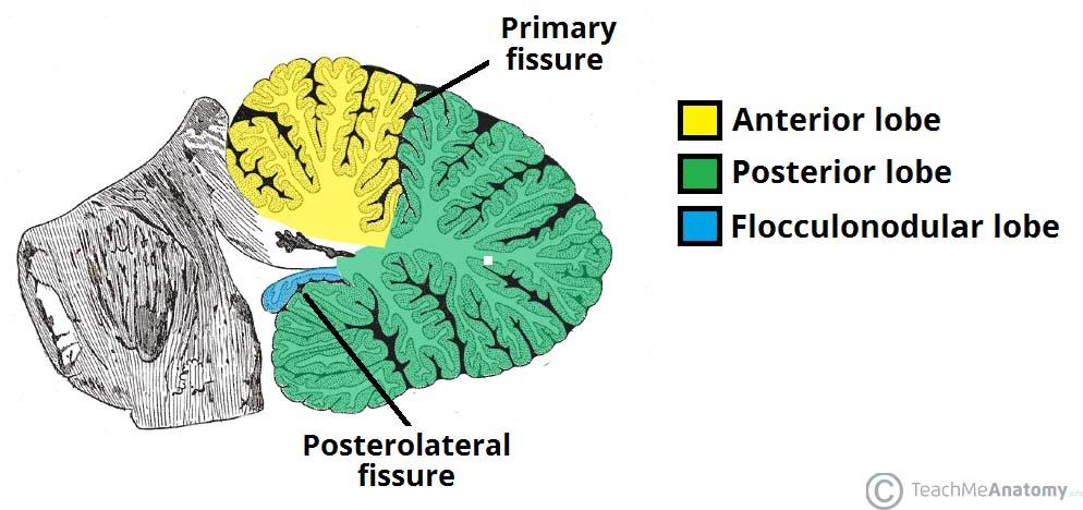
 Cerebellar tonsils Posterolateral fissure (uvulonodular fissure)")
45 Cerebellum: Gross Anatomy Three main lobes Anterior lobe Primary fissure Posterior lobe (middle lobe) Cerebellar tonsils Posterolateral fissure (uvulonodular fissure) Flocculonodular lobe
46
47 Cerebellum: Gross Anatomy
")

48 Cerebellum: Internal Structure Content Cerebellar cortex (folia) & central nuclei are grey matter Arbor vitae = tree of life = white matter

49 Cerebellum includes a cortex & deep nuclei The deep nuclei are the major source of output from the cerebellum Four nuclei from medial to lateral Fastigial Globose Emboliform Dentate Cerebellar Anatomy Interposed nuclei

50 Cerebellar cortex includes 5 cell types in 3 layers Five cell types Inhibitory cells Purkinje, basket, Golgi, and stellate cells Excitatory cells Granule cells Three layers Molecular layer Purkinje cell layer Granule cell layer Cerebellar Cortex
51 Inputs to the cerebellum Climbing fibers From inferior olivary complex (olivocerebellar fibers) Decussate Inferior cerebellar peduncle Mossy fibers All remaining inputs: spinal cord, vestibular n. & nuclei, & pontine nuclei Each type of input fibers branches Branch to deep nuclei Branch to cerebellar cortex Cerebellar Inputs
52 Cerebellar Circuit The basic cerebellar circuit includes Main excitatory loop Inhibitory cortical side loop
53 The Main Excitatory Loop Includes the input and the deep cerebellar nuclei Both the inputs & the cells of the deep nuclei are excitatory
54 The Inhibitory Cortical Side Loop Serves to modulate the activity in the deep cerebellar nuclei Mossy & climbing fibers are inputs to cerebellar cortex Climbing fibers contact Purkinje cells directly Mossy fibers contact granule cells Granule cells contact Purkinje cells Output of cerebellar cortex (Purkinje fibers) depend on the mossy & climbing fibers
55 The Inhibitory Cortical Side Loop Remaining cells (Golgi, basket & stellate) are inhibitory interneurons Alter granule & Purkinje cells Purkinje cells (cerebellar cortex output) are inhibitory Purkinje cells targets deep cerebellar nuclei & vestibular nuclei Thus cerebellar output is driven by the main excitatory loop and limited by the inhibitory cortical side loop
 Motor execution 3.")
56 Cerebellar Functional Divisions 1. Vestibulocerebellum Flocculonodular lobe & fastigial nu. Balance, eye movements 2. Spinocerebellum Vermis & paravermal parts of hemispheres & interposed nuclei (emboliform & globose) Motor execution 3. Cerebrocerebellum Lateral hemispheres & dentate nu. Motor planning
 Vestibular nuclei Reticular formation VL of thalamus Part of motor system targeted UMNs of medial pathway Major signs of damage Staggering or falling,")
57 Vestibulocerebellum Function Balance & eye movements Inputs Vestibular n. fibers Vestibular nuclei Inferior olive Deep nucleus Fastigial nucleus Outputs (From fastigial nu. & Purkinje cells) Vestibular nuclei Reticular formation VL of thalamus Part of motor system targeted UMNs of medial pathway Major signs of damage Staggering or falling, nystagmus

58 Function Spinocerebellum Execution of movement Compensates for changes in load, regulates muscle tone, guides limb movement, helps maintain posture Organized somatotopically Head & trunk vermis Limbs paravermal areas Inputs Spinal & trigeminal inputs Inferior olive Deep nucleus Fastigial & interposed nuclei Outputs Vermis Reticular formation & Vestibular nu. Paravermal Red nucleus, VL of thalamus & Inferior olive Part of motor system targeted UMNs of medial & lateral pathways Major signs of damage Staggering gait, intention tremor
 VL of thalamus Part of motor system targeted Motor cortex (via VL) Major signs of damage Decomposition of")
59 Cerebrocerebellum Function Coordination, planning of voluntary movements Inputs Pontine nuclei (relaying information from sensory & motor cerebral cortex) Inferior olive Deep nucleus Dentate nucleus Outputs Red nucleus (to inferior olive, back to cerebellum) VL of thalamus Part of motor system targeted Motor cortex (via VL) Major signs of damage Decomposition of movements
60 Cerebellar Peduncles Peduncle Inferior Restiform body Juxtarestiform body Middle (brachium pontis) Major inputs to cerebellum Fibers from: Inferior olive (climbing fibers) Dorsal spinocerebellar tract Cuneocerebellar tract Vestibular nerve Vestibular nuclei Pontine nuclei (relay inputs from cerebral cortex) Major outputs from cerebellum Fibers to: Vestibular nuclei None Superior (brachium conjunctivum) Ventral spinocerebellar Rostral spinocerebellar Red nucleus VL thalamus Reticular formation Inferior olive
61 Cerebellar Circuitry Cerebra l Cortex Motor cortex SCP V A V L Thalamus Red nu. Inferior olive Retic. Form. Sup. Colliculus Vestibular Nu. UMNs Lateral system Medial system Spinal cord & Brainstem γ α Pontine nuclei MCP Deep nuclei Cortex + + Cerebellum ICP Vestibular nerve Intrafusal Extrafusal

62 Blood Supply of Cerebellum SCA Superior cerebellar hemispheres Superior vermis Dentate nucleus Most of white matter Superior cerebellar peduncle AICA Middle cerebellar peduncle Flocculus Anteroinferior surface of the cerebellum PICA Posteroinferior cerebellar hemispheres Inferior portion of the vermis Inferior cerebellar peduncle
63 Motor System Intralaminar nu. Of thalamus SN compacta Basal Ganglia Globus pallidus Cerebral Cortex Glu + Striatum GABA GABA GPe Gpi SNr GABA Glu Subthalamic nucleus Motor cortex Glu VA VL Dorsal thalamus Red nu. GABA To other UMNs: Retic. Form. & Sup. Coll. Upper Motor Neurons Spinal cord & Brainstem SCP Inferior olive Retic. Form. Sup. Colliculus Vestibular Nu. γ α Pontine nuclei MCP Deep nuclei Cortex + + Cerebellum ICP Vestibular nerve Intrafusal Extrafusal
Basal Nuclei (Ganglia)
") Doctor said he will not go deep within these slides because we will take them in physiology, so he will explain the anatomical structures, and he will go faster in the functions sheet in yellow Basal Nuclei
Doctor said he will not go deep within these slides because we will take them in physiology, so he will explain the anatomical structures, and he will go faster in the functions sheet in yellow Basal Nuclei
Lecture : Basal ganglia & Cerebellum By : Zaid Al-Ghnaneem
 Lecture : Basal ganglia & Cerebellum By : Zaid Al-Ghnaneem Some notes in the beginning : #1 : there is a slides file contains the sheet info as notes for those who love slides more than word papers. #2
Located below tentorium cerebelli within posterior cranial fossa. Formed of 2 hemispheres connected by the vermis in midline.
 The Cerebellum Cerebellum Located below tentorium cerebelli within posterior cranial fossa. Formed of 2 hemispheres connected by the vermis in midline. Gray matter is external. White matter is internal,
I: To describe the pyramidal and extrapyramidal tracts. II: To discuss the functions of the descending tracts.
 Descending Tracts I: To describe the pyramidal and extrapyramidal tracts. II: To discuss the functions of the descending tracts. III: To define the upper and the lower motor neurons. 1. The corticonuclear
Voluntary Movement. Ch. 14: Supplemental Images
 Voluntary Movement Ch. 14: Supplemental Images Skeletal Motor Unit: The basics Upper motor neuron: Neurons that supply input to lower motor neurons. Lower motor neuron: neuron that innervates muscles,
Unit VIII Problem 5 Physiology: Cerebellum
 Unit VIII Problem 5 Physiology: Cerebellum - The word cerebellum means: the small brain. Note that the cerebellum is not completely separated into 2 hemispheres (they are not clearly demarcated) the vermis
Medial View of Cerebellum
 Meds 5371 System Neuroscience D. L. Oliver CEREBELLUM Anterior lobe (spinal) Posterior lobe (cerebral) Flocculonodular lobe (vestibular) Medial View of Cerebellum 1 Ventral View of Cerebellum Flocculus
Cerebellum. Steven McLoon Department of Neuroscience University of Minnesota
 Cerebellum Steven McLoon Department of Neuroscience University of Minnesota 1 Anatomy of the Cerebellum The cerebellum has approximately half of all the neurons in the central nervous system. The cerebellum
Cerebellum John T. Povlishock, Ph.D.
 Cerebellum John T. Povlishock, Ph.D. OBJECTIVES 1. To identify the major sources of afferent inputs to the cerebellum 2. To define the pre-cerebellar nuclei from which the mossy and climbing fiber systems
CN V! touch! pain! Touch! P/T!
 CN V! touch! pain! Touch! P/T! Visual Pathways! L! R! B! A! C! D! LT! E! F! RT! G! hypothalamospinal! and! ALS! Vestibular Pathways! 1. Posture/Balance!!falling! 2. Head Position! 3. Eye-Head Movements
The Cerebellum. Outline. Lu Chen, Ph.D. MCB, UC Berkeley. Overview Structure Micro-circuitry of the cerebellum The cerebellum and motor learning
 The Cerebellum Lu Chen, Ph.D. MCB, UC Berkeley 1 Outline Overview Structure Micro-circuitry of the cerebellum The cerebellum and motor learning 2 Overview Little brain 10% of the total volume of the brain,
Biological Bases of Behavior. 8: Control of Movement
 Biological Bases of Behavior 8: Control of Movement m d Skeletal Muscle Movements of our body are accomplished by contraction of the skeletal muscles Flexion: contraction of a flexor muscle draws in a
The Nervous System: Sensory and Motor Tracts of the Spinal Cord
 15 The Nervous System: Sensory and Motor Tracts of the Spinal Cord PowerPoint Lecture Presentations prepared by Steven Bassett Southeast Community College Lincoln, Nebraska Introduction Millions of sensory
Basal nuclei, cerebellum and movement
 Basal nuclei, cerebellum and movement MSTN121 - Neurophysiology Session 9 Department of Myotherapy Basal Nuclei (Ganglia) Basal Nuclei (Ganglia) Role: Predict the effects of various actions, then make
The Cerebellum. Little Brain. Neuroscience Lecture. Dr. Laura Georgescu
 The Cerebellum Little Brain Neuroscience Lecture Dr. Laura Georgescu Learning Objectives 1. Describe functional anatomy of the cerebellum- its lobes, their input and output connections and their functions.
The Cerebellum. The Little Brain. Neuroscience Lecture. PhD Candidate Dr. Laura Georgescu
 The Cerebellum The Little Brain Neuroscience Lecture PhD Candidate Dr. Laura Georgescu Learning Objectives 1. Describe functional anatomy of the cerebellum - its lobes, their input and output connections
1/2/2019. Basal Ganglia & Cerebellum a quick overview. Outcomes you want to accomplish. MHD-Neuroanatomy Neuroscience Block. Basal ganglia review
 This power point is made available as an educational resource or study aid for your use only. This presentation may not be duplicated for others and should not be redistributed or posted anywhere on the
PETER PAZMANY CATHOLIC UNIVERSITY Consortium members SEMMELWEIS UNIVERSITY, DIALOG CAMPUS PUBLISHER
 PETER PAZMANY CATHOLIC UNIVERSITY SEMMELWEIS UNIVERSITY Development of Complex Curricula for Molecular Bionics and Infobionics Programs within a consortial* framework** Consortium leader PETER PAZMANY
Functional Distinctions
 Functional Distinctions FUNCTION COMPONENT DEFICITS Start Basal Ganglia Spontaneous Movements Move UMN/LMN Cerebral Cortex Brainstem, Spinal cord Roots/peripheral nerves Plan Cerebellum Ataxia Adjust Cerebellum
The Cerebellum. Outline. Overview Structure (external & internal) Micro-circuitry of the cerebellum Cerebellum and motor learning
 Micro-circuitry of the cerebellum Cerebellum and motor learning") The Cerebellum P.T Ji Jun Cheol Rehabilitation Center 1 HansarangAsan Hospital. Outline Overview Structure (external & internal) Micro-circuitry of the cerebellum Cerebellum and motor learning 2 1 Cerebellum
The Cerebellum P.T Ji Jun Cheol Rehabilitation Center 1 HansarangAsan Hospital. Outline Overview Structure (external & internal) Micro-circuitry of the cerebellum Cerebellum and motor learning 2 1 Cerebellum
Abdullah AlZibdeh. Dr. Maha ElBeltagy. Maha ElBeltagy
 19 Abdullah AlZibdeh Dr. Maha ElBeltagy Maha ElBeltagy Introduction In this sheet, we discuss the cerebellum; its lobes, fissures and deep nuclei. We also go into the tracts and connections in which the
Connection of the cerebellum
 CEREBELLUM Connection of the cerebellum The cerebellum has external layer of gray matter (cerebellar cortex ), & inner white matter In the white matter, there are 3 deep nuclei : (a) dentate nucleus laterally
Chapter 8. Control of movement
 Chapter 8 Control of movement 1st Type: Skeletal Muscle Skeletal Muscle: Ones that moves us Muscles contract, limb flex Flexion: a movement of a limb that tends to bend its joints, contraction of a flexor
BASAL GANGLIA. Dr JAMILA EL MEDANY
 BASAL GANGLIA Dr JAMILA EL MEDANY OBJECTIVES At the end of the lecture, the student should be able to: Define basal ganglia and enumerate its components. Enumerate parts of Corpus Striatum and their important
Motor tracts Both pyramidal tracts and extrapyramidal both starts from cortex: Area 4 Area 6 Area 312 Pyramidal: mainly from area 4 Extrapyramidal:
 Motor tracts Both pyramidal tracts and extrapyramidal both starts from cortex: Area 4 Area 6 Area 312 Pyramidal: mainly from area 4 Extrapyramidal: mainly from area 6 area 6 Premotorarea: uses external
Brain Stem and cortical control of motor function. Dr Z Akbari
 Brain Stem and cortical control of motor function Dr Z Akbari Brain stem control of movement BS nuclear groups give rise to descending motor tracts that influence motor neurons and their associated interneurons
CASE 48. What part of the cerebellum is responsible for planning and initiation of movement?
 CASE 48 A 34-year-old woman with a long-standing history of seizure disorder presents to her neurologist with difficulty walking and coordination. She has been on phenytoin for several days after having
skilled pathways: distal somatic muscles (fingers, hands) (brainstem, cortex) are giving excitatory signals to the descending pathway
 (brainstem, cortex) are giving excitatory signals to the descending pathway") L15 - Motor Cortex General - descending pathways: how we control our body - motor = somatic muscles and movement (it is a descending motor output pathway) - two types of movement: goal-driven/voluntary
L15 - Motor Cortex General - descending pathways: how we control our body - motor = somatic muscles and movement (it is a descending motor output pathway) - two types of movement: goal-driven/voluntary
Teach-SHEET Basal Ganglia
 Teach-SHEET Basal Ganglia Purves D, et al. Neuroscience, 5 th Ed., Sinauer Associates, 2012 Common organizational principles Basic Circuits or Loops: Motor loop concerned with learned movements (scaling
Developmental sequence of brain
 Cerebellum Developmental sequence of brain Fourth week Fifth week Location of cerebellum Lies above and behind the medullar and pons and occupies posterior cranial fossa Location of cerebellum External
Brainstem. Steven McLoon Department of Neuroscience University of Minnesota
 Brainstem Steven McLoon Department of Neuroscience University of Minnesota 1 Course News Change in Lab Sequence Week of Oct 2 Lab 5 Week of Oct 9 Lab 4 2 Goal Today Know the regions of the brainstem. Know
FUNCTION: It COORDINATES movement HOW IT WORKS
 CEREBELLUM Chris Cohan, Ph.D. Dept. of Pathology/Anat Sci University at Buffalo Objectives: Describe the anatomy of the cerebellum, its 3 functions and associated regions Describe how the cerebellum influences
 Spinal Cord Tracts DESCENDING SPINAL TRACTS: Are concerned with somatic motor function, modification of ms. tone, visceral innervation, segmental reflexes. Main tracts arise form cerebral cortex and others
CNS consists of brain and spinal cord PNS consists of nerves
 CNS consists of brain and spinal cord PNS consists of nerves 1 As with sensory input, motor output is organized in central nervous system Peripheral Nervous system divides efferent signals somatotopically
Copy Right- Hongqi ZHANG-Department of Anatomy-Fudan University. Systematic Anatomy. Nervous system Cerebellum. Dr.Hongqi Zhang ( 张红旗 )
") Systematic Anatomy Nervous system Cerebellum Dr.Hongqi Zhang ( 张红旗 ) Email: zhanghq58@126.com 1 The Cerebellum Cerebellum evolved and developed with the complication of animal movement. Key points about
Systematic Anatomy Nervous system Cerebellum Dr.Hongqi Zhang ( 张红旗 ) Email: zhanghq58@126.com 1 The Cerebellum Cerebellum evolved and developed with the complication of animal movement. Key points about
Biology 218 Human Anatomy
 Chapter 21 Adapted form Tortora 10 th ed. LECTURE OUTLINE A. Overview of Sensations (p. 652) 1. Sensation is the conscious or subconscious awareness of external or internal stimuli. 2. For a sensation
For more information about how to cite these materials visit
 Author(s): Peter Hitchcock, PH.D., 2009 License: Unless otherwise noted, this material is made available under the terms of the Creative Commons Attribution Non-commercial Share Alike 3.0 License: http://creativecommons.org/licenses/by-nc-sa/3.0/
MODULE 6: CEREBELLUM AND BASAL GANGLIA
 MODULE 6: CEREBELLUM AND BASAL GANGLIA This module will summarize the important neuroanatomical and key clinical concepts from Chapters 15 and 16 of the textbook for the course. The first part of this
Cerebellum: little brain. Cerebellum. gross divisions
 Cerebellum The anatomy of the cerebellum and its gross divisions Its principal input and output pathways The organization of the cerebellar cortex Role of climbing vs. mossy fibre input The parallel-fibre/
Cerebellum: little brain. Cerebellum. gross divisions
 Cerebellum The anatomy of the cerebellum and its gross divisions Its principal input and output pathways The organization of the cerebellar cortex Role of climbing vs. mossy fibre input The parallel-fibre/
NS201C Anatomy 1: Sensory and Motor Systems
 NS201C Anatomy 1: Sensory and Motor Systems 25th January 2017 Peter Ohara Department of Anatomy peter.ohara@ucsf.edu The Subdivisions and Components of the Central Nervous System Axes and Anatomical Planes
COGNITIVE SCIENCE 107A. Motor Systems: Basal Ganglia. Jaime A. Pineda, Ph.D.
 COGNITIVE SCIENCE 107A Motor Systems: Basal Ganglia Jaime A. Pineda, Ph.D. Two major descending s Pyramidal vs. extrapyramidal Motor cortex Pyramidal system Pathway for voluntary movement Most fibers originate
Strick Lecture 3 March 22, 2017 Page 1
 Strick Lecture 3 March 22, 2017 Page 1 Cerebellum OUTLINE I. External structure- Inputs and Outputs Cerebellum - (summary diagram) 2 components (cortex and deep nuclei)- (diagram) 3 Sagittal zones (vermal,
A3.1.7 Motor Control. 10 November 2016 Institute of Psychiatry,Psychology and Neuroscience Marinela Vavla
 A3.1.7 Motor Control 10 November 2016 Institute of Psychiatry,Psychology and Neuroscience Marinela Vavla marinela.vavla@kcl.ac.uk Learning objectives Motor systems: components & organization Spinal cord
A. General features of the basal ganglia, one of our 3 major motor control centers:
 Reading: Waxman pp. 141-146 are not very helpful! Computer Resources: HyperBrain, Chapter 12 Dental Neuroanatomy Suzanne S. Stensaas, Ph.D. April 22, 2010 THE BASAL GANGLIA Objectives: 1. What are the
Faculty of Dental Medicine and Surgery. Sem 4 Cerebellum Dr. Abbas
 Faculty of Dental Medicine and Surgery Sem 4 Cerebellum Dr. Abbas Anatomy of the cerebellum Cerebellum Configurations External - located in posterior cranial fossa - communicate with other structure via
Spinal Cord Organization. January 12, 2011
 Spinal Cord Organization January 12, 2011 Spinal Cord 31 segments terminates at L1-L2 special components - conus medullaris - cauda equina no input from the face Spinal Cord, Roots & Nerves Dorsal root
Chapter 14: Integration of Nervous System Functions I. Sensation.
 Chapter 14: Integration of Nervous System Functions I. Sensation A. General Organization 1. General senses have receptors a. The somatic senses provide information about & 1. Somatic senses include: a.
A. General features of the basal ganglia, one of our 3 major motor control centers:
 Reading: Waxman pp. 141-146 are not very helpful! Computer Resources: HyperBrain, Chapter 12 Dental Neuroanatomy Suzanne S. Stensaas, Ph.D. March 1, 2012 THE BASAL GANGLIA Objectives: 1. What are the main
General Sensory Pathways of the Trunk and Limbs
 General Sensory Pathways of the Trunk and Limbs Lecture Objectives Describe gracile and cuneate tracts and pathways for conscious proprioception, touch, pressure and vibration from the limbs and trunk.
The Motor Systems. What s the motor system? Plan
 The Motor Systems What s the motor system? Parts of CNS and PNS specialized for control of limb, trunk, and eye movements Also holds us together From simple reflexes (knee jerk) to voluntary movements
Brainstem. By Dr. Bhushan R. Kavimandan
 Brainstem By Dr. Bhushan R. Kavimandan Development Ventricles in brainstem Mesencephalon cerebral aqueduct Metencephalon 4 th ventricle Mylencephalon 4 th ventricle Corpus callosum Posterior commissure
THE CEREBELLUM SUDIVISIONS, STRUCTURE AND CONNECTIONS
 THE CEREBELLUM Damage to the cerebellum produces characteristic symptoms primarily with respect to the coordination of voluntary movements. The cerebellum receives information from the skin, joints, muscles,
1. The cerebellum coordinates fine movement through interactions with the following motor-associated areas:
 DENT/OBHS 131 2009 Take-home test 4 Week 6: Take-home test (2/11/09 close 2/18/09) 1. The cerebellum coordinates fine movement through interactions with the following motor-associated areas: Hypothalamus
Arterial Blood Supply
 Arterial Blood Supply Brain is supplied by pairs of internal carotid artery and vertebral artery. The four arteries lie within the subarachnoid space Their branches anastomose on the inferior surface of
Making Things Happen 2: Motor Disorders
 Making Things Happen 2: Motor Disorders How Your Brain Works Prof. Jan Schnupp wschnupp@cityu.edu.hk HowYourBrainWorks.net On the Menu in This Lecture In the previous lecture we saw how motor cortex and
Lecturer. Prof. Dr. Ali K. Al-Shalchy MBChB/ FIBMS/ MRCS/ FRCS 2014
 Lecturer Prof. Dr. Ali K. Al-Shalchy MBChB/ FIBMS/ MRCS/ FRCS 2014 Dorsal root: The dorsal root carries both myelinated and unmyelinated afferent fibers to the spinal cord. Posterior gray column: Long
Spinal Interneurons. Control of Movement
 Control of Movement Spinal Interneurons Proprioceptive afferents have a variety of termination patterns in the spinal cord. This can be seen by filling physiologically-identified fibers with HRP, so their
Role of brainstem in somatomotor (postural) functions
 functions") Role of brainstem in somatomotor (postural) functions (vestibular apparatus) The muscle tone and its regulation VESTIBULAR SYSTEM (Equilibrium) Receptors: Otolith organs Semicircular canals Sensation (information):
Role of brainstem in somatomotor (postural) functions (vestibular apparatus) The muscle tone and its regulation VESTIBULAR SYSTEM (Equilibrium) Receptors: Otolith organs Semicircular canals Sensation (information):
Brain anatomy and artificial intelligence. L. Andrew Coward Australian National University, Canberra, ACT 0200, Australia
 Brain anatomy and artificial intelligence L. Andrew Coward Australian National University, Canberra, ACT 0200, Australia The Fourth Conference on Artificial General Intelligence August 2011 Architectures
Computational cognitive neuroscience: 8. Motor Control and Reinforcement Learning
 1 Computational cognitive neuroscience: 8. Motor Control and Reinforcement Learning Lubica Beňušková Centre for Cognitive Science, FMFI Comenius University in Bratislava 2 Sensory-motor loop The essence
By Dr. Saeed Vohra & Dr. Sanaa Alshaarawy
 By Dr. Saeed Vohra & Dr. Sanaa Alshaarawy 1 By the end of the lecture, students will be able to : Distinguish the internal structure of the components of the brain stem in different levels and the specific
Basal Ganglia George R. Leichnetz, Ph.D.
 Basal Ganglia George R. Leichnetz, Ph.D. OBJECTIVES 1. To understand the brain structures which constitute the basal ganglia, and their interconnections 2. To understand the consequences (clinical manifestations)
The Spinal Cord. The Nervous System. The Spinal Cord. The Spinal Cord 1/2/2016. Continuation of CNS inferior to foramen magnum.
 The Nervous System Spinal Cord Continuation of CNS inferior to foramen magnum Simpler than the brain Conducts impulses to and from brain Two way conduction pathway Reflex actions Passes through vertebral
THE CEREBELLUM. - anatomy of the cerebellum cerebellar nuclei cerebellar inputs and neuronal structure of the Purkinje cells outputs cerebellum
 CHAPTER THE CEREBELLUM Key Terms - anatomy of the cerebellum cerebellar nuclei cerebellar inputs and neuronal structure of the Purkinje cells outputs cerebellum cerebellar disorders Figure 14.9 For each
The Wonders of the Basal Ganglia
 Basal Ganglia The Wonders of the Basal Ganglia by Mackenzie Breton and Laura Strong /// https://kin450- neurophysiology.wikispaces.com/basal+ganglia Introduction The basal ganglia are a group of nuclei
HEAD AND NECK PART 2
 HEAD AND NECK PART 2 INTEGRATED CURRICULUM = Integrate Basic Science and Clinical Training 1- ENT PATIENT EXAM IN ICS COURSE - Today and next week - Review/Preview Anatomy underlying ENT exam 2- NEUROANATOMY/NEUROLOGY
Organization of Motor Functions 4.
 Organization of Motor Functions 4. Dr. Attila Nagy 2018 Sensory-motor system Limbic cortex Structure Subcortical Motivational sub areas Frontal cortex Task Motivation Sequence Plan Tim e Ascending system
Basal ganglia Sujata Sofat, class of 2009
 Basal ganglia Sujata Sofat, class of 2009 Basal ganglia Objectives Describe the function of the Basal Ganglia in movement Define the BG components and their locations Describe the motor loop of the BG
Brainstem: Midbrain. 1. Midbrain gross external anatomy 2. Internal structure of the midbrain:
 Brainstem: Midbrain 1. Midbrain gross external anatomy 2. Internal structure of the midbrain: cerebral peduncles tegmentum tectum (guadrigeminal plate) Midbrain Midbrain general features location between
OVERVIEW. Today. Sensory and Motor Neurons. Thursday. Parkinsons Disease. Administra7on. Exam One Bonus Points Slides Online
 OVERVIEW Today Sensory and Motor Neurons Thursday Parkinsons Disease Administra7on Exam One Bonus Points Slides Online 7 major descending motor control pathways from Cerebral Cortex or Brainstem
Introduction to the Central Nervous System: Internal Structure
 Introduction to the Central Nervous System: Internal Structure Objective To understand, in general terms, the internal organization of the brain and spinal cord. To understand the 3-dimensional organization
Ch 13: Central Nervous System Part 1: The Brain p 374
 Ch 13: Central Nervous System Part 1: The Brain p 374 Discuss the organization of the brain, including the major structures and how they relate to one another! Review the meninges of the spinal cord and
Anatomy of the basal ganglia. Dana Cohen Gonda Brain Research Center, room 410
 Anatomy of the basal ganglia Dana Cohen Gonda Brain Research Center, room 410 danacoh@gmail.com The basal ganglia The nuclei form a small minority of the brain s neuronal population. Little is known about
VL VA BASAL GANGLIA. FUNCTIONAl COMPONENTS. Function Component Deficits Start/initiation Basal Ganglia Spontan movements
 BASAL GANGLIA Chris Cohan, Ph.D. Dept. of Pathology/Anat Sci University at Buffalo I) Overview How do Basal Ganglia affect movement Basal ganglia enhance cortical motor activity and facilitate movement.
Non-cranial nerve nuclei
 Brainstem Non-cranial nerve nuclei Nucleus Gracile nucleus Cuneate nucleus Infeiro olivary nucleus Pontine nucleus inferior colliculus superior colliculus Red nucleus Substantia nigra Pretectal area Site
PETER PAZMANY CATHOLIC UNIVERSITY Consortium members SEMMELWEIS UNIVERSITY, DIALOG CAMPUS PUBLISHER
 PETER PAZMANY CATHOLIC UNIVERSITY SEMMELWEIS UNIVERSITY Development of Complex Curricula for Molecular Bionics and Infobionics Programs within a consortial* framework** Consortium leader PETER PAZMANY
Internal Organisation of the Brainstem
 Internal Organisation of the Brainstem Major tracts and nuclei of the brainstem (Notes) The brainstem is the major pathway for tracts and houses major nuclei, that contain sensory, motor and autonomics
Basal Ganglia. Introduction. Basal Ganglia at a Glance. Role of the BG
 Basal Ganglia Shepherd (2004) Chapter 9 Charles J. Wilson Instructor: Yoonsuck Choe; CPSC 644 Cortical Networks Introduction A set of nuclei in the forebrain and midbrain area in mammals, birds, and reptiles.
The Embryology and Anatomy of the Cerebellum
 The Embryology and Anatomy of the Cerebellum Maryam Rahimi Balaei, Niloufar Ashtari, and Hugo Bergen Abstract The cerebellum is an important structure in the central nervous system that controls and regulates
KINE 4500 Neural Control of Movement. Lecture #1:Introduction to the Neural Control of Movement. Neural control of movement
 KINE 4500 Neural Control of Movement Lecture #1:Introduction to the Neural Control of Movement Neural control of movement Kinesiology: study of movement Here we re looking at the control system, and what
Brainstem. Amadi O. Ihunwo, PhD School of Anatomical Sciences
 Brainstem Amadi O. Ihunwo, PhD School of Anatomical Sciences Lecture Outline Constituents Basic general internal features of brainstem External and Internal features of Midbrain Pons Medulla Constituents
Damage on one side.. (Notes) Just remember: Unilateral damage to basal ganglia causes contralateral symptoms.
 Just remember: Unilateral damage to basal ganglia causes contralateral symptoms.") Lecture 20 - Basal Ganglia Basal Ganglia (Nolte 5 th Ed pp 464) Damage to the basal ganglia produces involuntary movements. Although the basal ganglia do not influence LMN directly (to cause this involuntary
Lecture 20 - Basal Ganglia Basal Ganglia (Nolte 5 th Ed pp 464) Damage to the basal ganglia produces involuntary movements. Although the basal ganglia do not influence LMN directly (to cause this involuntary
Degree of freedom problem
 KINE 4500 Neural Control of Movement Lecture #1:Introduction to the Neural Control of Movement Neural control of movement Kinesiology: study of movement Here we re looking at the control system, and what
b. The groove between the two crests is called 2. The neural folds move toward each other & the fuse to create a
 Chapter 13: Brain and Cranial Nerves I. Development of the CNS A. The CNS begins as a flat plate called the B. The process proceeds as: 1. The lateral sides of the become elevated as waves called a. The
For more information about how to cite these materials visit
 Author(s): Peter Hitchcock, PH.D., 2009 License: Unless otherwise noted, this material is made available under the terms of the Creative Commons Attribution Non-commercial Share Alike 3.0 License: http://creativecommons.org/licenses/by-nc-sa/3.0/
Motor control. Proprioception and movement
 Motor control Proprioception and movement 2/24 in general we are not aware of information coming from proprioceptors though they belong to somatosensory receptors these receptors detect stretching of the
Auditory and Vestibular Systems
 Auditory and Vestibular Systems Objective To learn the functional organization of the auditory and vestibular systems To understand how one can use changes in auditory function following injury to localize
Control over movement is exerted by
 CHAPTER 16 INITIATION AND CONTROL OF MOVEMENT Control over movement is exerted by all parts of the nervous system, not just those identified as "motor" in textbooks. The participation of motoneurons in
Dr. Farah Nabil Abbas. MBChB, MSc, PhD
 Dr. Farah Nabil Abbas MBChB, MSc, PhD The Basal Ganglia *Functions in association with motor cortex and corticospinal pathways. *Regarded as accessory motor system besides cerebellum. *Receive most of
Somatic Nervous System: Motor Output *
 OpenStax-CNX module: m63208 1 Somatic Nervous System: Motor Output * Steven Telleen Based on Motor Responses by OpenStax This work is produced by OpenStax-CNX and licensed under the Creative Commons Attribution
Spinal Cord: Clinical Applications. Dr. Stuart Inglis
 Spinal Cord: Clinical Applications Dr. Stuart Inglis Tabes dorsalis, also known as syphilitic myelopathy, is a slow degeneration (specifically, demyelination) of the nerves in the dorsal funiculus of the
Motor Functions of Cerebral Cortex
 Motor Functions of Cerebral Cortex I: To list the functions of different cortical laminae II: To describe the four motor areas of the cerebral cortex. III: To discuss the functions and dysfunctions of
DISORDERS OF THE MOTOR SYSTEM. Jeanette J. Norden, Ph.D. Professor Emerita Vanderbilt University School of Medicine
 DISORDERS OF THE MOTOR SYSTEM Jeanette J. Norden, Ph.D. Professor Emerita Vanderbilt University School of Medicine THE MOTOR SYSTEM To understand disorders of the motor system, we need to review how a
The Central Nervous System I. Chapter 12
 The Central Nervous System I Chapter 12 The Central Nervous System The Brain and Spinal Cord Contained within the Axial Skeleton Brain Regions and Organization Medical Scheme (4 regions) 1. Cerebral Hemispheres
Nsci 2100: Human Neuroanatomy 2017 Examination 3
 Name KEY Lab Section Nsci 2100: Human Neuroanatomy 2017 Examination 3 On this page, write your name and lab section. On your bubble answer sheet, enter your name (last name, space, first name), internet
Lecture XIII. Brain Diseases I - Parkinsonism! Brain Diseases I!
 Lecture XIII. Brain Diseases I - Parkinsonism! Bio 3411! Wednesday!! Lecture XIII. Brain Diseases - I.! 1! Brain Diseases I! NEUROSCIENCE 5 th ed! Page!!Figure!!Feature! 408 18.9 A!!Substantia Nigra in
Motor systems.... the only thing mankind can do is to move things... whether whispering or felling a forest. C. Sherrington
 Motor systems... the only thing mankind can do is to move things... whether whispering or felling a forest. C. Sherrington 1 Descending pathways: CS corticospinal; TS tectospinal; RS reticulospinal; VS
Movement Disorders. Psychology 372 Physiological Psychology. Background. Myasthenia Gravis. Many Types
 Background Movement Disorders Psychology 372 Physiological Psychology Steven E. Meier, Ph.D. Listen to the audio lecture while viewing these slides Early Studies Found some patients with progressive weakness