Making Sense of Those Little Lines Advanced ECG Interpretation
|
|
|
- Julie Franklin
- 6 years ago
- Views:
Transcription

1 Making Sense of Those Little Lines Advanced ECG Interpretation Cara Solodky-Hardy, ND, ANP-BC, AACC MD24 House Call Cardiology My patients Image Sources The Alan E. Lindsay Ecg Learning Center The EKG of the week from NCEMI Normal EKG Axis determination Blocks Bundle branch blocks Nodal blocks Dysrhythmias Patterns of Infarction EKG CASES 1


2 Normal Electrical Pathway SA node AV node Bundle of His SA Bundle Branches AV NORMAL EKG P wave: atrial activity Q wave: first downward deflection from isoelectric line (t-p) R wave: first upward deflection from isoelectric line S wave: second downward deflection Basic Anatomy & Physiology Muscular pump. Left side is the larger and thicker. It does more work, pumps oxygenated blood into the aorta & throughout body. Right side of the heart, thinner, as it only has to send a short distance into the lungs via the pulmonary arteries. Each side of heart consists of two chambers, an atrium and a ventricle. The ventricles are the larger pumping chambers that have thicker wall size that expel blood from the heart with each beat (contraction/systole.) 2
3 NORMAL EKG qrs: small downward deflection, large upward deflection, and small downward deflection rs: small upward deflection, and large downward deflection Qr: large downward deflection, and small upward deflection Rs: large upward deflection, and small downward deflection AXIS: NORMAL EKG - positive polarity(tall R) in inferior and lateral leads with increasing positive polarity (r-wave progression) across the precordium V1-6 I AVR V1 V4 II AVL V2 V5 V3 V6 III AVF In a normal patient the only leads that should have negative polarity are AVR and V To determine axis: Look at leads I and AVF I AVR V1 V4 II AVL V2 V5 V3 V6 III AVF 3

 in AVF")

 in 1&")
4 LAD - negative polarity (rs) in AVF RAD: negative polarity(rs) in lead I Severe RAD, negative polarity(rs) in 1& AVF 4


5 Quick & Easy AXIS DETERMINATION Left axis deviation - negative QRS in lead AVF I I AVF Right axis deviation - negative QRS in lead I I AVF I AVF Severe Right axis deviation negative QRS in BOTH lead I and AVF I I AVF AVF AVF Why do we care about axis determination in the clinical practice? Differential Diagnosis LAD : LBBB, LAFB, Mechanical shift due to ascites or elevated diaphragm, left atrial hypertrophy RAD : RBBB, LPFB, right ventricular hypertrophy, dextrocardia, Pulmonary Embolism Both RAD and LAD can be caused by COPD, Hyperkalemia, MI, WPW LAD Note negative polarity in AVF 5
 in I & AVF BUNDLE BRANCH BLOCKS Unifascicular Right BBB Left")
 Right BBB PLUS Left anterior Left")

6 RAD Note negative polarity (rs) in I Severe RAD Note negative polarity (rs) in I & AVF BUNDLE BRANCH BLOCKS Unifascicular Right BBB Left Hemiblocks Left anterior OR Left posterior Bifascicular Left BBB (implies both hemiblocks present) Right BBB PLUS Left anterior Left posterior Trifascicular Bifasicular PLUS AV nodal block Right Bundle Branch Block QRS > 0.12 sec Predominantly positive rsr in V 1-2 Wide slurred S in lead I 6


7 LEFT BUNDLE BRANCH BLOCK Left bundle branch block (Both fascicles are blocked) QRS > 0.12 sec Deep S in V 1-3 Tall R and RsR in lateral leads: I, AVL, & V 5-6 Left bundle divides into anterior and posterior branches Left anterior fascicular block Left axis deviation: negative polarity (rs) of AVF rs waves in Inferior leads Small Q in I (qr) Left posterior fascicular block Right axis deviation RAD = negative polarity (rs) of Lead I Small Q in III (qr) 7


8 BIFASCICULAR BLOCKS Right bundle branch block associated with Left anterior fascicular block rs in AVF qr in I BIFASCICULAR BLOCKS Right bundle branch block associated with Left posterior fascicular block -- uncommon RBBB RAD rs I plus qr III SA BLOCK Sinus pause : 1-2 second pause sinus beat resumes Sinus arrest : > 2 seconds junctional escape beat intervenes at bpm ventricular escape beat at bpm 8


9 AV-BLOCKS 1st degree - PR > 0.2 sec AV-BLOCKS 2nd degree Mobitz I (Wenckebach) PR increases until a QRS is blocked dropped AV-BLOCKS 2nd degree Mobitz II - blocked QRS (2:1, 3:1, 4:1) PR interval is fixed and usually normal, then p-waves with dropped beats 9


10 AV-BLOCKS 3rd degree - disassociation of PP and RR, the PP intervals and RR intervals are constant. PP RR PEARLS Differential diagnosis for slow irregularly irregular rhythm Second Degree heart block : wenckebach Third Degree heart block If you see Left Axis Deviation, think about LAFB If you see Right Axis Deviation, think about LPFB TYPES OF DYSRHYTHMIAS Re-entry (SVT, WPW) Two parallel pathways with different rates and refractory periods Something alters the refractory period and the alternative pathway becomes dominant This causes a unidirectional conduction block, and a circuitous conduction pathway forms. PAC 10

 P waves of at least 3 different shapes No dominant atrial pacemaker Rate greater than 100 bpm Varying PR, RR,")
11 TYPES OF DYSRHYTHMIAS Enhanced or Triggered (PACs, PVCs, Afib, MFAT) Conduction cells act as Pacemaker cells Conduction cells can be enhanced and become dominant in the setting of ischemia, sepsis, electrolyte imbalance or toxins. Some dysrhythmias start with enhanced or triggered activity, but follow a circuitous pathway seen in reentry. (Atrial flutter, Vtach) A 60 yo with COPD c/o palpitations & SOB. The EKG shows: a. Atrial Fibrillation b. Premature Atrial Complexes c. Multi-Focal Atrial Tachycardia d. Paroxismal Atrial Tachycardia with block MULTIFOCAL ATRIAL TACHYCARDIA (MFAT) P waves of at least 3 different shapes No dominant atrial pacemaker Rate greater than 100 bpm Varying PR, RR, and PP intervals Enhanced or triggered automaticity 11

 P waves of at least 3 different shapes No dominant atrial pacemaker")

12 MFAT - CLINICAL SIGNIFICANCE Hypoxia COPD Methylxanthene toxicity CHF or sepsis Treat the underlying disease process causing the triggered automaticity OXYGENATION and PERFUSION Magnesium Sulfate Calcium channel blocker for rate control prn MULTIFOCAL ATRIAL TACHYCARDIA (MFAT) P waves of at least 3 different shapes No dominant atrial pacemaker Rate greater than 100 bpm Varying PR, RR, and PP intervals A 56 year old presents with palpitations. EKG shows: a. Atrial fibrillation b. Atrial flutter c. Left anterior fasicular block d. RBBB 12
 Sawtooth pattern of F waves in leads 2, 3 and AVF Little evidence")


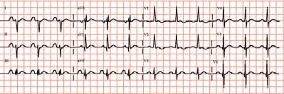
13 B. ATRIAL FLUTTER : Rapid, regular flutter (F) waves at per minute (ventricular conduction 1:2, ie ~150bpm) Sawtooth pattern of F waves in leads 2, 3 and AVF Little evidence of atrial activity in lead 1 AV conduction variable, QRS typically normal width Enhanced automaticity leading to circuitous conduction/reentry Atrial flutter is the most electrosensitive of all dysrhythmias therefore cardioversion is the treatment of choice for conversion to sinus rhythm. Drug of choice for rate control is Calcium channel blockers. Drug of choice for diagnostic purposes is Adenosine (as long as QRS is narrow ATRIAL FLUTTER - TREATMENT Atrial flutter with 2:1 conduction is often confused with SVT But, look for the sawtooth flutter waves in the inferior leads. 13


14 Same patient after adenosine, showing prominent flutter waves. A 46 year old presents with palpitations. EKG shows: a. Atrial fibrillation b. Atrial flutter c. Left anterior fasicular block d. RBBB EKG shows: a. Atrial fibrillation Prominent fibrillatory waves in V 1-3 & AVF Irregular ventricular response, greater than 100 / min Ventricular rate less than 100 implies AV block Triggered/enhanced automaticity 14


15 ATRIAL FIBRILLATION - treatment Cardiovert if unstable Ca Channel Blocker- Drug of choice for rate control Beta blocker Digitalis ASA alone for afib < 48h ASA & Anti-coagulate all others, if unknown or >48h» the longer the patient has been in afib, the less likely you will be able to convert to NSR Ashman s phenomenon short runs of wide complex tachycardia during rapid atrial fibrillation. The refractory period is rate-related, and when erratic changes in rate occur, an impulse conducted during the refractory period will have an aberrant (RBBB) pattern. The most common dysrhythmia associated with digitalis toxicity is: A. Paroxysmal atrial tachycardia with AV nodal block B. Premature ventricular contractions C. Second degree AV nodal blocks D. Ventricular tachycardia E. Junctional tachycardia 15

16 DIGITALIS TOXICITY - DYSRHYTHMIAS Most common : b. PVCs Most pathognomonic : PAT w/block Others AV nodal blocks sinus bradycardia, pause, SA block junctional escape beats or tachycardia Ectopic SVT, V-tach, V-fib Paroxysmal atrial tachycardia with block is pathognomonic for digitalis toxicity. Note the p waves at a rate > 100 & blocked QRS complexes. (Don t mistake for aflutter with variable conduction or 3 rd degree block) Note the blocked Impulses!! A 23 yo male with c/o palpitations, EKG shows: a. Atrial fibrillation b. MFAT c. SVT d. PAT with block 16

17 His EKG shows c. SVT or AV nodal reentry tachycardia with a rapid, regular rate, absent p waves & narrow QRS complexes AV nodal Re-entry tachycardia/svt Two parallel pathways with different rates and refractory periods Something alters the refractory period and the alternative pathway becomes dominant This causes a unidirectional conduction block, and a circuitous conduction pathway forms. SA AV AV nodal Re-entry tachycardia/svt The circuitous impulse is typically transmitted anterograde (forward) over the relatively slow AV nodal fibers, limiting the rate to 200bpm. WHAT S THE BIG DEAL??? Treat by blocking the AV node and allowing the normal pacemaker to resume. Adenosine Ca channel blocker Beta blocker SA AV 17
 SVT with aberrancy is treated by blocking the AV node and allowing the normal pacemaker to resume Adenosine Ca ch blocker Beta blocker It is very")
18 SVT with Aberrancy (rate-related block) SVT with aberrancy is a supraventricular tachycardia with a widecomplex QRS due to a raterelated bundle branch block. SA AV SVT with Aberrancy (rate-related block) SVT with aberrancy is treated by blocking the AV node and allowing the normal pacemaker to resume Adenosine Ca ch blocker Beta blocker It is very difficult to differentiate from Vtach if unsure, treat as stable Vtach amiodarone procainamide SA AV 44yo with complaint of palpitations and shortness of breath, ekg shows: a. SVT with aberrancy b. Ashman s phenomenon c. WPW d. V-tach 18
19 C. The EKG is WPW w/ retrograde conduction causing wide QRS. Because the etiology of a wide complex tachydysrhythmia is often unknown in the ER, treat with amiodarone, procainamide, lidocaine or cardioversion. (avoid procainamide in TCA OD or prolonged qt toursades) Pre-Excitation Syndromes- WPW & LGL Accessory pathway connects atria to the ventricles, bypassing the AV node Wolff-Parkinson-White: short PR (< 0.12 s), Delta wave (slurred upstroke QRS), slight wide QRS >0.10s, and frequently a psuedoinfarction pattern in the inferior leads and RBBB pattern. Lown-Ganong-Levine: short PR (< 0.12 s), NO Delta wave, normal QRS & episodes of tachydysrhythmias WPW LGL Delta waves, short pr interval, wide QRS The underlying ECG in WPW is a fusion of the accessory pathway (delta wave) and normal pathway of the QRS. During tachydysrhythmias, the electrical impulse follows only the accessory pathway in a circuitous fashion. 19
 SA AV Wide QRS b/c retrograde conduction 10% Accessory Pathways-WPW Wide")
 most AV nodal blockers stop only anterograde conduction and can allow the rate of retrograde conduction")

20 Underlying ECG Fusion of accessory & normal pathways Accessory pathway with circuitous impulses traveling retrograde (wide QRS) Accessory Pathways-WPW If narrow QRS d/t forward conduction, treat as SVT (Adenosine) SA AV Wide QRS b/c retrograde conduction 10% Accessory Pathways-WPW Wide QRS if retrograde conduction Adenosine, Ca channel blockers, B blockers and digitalis block the forward conduction, not the retrograde conduction. In a wide complex WPW (retrograde impulses) most AV nodal blockers stop only anterograde conduction and can allow the rate of retrograde conduction to speed up and deteriorate into Vfib! This is seen in wide complex WPW with Afib or Aflutter. Amiodarone and procainamide affect the forward and retrograde pathways as well as the ventricles and are safe in wide-complex tachydysrhythmias. SA AV (Caveat: Procainamide and Amiodarone not to be used in Toursades) 20
21 Evaluation of Re-entry Tachycardias -QRS Width Wide or Narrow If the QRS is narrow, it MUST have atrial origin and conduct through the AV node in a forward manner. If the QRS is wide, more than 0.12 seconds, consider : Bypass tract (WPW) with retrograde conduction SVT with aberrancy (rate-related bundle branch block) Junctional origin Ventricular origin Re-entry Tachycardias - Treatment Modalities Based on hemodynamic stability & QRS width Unstable : synchronized cardioversion Stable : Narrow complex vagal maneuvers, adenosine, calcium channel blockers or beta blockers Wide complex Amiodarone, Lidocaine or Procainamide to treat both anterograde and retrograde impulses and ventricular dysrhythmias Beware: it is very difficulty to tell the difference between the widecomplex tachy-dysrhythmias. It is safer to treat as presumed V-tach. PEARLS Wide complex QRS tachydysrhythmias of unknown etiology use amiodorone, procainamide, lidocaine Differential diagnosis for rapid, irregularly irregular rhythm MFAT Atrial Fib Atrial flutter with variable conduction SVT at 150 or 300, consider Atrial flutter 21

22 DYSRHYTHMIAS OF VENTRICULAR ORIGIN Idioventricular rhythms Ventricular Tachycardia Ventricular Fibrillation Torsades de pointes VENTRICULAR DYSRHYTHMIAS - Etiology V Tach, V Fib & Idioventricular rhythms typically caused by an ischemic focus which allows a rapid reentry dysrhythmia Torsades de pointes - caused by a prolonged QT interval Brugada syndrome sodium ion channelapathy IDIOVENTRICULAR RHYTHMS Mechanism : re-entry with unidirectional block due to myocardial ischemia QRS width > 0.12 sec and rate T waves typically have opposite polarity to QRS Treatment : Controversial, tends to be self-limited Supportive care & close observation 22


23 VENTRICULAR TACHYCARDIA Mechanism : re-entry with unidirectional block due to myocardial ischemia (Monomorphic) QRS width > 0.12 sec and rate > 140 bpm T waves have opposite polarity to QRS Treatment : Stable : Amiodarone, Procainamide, Sotolol, Lidocaine, Mag Unstable : Unsynchronized defibrillation plus meds VENTRICULAR FIBRILLATION Chaotic ventricular depolarization with loss of organized QRS complexes Life-threatening Immediate loss of consciousness Loss of blood pressure & death Treatment : immediate unsynchronized defibrillation at 200, 300, then 360 joules (if Biphasic use ½ dose or 150j) Brugada Syndrome: ST elevation V1-3 with RBBB-like pattern which predisposes to ventricular dysrhythmias. 30% mortality within 3 years. Brugada P & Brugada J. J Am Coll Cardiol 1992;20:


24 Brugada Syndrome: Look for ST elevation V1-3 part of the syncope or palpitation work-up immediate cardiology referral for ICD placement CARDIOVERSION PEARLS Atrial flutter is the most electro-responsive dysrhythmia joules ~ treatment of choice SVT and STABLE ventricular tachycardia often respond to 50 joules Atrial and Ventricular FIBRILLATION require 100 joules or more Biphasic defibrillators use half the joules or 150j TORSADES DE POINTES V-tach due to prolonged QT interval, in which the QRS axis alternates between positive and negative (Polymorphic) Often self-limited, but may deteriorate into ventricular fibrillation Treatment of Choice : Magnesium Overdrive pacing & Isoproterenol can be used to speed the heart and decrease QT interval Avoid procainamide and amiodarone, as can worsen QT prolongation If refractory, defibrillate 24


25 QUESTION ~ All of the following cause Torsades de pointes, except: A. Hypomagnesemia B. Tricyclic antidepressant overdose C. Procainamide D. Hyperkalemia E. Quinidine CAUSES OF PROLONGED QT INTERVAL Hypo -Mg, -Ca, -K, Type Ia antidysrhythmics - quinidine, procainamide Tricyclic antidepressant overdose drug reactions-ees, antihistamines, antifungals d is incorrect, hyperkalemia does not cause prolonged QT Prolonged qt interval 25


26 Shortened qt: hypercalcemia Peaked T waves ( > 1/3 QRS) Prolonged PR interval Hyperkalemia Widening of QRS Sine Wave U waves in Hypokalemia Potassium 3.5mEq/L Potassium 2mEq/L Potassium 3mEq/L Potassium 1mEq/L 26


27 Osborne J wave in hypothermia: notching at end of a slurred downstroke of QRS Tricyclic Antidepressant Overdose tall r in AVR slurring of the terminal portion of the rs in AVR Patterns of Infarction The LAD supplies the septal V1-2 and anterior leads V2-4 The RCA supplies the Inferior leads: II, III & AVF The Circumflex supplies the high and low Lateral leads: V5-6 and I &AVL 27


28 Inferior Wall MI ST segment elevation in II, III & avf Anterior Wall MI ST segment elevation in V2-4 Septal MI ST segment elevation V1-2 Lateral Wall MI ST segment elevation in V5-6 and/or I & avl 28

 TCA overdose : stach, widening QRS, slurring of the terminal rs in avr Axis deviation & Hemiblocks :")
29 Posterior Wall MI - Tall R in V1 & ST segment depression in V1-2 Pericarditis diffuse ST segment elevation & PR depression, with PR elevation in AVR EKG PEARLS When you see a normal looking EKG on a test, start looking for: Hyperkalemia :Peaked T waves Hypokalemia : U waves Hypomagnesimia : Prolonged QT Hypercalcemia: Shortened QT WPW : short PR, slurring of upstroke qrs Hypothermia : Osborne J waves (notched downstroke QRS; reversed delta waves) TCA overdose : stach, widening QRS, slurring of the terminal rs in avr Axis deviation & Hemiblocks : LAFB, LPFB 29
30 EKG PEARLS Usefulness of avr & V1 Tall R wave in V1 RBBB WPW Posterior wall MI Severe RV strain: PE, pneumothorax, severe COPD avr is normally flipped/negative polarity slurring terminal rs in TCA OD PR elevation in pericarditis Diffuse ST elevation: think pericarditis? 30
ECGs and Arrhythmias: Family Medicine Board Review 2009
 Rate Rhythm Intervals Hypertrophy ECGs and Arrhythmias: Family Medicine Board Review 2009 Axis Jess (Fogler) Waldura, MD University of California, San Francisco walduraj@nccc.ucsf.edu Ischemia Overview
Arrhythmic Complications of MI. Teferi Mitiku, MD Assistant Clinical Professor of Medicine University of California Irvine
 Arrhythmic Complications of MI Teferi Mitiku, MD Assistant Clinical Professor of Medicine University of California Irvine Objectives Brief overview -Pathophysiology of Arrhythmia ECG review of typical
12-Lead ECG Interpretation. Kathy Kuznar, RN, ANP
 12-Lead ECG Interpretation Kathy Kuznar, RN, ANP The 12-Lead ECG Objectives Identify the normal morphology and features of the 12- lead ECG. Perform systematic analysis of the 12-lead ECG. Recognize abnormalities
Huseng Vefali MD St. Luke s University Health Network Department of Cardiology
 Huseng Vefali MD St. Luke s University Health Network Department of Cardiology Learning Objectives Establish Consistent Approach to Interpreting ECGs Review Essential Cases for Paramedics and first responders
Cardiology Flash Cards
 Cardiology Flash Cards EKG in a nut shell www.brain101.info Conduction System www.brain101.info 2 Analyzing EKG Step by step Steps in Analyzing ECG'S 1. Rhythm: - Regular _ Sinus, Junctional or Ventricular.
ECGs and Arrhythmias: Family Medicine Board Review 2012
 Overview ECGs and Arrhythmias: Family Medicine Board Review 2012 Jess Waldura, MD University of California, San Francisco walduraj@nccc.ucsf.edu Bundle branch blocks Quick review of ischemia Arrhythmias
Conduction Problems / Arrhythmias. Conduction
 Conduction Problems / Arrhythmias Conduction Wolf-Parkinson White Syndrome (WPW) and Lown-Ganong-Levine (LGL): Atrial impulses bypass the AV node through an accessory pathway or bypass tract (bundle of
SIMPLY ECGs. Dr William Dooley
 SIMPLY ECGs Dr William Dooley Content Basic ECG interpretation pattern Some common (examined) abnormalities Presenting ECGs in context Setting up an ECG Setting up an ECG 1 V1-4 th Right intercostal space
2017 EKG Workshop Advanced. Family Medicine Review Course Lou Mancano, MD, FAAFP Reading Health System Family and Community Medicine Reading, PA
 2017 EKG Workshop Advanced Family Medicine Review Course Lou Mancano, MD, FAAFP Reading Health System Family and Community Medicine Reading, PA Part II - Objective Describe a useful approach to interpreting
Please check your answers with correct statements in answer pages after the ECG cases.
 ECG Cases ECG Case 1 Springer International Publishing AG, part of Springer Nature 2018 S. Okutucu, A. Oto, Interpreting ECGs in Clinical Practice, In Clinical Practice, https://doi.org/10.1007/978-3-319-90557-0
Cardiac Arrhythmia How to approach นพ.พ น จ แกวส วรรณะ หน วยโรคห วใจและหลอดเล อด
 Cardiac Arrhythmia How to approach นพ.พ น จ แกวส วรรณะ หน วยโรคห วใจและหลอดเล อด EKG paper is a grid where time is measured along the horizontal axis. Each small square is 1 mm in length and represents
REtrive. REpeat. RElearn Design by. Test-Enhanced Learning based ECG practice E-book
 Test-Enhanced Learning Test-Enhanced Learning Test-Enhanced Learning Test-Enhanced Learning based ECG practice E-book REtrive REpeat RElearn Design by S I T T I N U N T H A N G J U I P E E R I Y A W A
ECG Interpretation Cat Williams, DVM DACVIM (Cardiology)
") ECG Interpretation Cat Williams, DVM DACVIM (Cardiology) Providing the best quality care and service for the patient, the client, and the referring veterinarian. GOAL: Reduce Anxiety about ECGs Back to
ECG Interpretation Cat Williams, DVM DACVIM (Cardiology) Providing the best quality care and service for the patient, the client, and the referring veterinarian. GOAL: Reduce Anxiety about ECGs Back to
ECG Interpretation Made Easy
 ECG Interpretation Made Easy Dr. A Tageldien Abdellah, MSc MD EBSC Lecturer of Cardiology- Hull University Hull York Medical School 2007-2008 ECG Interpretation Made Easy Synopsis Benefits Objectives Process
SIMPLY ECGs. Dr William Dooley
 SIMPLY ECGs Dr William Dooley 1 No anatomy just interpretation 2 Setting up an ECG 3 Setting up an ECG 1 V1-4 th Right intercostal space at sternal border 2 V2-4 th Left intercostal space at sternal border
UNDERSTANDING YOUR ECG: A REVIEW
 UNDERSTANDING YOUR ECG: A REVIEW Health professionals use the electrocardiograph (ECG) rhythm strip to systematically analyse the cardiac rhythm. Before the systematic process of ECG analysis is described
Step by step approach to EKG rhythm interpretation:
 Sinus Rhythms Normal sinus arrhythmia Small, slow variation of the R-R interval i.e. variation of the normal sinus heart rate with respiration, etc. Sinus Tachycardia Defined as sinus rhythm with a rate
ECG Basics Sonia Samtani 7/2017 UCI Resident Lecture Series
 ECG Basics Sonia Samtani 7/2017 UCI Resident Lecture Series Agenda I. Introduction II.The Conduction System III.ECG Basics IV.Cardiac Emergencies V.Summary The Conduction System Lead Placement avf Precordial
CRC 431 ECG Basics. Bill Pruitt, MBA, RRT, CPFT, AE-C
 CRC 431 ECG Basics Bill Pruitt, MBA, RRT, CPFT, AE-C Resources White s 5 th ed. Ch 6 Electrocardiography Einthoven s Triangle Chest leads and limb leads Egan s 10 th ed. Ch 17 Interpreting the Electrocardiogram
How To Think About Rhythms and Conduction
 How To Think About Rhythms and Conduction Frank Yanowitz, MD Professor of Medicine (Cardiology) University of Utah School of Medicine Medical Director, IHC ECG Services Intermountain Healthcare http://ecg.utah.edu
ECG Cases and Questions. Ashish Sadhu, MD, FHRS, FACC Electrophysiology/Cardiology
 ECG Cases and Questions Ashish Sadhu, MD, FHRS, FACC Electrophysiology/Cardiology 32 yo female Life Insurance Physical 56 yo male with chest pain Terminology Injury ST elevation Ischemia T wave inversion
Pennsylvania Academy of Family Physicians Foundation & UPMC 43rd Refresher Course in Family Medicine CME Conference March 10-13, 2016
 Pennsylvania Academy of Family Physicians Foundation & UPMC 43rd Refresher Course in Family Medicine CME Conference March 10-13, 2016 Disclosures: EKG Workshop Louis Mancano, MD Speaker has no disclosures
2) Heart Arrhythmias 2 - Dr. Abdullah Sharif
 Heart Arrhythmias 2 - Dr. Abdullah Sharif") 2) Heart Arrhythmias 2 - Dr. Abdullah Sharif Rhythms from the Sinus Node Sinus Tachycardia: HR > 100 b/m Causes: o Withdrawal of vagal tone & Sympathetic stimulation (exercise, fight or flight) o Fever
2) Heart Arrhythmias 2 - Dr. Abdullah Sharif Rhythms from the Sinus Node Sinus Tachycardia: HR > 100 b/m Causes: o Withdrawal of vagal tone & Sympathetic stimulation (exercise, fight or flight) o Fever
If the P wave > 0.12 sec( 3 mm) usually in any lead. Notched P wave usually in lead I,aVl may be lead II Negative terminal portion of P wave in V1, 1
 usually in any lead. Notched P wave usually in lead I,aVl may be lead II Negative terminal portion of P wave in V1, 1") If the P wave > 0.12 sec( 3 mm) usually in any lead. Notched P wave usually in lead I,aVl may be lead II Negative terminal portion of P wave in V1, 1 mm depth and 3 mm width( most specific) Since Mitral
If the P wave > 0.12 sec( 3 mm) usually in any lead. Notched P wave usually in lead I,aVl may be lead II Negative terminal portion of P wave in V1, 1 mm depth and 3 mm width( most specific) Since Mitral
Supraventricular Arrhythmias. Reading Assignment. Chapter 5 (p17-30)
") Supraventricular Arrhythmias Reading Assignment Chapter 5 (p17-30) The Supraventricular Rhythms In Our Lives Site of Origin Single Events Slow Rates Intermediate Rates Fast Rates (>100 bpm) Sinus Sinus
Supraventricular Arrhythmias Reading Assignment Chapter 5 (p17-30) The Supraventricular Rhythms In Our Lives Site of Origin Single Events Slow Rates Intermediate Rates Fast Rates (>100 bpm) Sinus Sinus
Course Objectives. Proper Lead Placements. Review the ECG print paper. Review the mechanics of the Myocardium. Review basics of ECG Rhythms
 ECG Interpretations Course Objectives Proper Lead Placements Review the ECG print paper Review the mechanics of the Myocardium Review basics of ECG Rhythms How Leads Work The ECG Leads we use are Bipolar
Nathan Cade, MD Brandon Fainstad, MD Andrew Prouse, MD
 Nathan Cade, MD Brandon Fainstad, MD Andrew Prouse, MD OBJECTIVES 1. Identify the basic electrophysiology of the four causes of wide complex tachycardia. 2. Develop a simple framework for acute management
Review Packet EKG Competency This packet is a review of the information you will need to know for the proctored EKG competency test.
 Review Packet EKG Competency 2015 This packet is a review of the information you will need to know for the proctored EKG competency test. Normal Sinus Rhythm Rhythm: Regular Ventricular Rate: 60-100 bpm
ECG CONVENTIONS AND INTERVALS
 1 ECG Waveforms and Intervals ECG waveforms labeled alphabetically P wave== represents atrial depolarization QRS complex=ventricular depolarization ST-T-U complex (ST segment, T wave, and U wave)== V repolarization.
CORONARY ARTERIES. LAD Anterior wall of the left vent Lateral wall of left vent Anterior 2/3 of interventricluar septum R & L bundle branches
 CORONARY ARTERIES RCA Right atrium Right ventricle SA node 55% AV node 90% Posterior wall of left ventricle in 90% Posterior third of interventricular septum 90% LAD Anterior wall of the left vent Lateral
Section 3 and 4. Objectives. Bundle Branches 10/9/2018. LBBB, RBBB Bifascicular, Trifascicular Block
 Section 3 and 4 LBBB, RBBB Bifascicular, Trifascicular Block Objectives At the conclusion of this presentation the participant will be able to Outline a systematic approach to 12 lead ECG interpretation
ECG Interpretation. Introduction to Cardiac Telemetry. Michael Peters, RN, CCRN, CFRN CALSTAR Air Medical Services
 ECG Interpretation Introduction to Cardiac Telemetry Michael Peters, RN, CCRN, CFRN CALSTAR Air Medical Services Disclosures Nothing to disclose Objectives Describe the electrical conduction pathway in
Understanding the 12-lead ECG, part II
 Bundle-branch blocks Understanding the 12-lead ECG, part II Most common electrocardiogram (ECG) abnormality Appears as a wider than normal S complex Occurs when one of the two bundle branches can t conduct
10 ECGs No Practitioner Can Afford to Miss. Objectives
 10 ECGs No Practitioner Can Afford to Miss Mary L. Dohrmann, MD Professor of Clinical Medicine Division of Cardiovascular Medicine University of Missouri School of Medicine No disclosures Objectives 1.
You Don t Want to Miss This One! Focus on can t miss EKG tracings
 You Don t Want to Miss This One! Focus on can t miss EKG tracings Renaissance St. Louis Grand Hotel Convention Center October 23, 2014 David K. Tan, M.D., EMT-T, FAAEM EMS Section Chief, Division of Emergency
Paramedic Rounds. Tachyarrhythmia's. Sean Sutton Dallas Wood
 Paramedic Rounds Tachyarrhythmia's Sean Sutton Dallas Wood Objectives At the end of this session, the paramedic will be able to: State the key components of the cardiac conduction pathway, along with the
, David Stultz, MD.
 http://www.dilbert.com EKG Rounds Handouts available at http://www.drstultz.com January 5, 2004 David Stultz, MD Cardiology Fellow, PGY 4 Overview of Topics How to read an EKG Normal EKG Determination
General Introduction to ECG. Reading Assignment (p2-16 in PDF Outline )
") General Introduction to ECG Reading Assignment (p2-16 in PDF Outline ) Objectives 1. Practice the 5-step Method 2. Differential Diagnosis: R & L axis deviation 3. Differential Diagnosis: Poor R-wave progression
General Introduction to ECG Reading Assignment (p2-16 in PDF Outline ) Objectives 1. Practice the 5-step Method 2. Differential Diagnosis: R & L axis deviation 3. Differential Diagnosis: Poor R-wave progression
Appendix. Table 1: Causes for abnormal axis deviation Left axis deviation
 Appendix Table 1: Causes for abnormal axis deviation Left axis deviation Normal variant (2 5%) Left anterior fascicular block Left ventricular hypertrophy Inferior wall myocardial infarction Primum atrial
-RHYTHM PRACTICE- By Dr.moanes Msc.cardiology Assistant Lecturer of Cardiology Al Azhar University. OBHG Education Subcommittee
 -RHYTHM PRACTICE- By Dr.moanes Msc.cardiology Assistant Lecturer of Cardiology Al Azhar University The Normal Conduction System Sinus Node Normal Sinus Rhythm (NSR) Sinus Bradycardia Sinus Tachycardia
FLB s What Are Those Funny-Looking Beats?
 FLB s What Are Those Funny-Looking Beats? Reading Assignment (pages 27-45 in Outline ) The 5-Step Method ECG #: Mearurements: Rhythm (s): Conduction: Waveform: Interpretation: A= V= PR= QRS= QT= Axis=
Bundle Branch & Fascicular Blocks. Reading Assignment (p53-58 in Outline )
") Bundle Branch & Fascicular Blocks Reading Assignment (p53-58 in Outline ) Objectives 1. QRS analysis of Right and Left BBB 2. Uncomplicated vs complicated BBB 3. Diagnosis of RBBB with LAFB and LPFB 4.
Bundle Branch & Fascicular Blocks Reading Assignment (p53-58 in Outline ) Objectives 1. QRS analysis of Right and Left BBB 2. Uncomplicated vs complicated BBB 3. Diagnosis of RBBB with LAFB and LPFB 4.
Rhythm ECG Characteristics Example. Normal Sinus Rhythm (NSR)
") Normal Sinus Rhythm (NSR) Rate: 60-100 per minute Rhythm: R- R = P waves: Upright, similar P-R: 0.12-0.20 second & consistent P:qRs: 1P:1qRs Sinus Tachycardia Exercise Hypovolemia Medications Fever Substances
Normal Sinus Rhythm (NSR) Rate: 60-100 per minute Rhythm: R- R = P waves: Upright, similar P-R: 0.12-0.20 second & consistent P:qRs: 1P:1qRs Sinus Tachycardia Exercise Hypovolemia Medications Fever Substances
Return to Basics. ECG Rate and Rhythm. Management of the Hospitalized Patient September 25, 2009
 Management of the Hospitalized Patient September 25, 2009 ECG Refresher and Update 2009 Return to Basics Determine rate and rhythm Determine intervals and axes Define morphology of P-QRS-T-U Compare with
THE ELECTROCARDIOGRAM A UBIQUITOUS AND COST-EFFECTIVE DIAGNOSTIC TOOL FOR THE FAMILY MEDICINE REFRESHER COURSE MARCH 8, 2019
 THE ELECTROCARDIOGRAM A UBIQUITOUS AND COST-EFFECTIVE DIAGNOSTIC TOOL FOR THE FAMILY MEDICINE REFRESHER COURSE MARCH 8, 2019 Major Clinical Disorders Pulmonary Embolism 69 y/o woman with dyspnea and an
ECG S: A CASE-BASED APPROACH December 6,
 ECG S: A CASE-BASED APPROACH December 6, 2018 1 Faculty Disclosure Faculty: Lorne Gula MD, FRCPC Professor, Western University Cardiologist, Hearth Rhythm Specialist Director, Electrophysiology Laboratory,
12 Lead ECG. Presented by Rebecca Sevigny BSN, RN Professional Practice & Development Dept.
 12 Lead ECG Presented by Rebecca Sevigny BSN, RN Professional Practice & Development Dept. Two Main Coronary Arteries RCA LCA which branches into Left Anterior Descending Circumflex Artery Two Main Coronary
Chapter 16: Arrhythmias and Conduction Disturbances
 Complete the following. Chapter 16: Arrhythmias and Conduction Disturbances 1. Cardiac arrhythmias result from abnormal impulse, abnormal impulse, or both mechanisms together. 2. is the ability of certain
ECG ABNORMALITIES D R. T AM A R A AL Q U D AH
 ECG ABNORMALITIES D R. T AM A R A AL Q U D AH When we interpret an ECG we compare it instantaneously with the normal ECG and normal variants stored in our memory; these memories are stored visually in
Case #1. 73 y/o man with h/o HTN and CHF admitted with dizziness and SOB Treated for CHF exacerbation with Lasix Now HR 136
 Tachycardias Case #1 73 y/o man with h/o HTN and CHF admitted with dizziness and SOB Treated for CHF exacerbation with Lasix Now HR 136 Initial Assessment Check Telemetry screen if pt on tele Telemetry
3. AV Block 1. First-degree AV block 1. Delay in AV node 2. Long PR interval 3. QRS complex follows each P wave 4. Benign, no tx
 1. Rhythms & arrhythmias SA nodal rhythms Sinus rhythm Sinus tachycardia Sinus bradycardia Sinus arrhythmia Sick sinus syndrome SA block Sinus arrest AV blocks First-degree Second-degree Mobitz Type I
Pathologic ECG. Adelina Vlad, MD PhD
 Pathologic ECG Adelina Vlad, MD PhD Basic Interpretation of the ECG 1) Evaluate calibration 2) Calculate rate 3) Determine rhythm 4) Determine QRS axis 5) Measure intervals 6) Analyze the morphology and
PATIENT S NAME, DATE/TIME,
 ECG and Arrhythmias Dec, 1 st 2014 Doctor Mohammad Jarrah References: - Lecture and Slides - ECG Made Easy - Davidson Principles of Medicine - First Aid Cases for the USMLE Step 1 - Mini-OSCE Archive ECG
Dr. Schroeder has no financial relationships to disclose
 Valerie A Schroeder MD MS Assistant Professor University of Kansas Medical Center READING THE WAVES- THE HEART S ELECTRICAL MESSAGE FINANCIAL DISCLOSURE Dr. Schroeder has no financial relationships to
Electrical System Overview Electrocardiograms Action Potentials 12-Lead Positioning Values To Memorize Calculating Rates
 Electrocardiograms Electrical System Overview James Lamberg 2/ 74 Action Potentials 12-Lead Positioning 3/ 74 4/ 74 Values To Memorize Inherent Rates SA: 60 to 100 AV: 40 to 60 Ventricles: 20 to 40 Normal
Blocks & Dissociations. Reading Assignment (p47-52 in Outline )
") Blocks & Dissociations Reading Assignment (p47-52 in Outline ) Objectives Who are Wenckebach and Mobitz? Review SA and AV Blocks AV Dissociations: learning who s the boss and why 2 nd degree SA Block:
Blocks & Dissociations Reading Assignment (p47-52 in Outline ) Objectives Who are Wenckebach and Mobitz? Review SA and AV Blocks AV Dissociations: learning who s the boss and why 2 nd degree SA Block:
ABCs of ECGs. Shelby L. Durler
 ABCs of ECGs Shelby L. Durler Objectives Review the A&P of the cardiac conduction system Placement and obtaining 4-lead and 12-lead ECGs Overview of the basics of ECG rhythm interpretation Intrinsic
Family Medicine for English language students of Medical University of Lodz ECG. Jakub Dorożyński
 Family Medicine for English language students of Medical University of Lodz ECG Jakub Dorożyński Parts of an ECG The standard ECG has 12 leads: six of them are considered limb leads because they are placed
Basic electrocardiography reading. R3 lee wei-chieh
 Basic electrocardiography reading R3 lee wei-chieh The Normal Conduction System Lead Placement avf Limb Leads Precordial Leads Interpretation Rate Rhythm Interval Axis Chamber abnormality QRST change What
Return to Basics. Normal Intervals & Axes. ECG Rate and Rhythm
 Return to Basics Management of the Hospitalized Patient October 15, 2010 ECG Refresher and Update 2010 Determine rate and rhythm Determine intervals and axes Define morphology of P-QRS-T-U Compare with
402 Index. B β-blockers, 4, 5 Bradyarrhythmias, 76 77
 Index A Acquired immunodeficiency syndrome (AIDS), 126, 163 Action potentials, 1, 5, 27 Acute coronary syndromes, 123t, 129 Adenosine, intravenous, 277 Alcohol abuse, as T wave inversion cause, 199 Aneurysm,
Return to Basics. ECG Rate and Rhythm. Management of the Hospitalized Patient October 4, 2007
 Management of the Hospitalized Patient October 4, 2007 ECG Refresher for the Hospitalists Return to Basics Determine rate and rhythm Determine intervals and axes Define morphology of P-QRS-T-U Compare
ECG Interactive Session
 SA HEART 2018 PRE-CONGRESS WORKSHOP 4 OCTOBER 2018 ECG Interactive Session Ashley Chin Cardiologist/Electrophysiologist University of Cape Town Groote Schuur Hospital ECG 1 Which ONE of the following is
Emergency Medical Training Services Emergency Medical Technician Paramedic Program Outlines Outline Topic: WPW Revised: 11/2013
 Emergency Medical Training Services Emergency Medical Technician Paramedic Program Outlines Outline Topic: WPW Revised: 11/2013 Wolff-Parkinson-White syndrome (WPW) is a syndrome of pre-excitation of the
Ekg pra pr c a tice D.HAMMOUDI.MD
 Ekg practice D.HAMMOUDI.MD Anatomy Revisited RCA (Right Coronary Artery) Right ventricle Inferior wall of LV Posterior wall of LV (75%) SA Node (60%) AV Node (>80%) LCA (Left Coronary Artery) Septal wall
Miscellaneous Stuff Keep reading the Outline
 Miscellaneous Stuff Keep reading the Outline Welcome to the 5-Step Method ECG #: Mearurements: Rhythm (s): Conduction: Waveform: Interpretation: A= V= PR= QRS= QT= Axis= 1. Compute the 5 basic measurements:
BEDSIDE ECG INTERPRETATION
 BEDSIDE ECG INTERPRETATION Presented by: Ryan Dean, RN, MSN, CCRN, CCNS, CFRN Flight Nurse 2017 Based on presentations originally by Gennifer DePaoli, RN Objectives Hospital policies Electrical conduction
Appendix D Output Code and Interpretation of Analysis
 Appendix D Output Code and Interpretation of Analysis 8 Arrhythmia Code No. Description 8002 Marked rhythm irregularity 8110 Sinus rhythm 8102 Sinus arrhythmia 8108 Marked sinus arrhythmia 8120 Sinus tachycardia
ELECTROCARDIOGRAPHY KEVIN REBECK PA-C. For more presentations
 ELECTROCARDIOGRAPHY KEVIN REBECK PA-C For more presentations www.medicalppt.blogspot.com Objectives ECG History Pathophysiology Basics Case Historys Electrical activation of the heart In the heart
The ABCs of EKGs/ECGs for HCPs. Al Heuer, PhD, MBA, RRT, RPFT Professor, Rutgers School of Health Related Professions
 The ABCs of EKGs/ECGs for HCPs Al Heuer, PhD, MBA, RRT, RPFT Professor, Rutgers School of Health Related Professions Learning Objectives Review the basic anatomy of the heart Describe the cardiac conducting
ELECTROCARDIOGRAPH. General. Heart Rate. Starship Children s Health Clinical Guideline
 General Heart Rate QRS Axis T Wave Axis PR Interval according to Heart Rate & Age P Wave Duration and Amplitude QRS Duration according to Age QT Interval R & S voltages according to Lead & Age R/S ratio
Chapter 9. Learning Objectives. Learning Objectives 9/11/2012. Cardiac Arrhythmias. Define electrical therapy
 Chapter 9 Cardiac Arrhythmias Learning Objectives Define electrical therapy Explain why electrical therapy is preferred initial therapy over drug administration for cardiac arrest and some arrhythmias
CSI Skills Lab #5: Arrhythmia Interpretation and Treatment
 CSI 202 - Skills Lab #5: Arrhythmia Interpretation and Treatment Origins of the ACLS Approach: CSI 202 - Skills Lab 5 Notes ACLS training originated in Nebraska in the early 1970 s. Its purpose was to
Ben Taylor, PhD, PA-C
 Ben Taylor, PhD, PA-C The patient is a 23-year-old white male with a history of polysubstance abuse who was found unresponsive, last seen the day before. Classic signs of systemic hypothermia with prominent
Cardiac Arrhythmias. Cathy Percival, RN, FALU, FLMI VP, Medical Director AIG Life and Retirement Company
 Cardiac Arrhythmias Cathy Percival, RN, FALU, FLMI VP, Medical Director AIG Life and Retirement Company The Cardiovascular System Three primary functions Transport of oxygen, nutrients, and hormones to
2017 EKG Workshop Basic. Family Medicine Review Course Lou Mancano, MD, FAAFP Reading Health System Family and Community Medicine Reading, PA
 2017 EKG Workshop Basic Family Medicine Review Course Lou Mancano, MD, FAAFP Reading Health System Family and Community Medicine Reading, PA Part I - Objectives Discuss a systematic approach to EKG interpretation
Introduction to Electrocardiography
 Introduction to Electrocardiography Class Objectives: Introduction to ECG monitoring Discuss principles of interpretation Identify the components and measurements of the ECG ECG analysis ECG Monitoring
ECGs: Everything a finalist needs to know. Dr Amy Coulden As part of the Simply Finals series
 ECGs: Everything a finalist needs to know Dr Amy Coulden As part of the Simply Finals series Aims and objectives To be able to interpret basic ECG abnormalities To be able to recognise commonly tested
TEST BANK FOR ECGS MADE EASY 5TH EDITION BY AEHLERT
 Link download full: http://testbankair.com/download/test-bank-for-ecgs-made-easy-5thedition-by-aehlert/ TEST BANK FOR ECGS MADE EASY 5TH EDITION BY AEHLERT Chapter 5 TRUE/FALSE 1. The AV junction consists
EKG Competency for Agency
 EKG Competency for Agency Name: Date: Agency: 1. The upper chambers of the heart are known as the: a. Atria b. Ventricles c. Mitral Valve d. Aortic Valve 2. The lower chambers of the heart are known as
Myocardial Infarction. Reading Assignment (p66-78 in Outline )
") Myocardial Infarction Reading Assignment (p66-78 in Outline ) Objectives 1. Why do ST segments go up or down in ischemia? 2. STEMI locations and culprit vessels 3. Why 15-lead ECGs? 4. What s up with avr?
Myocardial Infarction Reading Assignment (p66-78 in Outline ) Objectives 1. Why do ST segments go up or down in ischemia? 2. STEMI locations and culprit vessels 3. Why 15-lead ECGs? 4. What s up with avr?
Acute Coronary Syndromes Unstable Angina Non ST segment Elevation MI (NSTEMI) ST segment Elevation MI (STEMI)
 ST segment Elevation MI (STEMI)") Leanna R. Miller, RN, MN, CCRN-CSC, PCCN-CMC, CEN, CNRN, CMSRN, NP Education Specialist LRM Consulting Nashville, TN Objectives Evaluate common abnormalities that mimic myocardial infarction. Identify
Leanna R. Miller, RN, MN, CCRN-CSC, PCCN-CMC, CEN, CNRN, CMSRN, NP Education Specialist LRM Consulting Nashville, TN Objectives Evaluate common abnormalities that mimic myocardial infarction. Identify
ARRHYTHMIAS IN THE ICU
 ARRHYTHMIAS IN THE ICU Nora Goldschlager, MD MACP, FACC, FAHA, FHRS SFGH Division of Cardiology UCSF IDENTIFIED VARIABLES IN ARRHYTHMOGENESIS Ischemia/infarction (scar) Electrolyte imbalance Proarrhythmia
Core Content In Urgent Care Medicine
 Palpitations/Arrhythmias Ebrahim Barkoudah, MD Clinical Instructor in Internal Medicine Harvard Medical School Assistant in Internal Medicine & Pediatrics Massachusetts General Hospital MGH Chelsea Chelsea,
Paroxysmal Supraventricular Tachycardia PSVT.
 Atrial Tachycardia; is the name for an arrhythmia caused by a disorder of the impulse generation in the atrium or the AV node. An area in the atrium sends out rapid signals, which are faster than those
Fast & Slow Tachy & Brady Arrhythmias DAVID STULTZ, MD, FACC KPN HEART & VASCULAR AUGUST 7, 2017
 Fast & Slow Tachy & Brady Arrhythmias DAVID STULTZ, MD, FACC KPN HEART & VASCULAR AUGUST 7, 2017 Normal EKG EKG boxes Heart Rate 1 big box = 200ms 1 small box = 40ms Big Boxes Between QRS complexes Heart
a lecture series by SWESEMJR
 Electrolyte disturbances Hypokalaemia Decreased extracellular potassium increases excitability in the myocardial cells and consequently the effect of very severe hypokalaemia is ventricular arrhythmia.
Dysrhythmias. Dysrythmias & Anti-Dysrhythmics. EKG Parameters. Dysrhythmias. Components of an ECG Wave. Dysrhythmias
 Dysrhythmias Dysrythmias & Anti-Dysrhythmics Rhythm bad in the heart: Whitewater rafting Electrical impulses coordinate heart Reduction in Cardiac Output PEA Asystole Components of an ECG Wave EKG Parameters
Section V. Objectives
 Section V Landscape of an MI Objectives At the conclusion of this presentation the participant will be able to Outline a systematic approach to 12 lead ECG interpretation Demonstrate the process for determining
The Efficient and Smart Methods for Diagnosis of SVT 대구파티마병원순환기내과정병천
 The Efficient and Smart Methods for Diagnosis of SVT 대구파티마병원순환기내과정병천 Differentiation Supraventricular Origin from Ventricular Origin on ECG. QRS-Complex Width. 1. Narrow QRS-Complex Tachycardia (
PAEDIATRIC ECG Dimosthenis Avramidis, MD.
 PAEDIATRIC ECG Dimosthenis Avramidis, MD. Consultant Mitera Children s Hospital Athens Greece S. Associate 1st Cardiology Dpt Evangelismos Hospital Athens Greece 5 y/o with sinus tach Background ECG changes
ECG Practice Strips Discussion part 1:
 ECG Practice Strips Discussion part 1: The first 20 strips are for teaching various abnormalities of the morphology of the waves of the ECG. Strips 21 and following are for teaching some abnormalities
Junctional Premature Contraction (JPC)
") Where s the PAC? Junctional Premature Contraction (JPC) A junctional premature contraction (JPC) is a beat that originates prematurely in the AV node. It can occur sporadically or in a grouped pattern.
Where s the PAC? Junctional Premature Contraction (JPC) A junctional premature contraction (JPC) is a beat that originates prematurely in the AV node. It can occur sporadically or in a grouped pattern.
EKG Rhythm Interpretation Exam
 as EKG Rhythm Interpretation Exam Name: Date: ID# Unit Assume each strip is a 6 second strip. Passing is 80%. 1. Identify the following rhythm: a. Asystole b. Ventricular fibrillation c. Atrial fibrillation
PEDIATRIC EKG WORKSHOP
 WHAT s NEW IN MEDICINE 2018 PEDIATRIC EKG WORKSHOP Salaam Sallaam, MD Asst. Prof, Division of Cardiology Department of Pediatrics Seattle Children s Hospital OVERVIEW Pediatric EKG interpretation Normal
ECG Interpretation. Best to have a system to methodically evaluate ECG (from Dubin) * Rate * Rhythm * Axis * Intervals * Hypertrophy * Infarction
 * Rate * Rhythm * Axis * Intervals * Hypertrophy * Infarction") ECG to save Babies ECG Interpretation Best to have a system to methodically evaluate ECG (from Dubin) * Rate * Rhythm * Axis * Intervals * Hypertrophy * Infarction Electrical Activity in the heart 5 events
ECG to save Babies ECG Interpretation Best to have a system to methodically evaluate ECG (from Dubin) * Rate * Rhythm * Axis * Intervals * Hypertrophy * Infarction Electrical Activity in the heart 5 events
12 Lead EKG. The Basics
 12 Lead EKG The Basics Objectives Demonstrate proper 12 EKG lead placement Determine electrical axis Identify ST and T wave changes as they relate to myocardial ischemia Describe possible complications
Acute Coronary Syndromes. Disclosures
 Acute Coronary Syndromes Disclosures I work for Virginia Garcia Memorial Health Center, Beaverton, OR. Jon Tardiff, BS, PA-C OHSU Clinical Assistant Professor And I am a medical editor for Jones & Bartlett
The ECG Course. Boone County Fire Protection District EMS Education
 The ECG Course Level I G rated material AV Blocks What Causes AV Block? Long list of bad things that includes ischemia and.. Old age / disease Medications or drugs Electrolyte imbalances Physiologic Blocks
EKG Abnormalities. Adapted from:
 EKG Abnormalities Adapted from: http://www.bem.fi/book/19/19.htm Some key terms: Arrhythmia-an abnormal rhythm or sequence of events in the EKG Flutter-rapid depolarizations (and therefore contractions)