CARDIO-VASCULAR SYNDROME
|
|
|
- Silas Butler
- 5 years ago
- Views:
Transcription

1 CARDIO-VASCULAR SYNDROME Dr Etienne Leroy-Terquem Centre hospitalier de Meulan les Mureaux. France French-cambodian association for pneumology (OFCP)
2
3
4


5 OFCP

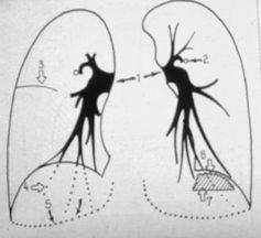
6 Right pulmonary artery Left pulmonary artery

7 Pulmonary vena Left auricle Left ventricle
8 L A L V

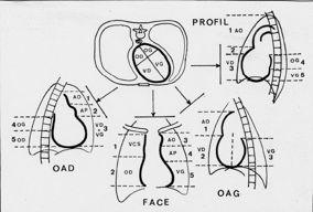
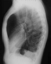
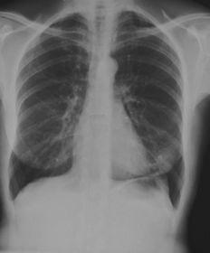
9 Estimate of cardiac volume: measure of the cardio-thoracic index: C/T
10 SVC AO PA AO RA LV RV PA LA LV Front view Lateral view LA RA LV RV


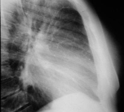
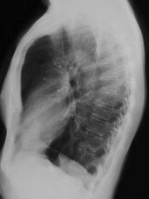
11 Hypertrophy of the right ventricle on the front view the RV pushes back the LV, and on the lateral view the RV fills the retro-sternal clear space
12 Hypertrophy of the right ventricle OFCP Normal chest x-ray
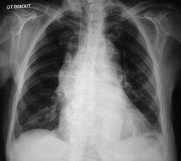
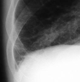
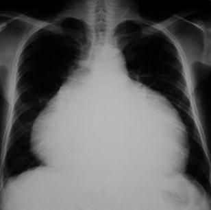
13 Left ventricle hypertrophy Increase of the inferior left arch. The cardiac apex sinks in the left diaphragm Increase of cardio-thoracic index

14 Left ventricle hypertrophy Normal chest x-ray


15 Left ventricle hypertrophy Normal chest x-ray

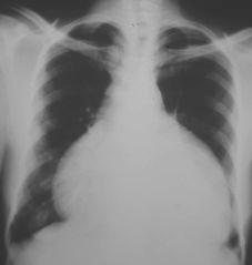
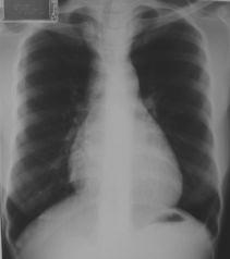
16 Hypertrophy of right auricle

17 thrombo-embolic disease with chronic pulmonary heart

18 Hypertrophy of right auricle OFCP Normal chest x-ray

19 Hypertrophy of the left auricle


20 Hypertrophy of the left auricle(rm) (courtesy of Dr. Anthoine)
21 Mitral disease. Notice the hypertrophy of the left middle arch, Hypertrophy of the LV. Hypertension of the pulmonary artery post capillar type.

22 Left auricle (Mitral stenosis)
23
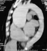
24 Ascending aorta dilatation (aneurysm)
25
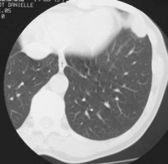
26 Pulmonary oedema Pulmonary oedema is the consequence of an abnormal accumulation of extra-vascular liquid, more or less rapidly, initially in the interstitial spaces (intersticial oedema), then in the alveolar cavities (alveolar oedema and acute pulmonary oedema).
27 Pulmonary oedema One can distinguish 2 physiopathological mechanisms: Hemodynamic oedema: cardiac origin, consequence of a left cardiac failure, with pulmonary arterial hypertension of post-capillar type. Usually the situation is reversible with adapted treatment (02, diuretic, TNT...). There is no anatomic lesion of the «alveolar-capillary barrier» Lesional oedema: diverse etiologies: infectious (viral or bacterial), toxic (inhalation of toxic gas or ingestion of toxic substances), or complication from shock regardless of the etiology. The prognosis is very often dismal: irreversible hypoxemy. The «alveolar -capillary» is definitively altered. Recovery is sometimes possible with sequela (pulmonary fibrosis).

28 Hemodynamic pulmonary oedema
29 Pulmonary hypertension Chronic pulmonary heart Pre-capillary hypertension Cardiac lung Post-capillary hypertension VC RA RV Pre-capillary obstacle Pulmonary capillaries PV LA LV aorta Intersticial oedema if cap. pressure> 20 mm hg Alveolar oedema if cap. pressure >30 mm hg
30 Pulmonary oedema If the capillary pressure is > 20mmhg: the oedema is intersticial: A and B Kerley lines Blurred opacities around the hilus Sometimes blurred opacities around bronchi (ring shadow sign) Small pleural effusion Round glass attenuation

31 Intersticial edema Kerley B lines


32 Intersticial edema Kerley B lines

33 Blurred opacities around the hilus. The vascular limits are not clear. Intersticial oedema

34 Ring shadow sign Around the bronchus Intersticial edema

")
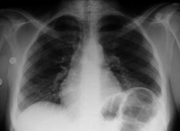
35 Mitral disease. Notice the hypertrophy of left superior arch (left auricle), left ventricle hypertrophy and intersticial oedema (ground glass attenuation) (hypertrophy and blurring of the hili).

36 Acute pulmonary oedema: alveolar oedema. The capillary pressure is > 30mmhg (butterfly wings picture)

37 Artery bronchus Pulmonary arterial hyperpression Pre-capillary type Pulmonary hyperpression Post-capillary type (ring shadow sign)

38 Post-capillary pulmonary hyperpressure
39 Pulmonary hypertension Chronic pulmonary heart Pre- capillary hypertension Cardiac lung Post-capillary hypertension VC RA RV Pre-capillary obstacle Pulmonary capillaries PV LA LV aorta Intersticial oedema if cap. pressure> 20 mm hg Alveolar oedema if cap. pressure >30 mm hg


40 Pre-capillary hypertension (emphysem) Chronic pulmonary heart
41 Pre-capillary hypertension
42
43 Pericarditis
44 Pericarditis After puncture
45

46 Pericarditis Cardiomegaly with hypertrophy of the left ventricle OFCP
47 Pulmonary embolism
48 Possible images of pulmonary embolism on the chest radiography: Nothing Pleural effusion Additional height of one hemi diaphragm Localised hypo-vascularisation Triangular picture with vertex toward the hilus (post-embolic pulmonary infarctus) Round picture (post-embolic pulmonary infarctus)
49 Don t forget: A normal chest radiography does not exclude Pulmonary embolism

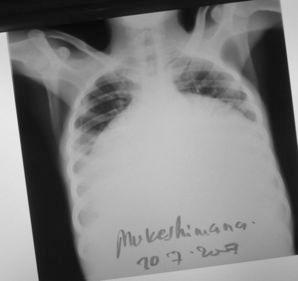
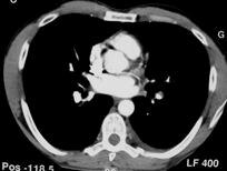
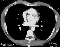
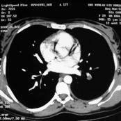
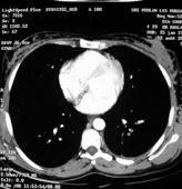
50 Male,45 years old, acute and severe dyspnea, thoracic pain. Tachycardia (110/mn), O2 Sa 89%.
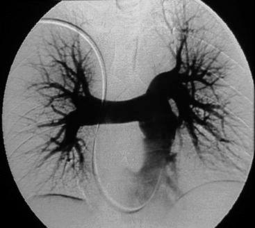
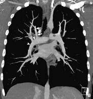
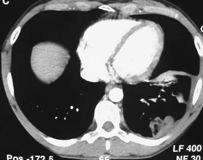
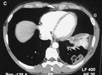
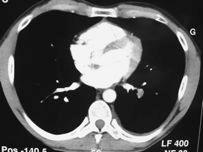
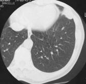
51 tomodensitometry Angioscanner

52 Angioscanner: severe and bilateral pulmonary embolism.


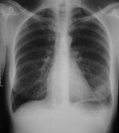
53 Additional height of the left diaphragm Additional height of the right diaphragm with small pleural effusion

54 Round pulmonary infarctus (decubitus position)


55 ! male, pain during deep inspiration! t C in evening. Initial treatment with amoxi./ Ac. Clav.! New chest x-ray 14 February 01!
56

57 Pulmonary embolism

.")
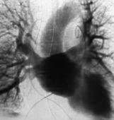
58 Female, 44 years old. Antecedent: phlebitis (familial deficiency in S protein). Pain in the left calf and acute dyspnea.

59 massive pulmonary embolism, in right inferior pulmonary artery


60 Female! flight Chicago-Paris! left thoracic pain! CXR in the emergency room! !
61 !
62 !


63 ! Pulmonary embolism


64 Woman,35 years old, thoracic pain left side, tachycardia and dyspnea. History of smoking 10 Pack-years. Has taken an oestro-progestative contraceptive for 5 years.
65
66
67 The diagnosis of pulmonary embolism is difficult without an angioscanner Indicative clinical context Indicative clinical signs (thoracic pain, tachycardia, acute dyspnea) and no other evident pathology (infection, cardiac disease.) Sometimes indicative ECG (acute pulmonary heart signs) Sometimes, but not always, the CXR may be indicative
Cardio-vascular syndrome. Etienne Leroy Terquem Pierre L Her SPI / ISP Soutien Pneumologique International / International Support for Pulmonology
 Cardio-vascular syndrome Etienne Leroy Terquem Pierre L Her SPI / ISP Soutien Pneumologique International / International Support for Pulmonology Left pulmonary artery Right pulmonary artery OFCP Left
Alveolar condensation syndrome
 Alveolar condensation syndrome Dr Etienne Leroy-Terquem Centre hospitalier de Meulan les Mureaux. France French-cambodian association for pneumology (OFCP) Lobule: morphological unit. Dimension: 10 to
Interstitial Syndrome Ground glass attenuation miliary and nodular images Linear images
 Interstitial Syndrome Ground glass attenuation miliary and nodular images Linear images Dr Etienne Leroy-Terquem Centre hospitalier de Meulan les Mureaux. France French-cambodian association for pneumology
Case N 1. Anterior thoracic paint with increasing dyspnea for few days. No cough. Decrease of cardiac sounds. Courtesy Dr Van den Homberg-Tanzania
 Case N 1 Anterior thoracic paint with increasing dyspnea for few days. No cough. Decrease of cardiac sounds Courtesy Dr Van den Homberg-Tanzania Case N 1 Enlargment of cardiac silhouette. Notice the symetry
Pleural syndrome. Tubercular pleurisy
 Pleural syndrome. Tubercular pleurisy Dr Etienne Leroy-Terquem Centre hospitalier de Meulan les Mureaux. France French-cambodian association for pneumology (OFCP) Pleurisy: Findings of fluid between visceral
Cardiac Radiography. Jared D. Christensen, M.D.
 Cardiac Radiography Jared D. Christensen, M.D. Cardiac radiography Jared D. Christensen, M.D. Overview Basic Concepts Technique Normal anatomy Cases Technique 3 Standard Views Posterior-Anterior (PA) Anterior-Posterior
Interstitial syndrome
 Interstitial syndrome Ground-glass attenuation Miliary and nodular images linear images Etienne Leroy Terquem Pierre L Her SPI / ISP Soutien Pneumologique International / International Support for Pulmonology
Bronchial syndrome. Atelectasis Draining bronchus Bronchiectasis
 Bronchial syndrome Atelectasis Draining bronchus Bronchiectasis Etienne Leroy Terquem Pierre L Her SPI / ISP Soutien Pneumologique International / International Support for Pulmonology Atelectasis Consequence
Key messages. CXR interpretation in TB/HIV setting. Training course
 Key messages CXR interpretation in TB/HIV setting Training course Normal CXR Front view and lateral view Good notions of technical conditions to obtain a good CXR Good knowledge of criteria for quality
Radiology of the respiratory/cardiac diseases (part 2)
") Cardiology Cycle - Lecture 6 436 Teams Radiology of the respiratory/cardiac diseases (part 2) Objectives Done By Team Leaders: Khalid Alshehri Hanin Bashaikh Team Members: Leena Alwakeel Aroob Alhuthail
Cardiology Cycle - Lecture 6 436 Teams Radiology of the respiratory/cardiac diseases (part 2) Objectives Done By Team Leaders: Khalid Alshehri Hanin Bashaikh Team Members: Leena Alwakeel Aroob Alhuthail
Pleural syndrome Tuberculous pleurisy
 Pleural syndrome Tuberculous pleurisy Etienne Leroy Terquem Pierre L Her SPI / ISP Soutien Pneumologique International / International Support for Pulmonology Pleural effusion: Findings of fluid between
Case 1. A 35-year-old male presented with fever, cough, and purulent sputum for one week. This was his CXR (Fig. 1.1). What is the diagnosis?
. What is the diagnosis?") 1 Interpreting Chest X-Rays CASE 1 Fig. 1.1 Case 1. A 35-year-old male presented with fever, cough, and purulent sputum for one week. This was his CXR (Fig. 1.1). What is the diagnosis? CASE 1 Interpreting
1 Interpreting Chest X-Rays CASE 1 Fig. 1.1 Case 1. A 35-year-old male presented with fever, cough, and purulent sputum for one week. This was his CXR (Fig. 1.1). What is the diagnosis? CASE 1 Interpreting
Echocardiography as a diagnostic and management tool in medical emergencies
 Echocardiography as a diagnostic and management tool in medical emergencies Frank van der Heusen MD Department of Anesthesia and perioperative Care UCSF Medical Center Objective of this presentation Indications
I particularly want to thank: -Pr Pierre L Her who has been my nearest partner in this adventure which began in Cambodia many years ago.
 Introduction and advices for users: CXR interpretation in TB/HIV high burden settings Clinical and radiological cases for training to positive and differential diagnosis Here are collected 144 clinical
The Chest X-ray for Cardiologists
 Mayo Clinic & British Cardiovascular Society at the Royal College of Physicians, London : 21-23-October 2013 Cases-Controversies-Updates 2013 The Chest X-ray for Cardiologists Michael Rubens Royal Brompton
Chest X-ray Interpretation
 Chest X-ray Interpretation Introduction Routinely obtained Pulmonary specialist consultation Inherent physical exam limitations Chest x-ray limitations Physical exam and chest x-ray provide compliment
Radiological syndroms. Alveolar syndrome Bronchial syndrome Interstitial syndrome Vascular syndrome Mediastinal Syndrome
 Radiological syndroms Alveolar syndrome Bronchial syndrome Interstitial syndrome Vascular syndrome Mediastinal Syndrome Alveolar syndrome Pulmonary architecture : Morphological unit is the lobule 15-25mm
Case N 1. Dyspnea and cough. Right thoracic paint. AFB in sputum not yet available. Courtesy Dr Van Den Homberg
 Case N 1 Dyspnea and cough. Right thoracic paint. AFB in sputum not yet available Courtesy Dr Van Den Homberg Case N 1 CXR: Right abundant pleural effusion. Notice the typical concave aspect of the superior
Pulmonary Embolism. Thoracic radiologist Helena Lauri
 Pulmonary Embolism Thoracic radiologist Helena Lauri 8.5.2017 Statistics 1-2 out of 1000 adults annually are diagnosed with deep vein thrombosis (DVT) and/or pulmonary embolism (PE) About half of patients
Interactive Lecture. Lecture 7 - Interactive. Radiology of cardiorespiratory disease. Editing File. Done By. Color Coding Important Notes Extra
 Lecture 7 - Interactive 436 Teams Interactive Lecture Radiology of cardiorespiratory disease Done By Team Leaders: Khalid Alshehri Hanin Bashaikh Team Members: Ghaida Alsaeed Maha Alissa Nawwaf AlHarbi
Undergraduate Teaching
 Prof. James F Meaney Undergraduate Teaching Chest X-Ray Understanding the normal anatomical by reference to cross sectional imaging Radiology? It s FUN! Cryptic puzzle Sudoku (Minecraft?) It s completely
Man 70 years old, chronic chonic exercice dyspnea, and past history of HTA. Acute and severe dyspnea, with non purulent sputum.
 Chapter 7 Man 70 years old, chronic chonic exercice dyspnea, and past history of HTA. Acute and severe dyspnea, with non purulent sputum. Auscultation: crepitant bilateral rales. Chest Xray: cardiomegaly
Radiological Anatomy of Thorax. Dr. Jamila Elmedany & Prof. Saeed Abuel Makarem
 Radiological Anatomy of Thorax Dr. Jamila Elmedany & Prof. Saeed Abuel Makarem Indications for Chest x - A chest x-ray may be used to diagnose and plan treatment for various conditions, including: Diseases/Fractures
HISTORY. Question: How do you interpret the patient s history? CHIEF COMPLAINT: Dyspnea of two days duration. PRESENT ILLNESS: 45-year-old man.
 HISTORY 45-year-old man. CHIEF COMPLAINT: Dyspnea of two days duration. PRESENT ILLNESS: His dyspnea began suddenly and has been associated with orthopnea, but no chest pain. For two months he has felt
Systemic lupus erythematosus (SLE): Pleuropulmonary Manifestations
: Pleuropulmonary Manifestations") 08/30/10 09/26/10 Systemic lupus erythematosus (SLE): Pleuropulmonary Manifestations Camila Downey S. Universidad de Chile, School of Medicine, Year VII Harvard University, School of Medicine Sept 17,
08/30/10 09/26/10 Systemic lupus erythematosus (SLE): Pleuropulmonary Manifestations Camila Downey S. Universidad de Chile, School of Medicine, Year VII Harvard University, School of Medicine Sept 17,
Tests Your Pulmonologist Might Order. Center For Cardiac Fitness Pulmonary Rehab Program The Miriam Hospital
 Tests Your Pulmonologist Might Order Center For Cardiac Fitness Pulmonary Rehab Program The Miriam Hospital BASIC ANATOMY OF THE LUNGS Lobes of Lung 3 lobes on the Right lung 2 lobes on the Left Blood
10/17/2016. Nuts and Bolts of Thoracic Radiology. Objectives. Techniques
 Nuts and Bolts of Thoracic Radiology October 20, 2016 Carleen Risaliti Objectives Understand the basics of chest radiograph Develop a system for interpreting chest radiographs Correctly identify thoracic
Restrictive Pulmonary Diseases
 Restrictive Pulmonary Diseases Causes: Acute alveolo-capillary sysfunction Interstitial disease Pleural disorders Chest wall disorders Neuromuscular disease Resistance Pathophysiology Reduced compliance
Interpreting thoracic x-ray of the supine immobile patient: Syllabus
 Interpreting thoracic x-ray of the supine immobile patient: Syllabus Johannes Godt Dep. of Radiology and Nuclear Medicine Oslo University Hospital Ullevål NORDTER 2017, Helsinki Content - Why bedside chest
Man, 65 years old, heavy smoker, cough, dyspnea and weight loss. AFB negative in sputum. Is TB possible?
 Chapter 10 Man, 65 years old, heavy smoker, cough, dyspnea and weight loss. AFB negative in sputum. Is TB possible? The opacity of the left upper lobe is not cavited and looks like a tissular mass, not
Pulmonary TB aspects
 Pulmonary TB aspects Nodule & infiltrate Cavern Pneumonia Etienne Leroy Terquem Pierre L Her SPI / ISP Soutien Pneumologique International / International Support for Pulmonology Nodules and infiltrates
Cardiac tamponade and Pericardiocentesis Made Easy
 Cardiac tamponade and Pericardiocentesis Made Easy www.cardiconcept.com Etiology of pericardial diseases. Non Infectious cause Infectious cause European Heart Journal (2015) 36, 2921 2964 Recommendations
J Somerville and V Grech. The chest x-ray in congenital heart disease 2. Images Paediatr Cardiol Jan-Mar; 12(1): 1 8.
: 1 8.") IMAGES in PAEDIATRIC CARDIOLOGY Images Paediatr Cardiol. 2010 PMCID: PMC3228330 The chest x-ray in congenital heart disease 2 J Somerville and V Grech Paediatric Department, Mater Dei Hospital, Malta Corresponding
IMAGES in PAEDIATRIC CARDIOLOGY Images Paediatr Cardiol. 2010 PMCID: PMC3228330 The chest x-ray in congenital heart disease 2 J Somerville and V Grech Paediatric Department, Mater Dei Hospital, Malta Corresponding
Human Anatomy and Physiology Chapter 19 Worksheet 1- The Heart
 Human Anatomy and Physiology Chapter 19 Worksheet 1- The Heart Name Date Period 1. The "double pump" function of the heart includes the right side, which serves as the circuit pump, while the left side
Chapter 2 Cardiac Interpretation of Pediatric Chest X-Ray
 Chapter 2 Cardiac Interpretation of Pediatric Chest X-Ray Ra-id Abdulla and Douglas M. Luxenberg Key Facts The cardiac silhouette occupies 50 55% of the chest width on an anterior posterior chest X-ray
ERDHEIM-CHESTER DISEASE LUNG & HEART ISSUES
 ERDHEIM-CHESTER DISEASE LUNG & HEART ISSUES GIULIO CAVALLI, M.D. INTERNAL MEDICINE AND CLINICAL IMMUNOLOGY IRCCS SAN RAFFAELE HOSPITAL VITA-SALUTE SAN RAFFAELE UNIVERSITY MILAN, ITALY cavalli.giulio@hsr.it
X-Rays. Prepared by Prof.Dr. Magda Hassab Allah Assist.lecturer Marwa Al Hady
 X-Rays Prepared by Prof.Dr. Magda Hassab Allah Assist.lecturer Marwa Al Hady CHEST X-RAYS Normal Chest X-ray Comments on chest X ray includes examination of 1- Bony cage (ribs,clavicles &vertebral column
ARIC HEART FAILURE HOSPITAL RECORD ABSTRACTION FORM. General Instructions: ID NUMBER: FORM NAME: H F A DATE: 10/13/2017 VERSION: CONTACT YEAR NUMBER:
 ARIC HEART FAILURE HOSPITAL RECORD ABSTRACTION FORM General Instructions: The Heart Failure Hospital Record Abstraction Form is completed for all heart failure-eligible cohort hospitalizations. Refer to
B-I-2 CARDIAC AND VASCULAR RADIOLOGY
 (YEARS 1 3) CURRICULUM FOR RADIOLOGY 13 B-I-2 CARDIAC AND VASCULAR RADIOLOGY KNOWLEDGE To describe the normal anatomy of the heart and vessels including the lymphatic system as demonstrated by radiographs,
Sheet lab 5 Anatomy: CT Scans
 Sheet lab 5 Anatomy: CT Scans In the orientation we see the picture from downward to upward. The first picture is a CT scan at the level of the heart. Left border of the heart is the left ventricle and
ECG Workshop. Nezar Amir
 ECG Workshop Nezar Amir Myocardial Ischemia ECG Infarct ECG in STEMI is dynamic & evolving Common causes of ST shift Infarct Localisation Left main artery occlusion: o diffuse ST-depression with ST elevation
Unilateral pulmonary oedema, a forgotten presentation.
 Unilateral pulmonary oedema, a forgotten presentation. Poster No.: C-2146 Congress: ECR 2018 Type: Educational Exhibit Authors: C. A. Arboleda Vallejo, M. I. carvajal, M. Perez ; Medellin, 1 2 1 1 2 Antioquia/CO,
Chest Radiology Interpretation: Findings of Tuberculosis
 Chest Radiology Interpretation: Findings of Tuberculosis Get out your laptops, smart phones or other devices pollev.com/chestradiology Case #1 1 Plombage Pneumonia Cancer 2 Reading the TB CXR Be systematic!
CHAPTER X - SECONDARY PULMONARY HYPERTENSION CHRONIC PULMONARY THROMBOEMBOLISM (HTP). PULMONARY THROMBENDARTERECTOMY
. PULMONARY THROMBENDARTERECTOMY") CHAPTER X - SECONDARY PULMONARY HYPERTENSION CHRONIC PULMONARY THROMBOEMBOLISM (HTP). PULMONARY THROMBENDARTERECTOMY Walter KLEPETKO, PhD, VIENNA - AUSTRIA Marian GASPAR, PhD, TIMISOARA 10. 1. Definition.
CHAPTER X - SECONDARY PULMONARY HYPERTENSION CHRONIC PULMONARY THROMBOEMBOLISM (HTP). PULMONARY THROMBENDARTERECTOMY Walter KLEPETKO, PhD, VIENNA - AUSTRIA Marian GASPAR, PhD, TIMISOARA 10. 1. Definition.
THE GOOFY ANATOMIST QUIZZES
 THE GOOFY ANATOMIST QUIZZES 7. LUNGS Q1. Fill in the blanks: the lung has lobes and fissures. A. Right, three, two. B. Right, two, one. C. Left, three, two. D. Left, two, three. Q2. The base of the lung
Chest X-ray (CXR) Interpretation Brent Burbridge, MD, FRCPC
 Interpretation Brent Burbridge, MD, FRCPC") Chest X-ray (CXR) Interpretation Brent Burbridge, MD, FRCPC An approach to reviewing a chest x-ray will create a foundation that will facilitate the detection of abnormalities. You should create your own
Chest X-ray (CXR) Interpretation Brent Burbridge, MD, FRCPC An approach to reviewing a chest x-ray will create a foundation that will facilitate the detection of abnormalities. You should create your own
Circulation and Respiration
 Circulation and Respiration Miller, Geoffrey. 2009. Spent: Sex, Evolution, and Consumer Behavior. Miller, Geoffrey. 2009. Spent: Sex, Evolution, and Consumer Behavior. Respiratory system moves O 2 into,
Pulmonary-Vascular Disease. Howard J. Sachs, MD.
 Pulmonary-Vascular Disease Howard J. Sachs, MD www.12daysinmarch.com Dyspnea Cardiac Pulmonary CAD Pump Failure Chest Wall Airways Valve Disease Pericardial Disease Alveoli Interstitium Rhythm Disturbance
PATENT DUCTUS ARTERIOSUS (PDA)
") PATENT DUCTUS ARTERIOSUS (PDA) It is a channel that connect the pulmonary artery with the descending aorta (isthumus part). It results from the persistence of patency of the fetal ductus arteriosus after
PATENT DUCTUS ARTERIOSUS (PDA) It is a channel that connect the pulmonary artery with the descending aorta (isthumus part). It results from the persistence of patency of the fetal ductus arteriosus after
Semiology of the Heart in the 21 st century
 Semiology of the Heart in the 21 st century Workshop Rodrigo Salgado Dept of Radiology Antwerp University Hospital - Belgium Question The cardiothoracic index a. Is something I always mention, because
cardiac imaging planes planning basic cardiac & aortic views for MR
 cardiac imaging planes planning basic cardiac & aortic views for MR Dianna M. E. Bardo, M. D. Assistant Professor of Radiology & Cardiovascular Medicine Director of Cardiac Imaging cardiac imaging planes
ARDS - a must know. Page 1 of 14
 ARDS - a must know Poster No.: C-1683 Congress: ECR 2016 Type: Authors: Keywords: DOI: Educational Exhibit M. Cristian; Turda/RO Education and training, Edema, Acute, Localisation, Education, Digital radiography,
October 2017 Pulmonary Embolism
 October 2017 Pulmonary Embolism Prof. Ahmed BaHammam, FRCP, FCCP Professor of Medicine College of Medicine King Saud University 1 Objectives Epidemiology Pathophysiology Diagnosis Massive PE Treatment
The External Anatomy of the Lungs. Prof Oluwadiya KS
 The External Anatomy of the Lungs Prof Oluwadiya KS www.oluwadiya.com Introduction The lungs are the vital organs of respiration Their main function is to oxygenate the blood by bringing inspired air into
Cardiac MRI: Clinical Application to Disease
 Cardiac MRI: Clinical Application to Disease Jessi Smith, MD Cardiothoracic imaging, Indiana University Slides courtesy of Stacy Rissing, MD Outline Imaging planes Disease findings Pulse sequences used
Cor pulmonale. Dr hamid reza javadi
 1 Cor pulmonale Dr hamid reza javadi 2 Definition Cor pulmonale ;pulmonary heart disease; is defined as dilation and hypertrophy of the right ventricle (RV) in response to diseases of the pulmonary vasculature
Pulmonary Hypertension: Another Use for Viagra
 Pulmonary Hypertension: Another Use for Viagra Kathleen Tong, MD Director, Heart Failure Program Assistant Clinical Professor University of California, Davis Disclosures I have no financial conflicts A
UERMMMC Department of Radiology. Basic Chest Radiology
 UERMMMC Department of Radiology Basic Chest Radiology PHYSICS DENSITIES BONE SOFT TISSUES WATER FAT AIR TELEROENTGENOGRAM Criteria for an Ideal Chest Radiograph 1. Upright 2. Posteroanterior View 3. Full
Pulmonary arterial hypertension in childhood: an unsual presentation with fibrosing mediastinitis
 ACTA BIOMED 2014; VOL. 85, N. 1: 68-72 Mattioli 1885 C A S E R E P O R T Pulmonary arterial hypertension in childhood: an unsual presentation with fibrosing mediastinitis Elisabetta Teopompi 1, Bertrand
SCLERODERMA LUNG DISEASE: WHAT THE PATIENT SHOULD KNOW
 SCLERODERMA LUNG DISEASE: WHAT THE PATIENT SHOULD KNOW Lung disease can be a serious complication of scleroderma. The two most common types of lung disease in patients with scleroderma are interstitial
Pulmonary veno-occlusive disease
 Disclosure Objectives Pulmonary veno-occlusive disease Tilman Humpl The Hospital for Sick Children University of Toronto, Canada Advisor/Research Grants Actelion Pfizer Historical aspects Epidemiology/Genetics
Chest XRay interpretation INTERPRETATIONS Identifications: Name & Date Technical evaluation Basic Interpretations
 Chest XRay interpretation INTERPRETATIONS Identifications: Name & Date Technical evaluation Basic Interpretations TECHNICAL EVALUATION 1. Projection: AP/PA view To differentiate between AP & PA films,
The Cardiovascular System. Chapter 15. Cardiovascular System FYI. Cardiology Closed systemof the heart & blood vessels. Functions
 Chapter 15 Cardiovascular System FYI The heart pumps 7,000 liters (4000 gallons) of blood through the body each day The heart contracts 2.5 billion times in an avg. lifetime The heart & all blood vessels
Anatomy. The respiratory system starts from the nose, mouth, larynx, trachea, and the two lungs.
 Respiratory System Anatomy The respiratory system starts from the nose, mouth, larynx, trachea, and the two lungs. Within the lungs, the bronchi transport air with oxygen to the alveoli on inspiration
Appendix to Gibson et al. Application of a decision rule and a D-dimer assay in the
 Appendix to Gibson et al. Application of a decision rule and a D-dimer assay in the diagnosis of pulmonary embolism (Thromb Haemost 2010; 103.4) Case 1 You are paged by an emergency room physician, who
Elderly Man with Dyspnoea
 Asia Pacific Family Medicine, 2004; 3 (1-2): 46-50 RADIOLOGY SERIES Elderly Man with Dyspnoea Wei-Yang LIM Faculty of Medicine National University of Singapore, Singapore Wilfred CG PEH Singapore Health
New murmur: acute valvular regurgitations. A.Pasquet, MD,PhD. UCL -Cliniques Saint Luc
 New murmur: acute valvular regurgitations. A.Pasquet, MD,PhD UCL -Cliniques Saint Luc Acute valvular regurgitation Clinical case Mr Dupont, a 53 y old men, without any particular medical history On Thursday
Shedding Light on Neonatal X-rays. Objectives. Indications for X-Rays 5/14/2018
 Shedding Light on Neonatal X-rays Barbara C. Mordue, MSN, NNP-BC Neonatal Nurse Practitioner LLUH Children s Hospital, NICU Objectives Utilize a systematic approach to neonatal x-ray interpretation Identify
Breakout Session: Transesophageal Echocardiography
 Breakout Session: Transesophageal Echocardiography Doris Ockert, MD Andrew Schroeder, MD University of Wisconsin School of Medicine and Public Health Jutta Novalija, MD, PhD Medical College of Wisconsin
Surgical repair of massive dilatation of the right atrium with tricuspid regurgitation
 Okada et al. Journal of Cardiothoracic Surgery (2018) 13:83 https://doi.org/10.1186/s13019-018-0769-7 CASE REPORT Open Access Surgical repair of massive dilatation of the right atrium with tricuspid regurgitation
Alexander A Schult, M.D., FCCP. October 21, 2017 Revised 1/10/18
 Alexander A Schult, M.D., FCCP October 21, 2017 Revised 1/10/18 Identifying normal anatomy Identifying various pathologic states Identifying placement of hardware Identifying limitations of portable CXR
The Dilated Pulmonary Artery: Is there a risk of Dissection?
 The Dilated Pulmonary Artery: Is there a risk of Dissection? Pastora Gallego, MD, PhD Inter-center Adult Congenital Heart Disease Unit Area del Corazón Seville, Spain No conflict of interest to disclose
Acute Coronary Syndromes Unstable Angina Non ST segment Elevation MI (NSTEMI) ST segment Elevation MI (STEMI)
 ST segment Elevation MI (STEMI)") Leanna R. Miller, RN, MN, CCRN-CSC, PCCN-CMC, CEN, CNRN, CMSRN, NP Education Specialist LRM Consulting Nashville, TN Objectives Evaluate common abnormalities that mimic myocardial infarction. Identify
Leanna R. Miller, RN, MN, CCRN-CSC, PCCN-CMC, CEN, CNRN, CMSRN, NP Education Specialist LRM Consulting Nashville, TN Objectives Evaluate common abnormalities that mimic myocardial infarction. Identify
Case 1 Organ Set 3. Case 1 (for Organ Sets 1 3) 10/2/2015 CARIOVASCULAR II LABORATORY
 10/2/2015 CARIOVASCULAR II LABORATORY") MHD I CRIOVSCULR II LORTORY 0/5/5 Case Organ Set Organ Set 2 Organ Set 3 Case (for Organ Sets 3) 72 year old man with a history of diabetes mellitus type 2, HTN, and hyperlipidemia presents with progressive
MHD I CRIOVSCULR II LORTORY 0/5/5 Case Organ Set Organ Set 2 Organ Set 3 Case (for Organ Sets 3) 72 year old man with a history of diabetes mellitus type 2, HTN, and hyperlipidemia presents with progressive
Cardiac MRI: Clinical Application to Disease
 Cardiac MRI: Clinical Application to Disease Stacy Rissing, MD! Cardiothoracic imaging, Indiana University! Outline Imaging planes Disease findings Pulse sequences used for each indication Pathophysiology
Chest X rays and Case Studies. No disclosures. Outline 5/31/2018. Carlo Manalo, M.D. Department of Radiology Loma Linda University Children s Hospital
 Chest X rays and Case Studies Carlo Manalo, M.D. Department of Radiology Loma Linda University Children s Hospital No disclosures. Outline Importance of history Densities delineated on radiography An approach
ACTIVITY 9: BLOOD AND HEART BLOOD
 ACTIVITY 9: BLOOD AND HEART OBJECTIVES: 1) How to get ready: Read Chapters 21 & 22, McKinley et al., Human Anatomy, 4e. All text references are for this textbook. Read dissection instructions BEFORE YOU
Chapter 38: Pulmonary Circulation, Pulmonary Edema, Pleural Fluid UNIT VII. Slides by Robert L. Hester, PhD
 UNIT VII Chapter 38: Pulmonary Circulation, Pulmonary Edema, Pleural Fluid Slides by Robert L. Hester, PhD Objectives Describe the pulmonary circulation Describe the pulmonary blood pressures List the
Can SCMR CMR protocol recommendations
 Can SCMR CMR protocol recommendations V1.3 - April 2009 CanSCMR CMR Protocol and SOP Recommendation 2009 (15 minutes) 2 Planning of LV fct. real time multiple axes Realtime 3 cine long axis 6 long axes
ACUTE RESPIRATORY DISTRESS SYNDROME
 ACUTE RESPIRATORY DISTRESS SYNDROME Angel Coz MD, FCCP, DCE Assistant Professor of Medicine UCSF Fresno November 4, 2017 No disclosures OBJECTIVES Identify current trends and risk factors of ARDS Describe
PATIENT CHARACTERISTICS AND PREOPERATIVE DATA (ecrf 1).
.") PATIENT CHARACTERISTICS AND PREOPERATIVE DATA (ecrf 1). 1 Inform Consent Date: / / dd / Mmm / yyyy 2 Patient identifier: Please enter the 6 digit Patient identification number from your site patient log
PATIENT CHARACTERISTICS AND PREOPERATIVE DATA (ecrf 1). 1 Inform Consent Date: / / dd / Mmm / yyyy 2 Patient identifier: Please enter the 6 digit Patient identification number from your site patient log
CT Chest. Verification of an opacity seen on the straight chest X ray
 CT Chest Indications: To assess equivocal plain x-ray findings Staging of lung neoplasm Merastatic workup of extra thoraces malignancies Diagnosis of diffuse lung diseases with HRCT Assessment of bronchietasis
ISUOG Basic Training. Obtaining & Interpreting Heart Views Correctly Alfred Abuhamad, USA. Basic training. Editable text here
 ISUOG Basic Training Obtaining & Interpreting Heart Views Correctly Alfred Abuhamad, USA Learning Objectives 6, 7 & 8 At the end of the lecture you will be able to: describe how to assess cardiac situs
What Is Valvular Heart Disease? Heart valve disease occurs when your heart's valves do not work the way they should.
 What Is Valvular Heart Disease? Heart valve disease occurs when your heart's valves do not work the way they should. How Do Heart Valves Work? MAINTAIN ONE-WAY BLOOD FLOW THROUGH YOUR HEART The four heart
Dr. Rami M. Adil Al-Hayali Assistant Professor in Medicine
 Dr. Rami M. Adil Al-Hayali Assistant Professor in Medicine Venous thromboembolism: pulmonary embolism (PE) deep vein thrombosis (DVT) 1% of all patients admitted to hospital 5% of in-hospital mortality
Heart Failure Dr ahmed almutairi Assistant professor internal medicin dept
 Heart Failure Dr ahmed almutairi Assistant professor internal medicin dept (MBBS)(SBMD) Introduction Epidemiology Pathophysiology diastolic/systolic Risk factors Signs and symptoms Classification of HF
Effects of mechanical ventilation on organ function. Masterclass ICU nurses
 Effects of mechanical ventilation on organ function Masterclass ICU nurses Case Male, 60 - No PMH - L 1.74 m and W 85 kg Pneumococcal pneumonia Stable hemodynamics - No AKI MV in prone position (PEEP 16
Thorax Lecture 2 Thoracic cavity.
 Thorax Lecture 2 Thoracic cavity. Spring 2016 Dr. Maher Hadidi, University of Jordan 1 Enclosed by the thoracic wall. Extends between (thoracic inlet) & (thoracic outlet). Thoracic inlet At root of the
Corso Base di Cardiologia Unisvet 2012
 Principi di Radiologia del torace Dr Luca Ferasin DVM PhD CertVC PGCert(HE) DipECVIM-CA (Cardiology) MRCVS European and RCVS Recognised Specialist in Veterinary Cardiology Introduction Thoracic radiography
Cath Lab Essentials: Basic Hemodynamics for the Cath Lab and ICU
 Cath Lab Essentials: Basic Hemodynamics for the Cath Lab and ICU Ailin Barseghian El-Farra, MD, FACC Assistant Professor, Interventional Cardiology University of California, Irvine Department of Cardiology
Level 2 Anatomy and Physiology Bite size revision. Respiratory System. The order of the passage of oxygen into the body (inhalation/inspiration) is: -
 is: -") Respiratory System Function of the respiratory system The lungs are located in the chest cavity and their function is to intake oxygen and remove carbon dioxide from the body. Structure of the respiratory
Respiratory System Function of the respiratory system The lungs are located in the chest cavity and their function is to intake oxygen and remove carbon dioxide from the body. Structure of the respiratory
Cardiac Pathology & Rehabilitation
 Cardiac Pathology & Rehabilitation Which of the following best describes the physical activity performed in my leisure time? A. I perform vigorous physical activity 3X/week for 20 minutes each time B.
Heart Valve disease: MR. AS tough patient When to echo, When to refer, What s new
 Heart Valve disease: MR. AS tough patient When to echo, When to refer, What s new B. Sonnenberg UAH Cardiology CME Day 5 May 2015 Disclosures Speaker s or Advisory Boards: none Research grants: none (co-investigator
A 50-year-old woman with syncope
 Hira Shahzad 1, Ali Bin Sarwar Zubairi 2 1 Medical College, Aga Khan University Hospital, Karachi 2 Department of Medicine, Aga Khan University Hospital, Karachi, Pakistan Ali Bin Sarwar Zubairi Associate
Lung ultrasound in the critically ill patient Pleural Effusions
 Lung ultrasound in the critically ill patient Pleural Effusions Rohit Patel, MD University of Florida Health Director, Critical Care Ultrasound Surgical ICU Center for Intensive Care Gainesville, Florida
Large veins of the thorax Brachiocephalic veins
 Large veins of the thorax Brachiocephalic veins Right brachiocephalic vein: formed at the root of the neck by the union of the right subclavian & the right internal jugular veins. Left brachiocephalic
Mediastinum It is a thick movable partition between the two pleural sacs & lungs. It contains all the structures which lie
 Dr Jamila EL medany OBJECTIVES At the end of the lecture, students should be able to: Define the Mediastinum. Differentiate between the divisions of the mediastinum. List the boundaries and contents of
FUNDAMENTALS OF CXR INTERPRETATION THE BASICS
 FUNDAMENTALS OF CXR INTERPRETATION THE BASICS PART I QUALITY ASSESSMENT 1 PATIENT-DEPENDENT FACTORS 3 REVIEW OF IMPORTANT ANATOMY 7 LUNGS AND PLEURA 11 DIAPHRAGMS 13 BONES AND SOFT TISSUES 14 A BRIEF LOOK
Cardiovascular manifestations of HIV
 Cardiovascular manifestations of HIV Prabhakar Rajiah, MBBS, MD, FRCR Associate Professor of Radiology Associate Director, Cardiac CT and MRI University of Texas Southwestern Medical Center, Dallas, USA
Assignable revenue codes: Explanation of services:
 computed tomography Chest/Cardiac Assignable revenue codes: Explanation of services: 0350 CT Scan General Classification 0351 CT Scan Head Scan 0352 CT Scan Body Scan 0359 CT Scan Other CT Scans Known