SKULL BASE AND. Bert De Foer A. Bernaerts, E. Loney +, J. Van Dinther, Th. Somers, E. Offeciers, A. Zarowski, J.W.Casselman / /* FACIAL TRAUMA
|
|
|
- Collin Wiggins
- 5 years ago
- Views:
Transcription


1 MANDIBULO- Bert De Foer A. Bernaerts, E. Loney +, J. Van Dinther, Th. Somers, E. Offeciers, A. Zarowski, J.W.Casselman / /* DEPARTMENT OF RADIOLOGY, GZA HOSPITALS SINT-AUGUSTINUS, ANTWERP, BELGIUM DEPARTMENT OF RADIOLOGY, AZ SINT-JAN AV, BRUGES, BELGIUM + COUNTY DURHAM AND DARLINGTON NHS FT MANDIBULO- DISCLOSURE OF CONFLICT OF INTEREST There are no potential conflicts of interest, relevant relationships or financial interests to report regarding this presentation. EUROPEAN INSTITUTE of ORL, GZA HOSPITALS SINT-AUGUSTINUS, ANTWERP, BELGIUM * UNIVERSITY OF GHENT, GHENT, BELGIUM EUROPEAN COURSE IN NEURORADIOLOGY DIAGNOSTIC AND INTERVENTIONAL 14 TH CYCLE, MODULE 4 TRAUMA, INFECTION AND DEGENERATIVE DISEASE 6 th -10 th MAY 2018 ANTWERP BELGIUM MANDIBULO- MANDIBULO- I. MANDIBULO- I. MANDIBULO- II. SKULL BASE / TEMPORAL BONE TRAUMA II. SKULL BASE / TEMPORAL BONE TRAUMA III. THE BEST OF THE REST III. THE BEST OF THE REST MANDIBULO- MANDIBULO- NASAL BONES : 30 G MANDIBULAR GENU: 40 G ZYGOMA: 50 G LOW G MANDIBULAR RAMI: 70 G Low G High G ETMOID: 80 G ALVEOLAR MARGINS: 100 G FRONTAL BONES: 200 G HIGH G Minor Injury 30% 32% Major Injury 21% 51% Mortality 2% 15% Maxillofacial Trauma. Luce et al Curr Probl Surg. 1984;21(2):1-68
 MOST USEFUL MODALITY IN : FAST + COMPLETE SUB-mm HELICAL NON CONTRAST BONE AND SOFT TISSUE SETTING MPR and 3D RECONSTRUCTION PLAIN FILMS EVALUATION OF")


2 MANDIBULO- MODALITIES CT (and not CBCT SOFT TISSUE VISUALISATION!!) MOST USEFUL MODALITY IN : FAST + COMPLETE SUB-mm HELICAL NON CONTRAST BONE AND SOFT TISSUE SETTING MPR and 3D RECONSTRUCTION PLAIN FILMS EVALUATION OF SIMPLE FRACTURES: NASAL BONES POOR IN COMPLEX INJURIES MANDIBULO- MODALITIES MRI NOT USED IN THE ACUTE SETTING COMPLEMENTARY IN UNEXPLAINED POST-TRAUMATIC CRANIAL NERVE DEFICITS EVALUATION OF INTRACRANIAL INJURY AND DIFFUSE AXONAL INJURIES MANDIBULO- THE PRINCIPLE OF FACIAL BUTTRESSES MANDIBULO- THE PRINCIPLE OF FACIAL BUTTRESSES: WHY DO BUTTRESSES MATTER? ALL LINKED DIRECTLY OR VIA ANOTHER BUTTRESS TO THE SKULL BASE Transverse Buttresses TRANSVERSE REDUCTION RESTORES FACIAL PROFILE VERTICAL REDUCTION RESTORES FACIAL HEIGHT REDUCTION ESTABLISHES FUNCTIONAL SUPPORT FOR TEETH AND GLOBES Vertical Buttresses MANDIBULO- WHERE DO WE START?? MANDIBULO- WHERE DO WE START?? STEP 1 : ARE THE PTERYGOID PLATES INVOLVED?


")


3 MANDIBULO- WHERE ARE THE PTERYGOID PLATES?? MANDIBULO- THE LE FORT FRACTURES R NAMED AFTER A FRENCH SURGEON: RENE LE FORT ( ) THREE DISTINCT INCT SUBTYPES S NO PTERYGOID PLATE FRACTURE = NO LE FORT FRACTURE MULTIPLE LE FORT FRACTURE PATTERNS CAN OCCUR AT THE SAME TIME DIFFERENT COMBINATIONS IONS CAN OCCUR ON THE TWO SIDES OF THE FACE MANDIBULO- MANDIBULO- THE LE FORT FRACTURES TYPE 1 : FLOATING TEETH / PALATE TYPE 2: FLOATING MAXILLA TYPE 3: FLOATING FACE LE FORT TYPE 1 : FLOATING TEETH / PALATE MANDIBULO- MANDIBULO- Male, 57-years-old Drunk, felt down when trying to get on his bicycle, leaving the pub to go home. LE FORT TYPE 2 : FLOATING MAXILLA PYRAMIDAL FRACTURE


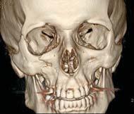
4 MANDIBULO- MANDIBULO- Male, 42-years-old Construction worker, Fall through a roof, 5 meters deep, face down. LE FORT TYPE 3 : FLOATING FACE MANDIBULO- MANDIBULO- STEP 2 : MEDIAL OR LATERAL BUTTRESS? NASO-ORBITO-ETHMOIDAL COMPLEX (NOE) OR ZYGOMATICOMAXILLARY COMPLEX (ZMC) NASO-ORBITO-ETHMOIDAL FRACTURE MANDIBULO- NASO-ORBITO-ETHMOIDAL FRACTURE CENTRAL UPPER MIDFACE FRACTURE INVOLVING NOSE, ETHMOIDS AND MEDIAL ORBITAL WALLS. WHAT TO REPORT ON CT DEGREE OF COMMINUTION OF MEDIAL MAXILLARY BUTTRESS ESPECIALLY THE LACRIMAL FOSSA DISTANCE BETWEEN THE LACRIMAL FOSSAE DISRUPTION OF NASOFRONTAL DUCTS (PREDISPOSES TO FRONTAL MUCOCELES) EVENTUAL FRONTAL LOBE CONTUSION MANDIBULO- NASO-ORBITO-ETHMOIDAL FRACTURE : COMPLICATIONS TELECANTHUS - HYPERTELORISM ENOPHTHALMOS MEDIAL CANTHAL ABNORMALITIES FORESHORTENED NOSE GLOBE DISPLACEMENT NASAL AIRWAY OBSTRUCTION LACRIMAL OBSTRUCTION


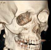
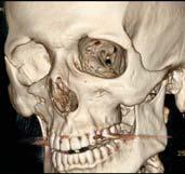
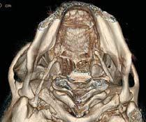
5 LO LO Male, 25-years-old Ô Stuck with the wheels of his bicycle in a tram track, lost his balance, felt on the right side of his face. Ô Ï Ð II I III Ï I LOFACIAL TRAUMA III II LO Ð Ô Ô Ò Ó Î Î Î Ï Ó ZYGOMATICOMAXILLARY COMPLEX FRACTURE (TRIPOD) FRACTURE I II Ð III LO LO ZYGOMATICOMAXILLARY COMPLEX (ZMC) FRACTURES PREVIOUSLY CALLED TRIMALAR OR TRIPOD FRACTURE FRACTURE COMPLEX WITH FRACTURE LINES INVOLVING ZYGOMATIC ARCH, LATERAL ORBITAL WALL, ANTERIOR AND LATERAL WALLS OF MAXILLARY SINUS AND ORBITAL FLOOR INFERIOR ORBITAL NERVE OFTEN INVOLVED RELATION TO CORONOID PROCESS OF MANDIBLE IS TO BE REPORTED


6 MANDIBULO- MANDIBULO- MANDIBULAR FRACTURES STUDIES VARY FROM 36-70% OF ALL MAXILLO-FACIAL FRACTURES. M > F STEP 3 : IF NONE OF THE FORMER IS THIS AN ISOLATED FRACTURE? NUMBER OF FRACTURES PER PATIENT 1 FRACTURE: 50 % 2 FRACTURES: 40% >2 FRACTURES: 10% 15% HAVE ANOTHER FACIAL FRACTURE RELATIONSHIP TO THE MANDIBULAR CANAL AND NERVE A MANDIBULE USUALLY NEVER FRACTURES ON ONE PLACE! MANDIBULO- MANDIBULO- Male, 23-years-old During sports, direct fall on his chin MANDIBULO- MANDIBULO- TYPES Symphyseal Parasymphyseal Body Ramus Angle Condylar Coronoid Alveolar DISPLACEMENT DUE TO MUSCLE PULL OF MEDIAL AND LATERAL PTERYGOID


7 LO LO 38-year old male Shot by his neighbour in the face after having had a discussion on loud music during a barbecue party. LOFACIAL TRAUMA FACIAL TRAU T RAUM UMA LO 38-year old male Rollover with his mountain bike. Hit the steering wheel with his teeth. LO LO


8 MANDIBULO- DENTOALVEOLAR FRACTURES: ELLIS AND DAVEY CLASSIFICATION I FRACTURE WITHIN ENAMEL II FRACTURE OF ENAMEL AND DENTINE III FRACTURE INVOLVING PULP IV TOOTH BECOMES NON-VITAL V AVULSION VI ROOT FRACTURE VII TOOTH DISPLACEMENT WITH NO FRACTURE IX CROWN EN MASSE FRACTURE AND AND ANN DISPLACEMENT X PRIMARY DENTITION INJURIES MANDIBULO- ORBITAL FRACTURES CAN BE ISOLATED OR ASSOCIATED WITH PREVIOUSLY DESCRIBED COMPLEXES. MANDIBULO- MANDIBULO- 2 young boys, involved in a fight. ORBITAL BLOW-OUT FRACTURE WITH DISPLACEMENT OF ORBITAL FLOOR AND DOWNWARDS HERNIATION OF ORBITAL FAT AND INFERIOR RECTUS MUSCLE MANDIBULO- MANDIBULO- ORBITAL FRACTURES CAN BE ISOLATED OR ASSOCIATED WITH PREVIOUSLY DESCRIBED COMPLEXES. BLOW-OUT FRACTURES LAMINA PAPYRACEA FRACTURES : MEDIAL RECTUS MUSCLE ORBITAL FLOOR FRACTURES : INFERIOR RECTUS MUSCLE FRACTURE OF LAMINA PAPYRACEA. NO ENTRAPMENT OF MEDIAL RECTUS MUSCLE


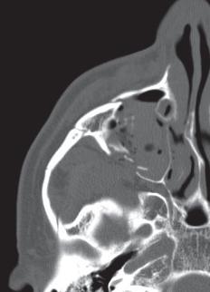
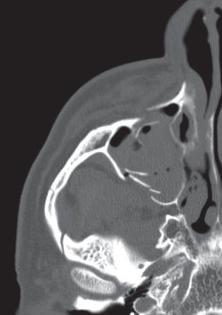
9 MANDIBULO- MANDIBULO- 3-year old female toddler fel from the stairs with a pencil in her mouth. The pencil perforated the hard palate. Parents withdraw the pencil before coming to the emergency department. MANDIBULO- MANDIBULO- Perforation of the hard palate and lamina papyracea with penetration of the orbital apex and optic nerve laceration and subsequent right-sided blindness. MANDIBULO- ORBITAL FRACTURES CAN BE ISOLATED OR ASSOCIATED WITH PREVIOUSLY DESCRIBED COMPLEXES. BLOW-OUT FRACTURES LAMINA PAPYRACEA FRACTURES : MEDIAL RECTUS MUSCLE ORBITAL FLOOR FRACTURES : INFERIOR RECTUS MUSCLE EXCLUDE FRACTURE FRAGMENT IMPINGEMENT LAMINA PAPYRACEA ON MEDIAL RECTAL MUSCLE ORBITAL APEX FRAGMENTS MANDIBULO- ORBITAL FRACTURES CAN BE ISOLATED OR ASSOCIATED WITH PREVIOUSLY DESCRIBED COMPLEXES. BLOW-OUT FRACTURES LAMINA PAPYRACEA FRACTURES : MEDIAL RECTUS MUSCLE ORBITAL FLOOR FRACTURES : INFERIOR RECTUS MUSCLE EXCLUDE FRACTURE FRAGMENT IMPINGEMENT LAMINA PAPYRACEA ON MEDIAL RECTAL MUSCLE ORBITAL APEX FRAGMENTS BEWARE OF DIPLOPIA


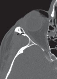
10 MANDIBULO- Lamina papyracea impinges on medial rectus. Muscle looks angulated. MANDIBULO- I. MANDIBULO- II. SKULL BASE / TEMPORAL BONE TRAUMA Medial wall fracture with enophthalmos. Medial rectus has lost flat profile and herniated into fracture site III. THE BEST OF THE REST MANDIBULO- MANDIBULO- 65-years-old female I. MANDIBULO- Fell down the stairs, drunk. II. SKULL BASE / TEMPORAL BONE TRAUMA III. THE BEST OF THE REST MANDIBULO- MANDIBULO- AX SE T1 - Gd AX RIGHT-SIDED CT TRANSVERSE FRACTURE LEFT-SIDED LONGITUDINAL FRACTURE WITH INVOLVEMENT OF THE OTIC CAPSULE AND INTRALABYRINTHINE AIR AX 3D FLAIR INTRALABYRINTHINE HEMORRAGHE ON THE RIGHT HEMOTYMPANUM ON THE LEFT


 OFTEN IMMEDIATE AND COMPLETE OSSICLES")


11 MANDIBULO- TRADITIONAL CLASSIFICATION : LONGITUDINAL T-BONE FRACTURES % CAUSED BY A TEMPOROPARIETAL IMPACT FORCES RUN PARALLEL TO THE LONG AXIS OF THE TEMPORAL BONE OSSICULAR AND TYMPANIC MEMBRANE INJURY FREQUENT WITH CONDUCTIVE HEARING LOSS FACIAL PARALYSIS RARE MANDIBULO- TRANSVERSE TEMPORAL BONE FRACTURES % CAUSED BY A FRONTAL/OCCIPITAL BLOW FORCES RUN PERPENDICULAR TO THE LONG AXIS OF THE TEMPORAL BONE LABYRINTH OFTEN INVOLVED WITH VERTIGO AND SNHL FACIAL PARALYSIS (40 50 %) OFTEN IMMEDIATE AND COMPLETE OSSICLES OFTEN SPARED (NO CHL) MANDIBULO- TRADITIONAL CLASSIFICATION : DRAWBACKS DOES NOT ADDRESS OBLIQUE or MIXED FRACTURES WHICH ARE MANDIBULO- 38-years-old male Left-sided deafness after a car crash. VERY FREQUENT DOES NOT CORRELATE WELL WITH CLINICAL OUTCOME / POTENTIAL COMPLICATIONS NEWER CLASSIFICATION: OTIC CAPSULE SPARING >< OTIC CAPSULE VIOLATING FRACTURES MANDIBULO- MANDIBULO- 42-years-old female LEFT-SIDED OTIC CAPSULE VIOLATING FRACTURE WITH AIR IN THE VESTIBULE Bilateral hearing loss after a car crash. Audiogram demonstrates bilateral conductive hearing loss.


12 MANDIBULO- MANDIBULO- 2 different patients with conductive hearing loss after a head trauma. BILATERAL AX CBCT OTIC CAPSULE SPARING FRACTURE WITH INCUDOMALLEAL SEPARATION MANDIBULO- DOUBLE OBLIQUE CT REFORMAT MANDIBULO- 47-years-old male Prior surgery with PORP placement on the left side. Severe left-sided conductive hearing loss after head trauma. DOUBLE OBLIQUE CBCT REFORMAT INCUDOSTAPEDIAL DISCONNECTION ON THE LEFT SIDE MANDIBULO- MANDIBULO- COMPLICATIONS OF TEMPORAL BONE TRAUMA: OSSICULAR INJURY OSSICULAR CHAIN DISRUPTION -> CONDUCTIVE HEARING LOSS REPEAT AUDIOGRAM AFTER RESORPTION OF BLOOD IN MIDDLE EAR PORP COR CBCT LUXATED OUT OF THE OVAL WINDOW STAPES LOST MALEUS ON TOP OF THE SCUTUM INCUS IN THE EAC DISLOCATION MUCH MORE FREQUENT THAN FRACTURE INCUDOSTAPEDIAL JOINT SEPARATION INCUDOMALLEAL JOINT SEPARATION INCUS DISLOCATION INCUDOMALLEAL COMPLEX DISLOCATION STAPEDIOVESTIBULAR DISLOCATION - > PERILYMPATHIC FISTULA


13 MANDIBULO- MANDIBULO- 25-years-old male Hearing loss after forcefull retraction of the index finger out of the external auditory canal. ISOLATED COR CBCT FRACTURE + SAG MIP OF MANUBRIUM MALEI REPAIRED USING BONECEMENT - OTOMIMIX MANDIBULO- COMPLICATIONS OF TEMPORAL BONE TRAUMA: OSSICULAR INJURY OSSICULAR CHAIN DISRUPTION -> CONDUCTIVE HEARING LOSS REPEAT AUDIOGRAM AFTER RESORPTION OF BLOOD IN MIDDLE EAR MANDIBULO- 26-years-old female Sudden onset of vertigo and conductive and sensorineural hearing loss after tympanic perforation with a scalp massage tool inserted by the patient s young child by accident DISLOCATION MUCH MORE FREQUENT THAN FRACTURE INCUDOSTAPEDIAL JOINT SEPARATION INCUDOMALLEAL JOINT SEPARATION INCUS DISLOCATION INCUDOMALLEAL COMPLEX DISLOCATION STAPEDIOVESTIBULAR DISLOCATION - > PERILYMPATHIC FISTULA ISOLATED OSSICULAR FRACTURE: MANUBRIUM MALEI MANDIBULO- MANDIBULO- + = AX CBCT


14 MANDIBULO- PROTRUSION IN THE VESTIBULE OF FOOTPLATE STAPES CAPITELLUM SURGERY WITH RETRACTION OF FOOTPLATE OUT OF VESTIBULE STAPES RECONSTRUCTION DISAPPEARANCE OF VERTIGO DURING THE DAYS FOLLOWING THE EVENT RECUPERATION OF 80% OF HEARING MANDIBULO- COMPLICATIONS OF TEMPORAL BONE TRAUMA: OSSICULAR INJURY OSSICULAR CHAIN DISRUPTION -> CONDUCTIVE HEARING LOSS REPEAT AUDIOGRAM AFTER RESORPTION OF BLOOD IN MIDDLE EAR DISLOCATION MUCH MORE FREQUENT THAN FRACTURE INCUDOSTAPEDIAL JOINT SEPARATION INCUDOMALLEAL JOINT SEPARATION INCUS DISLOCATION INCUDOMALLEAL COMPLEX DISLOCATION STAPEDIOVESTIBULAR DISLOCATION - > PERILYMPATHIC FISTULA ISOLATED OSSICULAR FRACTURE: MANUBRIUM MALEI FRACTURE OF STAPES: RARE PENETRATING INJURY MANDIBULO- MANDIBULO- 28-years-old male Severe conductive hearing loss after a head trauma with a left-sided temporal blow. AX CT + COR REFORMATS TEGMEN DEFECT WITH MENINGOCELE AND INCUDOMALLEAL SEPARATION MANDIBULO- COMPLICATIONS OF TEMPORAL BONE TRAUMA: CSF LEAKAGE % / TEGMEN INVOLVEMENT OTIC CAPSULE VIOLATING INJURY: 4x MORE FREQUENT MANDIBULO- 44-years-old male Fall out of a tree. Hearing loss with also an immediate left-sided facial nerve palsy. USUALLY WITHIN A WEEK AFTER TRAUMA CLINICALLY: CSF OTORRHEA or CSF RHINORRHEA RISK OF MENINGITIS IF LEAKAGE PERSISTS MORE THAN 1 WEEK MENINGOCELE / ENCEFALOCELE : LATE COMPLICATION


15 MANDIBULO- MANDIBULO- COMPLICATIONS OF TEMPORAL BONE TRAUMA: FACIAL NERVE INJURY TRANSSECTION: IMMEDIATE FACIAL PARALYSIS DELAYED FACIAL NERVE INJURY: EDEMA, HEMATOMA, CONTUSION AX CBCT LOW GRADE / INCOMPLETE PARALYSIS: WAIT AND SEE HIGH GRADE / COMPLETE PARALYSIS: AX 3D GRE T1 + Gd SAG REFORMAT CHECK FOR BONY FRAGMENTS INDICATION FOR IMMEDIATE DECOMPRESSIVE SURGERY OTIC CAPSULE SPARING FRACTURE WITH FACIAL NERVE THICKENING AND ENHANCEMENT. DUE TO HB GRADE 5 PALSY AND THE FINDINGS ON MRI, DECOMPRESSIVE SURGERY WAS PERFORMED MANDIBULO- MANDIBULO- I. MANDIBULO- I. MANDIBULO- II. SKULL BASE / TEMPORAL BONE TRAUMA II. SKULL BASE / TEMPORAL BONE TRAUMA III. THE BEST OF THE REST III. THE BEST OF THE REST MANDIBULO- MANDIBULO- Female, 34-years-old Recent episode of meningitis. Prior history of functional endoscopic sinus surgery (FESS). MENINGO-ENCEFALOCELE THROUGH CRIBRIFORM PLATE DEFECT.


16 MANDIBULO- MANDIBULO- Male, 25-years-old Anaesthesiologist request CT scan. Clinical information: unable to awake the patient after FESS MANDIBULO- MANDIBULO- Female, 47-years-old Maxillofacial surgeon plans sinus augmentation surgery prior to implant surgery. In the same session, he is also planning to treat the coincident sinusitis The day of the treatment, immediately after surgery, he request an urgent CT scan for right-sided facial swelling and visual loss PERFORATION OF THE POSTERIOR SPHENOID SINUS WALL WITH POSTERIOR COMMUNICANS ARTERY TRANSECTION AND SAH MANDIBULO- TAKE HOME MESSAGES NO LEFORT FRACTURE WITHOUT PTERYGOID PLATE FRACTURE I FLOATING TEETH / II FLOATING MAXILLA / III FLOATING FACE USUALLY MIXTURE OF DIFFERENT TYPES OF FRACTURES MANDIBULA: FRACTURES AT DIFFERENT PLACES ORBITAL FRACTURE: TOTAL PERFORATION RESOLUTION OF THE AFTER ORBITAL FLOOR ORBITAL WITH INTRACONAL FLOOR BLEEDING, RECONSTRUCTION PARTIAL HERNIATION AND OF THE RESORPTION INFERIOR RECTAL OF THE MUSCLE INTRACONAL AS WELL AS BLEEDING. INTRAORBITAL OPTIC FAT NERVE IN THE WAS DEFECT NOT TRANSSECTED NOR DAMAGED MEDIAL / INFERIOR RECTAL MUSCLE ORBITAL APEX INFRAORBITAL NERVE T BONE FRACTURE: OTIC CAPSULE SPARING OR NOT OSSICULAR DISLOCATION MORE FREQUENT THAN FRACTURE

17 MANDIBULO LO- THANK YOU FOR YOUR ATTENTION
CT of Maxillofacial Fracture Patterns. CT of Maxillofacial Fracture Patterns
 CT of Maxillofacial Fracture Patterns CT of Maxillofacial Fracture Patterns Stuart E. Mirvis, M.D., FACR Department of Radiology University of Maryland School of Medicine Viking 1 1976 MGS 2001 Technology
Core Curriculum Syllabus Emergencies in Otolaryngology-Head and Neck Surgery FACIAL FRACTURES
 Core Curriculum Syllabus Emergencies in Otolaryngology-Head and Neck Surgery A. General Considerations FACIAL FRACTURES Look for other fractures like skull and/or cervical spine fractures Test function
Facial and Temporal Bone Trauma Diagnostic imaging and therapeutic challenges in emergency
 Facial and Temporal Bone Trauma Diagnostic imaging and therapeutic challenges in emergency ATTYE A, KRAINIK A Department of Neuroradiology and MRI University Hospital Grenoble / University Grenoble Alpes
CT of Maxillofacial Injuries
 CT of Maxillofacial Injuries Stuart E. Mirvis, M.D., FACR Department of Radiology University of Maryland School of Medicine Viking 1 1976 MGS 2001 Technology changes the diagnosis Technologic Evolution
Maxillofacial Injuries Practical Tips
 Saturday, October 29, 2016 Maxillofacial Injuries Practical Tips Suyash Mohan MD, PDCC THE ROOTS OF PENN RADIOLOGY RADIOLOGICAL Assistant Professor of Radiology Assistant Professor of Neurosurgery Neuroradiology
Imaging Orbit/Periorbital Injury
 Imaging Orbit/Periorbital Injury 9 th Nordic Trauma Radiology Course 2016 Stuart E. Mirvis, M.D., FACR Department of Radiology University of Maryland School of Medicine Fireworks Topics to Cover Struts
North Oaks Trauma Symposium Friday, November 3, 2017
 + Evaluation and Management of Facial Trauma D Antoni Dennis, MD North Oaks ENT an Allergy November 3, 2017 + Financial Disclosure I do not have any conflicts of interest or financial interest to disclose
Thickened and thinner parts of the skull = important base for understanding of the functional structure of the skull - the transmission of masticatory
 Functional structure of the skull and Fractures of the skull Thickened and thinner parts of the skull = important base for understanding of the functional structure of the skull - the transmission of masticatory
Facial Trauma ASHNR. Disclosures: Acknowledgments: None. Edward P. Quigley, III, MD PhD University of Utah
 Disclosures: Facial Trauma ASHNR Edward P. Quigley, III, MD PhD University of Utah None Acknowledgments: Dr. Rebecca Cornelius Dr. Ilona M. Schmalfuss Dr. Richard Wiggins III Dr. Yoshimi Anzai Dr. Lindell
MAXILLOFACIAL TRAUMA. The on-call maxillofacial surgeons can be contacted through the switchboard at the Southern General Hospital
 MAXILLOFACIAL TRAUMA The on-call maxillofacial surgeons can be contacted through the switchboard at the Southern General Hospital Mandibular Injuries Mechanism of injury Assault, falls, RTA-Direct trauma
Computed-Tomography of maxillofacial fractures: What do surgeons want to know?
 Computed-Tomography of maxillofacial fractures: What do surgeons want to know? Poster No.: C-0968 Congress: ECR 2016 Type: Educational Exhibit Authors: A. Ammar, M. Jrad, I. KASRAOUI, A. Zoubli, H. Mizouni
McHenry Western Lake County EMS System Paramedic, EMT-B and PHRN Optional Continuing Education 2019 #1 Facial Trauma
 McHenry Western Lake County EMS System Paramedic, EMT-B and PHRN Optional Continuing Education 2019 #1 Facial Trauma The face is vital to human appearance and function. Facial injuries can impair a patient
TRAUMA TO THE FACE AND MOUTH
 Dr.Yahya A. Ali 3/10/2012 F.I.C.M.S TRAUMA TO THE FACE AND MOUTH Bailey & Love s 25 th edition Injuries to the orofacial region are common, but the majority are relatively minor in nature. A few are major
Dr. Esam Ahmad Z. Omar BDS, MSc-OMFS, FFDRCSI. Monitor the vital signs. Monitor the vital signs. Complications of Facial Traumas.
 Complications of Facial Traumas 1) Immediate Complications 2) Late Complications Dr. Esam Ahmad Z. Omar BDS, MSc-OMFS, FFDRCSI Assistant Professor Oral & Maxillofacial Surgeon Taibah University Monitor
Midface fractures; what the radiologist should know.
 Midface fractures; what the radiologist should know. Poster No.: C-1056 Congress: ECR 2013 Type: Educational Exhibit Authors: J. Garcia Villanego, E.-M. Heursen, A. Rodriguez Piñero; Cadiz/ES Keywords:
Infratemporal fossa: Tikrit University college of Dentistry Dr.Ban I.S. head & neck Anatomy 2 nd y.
 Infratemporal fossa: This is a space lying beneath the base of the skull between the lateral wall of the pharynx and the ramus of the mandible. It is also referred to as the parapharyngeal or lateral pharyngeal
Head and Neck Trauma. Disclosures: Acknowledgments: Introductory case. None
 Head and Neck Trauma None Disclosures: Edward P. Quigley III MD PhD Radiology and Imaging Sciences University of Utah Dr. Richard Wiggins III Dr. Yoshimi Anzai Dr. Lindell Gentry Dr. Blair Winegar Dr.
Major Anatomic Components of the Orbit
 Major Anatomic Components of the Orbit 1. Osseous Framework 2. Globe 3. Optic nerve and sheath 4. Extraocular muscles Bony Orbit Seven Bones Frontal bone Zygomatic bone Maxillary bone Ethmoid bone Sphenoid
Dr. Sami Zaqout, IUG Medical School
 The skull The skull is composed of several separate bones united at immobile joints called sutures. Exceptions? Frontal bone Occipital bone Vault Cranium Sphenoid bone Zygomatic bones Base Ethmoid bone
Maxillofacial and Ocular Injuries
 Maxillofacial and Ocular Injuries Objectives At the conclusion of this presentation the participant will be able to: Identify the key anatomical structures of the face and eye and the impact of force on
Diagnosis of Midface Fractures with CT: What the Surgeon Needs to Know 1
 Note: This copy is for your personal non-commercial use only. To order presentation-ready copies for distribution to your colleagues or clients, contact us at www.rsna.org/rsnarights. EDUCATION EXHIBIT
Oral and Maxillofacial Surgeons and the seriously injured patient. Barts and The London NHS Trust
 Oral and Maxillofacial Surgeons and the seriously injured patient Barts and The London NHS Trust How do you assess this? Primary Survey A B C D E Airway & Cervical Spine Breathing & Ventilation Circulation
Bones of the skull & face
 Bones of the skull & face Cranium= brain case or helmet Copyright The McGraw-Hill Companies, Inc. Permission required for reproduction or display. The cranium is composed of eight bones : frontal Occipital
Temporal fossa Infratemporal fossa Pterygopalatine fossa Terminal branches of external carotid artery Pterygoid venous plexus
 Outline of content Temporal fossa Infratemporal fossa Pterygopalatine fossa Terminal branches of external carotid artery Pterygoid venous plexus Boundary Content Communication Mandibular division of trigeminal
Older age, MVC and TBI higher incidence. Facial fractures a distracting injury? Carotid artery injury. Blindness may occur with facial fractures
 Dr Donald C. DeLisi Jr Oral & Maxillofacial Surgeon Multisystem injury 20 50% Nasal and mandibular fractures most common in community ED s Midface and zygomatic injuries most common in Trauma centers 25%
DR. SAAD AL-MUHAYAWI, M.D., FRCSC. ORL Head & Neck Surgery
 TRAUMA IN ORL DR. SAAD AL-MUHAYAWI, M.D., FRCSC Associate Professor & Consultant ORL Head & Neck Surgery TYPES OF TRAUMA EAR & TEMPORAL BONE TRAUMA NOSE & FACIAL BONES TRAUMA LARYNGEAL TRAUMA NECK TRAUMA
The orbit-1. Dr. Heba Kalbouneh Assistant Professor of Anatomy and Histology
 The orbit-1 Dr. Heba Kalbouneh Assistant Professor of Anatomy and Histology Orbital plate of frontal bone Orbital plate of ethmoid bone Lesser wing of sphenoid Greater wing of sphenoid Lacrimal bone Orbital
Chapter 7 Part A The Skeleton
 Chapter 7 Part A The Skeleton Why This Matters Understanding the anatomy of the skeleton enables you to anticipate problems such as pelvic dimensions that may affect labor and delivery The Skeleton The
RADIOLOGY TEACHING CONFERENCE
 RADIOLOGY TEACHING CONFERENCE John Athas, MD Monica Tadros, MD Columbia University, College of Physicians & Surgeons Department of Otolaryngology- Head & Neck Surgery September 27, 2007 CT SCAN IMAGING
Cranium Facial bones. Sternum Rib
 Figure 7.1 The human skeleton. Skull Thoracic cage (ribs and sternum) Cranium Facial bones Sternum Rib Bones of pectoral girdle Vertebral column Sacrum Vertebra Bones of pelvic girdle (a) Anterior view
Temporal region. temporal & infratemporal fossae. Zhou Hong Ying Dept. of Anatomy
 Temporal region temporal & infratemporal fossae Zhou Hong Ying Dept. of Anatomy Temporal region is divided by zygomatic arch into temporal & infratemporal fossae. Temporal Fossa Infratemporal fossa Temporal
Dr. Sami Zaqout Faculty of Medicine IUG
 Auricle External Ear External auditory meatus The Ear Middle Ear (Tympanic Cavity) Auditory ossicles Internal Ear (Labyrinth) Bony labyrinth Membranous labyrinth External Ear Auricle External auditory
Anatomy and Physiology. Bones, Sutures, Teeth, Processes and Foramina of the Human Skull
 Anatomy and Physiology Chapter 6 DRO Bones, Sutures, Teeth, Processes and Foramina of the Human Skull Name: Period: Bones of the Human Skull Bones of the Cranium: Frontal bone: forms the forehead and the
AXIAL SKELETON SKULL
 AXIAL SKELETON SKULL CRANIAL BONES (8 total flat bones w/ 2 paired) 1. Frontal forms forehead & upper portion of eyesocket (orbital) 2. Parietal paired bones; form superior & lateral walls of cranium 3.
The diagnostic value of Computed Tomography in evaluation of maxillofacial Trauma
 The diagnostic value of Computed Tomography in evaluation of maxillofacial Trauma Qais H. Muassa FICMS College of Dentistry, Babylon University Ibrahim S. Gataa, BDS, FICMS College of Dentistry, Sulaimania
Skeletal System -Axial System. Chapter 7 Part A
 Skeletal System -Axial System Chapter 7 Part A Skeleton Learn: Names of the s. Identify specific landmarks that allow: Bones to fit into each other, Organs to fit into the cavities, Muscles to attach,
MAXILLOFACIAL TRAUMATOLOGY Department of Maxillofacial Surgery Semmelweis University, Budapest. Dr. Huszár Tamás
 MAXILLOFACIAL TRAUMATOLOGY Department of Maxillofacial Surgery Semmelweis University, Budapest Dr. Huszár Tamás Maxillofacial injuries isolated maxillofacial injury multiple injuries polytrauma (injury
Epidemiology 3002). Epidemiology and Pathophysiology
. Epidemiology and Pathophysiology") Epidemiology Maxillofacial trauma or injuries are commonly encountered in the practice of emergency medicine and are presenting one of the most challenging problems to the attending surgeons or physicians
Epidemiology Maxillofacial trauma or injuries are commonly encountered in the practice of emergency medicine and are presenting one of the most challenging problems to the attending surgeons or physicians
Structure Location Function
 Frontal Bone Cranium forms the forehead and roof of the orbits Occipital Bone Cranium forms posterior and inferior portions of the cranium Temporal Bone Cranium inferior to the parietal bone forms the
Bruce Black MD EAC TRAUMA
 EAC TRAUMA Bruising in the deep canal due to cotton bud/q-tip selfcleaning attempts. No action required. A granuloma of the deep Lt. EAC. Superficial trauma has become secondarily infected. Clean thoroughly,
PTERYGOPALATINE FOSSA
 PTERYGOPALATINE FOSSA Outline Anatomical Structure and Boundaries Foramina and Communications with other spaces and cavities Contents Pterygopalatine Ganglion Especial emphasis on certain arteries and
Skull and Axial Skeleton
 Published on Second Faculty of Medicine, Charles University (http://www.lf2.cuni.cz ) Skull and Axial Skeleton Description of the test The examination of the skull skeleton is in oral format. It consists
Pictorial review of extraconal and osseous orbital pathology - what can be found 'around' the orbits?
 Pictorial review of extraconal and osseous orbital pathology - what can be found 'around' the orbits? Poster No.: C-2011 Congress: ECR 2013 Type: Educational Exhibit Authors: M. Meissnitzer, T. Meissnitzer,
Chapter 7. Skeletal System
 Chapter 7 Skeletal System 1 Skull A. The skull is made up of 22 bones: 8 cranial bones, 13 facial bones, and the mandible. B. The Cranium encloses and protects the brain, provides attachments for muscles,
APPENDICULAR SKELETON 126 AXIAL SKELETON SKELETAL SYSTEM. Cranium. Skull. Face. Skull and associated bones. Auditory ossicles. Associated bones.
 SKELETAL SYSTEM 206 AXIAL SKELETON 80 APPENDICULAR SKELETON 26 Skull Skull and associated s 29 Cranium Face Auditory ossicles 8 4 6 Associated s Hyoid Thoracic cage 25 Sternum Ribs 24 Vertebrae 24 column
Skull basic structures. Neurocranium
 Assoc. Prof. Květuše Lovásová, M.V.D., PhD. Skull basic structures Skull consists of two groups of bones: neurocranium (bones forming the brain box) splanchnocranium (bones forming the facial skeleton)
The Skull and Temporomandibular joint II Prof. Abdulameer Al-Nuaimi. E. mail:
 The Skull and Temporomandibular joint II Prof. Abdulameer Al-Nuaimi E-mail: a.al-nuaimi@sheffield.ac.uk E. mail: abdulameerh@yahoo.com Temporal fossa The temporal fossa is a depression on the temporal
Lesson Plans and Objectives: Review material for article Prep work for article Picture recovery Review for placement on-line.
 Lesson Plans and Objectives: Review material for article Prep work for article Picture recovery Review for placement on-line. After reading the article, the staff will be able to: Define facial trauma
By JOHN MARQUIS CONVERSE, M.D., and DAUBERT TELSEY, D.D.S.
 THE TRIPARTITE OSTEOTOMY OF THE MID-FACE FOR ORBITAL EXPANSION AND CORRECTION OF THE DEFORMITY IN CRANIOSTENOSIS By JOHN MARQUIS CONVERSE, M.D., and DAUBERT TELSEY, D.D.S. Center for Craniofacial Anomalies
Bones Ethmoid bone Inferior nasal concha Lacrimal bone Maxilla Nasal bone Palatine bone Vomer Zygomatic bone Mandible
 splanchnocranium - Consists of part of skull that is derived from branchial arches - The facial bones are the bones of the anterior and lower human skull Bones Ethmoid bone Inferior nasal concha Lacrimal
Anatomic Relations Summary. Done by: Sohayyla Yasin Dababseh
 Anatomic Relations Summary Done by: Sohayyla Yasin Dababseh Anatomic Relations Lecture 1 Part-1 - The medial wall of the nose is the septum. - The vestibule lies directly inside the nostrils (Nares). -
Chapter 7: Head & Neck
 Chapter 7: Head & Neck Osteology I. Overview A. Skull The cranium is composed of irregularly shaped bones that are fused together at unique joints called sutures The skull provides durable protection from
Human Anatomy and Physiology - Problem Drill 07: The Skeletal System Axial Skeleton
 Human Anatomy and Physiology - Problem Drill 07: The Skeletal System Axial Skeleton Question No. 1 of 10 Which of the following statements about the axial skeleton is correct? Question #01 A. The axial
Parotid Gland. Parotid Gland. Largest of 3 paired salivary glands (submandibular; sublingual) Ramus of Mandible. Medial pterygoid.
 Ramus of Mandible. Medial pterygoid.") Parotid region Parotid Gland Largest of 3 paired salivary glands (submandibular; sublingual) Ramus of Mandible Medial pterygoid Cross section of mandible Masseter D S SCM Parotid Gland Mastoid Process
Parotid region Parotid Gland Largest of 3 paired salivary glands (submandibular; sublingual) Ramus of Mandible Medial pterygoid Cross section of mandible Masseter D S SCM Parotid Gland Mastoid Process
Facial skeletal fractures are common,
 CE This symbol indicates that there is more content in the online version of this article. Computed Tomography of Facial Fractures Bryant Furlow, BA Facial skeletal fractures are common, potentially serious,
Dr. Sami Zaqout Faculty of Medicine IUG
 The Nose External Nose Nasal Cavity External Nose Blood and Nerve Supplies of the External Nose Blood Supply of the External Nose The skin of the external nose Branches of the ophthalmic and the maxillary
The Ear The ear consists of : 1-THE EXTERNAL EAR 2-THE MIDDLE EAR, OR TYMPANIC CAVITY 3-THE INTERNAL EAR, OR LABYRINTH 1-THE EXTERNAL EAR.
 The Ear The ear consists of : 1-THE EXTERNAL EAR 2-THE MIDDLE EAR, OR TYMPANIC CAVITY 3-THE INTERNAL EAR, OR LABYRINTH 1-THE EXTERNAL EAR Made of A-AURICLE B-EXTERNAL AUDITORY MEATUS A-AURICLE It consists
Skull-2. Norma Basalis Interna Norma Basalis Externa. Dr. Heba Kalbouneh Associate Professor of Anatomy and Histology
 Skull-2 Norma Basalis Interna Norma Basalis Externa Dr. Heba Kalbouneh Associate Professor of Anatomy and Histology Norma basalis interna Base of the skull- superior view The interior of the base of the
diagnosis Temporal bone fractures: a clinical
 Archives of Emergency Medicine, 1988, 5, 146-150 Temporal bone fractures: a clinical diagnosis J. WALDRON & S. E. J. HURLEY Department of Ear Nose and Throat Surgery, St Mary's Hospital, London, England
Introduction. patterns of injury. The injury pattern produced vanes with. j the object striking the face.
 Dolan et al. Facial fractures I Introduction Facial injury constitutes a frequent finding among emergency room patients. Schultz and Oldham estimate that 54% of such patients will have significant trauma.
Biology 218 Human Anatomy. Adapted from Martini Human Anatomy 7th ed. Chapter 6 The Skeletal System: Axial Division
 Adapted from Martini Human Anatomy 7th ed. Chapter 6 The Skeletal System: Axial Division Introduction The axial skeleton: Composed of bones along the central axis of the body Divided into three regions:
The sebaceous glands (glands of Zeis) open directly into the eyelash follicles, ciliary glands (glands of Moll) are modified sweat glands that open
 open directly into the eyelash follicles, ciliary glands (glands of Moll) are modified sweat glands that open") The Orbital Region The orbits are a pair of bony cavities that contain the eyeballs; their associated muscles, nerves, vessels, and fat; and most of the lacrimal apparatus upper eyelid is larger and more
The Orbital Region The orbits are a pair of bony cavities that contain the eyeballs; their associated muscles, nerves, vessels, and fat; and most of the lacrimal apparatus upper eyelid is larger and more
Fracture frontal bone and its management
 From the SelectedWorks of Balasubramanian Thiagarajan March 1, 2013 Fracture frontal bone and its management Balasubramanian Thiagarajan Available at: https://works.bepress.com/drtbalu/14/ ISSN: 2250-0359
Clues of a Ruptured Globe
 Definition any eye that has sustained a full thickness traumatic disruption of the cornea or sclera Overwhelmingly, rupture accidents occur in young men, small children and the elderly Corneal laceration
Skeletal System: Skull.
 Skeletal System: Skull www.fisiokinesiterapia.biz Bones of the Skull SPLANCHNOCRANIUM Nasal (2) Maxilla (2) Lacrimal (2) Zygomatic (2) Palatine (2) Inferior concha (2) Vomer Mandible NEUROCRANIUM Frontal
Dental Implants: A Predictable Solution for Tooth Loss. Reena Talwar, DDS PhD FRCD(C) Oral & Maxillofacial Surgeon Associate Clinical Professor
 Oral & Maxillofacial Surgeon Associate Clinical Professor") Dental Implants: A Predictable Solution for Tooth Loss Reena Talwar, DDS PhD FRCD(C) Oral & Maxillofacial Surgeon Associate Clinical Professor What are Dental Implants? Titanium posts used to replace missing
Dental Implants: A Predictable Solution for Tooth Loss Reena Talwar, DDS PhD FRCD(C) Oral & Maxillofacial Surgeon Associate Clinical Professor What are Dental Implants? Titanium posts used to replace missing
Blow-in fracture of both orbital roofs caused by shear strain to the skull. Department of Neurosurgery, Kanto Teishin Hospital, Tokyo, Japan
 J Neurosurg 49:734-738, 1978 Blow-in fracture of both orbital roofs caused by shear strain to the skull Case report OSAMU SATO, M.D., HIROSHI KAMITANI, M.D., AND TAKASHI KOKUNAI, M.D. Department of Neurosurgery,
Trigeminal Nerve (V)
") Trigeminal Nerve (V) Lecture Objectives Discuss briefly how the face is developed. Follow up the course of trigeminal nerve from its point of central connections, exit and down to its target areas. Describe
Trigeminal Nerve (V) Lecture Objectives Discuss briefly how the face is developed. Follow up the course of trigeminal nerve from its point of central connections, exit and down to its target areas. Describe
SKULL / CRANIUM BONES OF THE NEUROCRANIUM (7) Occipital bone (1) Sphenoid bone (1) Temporal bone (2) Frontal bone (1) Parietal bone (2)
 Occipital bone (1) Sphenoid bone (1) Temporal bone (2) Frontal bone (1) Parietal bone (2)") Important! 1. Memorizing these pages only does not guarantee the succesfull passing of the midterm test or the semifinal exam. 2. The handout has not been supervised, and I can not guarantee, that these
Important! 1. Memorizing these pages only does not guarantee the succesfull passing of the midterm test or the semifinal exam. 2. The handout has not been supervised, and I can not guarantee, that these
Extraoral radiography Introduction: Extraoral radiographs (outside the mouth) are taken when large areas of the skull or jaw must be examined or when
 are taken when large areas of the skull or jaw must be examined or when") Extraoral radiography Introduction: Extraoral radiographs (outside the mouth) are taken when large areas of the skull or jaw must be examined or when patients are unable to open their mouths for film placement.
Extraoral radiography Introduction: Extraoral radiographs (outside the mouth) are taken when large areas of the skull or jaw must be examined or when patients are unable to open their mouths for film placement.
 www.oralradiologists.com CONE BEAM CT REPORT CASE XXXX Patient information Patient Name: - Referring Doctor: - Patient DOB: - Scan Date: [Start date] Reason for Exam: Maxillary facial pain Doctor Notes:
University of Palestine. Midterm Exam 2013/2014 Total Grade:
 [ Course No: DNTS2208 Course Title: Head and Neck Anatomy Date: 17/11/1024 No. of Questions: (52) Time: 2hours Using Calculator (No) University of Palestine Midterm Exam 2013/2014 Total Grade: Instructor
Tracing the Cranial Nerves Osteologically
 CN I II III IV V 1 Supra-orbital ethmoidal nn. Ext. nasal V 2 Tracing the Cranial Nerves Osteologically Nucleus of Origin Olfactory tracts of frontal lobe of cerebrum Optic tracts from optic chiasma and
Maxilla, ORBIT and infratemporal fossa. Neophytos C Demetriades MD, DDS, MSc Associate professor European University of Cyprus School of Medicine
 Maxilla, ORBIT and infratemporal fossa Neophytos C Demetriades MD, DDS, MSc Associate professor European University of Cyprus School of Medicine MAXILLA Superior, middle, and inferior meatus Frontal sinus
Bisection of Head & Nasal Cavity 頭部對切以及鼻腔. 解剖學科馮琮涵副教授 分機
 Bisection of Head & Nasal Cavity 頭部對切以及鼻腔 解剖學科馮琮涵副教授 分機 3250 E-mail: thfong@tmu.edu.tw Outline: The structure of nose The concha and meatus in nasal cavity The openings of paranasal sinuses Canals, foramens
Parotid Gland, Temporomandibular Joint and Infratemporal Fossa
 M1 - Anatomy Parotid Gland, Temporomandibular Joint and Infratemporal Fossa Jeff Dupree Sanger 9-057 jldupree@vcu.edu Parotid gland: wraps around the mandible positioned between the mandible and the sphenoid
Dr.Sepideh Falah-kooshki
 Dr.Sepideh Falah-kooshki MAXILLA Premaxillary/median palatal suture (radiolucent). Incisive fossa and foramen (radiolucent). Nasal passages (radiolucent). Nasal septum (radiopaque). Anterior nasal spine
Dr.Ban I.S. head & neck anatomy 2 nd y جامعة تكريت كلية طب االسنان مادة التشريح املرحلة الثانية أ.م.د. بان امساعيل صديق 6102/6102
 جامعة تكريت كلية طب االسنان مادة التشريح املرحلة الثانية أ.م.د. بان امساعيل صديق 6102/6102 Pterygopalatine fossa: The pterygopalatine fossa is a cone-shaped depression, It is located between the maxilla,
Professor Dr.Muhammad Ajmal Dr.Tehmina Nazir. HOLY FAMILY HOSPITAL Rawalpindi
 Professor Dr.Muhammad Ajmal Dr.Tehmina Nazir HOLY FAMILY HOSPITAL Rawalpindi SCHEME OF PRESENTATION PLAIN X-RAYS CT SCAN MRI CONCLUSION IMAGING MODALITIES PLAIN X-RAYS CT SCAN MRI OCCIPITOMENTAL/WATER
Imaging of the Paranasal Sinuses
 14. Sommerschule Imaging of the Paranasal Sinuses Bettlach 24.08.2018 Christoph Schlegel Conventional Radiology NNH-Status: okzipito-frontal: frontal sinus, anterior ethmoid okzipito-nasal : maxillary
COMPLICATIONS IN ENDOSCOPIC SINUS SURGERY
 COMPLICATIONS IN ENDOSCOPIC SINUS SURGERY John M. DelGaudio, MD Professor and Vice Chair Chief of Rhinology and Sinus Surgery Department of Otolaryngology-Head and Neck Surgery Emory University School
Nose & Mouth OUTLINE. Nose. - Nasal Cavity & Its Walls. - Paranasal Sinuses. - Neurovascular Structures. Mouth. - Oral Cavity & Its Contents
 Dept. of Human Anatomy, Si Chuan University Zhou hongying eaglezhyxzy@163.com Nose & Mouth OUTLINE Nose - Nasal Cavity & Its Walls - Paranasal Sinuses - Neurovascular Structures Mouth - Oral Cavity & Its
The Temporal Bone Anatomy & Pathology
 Department of Radiology University of California San Diego The Temporal Bone Anatomy & Pathology John R. Hesselink, M.D. Temporal Bone Axial View Temporal Bone Coronal View Longitudinal Fracture The Temporal
Trigeminal Nerve Worksheets, Distributions Page 1
 Trigeminal Nerve Worksheet #1 Distribution by Nerve Dr. Darren Hoffmann Dental Gross Anatomy, Spring 2013 We have drawn out each of the branches of CN V in lecture and you have an idea now for their basic
NEUROCRANIUM VISCEROCRANIUM VISCEROCRANIUM VISCEROCRANIUM
 LECTURE 4 SKULL NEUROCRANIUM VISCEROCRANIUM VISCEROCRANIUM VISCEROCRANIUM CRANIUM NEUROCRANIUM (protective case around brain) VISCEROCRANIUM (skeleton of face) NASOMAXILLARY COMPLEX MANDIBLE (DESMOCRANIUM)
Omran Saeed. Luma Taweel. Mohammad Almohtaseb. 1 P a g e
 2 Omran Saeed Luma Taweel Mohammad Almohtaseb 1 P a g e I didn t include all the photos in this sheet in order to keep it as small as possible so if you need more clarification please refer to slides In
Research Article Length and Geometric Patterns of the Greater Palatine Canal Observed in Cone Beam Computed Tomography
 International Dentistry Volume 2010, Article ID 292753, 6 pages doi:10.1155/2010/292753 Research Article Length and Geometric Patterns of the Greater Palatine Canal Observed in Cone Beam Computed Tomography
Middle ear CT imaging: Review of anatomy and common pathology
 Middle ear CT imaging: Review of anatomy and common pathology Poster No.: C-0665 Congress: ECR 2017 Type: Educational Exhibit Authors: M. R. Campos Arenas, M. C. Sánchez-Porro, J. Garrido Rull ; 1 1 2
3-Deep fascia: is absent (except over the parotid gland & buccopharngeal fascia covering the buccinator muscle)
") The Face 1-Skin of the Face The skin of the face is: Elastic Vascular (bleed profusely however heal rapidly) Rich in sweat and sebaceous glands (can cause acne in adults) It is connected to the underlying
The Face 1-Skin of the Face The skin of the face is: Elastic Vascular (bleed profusely however heal rapidly) Rich in sweat and sebaceous glands (can cause acne in adults) It is connected to the underlying
University of Palestine. Midterm Exam 2013/2014 Total Grade:
 Course No: DNTS2208 Course Title: Head and Neck Anatomy Date: 09/11/2013 No. of Questions: (50) Time: 1hour Using Calculator (No) University of Palestine Midterm Exam 2013/2014 Total Grade: Instructor
Multidetector computed tomographic evaluation of maxillofacial trauma
 ORIGINAL ARTICLE ASIAN JOURNAL OF MEDICAL SCIENCES Multidetector computed tomographic evaluation of maxillofacial trauma Kaleem Ahmad 1, R. K. Rauniyar 2, Mukesh Kumar Gupta 3, Sajid Ansari 4, Ashok Raj
ANATOMY & PHYSIOLOGY I Laboratory Version B Name Section. REVIEW SHEET Exercise 10 Axial Skeleton
 ANATOMY & PHYSIOLOGY I Laboratory Version B Name Section REVIEW SHEET Exercise 10 Axial Skeleton 1 POINT EACH. THE SKULL MULTIPLE CHOICE 1. The major components of the axial skeleton include the 7. The
3. The Jaw and Related Structures
 Overview and objectives of this dissection 3. The Jaw and Related Structures The goal of this dissection is to observe the muscles of jaw raising. You will also have the opportunity to observe several
Introduction to Local Anesthesia and Review of Anatomy
 5-Sep Introduction and Anatomy Review 12-Sep Neurophysiology and Pain 19-Sep Physiology and Pharmacology part 1 26-Sep Physiology and Pharmacology part 2 Introduction to Local Anesthesia and Review of
CSF Leaks. Abnormal communication between the subarachnoid space and the tympanomastoid space or nasal cavity. Presenting symptoms:
 CSF Leaks Steven Wright, M.D. Faculty Advisor: Matthew Ryan, M.D. The University of Texas Medical Branch Department of Otolaryngology Grand Rounds Presentation January 5, 2005 CSF Leaks Abnormal communication
ACTIVITY 3: AXIAL SKELETON AND LONG BONE DISSECTION COW BONE DISSECTION
 ACTIVITY 3: AXIAL SKELETON AND LONG BONE DISSECTION Objectives: 1) How to get ready: Read Chapter 7, McKinley et al., Human Anatomy, 4e. All text references are for this textbook. Learning the meanings
AO COIAC COmprehensive Injury Automatic Classifier. Craniomaxillofacial Fracture Classification Module
 AO COIAC COmprehensive Injury Automatic Classifier Software for the classification and documentation of injuries User Manual Version 4.0.0 Craniomaxillofacial Fracture Classification Module Audigé L.,