Incidentally diagnosed of melorheostosis of upper Limb: case report
|
|
|
- Brittany Wright
- 5 years ago
- Views:
Transcription
1 Incidentally diagnosed of melorheostosis of upper Limb: case report Vyskocil Vaclav 1*, Koudela Karel jr. 1, Pavelka Tomas 1, Stajdlova Kristyna 2, Suchý David 3 1 Department of Orthopaedic Surgery, Faculty Hospital Plzen, Alej Svobody 80, Plzen, Czech Republic; vyskocil@fnplzen.cz, koudelak@fnplzen.cz, pavelka@fnplzen.cz 2 Department of Imaging Methods, Charles University Teaching Hospital Plzen and Medical Faculty Plzen, Alej Svobody 80, Plzen, Czech Republic, ohlidalova@fnplzen.cz 3 Department of Clinical Pharmacology Charles University Teaching Hospital E. Benese 13, Plzen, 30599, Czech Republic, suchyd@fnplzen.cz Corresponding author *: Assoc. prof. Vyskočil Václav, M.D. Ph.D. vyskocil@fnplzen.cz Abstract: Background: Melorheostosis is quite a rare bone disease with still unclear ethiology. Although multifocal affection is highly debilitating with unfavorable prognosis, there is no clear consensus about therapeutical approach. There is still insufficient evidence in the literature for almost a century after the first description. 1
2 Affected bone has a typical appearance of melting wax. Diagnosis is usually incidental with pain as a leading symptom. Diagnosis itself is relatively easy, routine X-ray examination is sufficient. Even though it could be easily overlooked and mistaken with other diseases. Melorheostosis is incurable, the therapy is mostly focused on maintaining patient quality of life. Presented case is unique in terms of extent of the affection (index finger, metacarp shaft, carpal bones, forearm, humerus and whole scapula) in combination with osteopoikilotic islands in other 3 regions (vertebrae, manubrium sterni and left collar bone). Currently there is only one such a case published in the literature (Campbell), but without osteopoikilotic islands. Case presentation: Melorheostosis was diagnosed in 26-year old female after injury as an incidental finding. This was quite surprising as the patient already suffered by limited movement in the upper limb and pain before the injury. Detailed examination were performed to confirm the diagnosis, no family history was found. Pharmacotherapy with bisphosphonates, nonsteroidal antirheumatics and vasodilatans/rheologic drugs seemed to be effective to maintain the relatively good quality of patient life and good performance in daily routine. Questionable is further development of patient performance status and sustainability of conservative treatment in the long term follow up. Conclusion: Conservative treatment with bisphopshonates and COX-2 inhibitors in combination with naftidrofuryl can delay surgery solution. 2
3 Key words: Melorheostosis, melting wax, joint contracture, swelling, osteopoikilotic islands, bisphosphonates, COX2-inhibitors Background: Melorheostosis is a rare bone disease. Only about 300 cases have been reported worldwide [1]. The etiology is still unknown. It is a developmental anomaly of bone formation with the evidence of inheritance. The small part of patients have mutations in LEMD3 group, but this mutations was not present in most of the cases. This anomaly was firstly described in 1922 by Leri and Joany [2]. Typical for melorheostosis is a presence of bone sclerosis with a linear pattern mainly affecting the cortex of tubular bones which is identifiable by plain radiography [3]. Melorheostosis can occur at any age and both sexes are affected equally. The linear hyperostosis of cortex can extend and affect medullar canal and periosteum resembling a typical melting wax appearance of the affected bone [4]. Hyperostosis is very often accompanied by hyperplasia and abnormalities of adjacent connective tissues [5]. Melorheostosis may be asymptomatic for a long time but often leads to joint contractures, swelling, stiffness and chronic pain [6]. Peak age of diagnosis is between 5 and 20 years [7]. Besides radiographic changes melorheostosis has 9 typical signs [8]: thickening of outer layer of bone, skin affection, intermittent joint swelling, joint pain, limb deformity, nerve oppression, pain, paresthesia and reduced range of motion. Other associated issues are dermal and soft tissue lesions such as linear scleroderma, vascular malformations, hemangioma, neurofibromatosis, arterial aneurysm, tuberous sclerosis and focal subcutaneous fibrosis [9]. 3
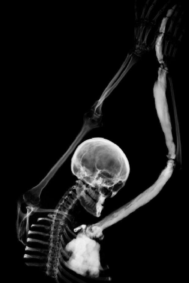
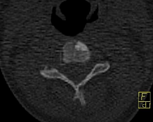
4 There are 2 theories for melorheostosis etiology: (a) early embryonic infection of a sensory nerve inducing the changes in sclerotome [10] and (b) concept of mosaicism which can better explain an asymmetric segmental pattern with variable expressivity and equal gender ratio of the disease [11]. Case presentation: 26-year old female patient underwent surgery of a cleft palate at the age of 5 years, later she had no significant health issue. Melorheostosis was diagnosed incidentally by X-ray after shoulder injury in sport. Clinical examination showed joint abduction to 60 degrees only in the shoulder, further movement was only in scapula, movement range in the elbow was limited to 20 degrees, in radiocarpal joint was volar flexion up to 40 degrees, dorsal flexion 0, pronation and supination was also 0. The patient reported pain in the whole upper limb at rest. Osteopoikilotic islands were identified in the body of third cervical vertebra and also in the right collar bone and sternum manubrium. CT of the upper limbs and upper chest was performed without contrast tracer. There was no family history of melorheostosis found. Diffuse thickening and sclerotisation was identified on index finger, II. metacarp shaft on the left hand, the lesion was less aparent at the I. metacarp of the left hand and on some carpal bones (Figure1). There was diffusely enlarged sclerotic radius, humerus and the scapula on the left side. The cavities on affected bones were completely filled with the sclerotic bone. Small sclerotic focus was in the ventral part of C3 vertebral body - size 7 mm (Figure 2), there was also a small island in the left manubrium sternum and in the sternal part of the left collarbone. The finding corresponded to melorheostosis Léri. Thickness of patient s left scapula was up to 31 mm, the contralateral scapula was unaffected with thickness of 2-3 4
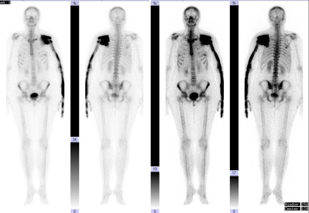
5 mm. The cortex of both collarbone and ribs had slightly higher bone density. The skull was without pathological finding. Bone scintigraphy showed normal level of overall metabolic activity in the skeleton. Significant, locally inhomogeneous increase of activity was evident in most of the scapula, humerus, radius and in II. shaft on the left, the highest intensity of changes was in the scapula, which showed 7-times higher activity compared to the contra lateral parts. Activity increase in the I. shaft left was more modest and slight accumulation was present in the left medial clavicle (Figure 3). The rest of skeleton was without significant pathological changes. DXA examination of the whole skeleton showed osteopenia in the lumbar spine: T score -1.2 SD, in both hips T-score was same and within the normal range. There was no abnormality found in the laboratory tests including bone markers except for increased level of osteocalcin and C-terminal telopeptide. The published cases reported good experience with bisphosphonate in terms of disease progression [12]. The patient was treated with weekly alendronate and COX2-inhibitor (celecoxib). After onset of sleep disorders and increased skin sensitivity on affected site naftidrofuryl in high doses (600 mg daily) was added, which led to symptoms relieve. The patient is currently without pain and is able to perform normal daily activities with nonprogressive restriction for 5 years after the diagnosis. Pharmacological treatment in described combination could delay surgery solution and eventually could prevent an excessive dosage of analgetics. Questionable is further development of patient performance status and sustainability of conservative treatment in the long term follow up. 5
6 Conclusion: X-ray is a sufficient method for diagnosis of melorheostosis. Other imaging techniques are essential for decision about therapeutic intervention (CT, MRI, scintigraphy and DXA). Laboratory findings are usually in physiological range (calcium, phosphorus, markers of bone formation and resorption, IGF-1). Symptomatic therapy proved to be sufficient in subjective symptoms management. The long term effect of conservative treatment remains questionable. Consent: Written informed consent was obtained from the patient for publication of this Case report and any accompanying images. A copy of the written consent is available for review by the Editor of this journal. List of abbreviation: CT Computer tomography, MRI Magnetic resonance imaging, DXA Dual X-ray absorptionmetry, SD Standard deviation Competing interest: All authors declare that they have no competing interests. Author s contributions: VV completed all examinations, decided about final diagnosis and and drafted the manuscript. KK performed first clinical examinations. PT and SK provided consultation 6
7 regarding interpretation of imaging methods. All authors read and approved the final manuscript. Authors information: VV is the head of Center of Metabolic Bone Diseases. His long term scientific interest is focused on rare bone metabolic diseases and related genetic disorders. PT is an international expert in orthopaedic traumatology of pelvis and acetabulum. Acknowledgement: We would like to express our gratitude to all of our collegues at the Department of Imaging Methods of Faculty Hospital. References: [1] Werner MS, Scheimer RA: Melorheostosis. A review of the literature and case report. J Am Podiatr Med Assoc 1987, 77(2): [2] Leri A, Joanny J: Une affection non decrite des os: hyperostose «en coulee» sur toute la longueur d un membre ou melorheostose. Bull Mem Soc Med Hosp Paris 1922, 46: [3] Benli IT, S. Akalin S, E. Boysan E, E. F. Mumcu EF, M. Kiş M, and D. Türkoğlu D:, Epidemiological, clinical and radiological aspects of osteopoikilosis, J. Bone Joint Surg. Br., vol. 74, no. 4, pp , Jul [4] A. Bansal, The Dripping Candle Wax Sign, Radiology, vol. 246, no. 2, pp , Feb
8 [5] R. Happle, Melorheostosis may originate as a type 2 segmental manifestation of osteopoikilosis, Am. J. Med. Genet. A., vol. 125A, no. 3, pp , Mar [6] T. Murano and M. Egarian, Case report: Emergency department diagnosis of melorheostosis in the upper extremity: a rare disease with an unusual presentation, J. Emerg. Med., vol. 43, no. 4, pp , Oct [7] C. Yildirim, S. Ozyürek, E. I. Ciçek, and M. Kuskucu, Melorheostosis in the upper extremity, Orthopedics, vol. 32, no. 4, Apr [8] V. K. Jain, R. K. Arya, M. Bharadwaj, and S. Kumar, Melorheostosis: Clinicopathological Features, Diagnosis, and Management, Orthopedics, vol. 32, no. 7, pp , Jul [9] C. H. Fernandes, L. R. Nakachima, J. B. G. Santos, A. R. C. Fernandes, M. G. Jannini, and F. Faloppa, Melorheostosis of the thumb and trapezium bone, Hand N. Y. N, vol. 6, no. 1, pp , Mar [10] R. O. Murray and J. McCredie, Melorheostosis and the sclerotomes: a radiological correlation, Skeletal Radiol., vol. 4, no. 2, pp , Jun [11] J. P. Fryns, Melorheostosis and somatic mosaicism, Am. J. Med. Genet., vol. 58, no. 2, p. 199, Aug [12] Donáth J1, Poór G, Kiss C, Fornet B, Genant H. Atypical form of active melorheostosis and its treatment with bisphosphonate. Skeletal Radiol Dec;31(12): Epub 2002 Oct 29. Figures: Figure 1. CT of left hand and forearm. Legend: Diffuse thickening and sclerotisation on index finger and II. metacarp shaft. The lesion is less aparent at the I. metacarp and on some carpal bones. There is diffusely enlarged 8
9 sclerotic radius and humerus without ulna bridging. Whole scapula on the left side is also affected. The cavities on the affected bones are completely filled with the sclerotic bone. Figure 2. CT of cervical spine. Legend: Small sclerotic focus is located in the ventral part of C3 vertebral body - size 7 mm. Figure 3. Whole body bone scintigraphy. Legend: Significant, locally inhomogeneous increase of activity is evident in most of the scapula, humerus, radius and in II. shaft on the left, the highest intensity of changes are in the scapula, which shows 7-times higher activity compared to the contra lateral parts. Activity increase in the I. shaft left is more modest and slight accumulation is present in the left medial clavicle and manubrium sterni. 9
10 Figure 1
11 Figure 2
12 Figure 3
The Skeletal System in Action!! The Skeletal System in Action!
 Skeletal System The Skeletal System in Action!! The Skeletal System in Action! 5 Functions of the Skeletal System 1. Movement: Skeletal system provides points of attachment for muscles. Your legs and arms
Anatomy. Anatomy deals with the structure of the human body, and includes a precise language on body positions and relationships between body parts.
 Anatomy deals with the structure of the human body, and includes a precise language on body positions and relationships between body parts. Proper instruction on safe and efficient exercise technique requires
Copyright 2004 Lippincott Williams & Wilkins. 2. Bone Structure. Copyright 2004 Lippincott Williams & Wilkins
 Chapter 7 The Skeleton: Bones and Joints The Skeleton Skeletal system is made up of bones and joints and supporting connective tissue. 1. Bone Functions 1. To store calcium salts 2. To protect delicate
Chapter 6 & 7 The Skeleton
 Chapter 6 & 7 The Skeleton Try this Make clockwise circles with your RIGHT foot, while doing this, draw the number 6 in the air with you RIGHT hand what happens to your foot???? Bony Background Adult body
Sports Medicine Part I : ANATOMY OF THE SPINE, ABDOMEN AND SHOULDER COMPLEX
 Sports Medicine 25 1.1 Part I : ANATOMY OF THE SPINE, ABDOMEN AND SHOULDER COMPLEX c.w.p. Wagner High School, Sports Medicine, A. Morgan, T. Morgan 2008 Anatomy of the Upper Body In this section of the
Human Skeletal System Glossary
 Acromegaly Apatite Acromegaly - is a condition which involves excessive growth of the jaw, hands, and feet. It results from overproduction of somatotropin in adults (after fusion of the ossification centres
Bio 103 Skeletal System 45
 45 Lecture Outline: SKELETAL SYSTEM [Chapters 7, 8] Introduction A. Components B. Functions 1. 2. 3. 4. Classification and Parts A. Bone Shapes 1. Long: 2. Short: 3. Flat: 4. Irregular: 5. Sesamoid: B.
YOGA ANATOMY. Part Three - Bones. Yoga Teacher Training Robin Bennett 200 RYT
 YOGA ANATOMY Yoga Teacher Training Part Three - Bones 2015 Robin Bennett 200 RYT THE HUMAN SKELETON BONE COMPOSITION A femur head with a cortex of compact bone and medulla of trabecular (spongy) bone OSTEOBLASTS
Melorheostosis. Deborah Wenkert, M.D.
 Melorheostosis Deborah Wenkert, M.D. Center for Metabolic Bone Disease and Molecular Research, Shriners Hospital for Children; St. Louis, Missouri, U.S.A. What is Melorheostosis? (OMIM 155950) A rare form
A Patient s Guide to Diffuse Idiopathic Skeletal Hyperostosis (DISH)
") A Patient s Guide to Diffuse Idiopathic Skeletal Hyperostosis (DISH) 6565 Fannin Street Houston, TX 77030 Phone: 713-790-3333 DISCLAIMER: The information in this booklet is compiled from a variety of sources.
A Patient s Guide to Diffuse Idiopathic Skeletal Hyperostosis (DISH) 6565 Fannin Street Houston, TX 77030 Phone: 713-790-3333 DISCLAIMER: The information in this booklet is compiled from a variety of sources.
TRAINING LAB SKELETAL REMAINS: IDENTIFYING BONES NAME
 TRAINING LAB SKELETAL REMAINS: IDENTIFYING BONES NAME Background: Skeletal remains are important pieces of evidence. The flesh, muscle, and organs of a victim rapidly decompose; however, the victim s skeleton
Bone Composition. Bone is very strong for its relatively light weight The major components of bone are:
 Human Bones Bone Composition Bone is very strong for its relatively light weight The major components of bone are: Calcium carbonate Calcium phosphate Collagen Water Cortical Bone Spongy Bone Medullary
Skeletal System. Supplementary Information
 Skeletal System Supplementary Information COMMON ANATOMICAL TERMS Planes run through the body side to side and front to back eg. median plane Surfaces of the body are also named eg. anterior surface This
Skeletal System Tour Lab. Station Label the bones on your answer sheet.
 Station 1 1. Label the bones on your answer sheet. Station 2 2. Label the joints on your answer sheet. Fixed Pivot Hinge Hinge Gliding Ball and Socket Hinge Swivel Gliding Gliding Ball and Socket Types
Osteopoikilosis: A Sign Mimicking Skeletal Metastases in a Cancer Patient
 Case Report Middle East Journal of Cancer 2011; 2(1): 37-41 Osteopoikilosis: A Sign Mimicking Skeletal Metastases in a Cancer Patient Sepideh Sefidbakht *, Yaghoub Ashouri-Taziani **, Sareh Hoseini **,
The Skeletal System. Chapter 7a. Skeletal System Introduction Functions of the skeleton Framework of bones The skeleton through life
 The Skeletal System Skeletal System Introduction Functions of the skeleton Framework of bones The skeleton through life Chapter 7a Support Protection Movement Storage areas Minerals Lipids Hemopoiesis
Skeletal System. Std. VIII
 Skeletal System Std. VIII The skeleton in our body serves following functions : 1. Support and shape : The skeleton provides a support or framework to all the soft parts and gives the body and its parts
36 1 The Skeletal System Slide 1 of 40
 1 of 40 The Skeleton All organisms need structural support. Unicellular organisms have a cytoskeleton. Multicellular animals have either an exoskeleton (arthropods) or an endoskeleton (vertebrates). 2
Principles of Anatomy and Physiology
 Principles of Anatomy and Physiology 14 th Edition CHAPTER 8 The Skeletal System: The Appendicular Skeleton The Appendicular Skeleton The 126 bones of the appendicular skeleton are primarily concerned
SKELETAL STRUCTURES Objectives for Exam #1: Objective for Portfolio #1: Part I: Skeletal Stations Station A: Bones of the Body
 SKELETAL STRUCTURES Objectives for Exam #1: 1. Provide information on the various structures and functions of the skeletal system. 2. Describe various skeletal system disorders, including imaging techniques
General osteology. General anatomy of the human skeleton. Development and classification of bones. The bone as a multifunctional organ.
 General osteology. General anatomy of the human skeleton. Development and classification of bones. The bone as a multifunctional organ. Composed by Natalia Leonidovna Svintsitskaya, Associate professor
Prof. Dr. NAGUI M. ABDELWAHAB,M.D.; MARYSE Y. AWADALLAH, M.D. AYA M. BASSAM, Ms.C.
 Role of Whole-body Diffusion MR in Detection of Metastatic lesions Prof. Dr. NAGUI M. ABDELWAHAB,M.D.; MARYSE Y. AWADALLAH, M.D. AYA M. BASSAM, Ms.C. Cancer is a potentially life-threatening disease,
Chapter 7 Skeletal System. Skeletal System: Bone Functions: Describe the role the skeletal system plays in each of the following functions.
 Chapter 7 Skeletal System Skeletal System: Bone Functions: Describe the role the skeletal system plays in each of the following functions. support protection muscle attachment - movement blood production
Bones of Thorax (Rib Cage)
") Musculoskeletal System (Part A-2) Module 7 -Chapter 10 Overview Muscles Attachments Bones Bone types Surface features of bones Divisions of the skeletal system Joints or Articulations Susie Turner, M.D.
Musculoskeletal System (Part A-2) Module 7 -Chapter 10 Overview Muscles Attachments Bones Bone types Surface features of bones Divisions of the skeletal system Joints or Articulations Susie Turner, M.D.
Lab-1. Miss. Lina Al-Onazy & samar Al-Wgeet =)
") Lab-1 Introduction The human skeleton is composed of 300 bones at birth and by the time adulthood is reached, some bones have fused together to give a total of 206 bones in the body. The human skeleton
Lab-1 Introduction The human skeleton is composed of 300 bones at birth and by the time adulthood is reached, some bones have fused together to give a total of 206 bones in the body. The human skeleton
RADIOGRAPHY OF THE WRIST
 RADIOGRAPHY OF THE WRIST Patient Position: WRIST PA Projection, elbow in same plane Part Position: Hand ; fingers centered to IR Central Ray: Structures Shown: NOTE: Optional AP projection best demonstrates
Overview of the Orthopedic Aspects of Melorheostosis: An Analysis of the Melorheostosis Association database Daniel Lewis, MD* Orthopedic Resident
 Overview of the Orthopedic Aspects of Melorheostosis: An Analysis of the Melorheostosis Association database Daniel Lewis, MD* Orthopedic Resident Jeffrey C. King, MD*# Clinical Assistant Professor *Michigan
Skeletal System. Chapter 7.1. Objective- Read 7.1 and understand that bones are alive and multifunctional. Introduction:
 Chapter 7.1 Skeletal System Objective- Read 7.1 and understand that bones are alive and multifunctional. Introduction: A. Bones are very active tissues B. Each bone is made up of several types of tissues
BLUE SKY SCHOOL OF PROFESSIONAL MASSAGE AND THERAPEUTIC BODYWORK. Musculoskeletal Anatomy & Kinesiology I TERMINOLOGY, STRUCTURES, & SKELETAL OVERVIEW
 BLUE SKY SCHOOL OF PROFESSIONAL MASSAGE AND THERAPEUTIC BODYWORK Musculoskeletal Anatomy & Kinesiology I TERMINOLOGY, STRUCTURES, & SKELETAL OVERVIEW MSAK101-I Session 1 Learning Objectives: 1. Define
Chapter 5 The Skeletal System
 Chapter 5 The Skeletal System The Skeletal System Parts of the skeletal system Bones (skeleton) Joints Cartilages Ligaments (bone to bone)(tendon=bone to muscle) Divided into two divisions Axial skeleton:
OHIO STATE UNIVERSITY EXTENSION
 Cloverbud Investigators: Career Detectives October Background: Today we are going to learn about our bones and how they join together to hold up our body, all the way from our head to our toes. Did you
Skeletal System worksheet
 Skeletal System worksheet Name Section A: Intro to Skeletal System The skeletal system performs vital functions that enable us to move through our daily lives. Support - The skeleton provides support and
Figure 1: Bones of the upper limb
 BONES OF THE APPENDICULAR SKELETON The appendicular skeleton is composed of the 126 bones of the appendages and the pectoral and pelvic girdles, which attach the limbs to the axial skeleton. Although the
Melorheostosis Current Understanding and Recent Developments
 Melorheostosis Current Understanding and Recent Developments Robert E. Fleming, M.D. Associate Professor of Pediatrics and Biochemistry & Molecular Biology Saint Louis University School of Medicine Clinical
Skin. the largest organ of the body 1 mm to 2 mm thick almost 2 square meters 6% of a person s body weight
 Skin the largest organ of the body 1 mm to 2 mm thick almost 2 square meters 6% of a person s body weight Functions of the Skin protection disease-causing organisms dangerous chemicals blood loss fluid
Chapter 8. The Pectoral Girdle & Upper Limb
 Chapter 8 The Pectoral Girdle & Upper Limb Pectoral Girdle pectoral girdle (shoulder girdle) supports the arm consists of two on each side of the body // clavicle (collarbone) and scapula (shoulder blade)
THE SKELETAL SYSTEM 7 TH GRADE SCIENCE
 THE SKELETAL SYSTEM 7 TH GRADE SCIENCE INTRODUCTION Skeletal system is made up of your bones, ligaments, and tendons. It determines : the shape and symmetry of the body acts as protection for your organs
A. Incorrect! The appendicular skeleton includes bones of the shoulder, arm, hand, pelvis, leg and foot.
 Anatomy and Physiology - Problem Drill 08: The Skeletal System III No. 1 of 10 1. Which of the following statements about the appendicular skeleton is correct? A. The appendicular skeleton includes bones
Osteomyelitis in infancy and childhood: A clinical and diagnostic overview M. Mearadji
 Osteomyelitis in infancy and childhood: A clinical and diagnostic overview M. Mearadji International Foundation for Pediatric Imaging Aid Introduction Osteomyelitis is a relative common disease in infancy
Skeletal metastases are the most common variety of bone tumors and should always be considered in the differential diagnosis, particularly in older
 Dr Brajesh Nandan Skeletal metastases are the most common variety of bone tumors and should always be considered in the differential diagnosis, particularly in older patients. Cancers of the breast, prostate,
Hands PA; Obl. Lat.; Norgaard s Thumb AP; Lat. PA. PA; Lat.: Obls.; Elongated PA with ulnar deviation
 Projections Region Basic projections Additional / Modified projections Upper Limbs Hands PA; Obl. Lat.; Norgaard s Thumb ; Lat. PA Fingers PA; Lat. Wrist PA; Lat. Obls. Scaphoid Lunate Trapezium Triquetral
Unit 5 Skeletal System
 Unit 5 Skeletal System Nov 21 10:24 PM I. Functions A. Support: > internal framework, structure, anchors & supports soft tissue organs B. Protection: > protects vital organs C. Movement: > provides attach
Lab Exercise #04 The Skeletal System Student Performance Objectives
 Lab Exercise #04 The Skeletal System Student Performance Objectives The material that you are required to learn in this exercise can be found in either the lecture text or the supplemental materials provided
L01:Name and locate the major bones within the skeletal system.
 L01:Name and locate the major bones within the skeletal system. All physical activity requires movement using bones and muscles. Name three major bones which are located in the leg. Bone 1 - Bone 2- Bone
The Musculoskeletal System
 The Musculoskeletal System Introduction The skeletal system and muscular system are often considered together because they are close in terms of structure and function. The two systems are referred to
Chapter 7 /8 pgs SKELETAL TISSUES AND THE SKELETAL SYSTEM
 Chapter 7 /8 pgs. 189-250 SKELETAL TISSUES AND THE SKELETAL SYSTEM Skeletal Tissue Introduction Bone and cartilage are a specialized types of connective tissue Individual Bones are considered separate
Ch. 5 - Skeletal System
 Ch. 5 - Skeletal System Bones are living, ever-changing structures. This allows them grow and adapt to new situations that the body encounters. The functions of the skeletal system: 1) support bones are
Upper Limb Muscles Muscles of Axilla & Arm
 Done By : Saleh Salahat Upper Limb Muscles Muscles of Axilla & Arm 1) Muscles around the axilla A- Muscles connecting the upper to thoracic wall (4) 1- pectoralis major Origin:- from the medial half of
The skeleton consists of: Bones: special connective tissue, hard. Cartilage: special connective tissue, less hard than bones. Joints: joint is the
 The skeleton consists of: Bones: special connective tissue, hard. Cartilage: special connective tissue, less hard than bones. Joints: joint is the location at witch two bones make contact, whereas ligaments
Case McCune Albright Syndrome (MAS) - polyostotic fibrous dysplasia
 - polyostotic fibrous dysplasia") Case 14477 McCune Albright Syndrome (MAS) - polyostotic fibrous dysplasia Lukasz Augsburg 1, Filip M. Vanhoenacker 1, 2, 3, Jan Gielen1 1. University Hospital Antwerp, Department of Radiology, University
Case 14477 McCune Albright Syndrome (MAS) - polyostotic fibrous dysplasia Lukasz Augsburg 1, Filip M. Vanhoenacker 1, 2, 3, Jan Gielen1 1. University Hospital Antwerp, Department of Radiology, University
Types of Body Movements
 Types of Body Movements Bởi: OpenStaxCollege Synovial joints allow the body a tremendous range of movements. Each movement at a synovial joint results from the contraction or relaxation of the muscles
Equine Skeletal System
 Equine Skeletal System EQS 110 Table of Contents Click on the different sections of the table of contents to jump through this document Functions of the Skeletal System... 3 Skeletal Strength... 3 Bone
Development of the Axial Skeleton and Limbs. Professor Alfred Cuschieri Department of Anatomy University of Malta
 Development of the Axial Skeleton and Limbs Professor Alfred Cuschieri Department of Anatomy University of Malta During the Fourth Week the Embryo Is Segmented. Each segment consists of: a segment of neural
October. Cloverbud Investigators: Career Detectives
 October OHIO STATE UNIVERSITY EXTENSION Cloverbud Investigators: Career Detectives Background: Today we are going to learn about our bones and how they join together to hold up our body all the way from
36.3 The Integumentary System The Skin. KEY CONCEPT The integumentary system has many tissues that protect the body.
 36.3 The Integumentary System The Skin KEY CONCEPT The integumentary system has many tissues that protect the body. 36.3 The Integumentary System The Skin The integument is the body system that surrounds
PRELIMINARY HSC PDHPE. CQ1 How do the musculoskeletal and cardiorespiratory systems of the body influence and respond to movement?
 PRELIMINARY HSC PDHPE CQ1 How do the musculoskeletal and cardiorespiratory systems of the body influence and respond to movement? How do the musculoskeletal and cardiorespiratory systems of the body influence
Presidency General Hospital, Calcutta
 CONGENITAL ANOMALY OF HAND: " MIRROR HAND " By M. MUKERJI, F.R.C.S. Presidency General Hospital, Calcutta Case Report.--S. B., aged 4 months, was born with eight fingers and no thumb on the left hand and
This presentation is the intellectual property of the author. Contact them for permission to reprint and/or distribute.
 The Stiff Hand: Manual Therapy Sylvia Dávila, PT, CHT San Antonio, Texas Orthopedic Manual Therapy Common Applications Passive stretch Tensile force to tissue to increase extensibility of length & ROM
Bell Work. Label the diagram with the layman s or everyday terms we use to talk about our bodies.
 Bell Work Label the diagram with the layman s or everyday terms we use to talk about our bodies. (Leave space to label the appropriate medical terms as we go). The Skeletal System Standards 13) Label on
THE SKELETAL SYSTEM. Focus on the Pectoral Girdle
 THE SKELETAL SYSTEM Focus on the Pectoral Girdle Appendicular Skeleton 126 bones Includes bones of the limbs (arms and legs) Pectoral girdle (shoulder) Pelvic girdle (hip) Pectoral Girdle (the shoulder)
Radiographic Positioning Summary (Basic Projections RAD 222)
") Lower Extremity Radiographic Positioning Summary (Basic Projections RAD 222) AP Pelvis AP Hip (Unilateral) (L or R) AP Femur Mid and distal AP Knee Lateral Knee Pt lies supine on table Align MSP to Center
Lower Extremity Radiographic Positioning Summary (Basic Projections RAD 222) AP Pelvis AP Hip (Unilateral) (L or R) AP Femur Mid and distal AP Knee Lateral Knee Pt lies supine on table Align MSP to Center
Parts of the skeletal system. Bones (skeleton) Joints Cartilages Ligaments (bone to bone)(tendon=bone to muscle)
 Joints Cartilages Ligaments (bone to bone)(tendon=bone to muscle)") The Skeletal System The Skeletal System Parts of the skeletal system Bones (skeleton) Joints Cartilages Ligaments (bone to bone)(tendon=bone to muscle) Divided into two divisions Axial skeleton Appendicular
The Skeletal System The Skeletal System Parts of the skeletal system Bones (skeleton) Joints Cartilages Ligaments (bone to bone)(tendon=bone to muscle) Divided into two divisions Axial skeleton Appendicular
Pediatric metabolic bone diseases
 Pediatric metabolic bone diseases Classification and overview of clinical and radiological findings M. Mearadji International Foundation for Pediatric Imaging Aid www.ifpia.com Introduction Metabolic bone
Joints. Vi Michelle Austin
 Joints Vi Michelle Austin Joints Overview A joint, otherwise known as an articulation, is a point at which points connect. They are constructed to allow movement (except for skull bones) and provide mechanical
Connects arm to thorax 3 joints. Glenohumeral joint Acromioclavicular joint Sternoclavicular joint
 Connects arm to thorax 3 joints Glenohumeral joint Acromioclavicular joint Sternoclavicular joint Scapula Elevation Depression Protraction (abduction) Retraction (adduction) Downward Rotation Upward Rotation
The Skeletal System. Mosby items and derived items 2010, 2006, 2002, 1997, 1992 by Mosby, Inc., an affiliate of Elsevier Inc.
 The Skeletal System Functions of Skeletal System Provides internal framework that supports the body Protects internal organs Helps fight disease by producing white blood cells 2 Functions of Skeletal System
Muscles of the Upper Limb
 Muscles of the Upper Limb anterior surface of ribs 3 5 coracoid process Pectoralis minor pectoral nerves protracts / depresses scapula Serratus anterior Subclavius ribs 1-8 long thoracic nerve rib 1 ----------------
Lecture 2. Statics & Dynamics of Rigid Bodies: Human body 30 August 2018
 Lecture 2. Statics & Dynamics of Rigid Bodies: Human body 30 August 2018 Wannapong Triampo, Ph.D. Static forces of Human Body Equilibrium and Stability Stability of bodies. Equilibrium and Stability Fulcrum
Skeletal System Notes
 Skeletal System Notes A. Introduction 1. Skeletal system is made of organs that are called bones 2. In the adult, there are 206 bones B. Functions of bones 1. Framework: support the body s muscle fat,
Skeletal System worksheet
 Skeletal System worksheet Name Section A: Intro to Skeletal System The skeletal system performs vital functions that enable us to move through our daily lives. Support - The skeleton provides support and
Chapter 7: Skeletal System
 Chapter 7: Skeletal System The Skeletal System Introduction P. 182 Bone is an organ made up of tissues: It is made up of the following components. Cartilage Blood Nerves Bone Connective Bone Classification
Exercise 11. The Appendicular Skeleton
 Exercise 11 The Appendicular Skeleton The Appendicular Skeleton The appendicular skeleton contains 126 bones. Consists of the upper and lower limbs, the pectoral girdles, and the pelvic girdles. The pectoral
Case report. Open Access. Abstract
 Open Access Case report Acute posterior neck pain in adult: a case series Yasuhiro Homma*, Akira Itoi, Tomoya Muta, Yoshio Shimamura, Kiyohito Naito, Atsuhiko Mogami, Osamu Obayashi, Colin G Murphy and
The role of multimodality imaging in Multiple Myeloma: Past, Present and Future
 The role of multimodality imaging in Multiple Myeloma: Past, Present and Future Poster No.: C-1661 Congress: ECR 2015 Type: Educational Exhibit Authors: J. Niza, R. Gil, P. Pereira, C. Oliveira ; Setúbal/PT,
Section 11.1 Your Skeletal System
 Objectives Identify the five main roles of the skeletal system. Describe the functions of bones and joints. Explain how you can keep your skeletal system healthy. Slide 1 of 24 Quick Quiz Complete each
UNIT 5 THE SKELETAL SYSTEM
 UNIT 5 THE SKELETAL SYSTEM Nov 20 12:02 PM I. Functions A. Support: Internal framework, Structure, Anchors & Supports soft tissue/organs B. Protection: Protects vital organs C. Movement: Provide attach
Nervous & Skeletal Systems. Virtual Science University
 Nervous & Skeletal Systems Virtual Science University 1 Nervous & Skeletal Systems Texas TEK B.10(A) The student will interpret the function of systems in organisms (humans) including the nervous and skeletal
Hand and wrist emergencies
 Chapter1 Hand and wrist emergencies Carl A. Germann Distal radius and ulnar injuries PEARL: Fractures of the distal radius and ulna are the most common type of fractures in patients younger than 75 years.
RADIOGRAPHY OF THE HAND, FINGERS & THUMB
 RADIOGRAPHY OF THE HAND, FINGERS & THUMB FINGERS (2nd 5th) - PA Projection Patient Position: Seated; hand ; elbow on IR table top Part Position: Fingers centered to IR unless protocol is Central Ray: Perpendicular
Chapter 8 The Skeletal System: The Appendicular Skeleton. Copyright 2009 John Wiley & Sons, Inc.
 Chapter 8 The Skeletal System: The Appendicular Skeleton Appendicular Skeleton It includes bones of the upper and lower limbs Girdles attach the limbs to the axial skeleton The pectoral girdle consists
Clinical examination of the wrist, thumb and hand
 Clinical examination of the wrist, thumb and hand 20 CHAPTER CONTENTS Referred pain 319 History 319 Inspection 320 Functional examination 320 The distal radioulnar joint.............. 320 The wrist.......................
Radiography Protocols
 Radiography Protocols Upper Limb Second through Fifth Digits (Standard 3 views) First Digit (Thumb) (Standard 3 views) Hand (Standard 3 views) Wrist (Standard 4 views) Forearm (Standard 2 views) Elbow
Country Health SA Medical Imaging
 Country Health SA Medical Imaging REMOTE OPERATORS POSITIONING GUIDE Contents Image Evaluation Page 4 Positioning Guides Section 1 - THORAX 1.1 Chest Page 5 1.2 Bedside Chest Page 7 1.3 Ribs Page 8 Section
Clinician s Guide to Prevention and Treatment of Osteoporosis
 Clinician s Guide to Prevention and Treatment of Osteoporosis Published: 15 August 2014 committee of the National Osteoporosis Foundation (NOF) Tipawan khiemsontia,md outline Basic pathophysiology screening
CHAPTER 7, PART II (BONES)
") Anatomy Name: CHAPTER 7, PART II (BONES) Entry #: INSTRUCTIONS: 1) READ Chapter 7, pg. 140-161. 2) Using the outline, make a note card for each underlined bone name or phrase. 3) On each note card, put
Anatomy Name: CHAPTER 7, PART II (BONES) Entry #: INSTRUCTIONS: 1) READ Chapter 7, pg. 140-161. 2) Using the outline, make a note card for each underlined bone name or phrase. 3) On each note card, put
10/12/2010. Upper Extremity. Pectoral (Shoulder) Girdle. Clavicle (collarbone) Skeletal System: Appendicular Skeleton
 Girdle. Clavicle (collarbone) Skeletal System: Appendicular Skeleton") Skeletal System: Appendicular Skeleton Pectoral girdle Pelvic girdle Upper limbs Lower limbs 8-1 Pectoral (Shoulder) Girdle Consists of scapula and clavicle Clavicle articulates with sternum (Sternoclavicular
Skeletal System: Appendicular Skeleton Pectoral girdle Pelvic girdle Upper limbs Lower limbs 8-1 Pectoral (Shoulder) Girdle Consists of scapula and clavicle Clavicle articulates with sternum (Sternoclavicular
11/25/2012. Chapter 7 Part 2: Bones! Skeletal Organization. The Skull. Skull Bones to Know Cranium
 Chapter 7 Part 2: Bones! 5) Distinguish between the axial and appendicular skeletons and name the major parts of each 6) Locate and identify the bones and the major features of the bones that compose the
ISPUB.COM. Spectrum Of MRI Findings In Musculoskeletal Tuberculosis: Pictoral Essay. P Chudgar INTRODUCTION SPINE
 ISPUB.COM The Internet Journal of Radiology Volume 8 Number 2 Spectrum Of MRI Findings In Musculoskeletal Tuberculosis: Pictoral Essay P Chudgar Citation P Chudgar.. The Internet Journal of Radiology.
Due in Lab. Due next week in lab - Scientific America Article Select one article to read and complete article summary
 Due in Lab 1. Skeletal System 33-34 2. Skeletal System 26 3. PreLab 6 Due next week in lab - Scientific America Article Select one article to read and complete article summary Cell Defenses and the Sunshine
5.1 BONES: AN OVERVIEW
 Unit 5 Skeletal System 5.1 BONES: AN OVERVIEW Section Objectives Identify the major structures and functions of the skeletal system. Differentiate between the two divisions (axial and appendicular) of
The Skeletal System: Articulations Pearson Education, Inc.
 8 The Skeletal System: Articulations Introduction The body cannot move without joints Movements are linked to the range of joint action Joints (arthroses) are connections between bones that may or may
NOTES SKELETAL SYSTEM
 NOTES for the SKELETAL SYSTEM Anatomy & Physiology 2016 Johnson The Skeletal System I. System includes 4 basic parts: A. Bones (206 of em) B. Joints C. Cartilages D. Ligaments II. Bones have 5 basic functions:
Biology 218 Human Anatomy
 Chapter 8 Adapted from Tortora 10 th ed. LECTURE OUTLINE A. Introduction (p. 203) 1. The appendicular skeleton contains 126 bones that form: i. two pectoral (shoulder) girdles two upper limbs i one pelvic
COURSE TITLE: Skeletal Anatomy and Fractures of the Lower Arm, Wrist, and Hand
 COURSE DESCRIPTION Few parts of the human body are required to pivot, rotate, abduct, and adduct like the wrist and hand. The intricate and complicated movements of the arm, wrist, and hand exist partly
PowerPoint Lecture Slides. Prepared by Patty Bostwick-Taylor, Florence-Darlington Technical College. The Skeletal System Pearson Education, Inc.
 PowerPoint Lecture Slides Prepared by Patty Bostwick-Taylor, Florence-Darlington Technical College CHAPTER 5 The Skeletal System 2012 Pearson Education, Inc. Title Classification of Bones and Gross Anatomy
Musculoskeletal Assessment ANATOMY AND PHYSIOLOGY
 Health Assessment - 1 - Lab 8: Musculoskeletal Assessment University of Kerbala / College of Nursing Adult Nursing Department Health Assessment Musculoskeletal Assessment ANATOMY AND PHYSIOLOGY The musculoskeletal
Anatomy and Physiology II. Review Shoulder Girdle New Material Upper Extremities - Bones
 Anatomy and Physiology II Review Shoulder Girdle New Material Upper Extremities - Bones Anatomy and Physiology II Shoulder Girdle Review Questions From Last Lecture Can you identify the following muscles?
ANATOMY AND PHYSIOLOGY
 WHY? ANATOMY AND PHYSIOLOGY VETERINARY SCIENCE PROGRAM RECOGNIZE AND UNDERSTAND BASIC DIRECTIONAL AND ANATOMICAL TERMS UNDERSTAND AND SPEAK THE LANGUAGE OF ANATOMY EXPECTED TO BE ABLE TO COMMUNICATE INTELLIGENTLY
Unit 5 Skeletal System
 Unit 5 Skeletal System I. Functions A. Support: > Internal framework, structure, anchors & supports soft tissue organs B. Protection: > Protects vital organs C. Movement: > Provides attach point for muscles