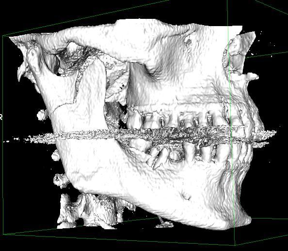
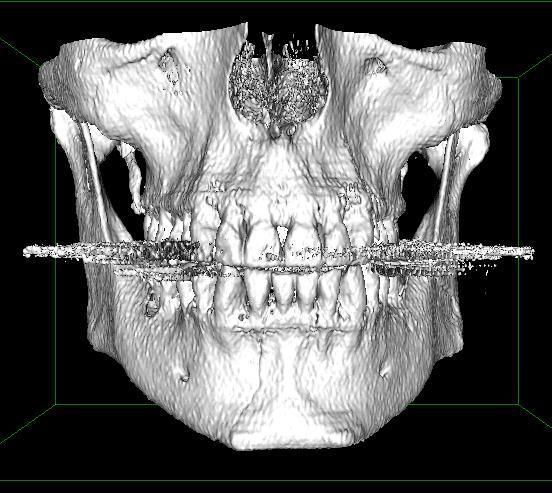
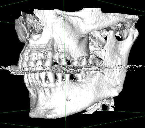
Comprehensive AOCMF Classification System. Cornelius CP, Kunz C, Prein J, Audigé L. Mandibular fractures Level-2 system (cases 1 to 18)
|
|
|
- Alicia Roberts
- 5 years ago
- Views:
Transcription
1 Comprehensive AOCMF Classification System Cornelius CP, Kunz C, Prein J, Audigé L Mandibular fractures Level-2 system (cases 1 to 18)
: anterior")
 view.")


2 Case 1: Body fracture traversing anterior transition zone Imaging: 3 D Reformatted CT scans (surface rendering): anterior oblique view, oblique basal view with making of anterior transitional zone, basal view, inside (lingual) view. Description: Oblique fracture starting in interdental space between lateral lower incisor and canine, travelling back and downwards across the transitional zone. Lingual view confirms the course of the fracture line over the transitional zone. Level 2 Code: 91 m.b AOCOIAC case CMTR

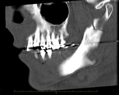
3 Case 2: Body region fracture Imaging: Panorama x-ray Description: vertical fracture line along the periodontium of first molar to inferior mandibular border. Level 2 Code: 91 B.m AOCOIAC case CMTR


4 Case 3: Angle fracture in posterior transition zone Imaging: Panorama x-ray, a.p.15 -Clementschisch Description: Description: Vertical fracture line just behind the posterior transition zone on the left. The wisdom tooth (38) is involved. Level 2 Code: 91 m.a AOCOIAC case CMTR
. Level 2 Code: 91 m.")
5 Case 4: Fracture fully within posterior transition zone Imaging: Panoramic x-ray Description: Almost vertically running fracture within the posterior transition zone on the left, transsecting the root bifurcation of the wisdom tooth (38). Level 2 Code: 91 m.a AOCOIAC case CMTR

6 Case 5: Horizontal angle / ramus fracture Imaging: Panorama x-ray Description: Single horizontal fracture within the angle / ramus region on the left side Level 2 Code: 91 m.a AOCOIAC case CMTR

7 Case 6: Vertical angle / ramus fracture Imaging: 3D CT reformation Description: A vertical fracture line running from the coronoid process to the inferior border of the mandible. The fracture does not separate the coronoid process from its base. Therefore it is not classified as a coronoid fracture. It corresponds to a typical vertical ramus fracture, which is encompassed in the angle/ ramus region. Level 2 Code: 91 m.a AOCOIAC case CMTR


8 Case 7: Isolated coronoid fracture Imaging: CT Scans coronal, sagittal, axial, 3D Reformation: volume and surface rendering Description: single fracture line separating the coronoid process on the left from its base. Level 2 Code: 91 m.c AOCOIAC case CMTR


9 Case 8: Condylar process fracture Imaging: CT Scans coronal, axial. 3 D Reformation volume rendering Description: Fracture line running through the base of the condylar process on the left side (Level 3). The small fragment is laterally displaced (lateral override) and angulated medially. The bony contact between the fragments is not completely lost. The condylar head is orthtotopically distorted within the fossa. Level 2 Code: 91 m.p AOCOIAC case CMTR Level 3 P (left) Code: B0
 to the inferior mandibular")
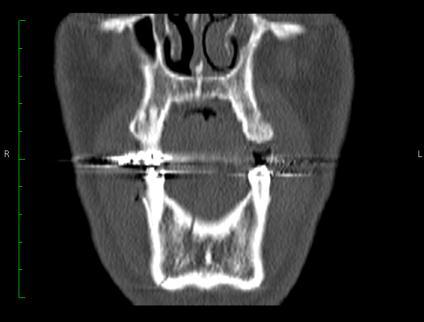
10 Case 9: Double fracture of the mandible Imaging: Panoramic X-ray Description (Level-2): Double fracture of the mandible - Condylar process fracture with displaced fragment of the condylar head on the right, and symphyseal fracture, with vertical fracture line within the symphyseal region extending from the interdental space between he medial and lateral incisors (21/22) to the inferior mandibular border. Level 2 Code: 91 P.S AOCOIAC case CMTR


11 Case 10: Double fracture Imaging: 3D CT Reformation Description: Double fracture of the mandible - Angle / ramus fracture with fracture line from the retromolar area on the right downward to the inferior border within the posterior transition zone, and body fracture with oblique fracture line across the anterior transition zone on the left. Level 2 Code: 91 A.m.B AOCOIAC case CMTR

")


12 Case 11: Multifragmented fracture within right ascending ramus with condylar process involvement Imaging: 3D CT Reformations of mandibular ramus on the right (lateral view, anterior view, inside view) coronal CT scan at midlevel of the ramus Description: Multifragmentation within right ascending ramus. One fracture line separates the condylar process at its base from the ramus Level 2 Code: 91 P.C AOCOIAC case CMTR


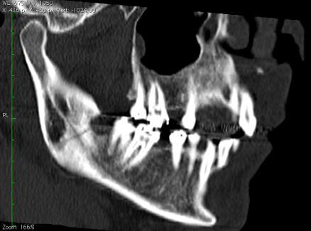
13 Case 12: Triple mandibular fracture (1) Imaging: Panoramic xray, 3D reformatted CTs, sagittal CT slices at the level of the right condylar process Description: Fracture one: Fracture line from the (sigmoid) mandibular notch on the right running backwards along the base of the condylar process to the outer angle - angle/ ramus. From a functional view point it can be regarded as condylar process fracture. Fracture two: Fracture line extending from the toothless molar area on the right posteriorly downward crossing the posterior transition zone and ending in the inferior border of the angle. Basal wedge at the lingual aspect. Fracture three: Fracture line from the posterior aspect of 35 to lower mandibular border - body fracture.
14 Level 2 Code: 91 A-B.m.B AOCOIAC case CMTR

15 Case 13: Triple mandibular fracture (2)
16 Imaging: Panoramic x-ray and CT scans (3D Volume Rendering, MPR coronal, sagittal right, MPR sagittal left, 3D Surface Rendered Views) Description: Fracture one: Fracture starting from the anterior part of the sigmoid notch separating the base of the condylar process in a rectangular fashion - condylar process fracture. Fracture two: Fracture line running from the interdental space between the medial and lateral incisors on the right (41/42) halfway vertically downward into the anterior transition zone - symphyseal fracture. Fracture three: Oblique fracture from the left retromolar area to the outer angle - angle fracture. Level 2 Code: 91 P.S.A AOCOIAC case CMTR


17 Case 14: Triple mandibular fracture (3) Imaging: 3 D CT Reformations Description: Fracture one and three: Sagittal frature line separating the condylar head including the pole zone on the right and left (condylar head bearing fragments in red) - level 2: bilateral condylar process fractures. Fracture two: vertical midline fracture - symphyseal fracture. Level 2 Code: 91 P.S.P AOCOIAC case CMTR


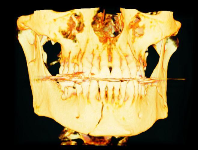
18 Case 15: Multiple mandible fractures Imaging: 3 D CT Reformations Description: Panfacial fracture with multiple fractures in the mandible: condylar process right, angle/ramus right, symphysis contiguous with the body on the left, coronoid process left and condylar process left. Level 2 Code: 91 P.A.S-B.C.P AOCOIAC case CMTR


19 Case 16: Mandibular fracture within the posterior transitional zone Imaging: 3D CT Reformations Description: Oblique fracture line from the tooth gap anterior to the second molar downwards and backwards into the posterior transition zone on the left. - body fracture Level 2 Code: 91 m.b AOCOIAC case CMTR


 Level 2 Code:")
20 Case 17: Condylar process fracture on edentulous mandible Imaging:Panoramic x-ray and 3D CT Reformations Description: Fracture from sigmoid notch posteriorly downwards within the condylar base (Level 2) Level 2 Code: 91 m.p AOCOIAC case CMTR


21 Case 18: Mandibular body fracture Imaging: 3D CT Reformations Description: Sagittal fracture reaching from the lower canine on the outer table to the molar area on the inner table. Body fracture (Level 2). Level 2 Code: 91 m.b AOCOIAC case CMTR
Comprehensive AOCMF Classification System. Cornelius CP, Kunz C, Prein J, Audigé L. Mandibular fractures Level-3 system (cases 22 to 33)
") Comprehensive AOCMF Classification System Cornelius CP, Kunz C, Prein J, Audigé L Mandibular fractures Level-3 system (cases 22 to 33) Case 22: Alvelolar process fracture Imaging: axial CT scan & 3D CT
Comprehensive AOCMF Classification System Cornelius CP, Kunz C, Prein J, Audigé L Mandibular fractures Level-3 system (cases 22 to 33) Case 22: Alvelolar process fracture Imaging: axial CT scan & 3D CT
Dr.Sepideh Falah-kooshki
 Dr.Sepideh Falah-kooshki MAXILLA Premaxillary/median palatal suture (radiolucent). Incisive fossa and foramen (radiolucent). Nasal passages (radiolucent). Nasal septum (radiopaque). Anterior nasal spine
Upper arch. 1Prosthodontics. Dr.Bassam Ali Al-Turaihi. Basic anatomy & & landmark of denture & mouth
 1Prosthodontics Lecture 2 Dr.Bassam Ali Al-Turaihi Basic anatomy & & landmark of denture & mouth Upper arch Palatine process of maxilla: it form the anterior three quarter of the hard palate. Horizontal
AO COIAC COmprehensive Injury Automatic Classifier. Craniomaxillofacial Fracture Classification Module
 AO COIAC COmprehensive Injury Automatic Classifier Software for the classification and documentation of injuries User Manual Version 4.0.0 Craniomaxillofacial Fracture Classification Module Audigé L.,
Extraoral radiography Introduction: Extraoral radiographs (outside the mouth) are taken when large areas of the skull or jaw must be examined or when
 are taken when large areas of the skull or jaw must be examined or when") Extraoral radiography Introduction: Extraoral radiographs (outside the mouth) are taken when large areas of the skull or jaw must be examined or when patients are unable to open their mouths for film placement.
Extraoral radiography Introduction: Extraoral radiographs (outside the mouth) are taken when large areas of the skull or jaw must be examined or when patients are unable to open their mouths for film placement.
高雄醫學大學 口腔醫學院 口腔病理影像科 牙科 X 光影像判讀 教學範例
 高雄醫學大學 口腔醫學院 口腔病理影像科 牙科 X 光影像判讀 教學範例 Content Image No. 001 Dentigerous cyst over left upper embedded canine--------------------- 頁 1 Image No. 002---------------------------------------------------------------
Tikrit University collage of dentistry Dr.Ban I.S. head & neck anatomy 2 nd y. Lec [5] / Temporal fossa :
![Tikrit University collage of dentistry Dr.Ban I.S. head & neck anatomy 2 nd y. Lec [5] / Temporal fossa :](/thumbs/88/115294566.jpg "Tikrit University collage of dentistry Dr.Ban I.S. head & neck anatomy 2 nd y. Lec [5] / Temporal fossa :") Lec [5] / Temporal fossa : Borders of the Temporal Fossa: Superior: Superior temporal line. Inferior: gap between zygomatic arch and infratemporal crest of sphenoid bone. Anterior: Frontal process of the
Lec [5] / Temporal fossa : Borders of the Temporal Fossa: Superior: Superior temporal line. Inferior: gap between zygomatic arch and infratemporal crest of sphenoid bone. Anterior: Frontal process of the
Arrangement of the artificial teeth:
 Lecture Prosthodontic Dr. Osama Arrangement of the artificial teeth: It s the placement of the teeth on a denture with definite objective in mind or it s the setting of teeth on temporary bases. Rules
Muscles of mastication [part 1]
![Muscles of mastication [part 1]](/thumbs/76/73586850.jpg "Muscles of mastication [part 1]") Muscles of mastication [part 1] In this lecture well have the muscles of mastication, neuromuscular function, and its relationship to the occlusion morphology. The fourth determinant of occlusion is the
Muscles of mastication [part 1] In this lecture well have the muscles of mastication, neuromuscular function, and its relationship to the occlusion morphology. The fourth determinant of occlusion is the
Research report for MSc Dent. University of Witwatersrand. Faculty of health science. Dr J Beukes. Student number: h
 Research report for MSc Dent University of Witwatersrand Faculty of health science Dr J Beukes Student number: 9507510h Supervisor: Prof JP Reyneke October 2011 1 1. Title 2. Aim 3. Introduction 4. Objectives
Variations in the anatomical dimensions of the mandibular ramus and the presence of third molars: its effect on the sagittal split ramus osteotomy
 1 Variations in the anatomical dimensions of the mandibular ramus and the presence of third molars: its effect on the sagittal split ramus osteotomy J. Beukes 1,, J. P. Reyneke 1,2,3,4, P. J. Becker 5,6
Techniques of local anesthesia in the mandible
 Techniques of local anesthesia in the mandible The technique of choice for anesthesia of the mandible is the block injection and this is attributed to the absence of the advantages which are present in
Classification proposal for fractures of the processus condylaris mandibulae
 Clinical Oral Investigations https://doi.org/10.1007/s00784-018-2459-1 ORIGINAL ARTICLE Classification proposal for fractures of the processus condylaris mandibulae Marcin Kozakiewicz 1 Received: 5 November
Extraoral Radiology October 10th, 2008
 Extraoral Radiology October 10th, 2008 Steven R. Singer, DDS srs2@columbia.edu 212.305.5674 November 8 th, 1895 Extraoral Projections Images can be produced in the dental office X-ray source can be Intraoral
Definition of Anatomy. Anatomy is the science of the structure of the body and the relation of its parts.
 Definition of Anatomy Anatomy is the science of the structure of the body and the relation of its parts. Basic Anatomical Terms Anatomical terms for describing positions: Anatomical position: Supine position:
Dentalelle Tutoring - Faulty Radiographs
 Dentalelle Tutoring - Faulty Radiographs Errors in improperly exposing or processing dental films can produce undesirable dental radiographs of nondiagnostic quality. These are known as faulty radiographs.
Clinical details: Details of scan: CONE BEAM CT REPORT: Name: H. B. Gender: Reason for referral: Referred by:
 Name: H. B. Gender: Male DOB: 11/12/1950 Age: 64 Date taken: 16/11/2015 Date reported: 19/11/2015 Clinical details: Reason for referral: Referred by: Investigate symptoms related to left TMJ. Reconstructed
Mandibular and Maxillary Anesthesia
 Mandibular and Maxillary Anesthesia Uses of the Conduction Technique JACK H. SELTSAM, D.D.S., M.D., Los Angeles THE ARMAMENTARIUM of a surgeon who operates on the head and neck should include the ability
Oral Surgery. Basic Techniques of Dental Local Anesthesia. A variety of techniques used in administration and deposition of local anesthesia:
 Oral Surgery Lecture: 9 Dr. Saif Saadedeen Basic Techniques of Dental Local Anesthesia A variety of techniques used in administration and deposition of local anesthesia: 1. Topical anesthesia 2. Infiltration
Fundamentals of technique Types of local anaesthesia Topical or surface anaesthesia
 Fundamentals of technique The importance of a quiet, confident, and friendly manner towards all patients so physical comfort is also essential for the co-operation of the patient and the ease of operation
The Application of Cone Beam CT Image Analysis for the Mandibular Ramus Bone Harvesting
 44 The Application of Cone Beam CT Image Analysis for the Mandibular Ramus Bone Harvesting LivingWell Institute of Dental Research Lee, Jang-yeol, Youn, Pil-sang, Kim, Hyoun-chull, Lee Sang-chull Ⅰ. Introduction
Key words: Third molar, Impacted tooth, Tooth Eruption, Molar, Mandible, Unerupted Tooth.
 JOURNAL OF CASE REPORTS 2014;4(2):286-290 OPG and CBCT Finding s of an Ectopic Third Molar in the Sub-condylar Region Tatu Joy E 1, Farakath Khan 1, Shameel Mohammed 2 From the Department of Oral Medicine
The Comprehensive AOCMF Classification System: Glossary of Common Terminology
 S136 Glossary The Comprehensive AOCMF Classification System: Glossary of Common Terminology Carl-Peter Cornelius, MD, DDS 1 Laurent Audigé, DVM, PhD 2,3 Christoph Kunz, MD, DDS 4 Joachim Prein, MD, DDS
1B Getting Ready for Instrumentation: Mathematical Principles and Anatomic Descriptors
 MODULE 1B Getting Ready for Instrumentation: Mathematical Principles and Anatomic Descriptors Module Overview This module contains a review of the mathematical principles and anatomic descriptors used
King's College Hospital Dental School, London, S.E. 5.
 OSTECTOMY AT THE MANDIBULAR SYMPHYSIS J. H. SOWRAY, B.D.S., F.D.S.R.C.S. (Eng.), L.R.C.P., M.R.C.S. and R. HASKELL, M.B., B.S., F.D.S.R.C.S. (Eng.). King's College Hospital Dental School, London, S.E.
DEFINATION Growth was concieved by an anatomist, born to a biologist, delivered by a physician, left on a chemist doorstep, and adopted by a physiolog
 DEFINATION Growth was concieved by an anatomist, born to a biologist, delivered by a physician, left on a chemist doorstep, and adopted by a physiologist.at an early age- she eloped with a statistician,
DEVELOPING ANALOGUE/SUBTITUTE FOR THE MANDIBULAR DENTURE BEARING AREA. Dr Muhammad Rizwan Memon FCPS Assistant Professor
 DEVELOPING ANALOGUE/SUBTITUTE FOR THE MANDIBULAR DENTURE BEARING AREA Dr Muhammad Rizwan Memon FCPS Assistant Professor Crest of Residual Ridge Buccal Shelf Shape of supporting structure Mylohyoid Ridge
JMSCR Vol 04 Issue 08 Page August 2016
 www.jmscr.igmpublication.org Impact Factor 5.244 Index Copernicus Value: 83.27 ISSN (e)-2347-176x ISSN (p) 2455-0450 DOI: http://dx.doi.org/10.18535/jmscr/v4i8.54 Spectrum of Mandibular Fractures in Motor
ORAL FREEDOM: HYBRID PROSTHESES ARE BEST LINGUALIZED
 ORAL FREEDOM: HYBRID PROSTHESES ARE BEST LINGUALIZED 2 ORAL FREEDOM: HYBRID PROSTHESES ARE BEST LINGUALIZED MDT HENRYK JURZYCA Abutment teeth and implants do not tolerate horizontal forces over the long
Dr. Sami Zaqout Faculty of Medicine IUG
 Auricle External Ear External auditory meatus The Ear Middle Ear (Tympanic Cavity) Auditory ossicles Internal Ear (Labyrinth) Bony labyrinth Membranous labyrinth External Ear Auricle External auditory
EUROPEAN SOCIETY OF LINGUAL ORTHODONTICS
 EUROPEAN SOCIETY OF LINGUAL ORTHODONTICS CANDIDATE NUMBER: Dr. Stefan Blasius Year: 2010 WBLO 01 EUROPEAN SOCIETY OF LINGUAL ORTHODONTICS CANDIDATE NUMBER: Dr. Stefan Blasius Year: 2010 WBLO 01 RÉSUMÉ
3. The Jaw and Related Structures
 Overview and objectives of this dissection 3. The Jaw and Related Structures The goal of this dissection is to observe the muscles of jaw raising. You will also have the opportunity to observe several
Normal Radiographic Anatomy Maxillary Lateral Area. Carmen Elena Georgescu1, Gabriela Tãnase 2, Augustin Mihai 3. Objectives.
 Normal Radiographic Anatomy Maxillary Lateral Area Carmen Elena Georgescu1, Gabriela Tãnase 2, Augustin Mihai 3 Bucharest, Romania Summary Intraoral examinations are the backbone of dental radiography.
Figure (2-6): Labial frenum and labial notch.
: Labial frenum and labial notch.") The anatomy of the edentulous ridge in the maxilla and mandible is very important for the design of a complete denture. The consistency of the mucosa and architecture of the underlying bone is different
The anatomy of the edentulous ridge in the maxilla and mandible is very important for the design of a complete denture. The consistency of the mucosa and architecture of the underlying bone is different
Large Dentigerous Cyst
 Volume 16.2.1 Feb 2016 This Lecture Series qualifies for 0.5 Informal CPD Learning Hours Large Dentigerous Cyst By Dr Hassem Geha A 55 year-old male presented with a painless swelling in the right mandible.
Core Curriculum Syllabus Emergencies in Otolaryngology-Head and Neck Surgery FACIAL FRACTURES
 Core Curriculum Syllabus Emergencies in Otolaryngology-Head and Neck Surgery A. General Considerations FACIAL FRACTURES Look for other fractures like skull and/or cervical spine fractures Test function
Conventional radiograph verses CT for evaluation of sagittal fracture of mandibular condyle
 Case Report: Conventional radiograph verses CT for evaluation of sagittal fracture of mandibular condyle Dr Anjali Wadhwa, Dr Gaurav Shah, Dr Shweta Sharma, Dr Anand Bhatnagar, Dr Pallavi Malaviya NIMS
Novel three-dimensional position analysis of the mandibular foramen in patients with skeletal class III mandibular prognathism
 Imaging Science in Dentistry 2016; 46: 77-85 http://dx.doi.org/10.5624/isd.2016.46.2.77 Novel three-dimensional position analysis of the mandibular foramen in patients with skeletal class III mandibular
Imaging the musculoskeletal system. An Introduction
 Imaging the musculoskeletal system An Introduction Objectives Discuss: commonly used imaging modalities in the musculoskeletal system normal imaging anatomy in the extremities fracture description Imaging
The relative position of the inferior alveolar nerve in cadaveric hemi-mandibles
 SHORT REPORT Eur J Anat, 9 (1): 49-53 (2005) The relative position of the inferior alveolar nerve in cadaveric hemi-mandibles V. Saralaya and K. Narayana Department of Anatomy, Centre for Basic Sciences,
Dr.Ban I.S. head & neck anatomy 2 nd y. جامعة تكريت كلية طب االسنان املرحلة الثانية أ.م.د. بان امساعيل صديق 6102/6102
 جامعة تكريت كلية طب االسنان التشريح مادة املرحلة الثانية أ.م.د. بان امساعيل صديق 6102/6102 Parotid region The part of the face in front of the ear and below the zygomatic arch is the parotid region. The
Sample Case #1. Disclaimer
 ABO Sample Cases Disclaimer Sample Case #1 The following sample questions and answers were composed and vetted by a panel of experts in orthodontics and are intended to provide an example of the types
Infratemporal fossa: Tikrit University college of Dentistry Dr.Ban I.S. head & neck Anatomy 2 nd y.
 Infratemporal fossa: This is a space lying beneath the base of the skull between the lateral wall of the pharynx and the ramus of the mandible. It is also referred to as the parapharyngeal or lateral pharyngeal
Introduction to Occlusion and Mechanics of Mandibular Movement
 Introduction to Occlusion and Mechanics of Mandibular Movement Dr. Pauline Hayes Garrett Department of Endodontics, Prosthodontics, and Operative Dentistry University of Maryland, Baltimore Assigned reading
Case Report. Orthognathic Correction of Class II Open Bite. Using the Piezoelectric System and MatrixORTHOGNATHIC Plating System.
 Case Report Orthognathic Correction of Class II Open Bite. Using the Piezoelectric System and MatrixORTHOGNATHIC Plating System. Orthognathic Correction of Class II Open Bite. Using the Piezoelectric System
The Neck the lower margin of the mandible above the suprasternal notch and the upper border of the clavicle
 The Neck is the region of the body that lies between the lower margin of the mandible above and the suprasternal notch and the upper border of the clavicle below Nerves of the neck Cervical Plexus Is formed
Oral Surgery Dr. Labeed Sami جامعة تكريت كلية طب االسنان مادة جراحة الفم املرحلة اخلامسة م.د. لبيد سامي حسن
 جامعة تكريت كلية طب االسنان مادة جراحة الفم املرحلة اخلامسة م.د. لبيد سامي حسن 6102-6102 Mandibular fractures And Dentoalveolar fractures MANDIBULAR ANATOMY The mandible is a U- or V-shaped structure consisting
10/17/2016. Nuts and Bolts of Thoracic Radiology. Objectives. Techniques
 Nuts and Bolts of Thoracic Radiology October 20, 2016 Carleen Risaliti Objectives Understand the basics of chest radiograph Develop a system for interpreting chest radiographs Correctly identify thoracic
TRAUMA TO THE FACE AND MOUTH
 Dr.Yahya A. Ali 3/10/2012 F.I.C.M.S TRAUMA TO THE FACE AND MOUTH Bailey & Love s 25 th edition Injuries to the orofacial region are common, but the majority are relatively minor in nature. A few are major
Transmigration of Mandibular Canines - Clinical Implications
 CASE REPORT Transmigration of Mandibular Canines - Clinical Implications Preema Melani Pinto 1, Vijay R Naik 2, Vikram Pai 3, Kalpana S Jadhav 4, Siddharth Revankar 5 Quick Response Code ABSTRACT: Transmigration
Human, Female, Black, Shotgun wound
 Human, Female, Black, Shotgun wound Product Number: Specimen Evaluated: Skeletal Inventory: BC-196 Bone Clones replica 1 intact cranium 2 fragments of mandible: - portion of left body, ramus, coronoid
Oral & Maxillofacial Surgery
 Chapter 2 Oral & Maxillofacial Surgery Ruchi Singhal 1 ; Virendra Singh 2 ; Amrish Bhagol* 1 Jaipur Dental College, Jaipur, India 2 Senior Professor, PGIDS, Rohtak, India Amrish Bhagol Condylar Fractures
Incidence of canine impaction and transmigration in a patient population
 (2004) 33, 164 169 q 2004 The British Institute of Radiology http://dmfr.birjournals.org RESEARCH Incidence of canine impaction and transmigration in a patient population U Aydin*, HH Yilmaz and D Yildirim
Unitek Temporary Anchorage Device (TAD) System
 System") Planning and Placement Guide Treatment Planning Panoramic or P.A. X-ray - visualize root proximity Curved explorer tip - outline roots intraorally Bone sounding - measure tissue thickness using probe with
Planning and Placement Guide Treatment Planning Panoramic or P.A. X-ray - visualize root proximity Curved explorer tip - outline roots intraorally Bone sounding - measure tissue thickness using probe with
X-Rays. Kunal D Patel Research Fellow IMM
 X-Rays Kunal D Patel Research Fellow IMM The 12-Steps } 1: Name 2: Date 3: Old films 4: What type of view(s) 5: Penetration } Pre-read 6: Inspiration 7: Rotation Quality Control 8: Angulation 9: Soft tissues
A correlation between a new angle (S-Gn-Go angle) with the facial height
 with the facial height") A correlation between a new angle (S-Gn-Go angle) with the facial height Esraa S. Jassim B.D.S., M.Sc. (1) Marwan S. Al-Daggistany B.D.S., M.Sc. (1) Jinan E. Saloom B.D.S., M.Sc. (1) ABSTRACT Background:
A correlation between a new angle (S-Gn-Go angle) with the facial height Esraa S. Jassim B.D.S., M.Sc. (1) Marwan S. Al-Daggistany B.D.S., M.Sc. (1) Jinan E. Saloom B.D.S., M.Sc. (1) ABSTRACT Background:
Objective: The antilingular prominence (AP) is a well-known landmark used during planning
 is a well-known landmark used during planning") *Revised Manuscript without title page Abstract Objective: The antilingular prominence (AP) is a well-known landmark used during planning of intraoral vertical ramus osteotomy (IVRO) in order to prevent
*Revised Manuscript without title page Abstract Objective: The antilingular prominence (AP) is a well-known landmark used during planning of intraoral vertical ramus osteotomy (IVRO) in order to prevent
Imaging of Cervical Spine Trauma Tudor H Hughes, M.D.
 Imaging of Cervical Spine Trauma Tudor H Hughes, M.D. General Considerations Most spinal fractures are due to a single episode of major trauma. Fatigue fractures of the spine are unusual except in the
Unusual transmigration of canines report of two cases in a family
 ISSN: Electronic version: 1984-5685 RSBO. 2014 Jan-Mar;11(1):88-92 Case Report Article Unusual transmigration of canines report of two cases in a family Sulabha A. Narsapur 1 Sameer Choudhari 2 Shrishal
Dr Robert Drummond. BChD, DipOdont Ortho, MChD(Ortho), FDC(SA) Ortho. Canad Inn Polo Park Winnipeg 2015
, FDC(SA) Ortho. Canad Inn Polo Park Winnipeg 2015") Dr Robert Drummond BChD, DipOdont Ortho, MChD(Ortho), FDC(SA) Ortho Canad Inn Polo Park Winnipeg 2015 Severely compromised FPM with poor prognosis Children often present with a developing dentition affected
Dr Robert Drummond BChD, DipOdont Ortho, MChD(Ortho), FDC(SA) Ortho Canad Inn Polo Park Winnipeg 2015 Severely compromised FPM with poor prognosis Children often present with a developing dentition affected
Fig. 12 Trigonias?osborni Lucas, SMNH PI637.2, left maxillary fragment with pi to MI; occlusal view, x I. DESCRIPTION
 Fig. 12 Trigonias?osborni Lucas, SMNH PI637.2, left maxillary fragment with pi to MI; occlusal view, x I. DESCRIPTION SMNH P1637.1 has the teeth somewhat cracked but otherwise well preserved. p 2 is very
Temporomandibular Joint. Dr Noman ullah wazir
 Temporomandibular Joint Dr Noman ullah wazir Type of Joint TMJ is a Synovial joint between : The condylar head of the mandible. The mandibular fossa of squamous part of temporal bone. The joint cavity
Postoperative Evaluation on SSRO performed by Short Lingual Osteotomy and IVRO
 140 J Meikai Dent Med 43 2, 140 147, 2014 Short Lingual Osteotomy SSRO IVRO 1 1 1 1 1 1 2 2 1 2 1 1 2 SSRO SSRO IVRO SSRO short lingual osteotomy SL SL IVRO SL 4 6 IVRO SL IVRO SL 1 IVRO SL short lingual
Lips and labial mucosa
 Lips and labial mucosa External portion of the lips: the vermilion border and the skin Vermilion border : the exposed red portion of the lip, covered by mucous membrane, no mucous glands Boundary: the
Maxillofacial Injuries Practical Tips
 Saturday, October 29, 2016 Maxillofacial Injuries Practical Tips Suyash Mohan MD, PDCC THE ROOTS OF PENN RADIOLOGY RADIOLOGICAL Assistant Professor of Radiology Assistant Professor of Neurosurgery Neuroradiology
We are IntechOpen, the world s leading publisher of Open Access books Built by scientists, for scientists. International authors and editors
 We are IntechOpen, the world s leading publisher of Open Access books Built by scientists, for scientists 4,000 116,000 120M Open access books available International authors and editors Downloads Our
Dr Mohammed Alfarsi Page 1 9 December Principles of Occlusion
 Dr Mohammed Alfarsi Page 1 9 December 2013 Principles of Occlusion Overview: The occlusion is a very large, yet easy to manage once properly understood, topic. Thus, no one handout is enough to fully understand
Periodontal Disease. Radiology of Periodontal Disease. Periodontal Disease. The Role of Radiology in Assessment of Periodontal Disease
 Radiology of Periodontal Disease Steven R. Singer, DDS srs2@columbia.edu 212.305.5674 Periodontal Disease! Includes several disorders of the periodontium! Gingivitis! Marginal Periodontitis! Localized
The Comprehensive AOCMF Classification System: Radiological Issues and Systematic Approach
 Tutorial Article S123 The Comprehensive AOCMF Classification System: Radiological Issues and Systematic Approach Carlos H. Buitrago-Téllez, MD 1,2 Carl-Peter Cornelius, MD, DDS 3 Joachim Prein, MD, DDS
Anatomy and physiology of Temporomandibular Joint
 Anatomy and physiology of Temporomandibular Joint Temporomandibular joint (TMJ): It is the articulation of the condyle of the mandible, and the inter-articular disc; with the mandibular fossa (glenoid
Oral cavity landmarks
 By: Dr. Ahmed Rabah Oral cavity landmarks The knowledge of oral anatomy and physiology will help the operator and provides enough landmarks to act as positive guide during denture construction. This subject
Computed tomography for dental implants: the influence of the gantry angle and mandibular positioning on the bone height and width
 (2005) 34, 9 15 q 2005 The British Institute of Radiology http://dmfr.birjournals.org RESEARCH Computed tomography for dental implants: the influence of the gantry angle and mandibular positioning on the
The Skull and Temporomandibular joint II Prof. Abdulameer Al-Nuaimi. E. mail:
 The Skull and Temporomandibular joint II Prof. Abdulameer Al-Nuaimi E-mail: a.al-nuaimi@sheffield.ac.uk E. mail: abdulameerh@yahoo.com Temporal fossa The temporal fossa is a depression on the temporal
TitleTemporomandibular joint ankylosis: Mitarashi, S; Abe, S; Watanabe, H; Author(s) Hashimoto, M; Ide, Y
 Hashimoto, M; Ide, Y") TitleTemporomandibular joint ankylosis: Mitarashi, S; Abe, S; Watanabe, H; Author(s) Hashimoto, M; Ide, Y Cranio : the journal of craniomandi Journal 20(1): 67-71 URL http://hdl.handle.net/10130/1098 Right
TitleTemporomandibular joint ankylosis: Mitarashi, S; Abe, S; Watanabe, H; Author(s) Hashimoto, M; Ide, Y Cranio : the journal of craniomandi Journal 20(1): 67-71 URL http://hdl.handle.net/10130/1098 Right
radiologymasterclass.co.uk
 http://radiologymasterclass.co.uk Hip X-ray anatomy - Normal AP (anterior-posterior) Shenton's line is formed by the medial edge of the femoral neck and the inferior edge of the superior pubic ramus Loss
Intraoral radiographic techniques Introduction There are three main types of intraoral radiographs: Periapical radiograph Bitewing radiograph
 Intraoral radiographic techniques Introduction There are three main types of intraoral radiographs: Periapical radiograph Bitewing radiograph Occlusal radiograph The anatomic area of interest and type
Human Anatomy and Physiology - Problem Drill 07: The Skeletal System Axial Skeleton
 Human Anatomy and Physiology - Problem Drill 07: The Skeletal System Axial Skeleton Question No. 1 of 10 Which of the following statements about the axial skeleton is correct? Question #01 A. The axial
Bones Ethmoid bone Inferior nasal concha Lacrimal bone Maxilla Nasal bone Palatine bone Vomer Zygomatic bone Mandible
 splanchnocranium - Consists of part of skull that is derived from branchial arches - The facial bones are the bones of the anterior and lower human skull Bones Ethmoid bone Inferior nasal concha Lacrimal
Cephalometric Analysis
 Cephalometric Analysis of Maxillary and Mandibular Growth and Dento-Alveolar Change Part III In two previous articles in the PCSO Bulletin s Faculty Files, we discussed the benefits and limitations of
ALTERNATE OCCLUSAL SCHEMES
 ALTERNATE OCCLUSAL SCHEMES The same basic concepts need to be applied to all occlusal schemes. Some challenges include missing teeth, transposed teeth, crossbites, and anterior open bites. POSTERIOR CROSSBITES
IDC COURSE BASIC LOCAL DENTAL ANESTHESIA LULU F. SCHAEFER LT, DC, USN
 IDC COURSE BASIC LOCAL DENTAL ANESTHESIA LULU F. SCHAEFER LT, DC, USN Lulu.F.Schaefer.mil@mail.mil LOCAL ANESTHETIC AND ARMAMENTARIUM Aspirating syringe Needle Local Anesthetic Carpules LOCAL ANESTHETIC
European Veterinary Dental College
 European Veterinary Dental College EVDC Training Support Document Preparation of Radiograph Sets (Cat and Dog) Document version : evdc-tsd-radiograph_positioning_(dog_and_cat)-20120121.docx page 1 of 13
It is formed by fusion of 3 bones: I. Ilium (superior bone). II. Pubis (antero-inferior bone). III. Ischium (postero-inferior bone).
. II. Pubis (antero-inferior bone). III. Ischium (postero-inferior bone).") It is formed by fusion of 3 bones: I. Ilium (superior bone). II. Pubis (antero-inferior bone). III. Ischium (postero-inferior bone). Pubis Acetabulum Ana (242 ) The three constituent of bones of the hip
It is formed by fusion of 3 bones: I. Ilium (superior bone). II. Pubis (antero-inferior bone). III. Ischium (postero-inferior bone). Pubis Acetabulum Ana (242 ) The three constituent of bones of the hip
Maxillo-facial and Oral Surgery Department, Withington Hospital, Manchester
 FRACTURE OF THE MIDLINE OF THE MANDIBLE ASSOCIATED WITH COMPLETE UNILATERAL DISLOCATION OF THE JAW By IAN H. HESLOV, M.B., B.S., B.D.S., F.D.S.R.C.S.(Eng.) Maxillo-facial and Oral Surgery Department, Withington
Principle of Occlusion
 Principle of Occlusion Mohammed Alfarsi BDS, MDSc(Pros), PhD www.drmohdalfarsi.com com.+*()ا&%$ر"!. www Overview Principle of Occlusion Overview Principle of Occlusion Point centric Long centric Freedom
TIGHTEN YOUR DENTISTRY KNOWLEDGE Jeanne Perrone, CVT VTS (Dentistry)
") TIGHTEN YOUR DENTISTRY KNOWLEDGE Jeanne Perrone, CVT VTS (Dentistry) DENTISTRY Skeletal Anatomy Skull Types There are three common head shapes in the dog and cat. Mesocephalic or mesaticephalic: the most
TIGHTEN YOUR DENTISTRY KNOWLEDGE Jeanne Perrone, CVT VTS (Dentistry) DENTISTRY Skeletal Anatomy Skull Types There are three common head shapes in the dog and cat. Mesocephalic or mesaticephalic: the most
Interpretation Basics of Cone Beam Computed Tomography COPYRIGHTED MATERIAL
 Interpretation Basics of Cone Beam Computed Tomography COPYIGHTED MATEIA 1 Introduction to Cone Beam Computed Tomography Shawneen M. Gonzalez Introduction This chapter will cover basics of cone beam computed
CT of Maxillofacial Fracture Patterns. CT of Maxillofacial Fracture Patterns
 CT of Maxillofacial Fracture Patterns CT of Maxillofacial Fracture Patterns Stuart E. Mirvis, M.D., FACR Department of Radiology University of Maryland School of Medicine Viking 1 1976 MGS 2001 Technology
Samantha W. Chou, D.M.D N. Southport Ave. Chicago, Illinois Phone: Fax:
 Samantha W. Chou, D.M.D. 2325 N. Southport Ave. Chicago, Illinois 60614 Phone: 312-608-6881 Fax: 773-296-0601 Samanthawchou@gmail.com What is our role as the dentist? "We live in a culture in which people
Basic Anatomy and Physiology of the Lips and Oral Cavity. Dr. Faghih
 Basic Anatomy and Physiology of the Lips and Oral Cavity Dr. Faghih It is divided into seven specific subsites : 1. Lips 2. dentoalveolar ridges 3. oral tongue 4. retromolar trigone 5. floor of mouth 6.
The ASE Example Case Report 2010
 The ASE Example Case Report 2010 The Requirements for Case Presentation in The Angle Society of Europe are specified in the Appendix I to the Bylaws. This example case report exemplifies how these requirements
Orthopantomographic Analysis for Assessment of Mandibular Asymmetry
 10.5005/jp-journals-10021-1054 ORIGINAL ARTICLE JIOS Orthopantomographic Analysis for Assessment of Mandibular Asymmetry 1 Shreya Gupta, 2 Sandhya Jain ABSTRACT Objective: The objective of this article
An Anatomical Study of Mandibular and Accessory Mandibular Foramen in Dry Adult Human Mandibles of South Indian Origin
 IOSR Journal of Dental and Medical Sciences (IOSR-JDMS) e-issn: 2279-0853, p-issn: 2279-0861. Volume 13, Issue 4 Ver. II. (Apr. 2014), PP 83-88 An Anatomical Study of Mandibular and Accessory Mandibular
Arrangement of posterior artificial teeth Standardized parameters Curve of Wilson Curve of Spee
 . Arrangement of posterior artificial teeth Posterior teeth are set up in tight centric occlusion. The mandibular teeth are set in the wax occlusion rim over the residual ridge in their ideal buccolingual
Fundamental & Preventive Curvatures of Teeth and Tooth Development. Lecture Three Chapter 15 Continued; Chapter 6 (parts) Dr. Margaret L.
 Dr. Margaret L.") Fundamental & Preventive Curvatures of Teeth and Tooth Development Lecture Three Chapter 15 Continued; Chapter 6 (parts) Dr. Margaret L. Dennis Proximal contact areas Contact areas are on the mesial and
Fundamental & Preventive Curvatures of Teeth and Tooth Development Lecture Three Chapter 15 Continued; Chapter 6 (parts) Dr. Margaret L. Dennis Proximal contact areas Contact areas are on the mesial and
Crowded Class II Division 2 Malocclusion
 Class II Division 2 Malocclusion Crowded Class II Division 2 Malocclusion Clinicians: Drs. Chris Chang, Hsin-Yin Yeh, Sophia Pei-Wen Shu, W. Eugene Roberts Patient: Miss Jhan Pre-treatment Diagnosis An
Class II correction with Invisalign - Combo treatments. Carriere Distalizer.
 Tips from your peers to help you treat with confidence. Class II correction with Invisalign - Combo treatments. Carriere Distalizer. Dr. Clark D. Colville. Carriere Distalizer and Invisalign Combo. A distalization
The Pharynx. Dr. Nabil Khouri MD. MSc, Ph.D
 The Pharynx Dr. Nabil Khouri MD. MSc, Ph.D Introduction The pharynx is the Musculo-fascial halfcylinder that links the oral and nasal cavities in the head to the larynx and esophagus in the neck Common
PELVIS & SACRUM Dr. Jamila El-Medany Dr. Essam Eldin Salama
 PELVIS & SACRUM Dr. Jamila El-Medany Dr. Essam Eldin Salama Learning Objectives At the end of the lecture, the students should be able to : Describe the bony structures of the pelvis. Describe in detail