The Biomechanics of the Human Lower Extremity DR.AYESH BASHARAT BSPT, PP.DPT. M.PHIL
|
|
|
- Loreen Todd
- 6 years ago
- Views:
Transcription

1
2 The Biomechanics of the Human Lower Extremity DR.AYESH BASHARAT BSPT, PP.DPT. M.PHIL
3 Hip joint One of the largest and most stable joint: The hip joint Rigid ball-and-socket configuration (Intrinsic stability)

4

5
6 The femoral head Femoral head : convex component Two-third of a sphere Cover with cartilage Rydell (1965) suggested : most load----- superior quadrant

7 Acetabulum Concave component of ball and socket joint Facing obliquely forward, outward and downward Covered with articular cartilage Provide static stability

8 Acetabulum Labrum: a flat rim of fibro cartilage Transverse acetabular ligament
9 Ligaments and Bursae Iliofemoral ligament: Y shaped extremely strong= anterior stability Pubofemoral ligament: anterior stability

10
11 Ischiofemoral ligament: posterior stability Ligamentum teressupplies a direct attachment from rim of acetabulum to head of femur
12


13 Iliopsoas burs b/w illiopsoas & capsule

14


15 Deep trochanteric bursa b/w G.maximus & G.trochanter


16 Trochantric bursitis

17
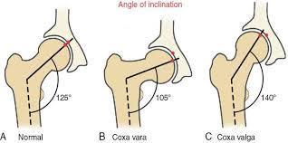
18 The femoral neck Frontal plane (the neck-to-shaft angle/ angle of inclination), Transverse plane (the angle of anteversion)
19 Neck-toshaft angle : 125º, vary from 90º to 135º Effect : lever arms
20


21

22
23 Angle of anteversion :12º Effect : during gait >12º :internal rotation <12º :external rotation
24

25
26 Kinematics Rang of motion in all three planes: sagittal, frontal, transverse 0~140 0~15 0~30 0~25 0~90 0~70


27 Structure of the Hip Sacrum Ilium Acetabulum Femoral head Pubis Ischium Femur The pelvic girdle includes the two ilia and the sacrum,. It can be rotated forward, backward, and laterally to optimize positioning of the hip.
28 Movements at the Hip What movements of the femur are facilitated by pelvic tilt? Pelvic tilt direction Femoral movement posterior anterior lateral (to opposite side) flexion extension abduction
29 Movements at the Hip What muscles contribute to flexion at the hip? iliacus Psoas Major Assisted by: Pectineus Rectus femoris Sartorius Tensor fascia latae
30 Movements at the Hip extension at the hip joint? Gluteus maximus Hamstrings Biceps Femoris Semimembranosus Semitendinosus
31 Movements at the Hip abduction at the hip joint gluteus medius assisted by: gulteus minimus
32 Movements at the Hip adduction at the hip joint? adductor magnus adductor longus adductor brevis assisted by: gracilis
33 Movements at the Hip lateral rotation at the hip joint? Piriformis Gemellus superior Gemellus inferior Obturator internus Obturator externus Quadratus femoris
34 Movements at the Hip medial rotation at the hip joint? gluteus minimus Assisted by: TFL Semimembranosus Semitendinosus Gluteus medius
35 LOADS ON THE HIP Highly specialized and well designed Compressive forces due to following:::: Amount of load (more than ½ of body weight above hip +tension in surrounding muscles).. Effect of speed..foot wear Training surface.. Painful conditions.
 Loss of balance and fall fracture Regular physical")
36 COMMON INJURIES OF THE HIP Fractures Hip is subjected to high repetitive loads times the body weight during locomotion Fractures of femoral neck (aging, osteoporosis) Loss of balance and fall fracture Regular physical activity

37
38 Injuries contd CONTUSIONS Anterior aspect muscles--- prime location for direct injury in Contact sports--- - Internal hemorrhaging--- Appearance of bruises mild to severe Uncommon but serious complication compartment syndrome in which internal hemorrhage- compression on nerves, vessels, muscle---- tissue death
39 Injuries contd STRAINS Hamstring strain.late stance or late swing phase as ecentric contraction.(simultaneous hip flexion &knee extension) Groin Strain.forceful thigh movement in abduction causes strain in adductors(ice hockey players
40
41 KNEE BIOMECHANICS

42 Structure of the Knee Modified hinge joint. Formed by Tibofemoral & patello femoral joit Tibiofemoral joint? Dual condyloid articulations between medial and lateral condyles of tibia and the femur; composing the main hinge joint of the knee
43 Screw home locking mechanism Open chain and closed chain movements??? Close pack position= full extension Open pack position= 25


44 Femur Medial and lateral condyles Convex, asymmetric Medial larger than lateral 44

45 Tibia Medial tibial condyle: concave Lateral tibial condyle: flat or concax Medial 50% larger than lateral 1 45


46 Structure of the Knee Femur Patella Tibia Fibula Bony structure of the tibiofemoral joint.

47 Structure of the Knee Lateral meniscus Transverse ligament Medial meniscus Posterior cruciate ligament Anterior cruciate ligament Superior view The menisci of the knee. Medial meniscus is also attached directly to the medial collateral ligament
48 Deepens the articulating depression of tibial plateaus Load transmission and shock absorption If menisci are removed stress may reach up to 3 times Increased likelihood of degenerative conditions

49 Knee Ligaments:
50 Medial & lateral stabilizers (mostly ligaments) Ligaments most important static stabilizers & dynamic tensile strength - related to composition Primary valgas restraint % restraining moment of knee(mcl) Tense in lateral rotation, lax in flexion
51 Lateral side LCL Primary Varus restraint lax in flexion
52 Cruciates ACL Primary static restraint to anterior displacement tense in extension, lax in flexion PCL Primary restraint to post Displacement - 90% relaxed in extension, tense in flexion restraint to Varus/ valgus force resists rotation, esp.int rot of tibia on femur

53
54 Structure of the Knee What is the patellofemoral joint? articulation between the patella and the femur (the patella improves the mechanical advantage of the knee extensors by as much as 50%) Movement at knee joint & muscles
55 Patellofemoral joint motion Gliding movements== 7 cm in vertical direction Superior glide Inferior glide Lateral and medial shifting (v.little)
56 Loads on the knee joint Tibiofemoral joint: Compression loading more in stance phase Shear loading= tendency of the femur to displace anteriorly on tibial plateaus(glide) Knee flexion angle exceeding than 90 degree result in larger shear forces. Full squats not recommended for novice athletes

57
58 forces at Patellofemoral joint 1/3 rd of body weight compressive forces during normal walking 3 times the body weight during stair climbing-- High compressive forces during knee flexion Squatting highly stressful to the knee complex

59


60
61 Common Injuries of the Knee & Lower Leg

62 Knee Anatomy

63 Patella Fractures Result from direct blow such as knee hitting dashboard in MVA, fall on flexed knee, forceful contraction of quad. Muscle. Transverse fractures most common

64 Femoral Condyle Fractures These injuries secondary to direct trauma from fall w/axial loading or blow to distal femur.

65 Anterior Cruciate Ligament a deceleration, hyperextension or internal rotation of tibia on femur May hear pop, swelling, assoc. w/medial meniscal tear Excessive anterior translation or rotation of femur on the tibia

66 Incidence of ACL injuries is more in females Notable lessening of flexion extension range of motion at the knee due to quadriceps avoiding Altered joint kinetics== subsequent inset of osteoarthritis
67 Surgical repair through middle third of patellar tendon Notable weakness in quadriceps, impaired joint range and proprioception Muscle inhibition: inability to activate all motor units of a muscle during maximal voluntary contraction

68 Posterior Cruciate Ligament Less common than ACL injury Mechanism is hyperflexion of knee with foot plantarflexed Impact with dash board during motor vehicle accident Direct force on proximal anterior tibia
69 Medial collateral ligament injury Blows to the lateral side more common Valgus stress Contact sports= football= MCL injury more common Both MCL and LCL injured in wrestling

70
71 Prophylactic knee bracing To prevent knee ligament injuries in contact sports.(matter of contention Protection from torsional loads Reduced sprinting speed and earlier onset of fatigue
72 Meniscus Injuries Mechanism is usually squatting or twisting maneuvers. There is locking of the knee on flexion or extension that is painful or limits activity. Medial meniscus more commonly damaged due to its attachment with the MCL Combination injuries
73 Iliotibial band friction syndrome Friction of posterior edge of Iliotibial band against the lateral condyle of the femur during foot strike Very common in distance runners, hence referred as runner s knee Training errors and anatomical malalignments Excessive tibial lateral torsion, femoral anteversion, genu valgum, genu varum, increased Q angle etc,
74

75 Breaststroker's knee Forceful whipping together of the lower leg produces propulsive thrust Excessive abduction of the knee Irritation of the MCL and medial border of the patella Hip abduction less than 37 or greater than 42 degree == increased onset of knee pain
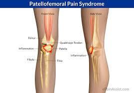
76 Patellofemoral pain syndrome Painful Patellofemoral joint motion involving anterior knee pain after activities requiring repeated flexion at the knee Anatomical malalignments Vastus Medialis Oblique and Vastus Lateralis in strength Large Q angle responsible Patellar maltracking

77

78

79

80

81
82 Chondromalacia Patellae Overuse syndrome of patellar cartilage Caused by patello-femoral malalignments which leads to tracking abnormality of patella putting excessive lateral pressure on articular cartilage Seen in young active women, pain worse w/stair climbing and rising from a chair


83

84 Shin Splints Generalized pain along the anterolateral or posteromedial aspect of the lower leg is commonly known as shin splints Overuse injury often associated with running, dancing on the hard surface and running uphill
85 Structure of the Ankle Tibiotalar joint Hinge joint where the convex surface of the superior talus articulates with the concave surface of the distal tibia considered to be the ankle joint
86 Fibula Structure of the Ankle Tibia Talus Calcaneus Posterior view The bony structure of the ankle.
87 Movements at the Ankle Dorsiflexion at the ankle Tibialis anterior extensor digitorum longus peroneus tertius assisted by: extensor hallucis longus
88 Movements at the Ankle plantar flexion at the ankle Gastrocnemius soleus assisted by: Tibialis posterior, Plantaris, peroneus longus, flexor hallucis longus, peroneus brevis, flexor digitorum longus
89 Structure of the Foot Fibula Subtalar joint (the anterior and posterior facets of the talus articulate with the sustentaculum tali on the superior calcaneus) Tibia Talus Calcaneus Posterior view

90
91 Structure of the Foot tarsometatarsal and intermetatarsal joints Nonaxial joints that permit only gliding movements Enable the foot to function as a semirigid unit and to adapt flexibly to uneven surfaces during weight bearing
92 Structure of the Foot metatarsophalangeal and interphalangeal joints Condyloid and hinge joints, respectively Toes function to smooth weight shift to the opposite foot during walking and help maintain stability during weight bearing by pressing against the ground when necessary
93 Structure of the Foot plantar arches The medial and lateral longitudinal arches stretch form the calcaneus to the metatarsals and tarsals The transverse arch is formed by the base of the metatarsal bones

94

95

96
97 Plantar Fascia Thick bands of fascia that cover the plantar aspects of the foot During weight bearing= mechanical energy is stored in the stretched ligaments, tendons, and plantar fascia of the foot. This energy is released to assist with push-off of the foot from the surface.


98 Structure of the Foot Lateral view Plantar fascia Plantar view The plantar fascia.
99 Movements of the Foot Toe flexion and extension Flexion - flexor digitorum longus, flexor digitorum brevis, lumbricals, Interossei Extension - extensor hallucis longus, extensor digitorum longus, extensor digitorum brevis
100 Movements of the Foot Inversion and eversion Inversion - Tibialis posterior, Tibialis anterior Eversion - peroneus longus, peroneus brevis, assisted by peroneus tertius


101
102 Common injuries of the ankle and foot Ankle injuries Inversion sprains= stretching or rupture of lateral ligaments (ATFL, PTFL, CFL) Medial = deltoid ligament very strong (ATTL, PTTL, TCL, TNL) Ankle bracing or taping (Mild injury treatment)

103

104

105


106 OVERUSE INJURIES Achilles tendinitis Plantar fascitis Stress fractures Dancing en pointe = stressed second metatarsal


107 Alignment anomalies of the foot Forefoot Valgus Forefoot Varus Hallux Valgus Hallux Varus
108 Injuries related to high and low arch structures High arches(pes cavus)= increased incidence of ankle sprains, plantar fascitis, ITB friction syndrome, 5 th metatarsal fracture Low arches (pes planus)= knee pain, patellar tendinitis, plantarfascitis,


109 109
CHAPTER 8: THE BIOMECHANICS OF THE HUMAN LOWER EXTREMITY
 CHAPTER 8: THE BIOMECHANICS OF THE HUMAN LOWER EXTREMITY _ 1. The hip joint is the articulation between the and the. A. femur, acetabulum B. femur, spine C. femur, tibia _ 2. Which of the following is
Muscles of the Hip 1. Tensor Fasciae Latae O: iliac crest I: lateral femoral condyle Action: abducts the thigh Nerve: gluteal nerve
 Muscles of the Hip 1. Tensor Fasciae Latae O: iliac crest I: lateral femoral condyle Action: abducts the thigh Nerve: gluteal nerve 2. Gluteus Maximus O: ilium I: femur Action: abduct the thigh Nerve:
Muscles of the Gluteal Region
 Muscles of the Gluteal Region 1 Some of the most powerful in the body Extend the thigh during forceful extension Stabilize the iliotibial band and thoracolumbar fascia Related to shoulders and arms because
The Muscular System. Chapter 10 Part D. PowerPoint Lecture Slides prepared by Karen Dunbar Kareiva Ivy Tech Community College
 Chapter 10 Part D The Muscular System Annie Leibovitz/Contact Press Images PowerPoint Lecture Slides prepared by Karen Dunbar Kareiva Ivy Tech Community College Table 10.14: Muscles Crossing the Hip and
Recognizing common injuries to the lower extremity
 Recognizing common injuries to the lower extremity Bones Femur Patella Tibia Tibial Tuberosity Medial Malleolus Fibula Lateral Malleolus Bones Tarsals Talus Calcaneus Metatarsals Phalanges Joints - Knee
Copyright 2012 by The McGraw-Hill Companies, Inc. All rights reserved. McGraw-Hill/Irwin
 CHAPTER 8: THE LOWER EXTREMITY: KNEE, ANKLE, AND FOOT KINESIOLOGY Scientific Basis of Human Motion, 12 th edition Hamilton, Weimar & Luttgens Presentation Created by TK Koesterer, Ph.D., ATC Humboldt State
Main Menu. Joint and Pelvic Girdle click here. The Power is in Your Hands
 1 Hip Joint and Pelvic Girdle click here Main Menu K.6 http://www.handsonlineeducation.com/classes//k6entry.htm[3/23/18, 2:01:12 PM] Hip Joint (acetabular femoral) Relatively stable due to : Bony architecture
Muscles of the lower extremities. Dr. Nabil khouri MD, MSc, Ph.D
 Muscles of the lower extremities Dr. Nabil khouri MD, MSc, Ph.D Posterior leg Popliteal fossa Boundaries Biceps femoris (superior-lateral) Semitendinosis and semimembranosis (superior-medial) Gastrocnemius
Hip joint Type: Articulating bones:
 Ana (242 ) Hip joint Type: Synovial, ball & socket Articulating bones: Formed between head of femur and lunate surface of acetabulum of hip bone. Capsule: it is a strong fibrous sleeve connecting the articulating
lesser trochanter of femur lesser trochanter of femur iliotibial tract (connective tissue) medial surface of proximal tibia
 medial surface of proximal tibia") LOWER LIMB MUSCLES OF THE APPENDICULAR SKELETON The muscles that act on the lower limb fall into three groups: those that move the thigh, those that move the lower leg, and those that move the ankle, foot,
LOWER LIMB MUSCLES OF THE APPENDICULAR SKELETON The muscles that act on the lower limb fall into three groups: those that move the thigh, those that move the lower leg, and those that move the ankle, foot,
Anatomy & Physiology. Muscles of the Lower Limbs.
 Anatomy & Physiology Muscles of the Lower Limbs http://www.ishapeup.com/musclecharts.html Muscles of the Lower Limbs Among the strongest muscles in the body. Because pelvic girdle is composed of heavy,
Muscles of Lesson Five. Muscular Nomenclature and Kinesiology - Two. Muscles of Lesson Five, cont. Chapter 16
 Chapter 16 Muscular Nomenclature and Kinesiology - Two Lessons 5-6 Muscles of Lesson Five Iliopsoas (psoas major, iliacus) Hip outward rotators (piriformis, gemellus superior, gemellus inferior, obturator
Lower limb summary. Anterior compartment of the thigh. Done By: Laith Qashou. Doctor_2016
 Lower limb summary Done By: Laith Qashou Doctor_2016 Anterior compartment of the thigh Sartorius Anterior superior iliac spine Upper medial surface of shaft of tibia 1. Flexes, abducts, laterally rotates
MUSCLES OF THE LOWER LIMBS
 MUSCLES OF THE LOWER LIMBS Naming, location and general function Dr. Nabil khouri ROLES THAT SHOULD NOT BE FORGOTTEN Most anterior compartment muscles of the hip and thigh Flexor of the femur at the hip
The Hip (Iliofemoral) Joint. Presented by: Rob, Rachel, Alina and Lisa
 Joint. Presented by: Rob, Rachel, Alina and Lisa") The Hip (Iliofemoral) Joint Presented by: Rob, Rachel, Alina and Lisa Surface Anatomy: Posterior Surface Anatomy: Anterior Bones: Os Coxae Consists of 3 Portions: Ilium Ischium Pubis Bones: Pubis Portion
The Hip (Iliofemoral) Joint Presented by: Rob, Rachel, Alina and Lisa Surface Anatomy: Posterior Surface Anatomy: Anterior Bones: Os Coxae Consists of 3 Portions: Ilium Ischium Pubis Bones: Pubis Portion
this makes sense, however this is lower order thinking and does not solve the lower leg
 Functional Knee Valgus in a Barbell Squat 1 One of the most common lower leg dysfunction we see in athletes, particularly general population is functional knee valgus, or better referred to as the knees
Human Anatomy Biology 351
 Human Anatomy Biology 351 Lower Limb Please place your name on the back of the last page of this exam. You must answer all questions on this exam. Because statistics demonstrate that, on average, between
THE LOWER EXTREMITY EXAM FOR THE FAMILY PRACTITIONER
 THE LOWER EXTREMITY EXAM FOR THE FAMILY PRACTITIONER Melinda A. Scott, D.O. Orthopedic Associates of Dayton Board Certified in Primary Care Sports Medicine GOALS Identify landmarks necessary for exam of
The Hip Joint. Shenequia Howard David Rivera
 The Hip Joint Shenequia Howard David Rivera Topics Of Discussion Movement Bony Anatomy Ligamentous Anatomy Muscular Anatomy Origin/Insertion/Action/Innervation Common Injuries MOVEMENT Flexion Extension
Understanding Leg Anatomy and Function THE UPPER LEG
 Understanding Leg Anatomy and Function THE UPPER LEG The long thigh bone is the femur. It connects to the pelvis to form the hip joint and then extends down to meet the tibia (shin bone) at the knee joint.
DISSECTION SCHEDULE. Session I - Hip (Front) & Thigh (Superficial)
 & Thigh (Superficial)") DISSECTION SCHEDULE Session I - Hip (Front) & Thigh (Superficial) Surface anatomy Inguinal region Gluteal region Thigh Leg Foot bones Hip bone Femur Superficial fascia Great saphenous vein Superficial
DISSECTION SCHEDULE Session I - Hip (Front) & Thigh (Superficial) Surface anatomy Inguinal region Gluteal region Thigh Leg Foot bones Hip bone Femur Superficial fascia Great saphenous vein Superficial
BONES JOINTS MUSCLES OF THE LOWER LIMB
 BONES JOINTS MUSCLES OF THE LOWER LIMB LOWER LIMB: BONES LOWER LIMB GLUTEAL REGION consists of 6 major segments: FEMORAL REGION (THIGH) KNEE REGION LEG REGION TALOCRURAL REGION (ANKLE) FOOT REGION LOWER
Topic 7: Hip and pelvis. Parts of the hip. Parts of the femur
 Topic 7: Hip and pelvis Parts of the hip Parts of the femur Classifying the hip joint Ball and socket Synovial Multiaxial Movements of the hip: Abduction/adduction Flexion/extension Medial/lateral rotation
From Childhood to Adulthood OMT for LOWER EXTREMITY Hip, Knee, Ankle, Foot. Objectives
 From Childhood to Adulthood OMT for LOWER EXTREMITY Hip, Knee, Ankle, Foot Jan Hendryx, DO, FAAO Peek n Peak CME March 1, 2019 Objectives 1. Demonstrate knowledge of the anatomy of the lower extremity-
Clarification of Terms
 Clarification of Terms The plantar aspect of the foot refers to the role or its bottom The dorsal aspect refers to the top or its superior portion The ankle and foot perform three main functions: 1. shock
The Knee. Clarification of Terms. Osteology of the Knee 7/28/2013. The knee consists of: The tibiofemoral joint Patellofemoral joint
 The Knee Clarification of Terms The knee consists of: The tibiofemoral joint Patellofemoral joint Mansfield, p273 Osteology of the Knee Distal Femur Proximal tibia and fibula Patella 1 Osteology of the
Lectures of Human Anatomy
 Lectures of Human Anatomy Lower Limb Gluteal Region and Hip Joint By DR. ABDEL-MONEM AWAD HEGAZY M.B. with honor 1983, Dipl."Gynecology and Obstetrics "1989, Master "Anatomy and Embryology" 1994, M.D.
Human Anatomy Biology 351
 Human Anatomy Biology 351 Lower Limb Please place your name on the back of the last page of this exam. You must answer all questions on this exam. Because statistics demonstrate that, on average, between
Hip joint and pelvic girdle. Lower Extremity. Pelvic Girdle 6/5/2017
 Hip joint and pelvic girdle Lower Extremity The relationship between the pelvic girdle and hip is similar to that between the shoulder girdle and shoulder joint. The lower limbs are attached to the axial
RN(EC) ENC(C) GNC(C) MN ACNP *** MECHANISM OF INJURY.. MOST IMPORTANT *** - Useful in determining mechanism of injury / overuse
 ENC(C) GNC(C) MN ACNP *** MECHANISM OF INJURY.. MOST IMPORTANT *** - Useful in determining mechanism of injury / overuse") HISTORY *** MECHANISM OF INJURY.. MOST IMPORTANT *** Age of patient Sport / Occupation - Certain conditions are more prevalent in particular age groups (Osgood Schlaters in youth / Degenerative Joint Disease
HISTORY *** MECHANISM OF INJURY.. MOST IMPORTANT *** Age of patient Sport / Occupation - Certain conditions are more prevalent in particular age groups (Osgood Schlaters in youth / Degenerative Joint Disease
Pelvic cavity. Gross anatomy of the lower limb. Walking. Sándor Katz M.D.,Ph.D.
 Pelvic cavity. Gross anatomy of the lower limb. Walking. Sándor Katz M.D.,Ph.D. Lower limb Pelvic girdle Free lower extremity Hip bone Definitive fusion of the Y- shaped growth plate occurs 16th -18th
Sports Medicine 15. Unit I: Anatomy. The knee, Thigh, Hip and Groin. Part 4 Anatomies of the Lower Limbs
 Sports Medicine 15 Unit I: Anatomy Part 4 Anatomies of the Lower Limbs The knee, Thigh, Hip and Groin Anatomy of the lower limbs In Part 3 of this section we focused upon 11 of the 12 extrinsic muscles
Figure 1 - Hip and Pelvis
 Hip Figure 1 - Hip and Pelvis The terms hip and pelvis are frequently used interchangeably, but strictly speaking, the pelvis is a girdle of bones and the hip is a joint. The pelvis consists of The sacrum
Human Anatomy Biology 255
 Human Anatomy Biology 255 Exam #4 Please place your name and I.D. number on the back of the last page of this exam. You must answer all questions on this exam. Because statistics demonstrate that, on average,
Lesson 24. A & P Hip
 Lesson 24 A & P Hip 1 Aims of the Session This session will allow candidates to have an understanding of the bony prominences and soft tissues of the hip 2 Learning Outcomes By the end of the lesson the
Balanced Body Movement Principles
 Balanced Body Movement Principles How the Body Works and How to Train it. Module 3: Lower Body Strength and Power Developing Strength, Endurance and Power The lower body is our primary source of strength,
The Lower Limb II. Anatomy RHS 241 Lecture 3 Dr. Einas Al-Eisa
 The Lower Limb II Anatomy RHS 241 Lecture 3 Dr. Einas Al-Eisa Tibia The larger & medial bone of the leg Functions: Attachment of muscles Transfer of weight from femur to skeleton of the foot Articulations
Main Menu. Ankle and Foot Joints click here. The Power is in Your Hands
 1 The Ankle and Foot Joints click here Main Menu Copyright HandsOn Therapy Schools 2009 K.8 http://www.handsonlineeducation.com/classes/k8/k8entry.htm[3/27/18, 1:40:03 PM] Ankle and Foot Joint 26 bones
The Dance Hall by Vincent van Gogh,1888
 The Dance Hall by Vincent van Gogh,1888 Articulations of the pelvic girdle Lumbosacral joints, sacroiliac joints & pubic symphysis The remaining joints of the lower limb Hip joint Knee joint Tibiofibular
The Knee. Tibio-Femoral
 The Knee Tibio-Femoral Osteology Distal Femur with Proximal Tibia Largest Joint Cavity in the Body A modified hinge joint with significant passive rotation Technically, one degree of freedom (Flexion/Extension)
The psoas minor is medial to the psoas major. The iliacus is a fan-shaped muscle that when contracted helps bring the swinging leg forward in walking
 1 p.177 2 3 The psoas minor is medial to the psoas major. The iliacus is a fan-shaped muscle that when contracted helps bring the swinging leg forward in walking and running. The iliopsoas and adductor
Anatomage Table Instructors Guide- Lower Limb
 The Lower Limb Anatomage Table Instructors Guide- Lower Limb Table of Contents Lower Limb 1- The Skeletal System...3 1: Hip Bone...3 2: Hip Joint and Femur...4 3: Patella and Knee Joint...7 4: Tibia, Fibula,
Practical 1 Worksheet
 Practical 1 Worksheet ANATOMICAL TERMS 1. Use the word bank to fill in the missing words. reference side stand body arms palms anatomical forward All anatomical terms have a(n) point which is called the
Lower Limb Dr. Robin Paudel
 Lower Limb n What is a limb? n Skeleton n Joints n Pelvis or limb girdle n Hip/Hip Muscles n Lumber and sacral plexus getting spinal nerves out onto limb n Muscles anterior and posterior compartments n
Muscles to know. Lab 21. Muscles of the Pelvis and Lower Limbs. Muscles that Position the Lower Limbs. Generally. Muscles that Move the Thigh
 Muscles to know Lab 21 Muscles of the Pelvis, Leg and Foot psoas major iliacus gluteus maximus gluteus medius sartorius quadriceps femoris (4) gracilus adductor longus biceps femoris semitendinosis semimembranosus
Lowe w r e r B ody Resistance Training
 Lower Body Resistance Training Tibialis Anterior Extensor Hallucis Longus Extensor Digitorum Longus Proneus Tertius AROM 25-40 degrees Extensor active-insufficiency Flexion & Eversion (Pronation) Flexion
Copyright 2004, Yoshiyuki Shiratori. All right reserved.
 Ankle and Leg Evaluation 1. History Chief Complaint: A. What happened? B. Is it a sharp or dull pain? C. How long have you had the pain? D. Can you pinpoint the pain? E. Do you have any numbness or tingling?
Joints of the Lower Limb II
 Joints of the Lower Limb II Lecture Objectives Describe the components of the knee and ankle joint. List the ligaments associated with these joints and their attachments. List the muscles acting on these
The Knee. Two Joints: Tibiofemoral. Patellofemoral
 Evaluating the Knee The Knee Two Joints: Tibiofemoral Patellofemoral HISTORY Remember the questions from lecture #2? Girth OBSERVATION TibioFemoral Alignment What are the consequences of faulty alignment?
ChiroCredit.com Presents Biomechanics: Focus on
 ChiroCredit.com Presents Biomechanics: Focus on the Knee Presented by: Ivo Waerlop, DC Shawn Allen, DC 1 Focus on The Knee 2 Pertinent Anatomy Femur Tibia Fibula Patella Prepatellar bursa Infrapatellar
Myology of the Knee. PTA 105 Kinesiology
 Myology of the Knee PTA 105 Kinesiology Objectives Describe the planes of motion and axes of rotation of the knee joint Visualize the origins and insertions of the muscles about the knee List the innervations
Knee Joint Assessment and General View
 Knee Joint Assessment and General View Done by; Mshari S. Alghadier BSc Physical Therapy RHPT 366 m.alghadier@sau.edu.sa http://faculty.sau.edu.sa/m.alghadier/ Functional anatomy The knee is the largest
The Knee. Prof. Oluwadiya Kehinde
 The Knee Prof. Oluwadiya Kehinde www.oluwadiya.sitesled.com The Knee: Introduction 3 bones: femur, tibia and patella 2 separate joints: tibiofemoral and patellofemoral. Function: i. Primarily a hinge joint,
Muscles of the Thigh. 6.1 Identify, describe the attachments of and deduce the actions of the muscles of the thigh: Anterior group
 Muscles of the Thigh 6.1 Identify, describe the attachments of and deduce the actions of the muscles of the thigh: Anterior group Sartorius: This is a long strap like muscle with flattened tendons at each
In-Depth Foundations: Anatomy Terms to Know
 Be familiar with / able to identify and define all the following parts. The Spine Cranium Vertebrae Cervical, Thoracic, Lumbar Sacrum Coccyx Bones of Upper Body Cranium Mastoid process; Occipital condyle,
UNIT 7 JOINTS. Knee and Ankle Joints DR. ABDEL-MONEM A. HEGAZY
 UNIT 7 JOINTS Knee and Ankle Joints BY DR. ABDEL-MONEM A. HEGAZY (Degree in Bachelor of Medicine and Surgery with honor 1983, Dipl."Gynaecology and Obstetrics "1989, Master "Anatomy and Embryology "1994,
Prevention and Treatment of Injuries. Anatomy. Anatomy. Chapter 20 The Knee Westfield High School Houston, Texas
 Prevention and Treatment of Injuries Chapter 20 The Knee Westfield High School Houston, Texas Anatomy MCL, Medial Collateral Ligament LCL, Lateral Collateral Ligament PCL, Posterior Cruciate Ligament ACL,
Organization of the Lower Limb
 Organization of the Lower Limb Most illustrations from: Thieme Atlas of Anatomy: Musculoskeletal System. M Schuenke, et al, 2006. Anatomy: A Regional Atlas of the Human Body. Carmine Clemente, 4th edition.
Ligamentous and Meniscal Injuries: Diagnosis and Management
 Ligamentous and Meniscal Injuries: Diagnosis and Management Daniel K Williams, MD Franciscan Physician Network Orthopedic Specialists September 29, 2017 No Financial Disclosures INTRODUCTION Overview of
Joints of the lower limb
 Joints of the lower limb 1-Type: Hip joint Synovial ball-and-socket joint 2-Articular surfaces: a- head of femur b- lunate surface of acetabulum Which is deepened by the fibrocartilaginous labrum acetabulare
Scapula Spine Lateral edge of clavicle. Medial border Scapula. Medial border of Scapula, between superior angle and root of spine. Scapula.
 Muscle attachments and actions answer sheet Muscle Origins insertions Movements Joints crossed Trapezius Base of skull Spinous process of C7 Thoracic Spine Lateral edge of clavicle Elevation Retraction
BLUE SKY SCHOOL OF PROFESSIONAL MASSAGE AND THERAPEUTIC BODYWORK Musculoskeletal Anatomy & Kinesiology KNEE & ANKLE MUSCLES
 BLUE SKY SCHOOL OF PROFESSIONAL MASSAGE AND THERAPEUTIC BODYWORK Musculoskeletal Anatomy & Kinesiology KNEE & ANKLE MUSCLES MSAK201-I Session 3 1) REVIEW a) THIGH, LEG, ANKLE & FOOT i) Tibia Medial Malleolus
Chapter 20 The knee and related structures
 Chapter 20 The knee and related structures Athletic Training Spring 2014 Jihong Park Bones & joints Femur, tibia, fibula, & patella Femur & tibia Weight bearing & muscle attachment Patella functions Anterior
5.1 Identify, describe the attachments of and deduce the actions of the muscles of the thigh:
 5.1 Identify, describe the attachments of and deduce the actions of the muscles of the thigh: Anterior group Proximal attachment Distal attachment Sartorius ASIS» Upper part of shaft tibia (middle surface)»
Hip Anatomy. Bony. The Athletic Hip: Anatomy and Common Injuries. Kyle Wilkens MSPAS, PA-C, ATC/L October 8, 2013
 The Athletic Hip: Anatomy and Common Injuries Kyle Wilkens MSPAS, PA-C, ATC/L October 8, 2013 Hip Anatomy Bony Femur Ischium Ilium Pubis Sacrum Coccyx Soft Tissue Joints Muscles -Pubic Symphisis Labrum
The Hay is in the Barn
 Anatomy 1 Practical 1 Review Made by Forrest Allen (nerd) Edited by TJ Williamson (not nerd) The Hay is in the Barn 2019 Thunderbringers Too much to handle https://www.youtube.com/watch?v=glii-kaza d8
Walking Theory DR.MURALI KRISHNA P.V M.S.(ORTHO)
") Walking Theory DR.MURALI KRISHNA P.V M.S.(ORTHO) G E N E R A L H O S P I T A L A N E K A L, D E P A R T M E N T O F O R T H O P E D I C S, B A N G A L O R E G O V E R N M E N T O F K A R N A T A K A Walking
Walking Theory DR.MURALI KRISHNA P.V M.S.(ORTHO) G E N E R A L H O S P I T A L A N E K A L, D E P A R T M E N T O F O R T H O P E D I C S, B A N G A L O R E G O V E R N M E N T O F K A R N A T A K A Walking
ANKLE PLANTAR FLEXION
 ANKLE PLANTAR FLEXION Evaluation and Measurements By Isabelle Devreux 1 Ankle Plantar Flexion: Gastrocnemius and Soleus ROM: 0 to 40-45 A. Soleus: Origin: Posterior of head of fibula and proximal1/3 of
Organization of the Lower Limb Audrone Biknevicius, Ph.D. Dept. Biomedical Sciences, OU HCOM at Dublin Clinical Anatomy Immersion 2014
 Organization of the Lower Limb Audrone Biknevicius, Ph.D. Dept. Biomedical Sciences, OU HCOM at Dublin Clinical Anatomy Immersion 2014 www.thestudio1.co.za LIMB FUNCTION choco-locate.com blog.coolibar.com
Muscle Testing of Knee Extensors. Yasser Moh. Aneis, PhD, MSc., PT. Lecturer of Physical Therapy Basic Sciences Department
 Muscle Testing of Knee Extensors Yasser Moh. Aneis, PhD, MSc., PT. Lecturer of Physical Therapy Basic Sciences Department Muscle Testing of Knee Extensors othe Primary muscle Quadriceps Femoris -Rectus
Temporalis Elevates & retracts mandible. Masseter Elevates mandible. Sternocleidomastoid Neck flexion. Trapezius Elevates & depresses shoulders
 Anterior Posterior Temporalis Elevates & retracts mandible Masseter Elevates mandible Sternocleidomastoid Neck flexion Trapezius Elevates & depresses shoulders Masseter Elevates mandible Temporalis Elevates
Where should you palpate the pulse of different arteries in the lower limb?
 Where should you palpate the pulse of different arteries in the lower limb? The femoral artery In the femoral triangle, its pulse is easily felt just inferior to the inguinal ligament midway between the
MUSCLES OF THE LOWER EXTREMITY
 MUSCLES OF THE LOWER EXTREMITY Muscles of the lower extremity are divisible into groups, corresponding with the different regions of the limb. I. Muscles of the Iliac Region II. Muscles of the Thigh III.
LOWER LIMB. As we know the bony part of the body is divided into Axial and Appendicular (upper and lower Limbs)
") LOWER LIMB As we know the bony part of the body is divided into Axial and Appendicular (upper and lower Limbs) Bones of the Lower limb: 1-Pelvic Girdle: composed of: 1. Right hip bone : is formed by 3
LOWER LIMB As we know the bony part of the body is divided into Axial and Appendicular (upper and lower Limbs) Bones of the Lower limb: 1-Pelvic Girdle: composed of: 1. Right hip bone : is formed by 3
To describe he knee joint, ligaments, structure & To list the main features of other lower limb joints
 To describe he knee joint, ligaments, structure & neurovascular supply To demonstrate the ankle joint anatomy To list the main features of other lower limb joints To list main groups of lymph nodes in
Human anatomy reference:
 Human anatomy reference: Weak Glut Activation Weak gluteal activation comes from poor biomechanics, poor awareness when training or prolonged exposure in deactivated positions such as sitting. Weak Glut
Gluteal region DR. GITANJALI KHORWAL
 Gluteal region DR. GITANJALI KHORWAL Gluteal region The transitional area between the trunk and the lower extremity. The gluteal region includes the rounded, posterior buttocks and the laterally placed
Leg. Dr. Heba Kalbouneh Associate Professor of Anatomy and Histology
 Leg Dr. Heba Kalbouneh Associate Professor of Anatomy and Histology Skin of the Leg Cutaneous Nerves Medially: The saphenous nerve, a branch of the femoral nerve supplies the skin on the medial surface
musculoskeletal system anatomy nerves of the lower limb 2 done by: Dina sawadha & mohammad abukabeer
 musculoskeletal system anatomy nerves of the lower limb 2 done by: Dina sawadha & mohammad abukabeer #Sacral plexus : emerges from the ventral rami of the spinal segments L4 - S4 and provides motor and
In the name of god. Knee. By: Tofigh Bahraminia Graduate Student of the Pathology Sports and corrective actions. Heat: Dr. Babakhani. Nov.
 In the name of god Knee By: Tofigh Bahraminia Graduate Student of the Pathology Sports and corrective actions Heat: Dr. Babakhani Nov. 2014 1 Anatomy-Bones Bones Femur Medial/lateral femoral condyles articulate
Peripheral Nervous System: Lower Body
 Peripheral Nervous System: Lower Body MSTN121 - Neurophysiology Session 11 Department of Myotherapy Lumbar Plexus Iliohypogastric nerve (T12-L1) Motor: Transverse abdominis and internal obliques Sensory:
Bony Anatomy. Femur. Femoral Head Femoral Neck Greater Trochanter Lesser Trochanter Intertrochanteric Crest Intertrochanteric Line Gluteal Tuberosity
 Hip Anatomy Bony Anatomy Femur Femoral Head Femoral Neck Greater Trochanter Lesser Trochanter Intertrochanteric Crest Intertrochanteric Line Gluteal Tuberosity Bony Anatomy Pelvic Girdle Acetabulum 3 bones
The Lower Limb. Anatomy RHS 241 Lecture 2 Dr. Einas Al-Eisa
 The Lower Limb Anatomy RHS 241 Lecture 2 Dr. Einas Al-Eisa The bony pelvis Protective osseofibrous ring for the pelvic viscera Transfer of forces to: acetabulum & head of femur (when standing) ischial
Anatomy of the lower limb
 Anatomy of the lower limb 1. Bones of the lower limb Pelvis Hip bone/coxal bone Acetabulum o Acetabular margin o Acetabular fossa o Acetabular notch o Lunate surface Ischiopubic ramus Obturator foramen
Organization of the Lower Limb
 Organization of the Lower Limb Limb Development Lower limb develops in an aterolateral position at the level of the L2 to S3 trunk segments Great toe positioned cephalic direction with the soles of the
Therapeutic Foot Care Certificate Program Part I: Online Home Study Program
 Therapeutic Foot Care Certificate Program Part I: Online Home Study Program 1 Anatomy And Terminology Of The Lower Extremity Joan E. Edelstein, MA, PT, FISPO Associate Professor of Clinical Physical Therapy
RN(EC) ENC(C) GNC(C) MN ACNP *** MECHANISM OF INJURY.. MOST IMPORTANT ***
 ENC(C) GNC(C) MN ACNP *** MECHANISM OF INJURY.. MOST IMPORTANT ***") HISTORY *** MECHANISM OF INJURY.. MOST IMPORTANT *** Age of patient - Certain conditions are more prevalent in particular age groups (Hip pain in children may refer to the knee from Legg-Calve-Perthes
HISTORY *** MECHANISM OF INJURY.. MOST IMPORTANT *** Age of patient - Certain conditions are more prevalent in particular age groups (Hip pain in children may refer to the knee from Legg-Calve-Perthes
Section Three: The Leg, Ankle, and Foot Lecture: Review of Clinical Anatomy, Patterns of Dysfunction and Injury, and
 Section Three: The Leg, Ankle, and Foot Lecture: Review of Clinical Anatomy, Patterns of Dysfunction and Injury, and Treatment Implications for the Leg, Ankle, and Foot Levels I and II Demonstration and
Functional biomechanics of the lower limb
 Functional biomechanics of the lower limb Ben and Matt. 24th July 2011 Principles of function Gravity Ground reaction Eco-concentric eccentric loading (preload) of a muscle (or group) is essential for
HUMAN BODY COURSE LOWER LIMB NERVES AND VESSELS
 HUMAN BODY COURSE LOWER LIMB NERVES AND VESSELS October 22, 2010 D. LOWER LIMB MUSCLES 2. Lower limb compartments ANTERIOR THIGH COMPARTMENT General lfunction: Hip flexion, knee extension, other motions
Pelvic Girdle
 ARTICULATIONS OF LOWER EXTREMITY Pages 429-437 Pelvic Girdle formed by connection of the hip bones and the sacrum Sacroiliac Joints compound joints synovial joint - anterior, between the auricular surfaces
rotation of the hip Flexion of the knee Iliac fossa of iliac Lesser trochanter Femoral nerve Flexion of the thigh at the hip shaft of tibia
 Anatomy of the lower limb Anterior & medial compartments of the thigh Dr. Hayder The fascia lata encloses the entire thigh like a sleeve/stocking. Three intramuscular fascial septa (lateral, medial, and
The Leg. Prof. Oluwadiya KS
 The Leg Prof. Oluwadiya KS www.oluwadiya.sitesled.com Compartments of the leg 4 Four Compartments: 1. Anterior compartment Deep fibular nerve Dorsiflexes the foot and toes 2. Lateral Compartment Superficial
Biomechanics of the Knee. Valerie Nuñez SpR Frimley Park Hospital
 Biomechanics of the Knee Valerie Nuñez SpR Frimley Park Hospital Knee Biomechanics Kinematics Range of Motion Joint Motion Kinetics Knee Stabilisers Joint Forces Axes The Mechanical Stresses to which
Exercise 13. Articulations and Body Movements
 Exercise 13 Articulations and Body Movements Articulations Articulations, or joints, are points where a bone is connected to one or more other bones. Articulations hold the skeleton together. Articulations
Evaluation of Gait Mechanics Using Computerized Plantar Surface Pressure Analysis and it s Relation to Common Musculoskeletal Problems
 Evaluation of Gait Mechanics Using Computerized Plantar Surface Pressure Analysis and it s Relation to Common Musculoskeletal Problems Laws of Physics effecting gait Ground Reaction Forces Friction Stored
Biomechanics of the Upper Extremity Shoulder and Hip
 Biomechanics of the Upper Extremity Shoulder and Hip www.fisiokinesiterapia.biz The Shoulder Common Injuries Shoulder Joint - Bones Anatomical Structures Bursa - Fibrous, fluid-filled sac that reduces
Lumbar Plexus. Ventral rami L1 L4 Supplies: Major nerves.. Abdominal wall External genitalia Anteromedial thigh
 Lower Limb Nerves Lectures Objectives Describe the structure and relationships of the plexuses of the lower limb. Describe the course, relationships and structures supplied for the major nerves of the
Due in Lab weeks because of Thanksgiving Prelab #10. Homework #8. Both sides! Both sides!
 Lab 8 MUSCLES Due in Lab 10 2 weeks because of Thanksgiving Prelab #10 Both sides! Homework #8 Both sides! Refer to Muscles 22-23 Naming of muscles Origin Site of muscle attachment that doesn t move during
Biology 325 Fall 2003
 Name: pre-lab exercise due at beginning of your lab session Matching a. fibrous joints b. cartilaginous joints c. synovial joints 1. exhibit a joint cavity 2. types are sutures and syndesmoses 3. bones