Diagnostic Test Skeletal Words Made Easy Match the terms on the right with the terms on the left.
|
|
|
- Alice Hubbard
- 6 years ago
- Views:
Transcription

1 CHAPTER 5 Thompson Educational Publishing, Inc All material is copyright protected. It is illegal to copy any of this material. This material may be used only in a course of study in which Exercise Science: An Introduction to Health and Physical Education (Temertzoglou/Challen) is the

2 Diagnostic Test Skeletal Words Made Easy Match the terms on the right with the terms on the left. 1. Breastbone 2. Shinbone 3. Skull 4. Kneecap 5. Shoulder blade 6. Lower jawbone 7. Upper jawbone 8. Thigh bone 9. Tail bone 10. Fingers 11. Backbones 12. Upper arm 13. Toes 14. collarbone 1. Clavicle 2. Coccyx 3. Cranium 4. Femur 5. Humerus 6. Mandible 7. Maxilla 8. Patella 9. Scapula 10. Phalanges 11. Vertebrae 12. Sternum 13. Tibia 14. *one word is used twice
3 Diagnostic Test Answers 1. Breastbone 2. Shinbone 3. Skull 4. Kneecap 5. Shoulder blade 6. Lower jawbone 7. Upper jawbone 8. Thigh bone 9. Tail bone 10. Fingers 11. Backbones 12. Upper arm 13. Toes 14. collarbone 1. Sternum 2. Tibia 3. Cranium 4. Patella 5. Scapula 6. Mandible 7. Maxilla 8. Femur 9. Coccyx 10. phalanges 11. Vertebrae 12. Humerus 13. Phalanges 14. clavicle
4 Pg.118 Lesson 5.1 ANATOMICAL TERMINOLOGY ~ ~ ~ TOPICS COVERED IN THIS LESSON (a) The Anatomical Position, Planes, and Axes (b) Describing Movements at Joints 2015 Thompson Educational Publishing, Inc. 4

5 The Anatomical Position The anatomical position is the standard position (standing straight, looking forward, arms at your side, and hands facing forward) used to describe the locations and relationships of anatomical parts on your body. It is used in similar way to how the markings on a compass are used to describe locations in geography.
6 Characteristics of the Anatomical Position The key features of the anatomical position are as follows: The person is in an upright, standing position with his or her head, eyes, and toes pointing forward. The feet are together and the arms are slightly out to the side. The forearms are fully supinated; in other words, the palms of the hands are facing forward Thompson Educational Publishing, Inc. 6
7 Examples of Anatomical Relationships Anterior / Posterior The sternum is anterior to the heart (so anterior also means in front of ). The heart also has an anterior surface. Superior / Inferior Superior refers to upward surfaces, inferior refers to downward surfaces. Medial / Lateral Medial means towards the midline or towards the median plane, whereas lateral means away from the midline or away from the median plane. Proximal / Distal Proximal means towards the point of attachment of the limb to the body, whereas distal means farther away from the point of attachment Thompson Educational Publishing, Inc. 7
8 Pg. 120 Anatomical Planes and Anatomical Axes The anatomical position is further standardized by dividing the body into Anatomical planes and Anatomical axes 2015 Thompson Educational Publishing, Inc. 8
9 Anatomical Planes Frontal plane The frontal (coronal) plane is vertical and extends from one side of the body to the other. Transverse plane The transverse (horizontal) plane is horizontal and divides the body into upper and lower segments. Sagittal plane The sagittal (median) plane is vertical and extends from the front of the body to the back Thompson Educational Publishing, Inc. 9


10 Sagittal Plane A vertical plane that bisects the body into right and left halves. Sagittal Plane Flexion/extension

11 The Sagittal Plane

12 Sagittal plane The plane dividing the body into right and left portions

13 Frontal plane A vertical plane that bisects the body into front and back It is at right angles to the sagittal plane Frontal Plane

14 Frontal Plane

15 Frontal plane The plane dividing the body into front and back portions Also called the Coronal plane
16 Transverse plane A transverse plane that bisects the body into top and bottom It s at right angles to both the sagittal and frontal planes Transvers e Plane

17 Transverse Plane

18 Transverse plane The horizontal plane dividing the body into upper and lower portions Also called the Horizontal plane
 is vertical, running from head to toe.")
19 Anatomical Axes Horizontal axis The horizontal axis extends from one side of the body to the other. Longitudinal axis The longitudinal axis (also known as the polar axis) is vertical, running from head to toe. Antero-posterior axis The antero-posterior axis extends from the front of the body to the back Thompson Educational Publishing, Inc. 19

20 Horizontal Axis Horizontal or (bilateral axis) is in an east-west relationship to the anatomical position.
21 Antero-Posterior Axis Antero-posterior axis is in a front-to-back relationship to the anatomical position.

22 Longitudinal or (polar axis) is in a north-south relationship to the anatomical position. Longitudinal Axis

23 Planes and Corresponding Axes


24 Planes and Corresponding Axes
25 Describing a Body Movement: General Rule A body movement can be described in terms of the anatomical plane through which it occurs and the anatomical axis around which it rotates. THE GENERAL RULE FOR DESCRIBING A BODY MOVEMENT: The axis of rotation is always perpendicular to the plane of movement Thompson Educational Publishing, Inc. 10
26 Relationship between Planes and Axes Axis of Rotation Plane of Motion Example Horizontal (Bilateral) Longitudinal (Polar) Antero-Posterior
27 Relationship between Planes and Axes Axis of Rotation Plane of Motion Example Horizontal (Bilateral) Sagittal Flexion, extension Longitudinal (Polar) Transverse Rotation of extremities, axial rotation Antero-Posterior Frontal (Coronal) Abduction, adduction


28 Various Movements and Planes of Movement Activity Axis Plane Stride Jump Cartwheel Elbow extension Nodding yes Tuck Somersault Twirling Shaking head no
29 Various Movements and Planes of Movement Activity Axis Plane Stride Jump Horizontal Sagittal Cartwheel Antero-Posterior Frontal Elbow extension Horizontal Sagittal Nodding yes Horizontal Sagittal Tuck Horizontal Sagittal Somersault Horizontal Sagittal Twirling Longitudinal Transverse Shaking head no Longitudinal Transverse

30 #1. Guess the plane and corresponding axis. Sagittal Plane/Horizontal Axis

31 #2. Guess the plane and corresponding axis. Transverse Plane/Longitudinal Axis

32 #3. Guess the plane and corresponding axis. Frontal Plane/Antero-posterior Axis
 occurs when you move a body segment to the side and away from your body (e.g., moving your arm out to the side and bringing it level with your shoulder).")
33 Terms Used to Describe Movement Flexion is the action of bending at a joint such that the joint angle decreases (e.g., when you bend your elbow to bring your palm up towards your face). Extension is the opposite of flexion. Abduction ( ab = from ) occurs when you move a body segment to the side and away from your body (e.g., moving your arm out to the side and bringing it level with your shoulder). Adduction ( ad = to ) is the opposite of abduction Thompson Educational Publishing, Inc. 33
34 Terms Used to Describe Movement Plantar flexion is specific to the ankle joint. It occurs when you point your toes (e.g., when you stand on your tip toes). Dorsiflexion occurs when you bend the ankle to bring the top of your foot closer to your shin. Supination is rotating the wrist such that the palm of your hand is facing forward (e.g., when you catch a softball underhanded with one hand). Pronation occurs in the opposite direction Thompson Educational Publishing, Inc. 34
.")
.")
35 Terms Used to Describe Movement Inversion is associated with the ankle joint. It is a result of standing on the outer edge of your foot (e.g., when you twist your ankle). Eversion is a result of standing on the inner edge of your foot. External rotation results when you twist or turn a body part outward from the midline (eg., turning your toes outward). Internal rotation results when you twist or turn a body part inward towards the midline Thompson Educational Publishing, Inc. 35
. Depression is the opposite motion movement in a downwards direction (e.g., slouching your shoulders).")

36 Terms Used to Describe Movement Elevation refers to movement in an upwards direction (e.g., hunching your shoulders). Depression is the opposite motion movement in a downwards direction (e.g., slouching your shoulders). Circumduction is a combination of flexion, extension, abduction, and adduction. An example of this movement occurs when a softball pitcher throws a ball with a windmill action Thompson Educational Publishing, Inc. 36


37 Supine Lying on the back when performing a bench press Prone Lying face down when preparing to perform a push-up


38


39 Ankle Sprains Inversion Ankle Sprain Eversion Ankle Sprain (Potts Fracture)

40 Guess the movements Abduction

41 Guess the movements Supination

42 Guess the movements Dorsiflexion
43 Pg.124 Lesson 5.2 THE MUSCULOSKELETAL SYSTEM ~ ~ ~ TOPICS COVERED IN THIS LESSON (a) The Human Skeleton: An Overview (b) The Axial and Appendicular Skeleton 2015 Thompson Educational Publishing, Inc. 43
.")
44 Pg.124 What Is the Human Skeleton? The adult human skeleton is made up of 206 bones, accounting for about 14 percent of total body weight. Humans start life with more bones than that about 300 bones at birth. Over time, several bones fuse as growth takes place (such as in the skull and lower part of the vertebral column) Thompson Educational Publishing, Inc. 44

45 The Human Skeleton 2015 Thompson Educational Publishing, Inc. 20
46 The Human Skeletal System Bones are made up of living tissue bone cells, fat cells, and blood vessels. Compared to other body systems, the human skeletal system is extremely hard and durable. Bones themselves are composed primarily of the mineral calcium. People whose diet is low in calcium may find their bones becoming increasingly brittle and breakable a major concern for older people (osteoporosis) Thompson Educational Publishing, Inc. 46

47 Main Functions of the Skeletal System The skeletal system provides structural support to the body, protects vital organs, serves as a growth centre for cells, acts as a reserve for minerals, and of course plays a major role in movement Thompson Educational Publishing, Inc. 47
48 Classification of Bones in the Human Body Pg.125 Bones are normally classified according to their shape long, short, flat, irregular, and a fifth type (sesamoid) that is found within tendons. Long bones are found in the arms and legs (e.g., the femur). Short bones are most common in the wrists (e.g., the carpal bones). Flat bones are flat and thin and are found in the roof of the skull. Irregular bones include odd-looking bones such as the bones in the vertebrae. Sesamoid bones are unusual, small, flat bones wrapped within tendons that move over bony surfaces (e.g., the patella) Thompson Educational Publishing, Inc. 48

49 2015 Thompson Educational Publishing, Inc. 49

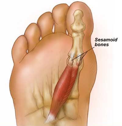
50 Sesamoid Bones

51 Thompson Educational Publishing, Inc All material is copyright protected. It is illegal to copy any of this material. This material may be used only in a course of study in which Exercise Science: An Introduction to Health and Physical Education (Temertzoglou/Challen) is the


52 Thompson Educational Publishing, Inc All material is copyright protected. It is illegal to copy any of this material. This material may be used only in a course of study in which Exercise Science: An Introduction to Health and Physical Education (Temertzoglou/Challen) is the
53 See H.O. Classify each bone


54 Classify each bone. 1.FLAT 2.SHORT 3.LONG 4.SHORT 5.SESAMOID 6.LONG 7.IRREGULAR 8. FLAT
55 Pg.126 The Structure of the Skeleton The human skeletal system is generally divided into two main parts: The axial skeleton (shown in orange), and The appendicular skeleton (shown in green) Thompson Educational Publishing, Inc. 55
56 The Axial Skeleton 80 Bones The axial skeleton is comprised mainly of the vertebral column (the spine), much of the skull, and the rib cage. Most of the body s core muscles originate from the axial skeleton. These core muscles help stabilize and support the axial skeleton, thus providing proper posture and alignment Thompson Educational Publishing, Inc. 56
57 The Appendicular Skeleton 126 Bones The appendicular skeleton includes the movable limbs and their supporting structures (girdles), which play a key role in allowing us to move. The appendicular skeleton can be divided into six major regions: pectoral girdle; arms and forearms; hands; pelvis; thighs and legs; and feet and ankles Thompson Educational Publishing, Inc. 57
58 Axial vs. Appendicular Skeleton The axial skeleton consists of 80 bones: 26 vertebral column 1 hyoid 22 skull 6 auditory 25 ribs The appendicular skeleton consists of 126 bones: 64 upper extremity 62 lower extremity Axial (80) + Appendicular (126) = 206 bones Thompson Educational Publishing, Inc All material is copyright protected. It is illegal to copy any of this material. This material may be used only in a course of study in which Exercise Science: An Introduction to Health and Physical Education (Temertzoglou/Challen) is the required textbook.
59 Pg.126 Bone Landmarks All the bones in the human skeleton have features known as landmarks. A landmark is a ridge, bump, groove, depression, or prominence on the surface of the bone that serves as a guide to the locations of other body structures. For example, the quadriceps muscles of the front thigh ultimately wrap around the patella (kneecap) and insert on the tibial tuberosity (a landmark at the top of the tibia) Thompson Educational Publishing, Inc. 59


60 See H.O. The Anatomy of a Long Bone 2015 Thompson Educational Publishing, Inc. 60

61 Section through the head of the femur, showing the cortex, the red bone marrow and a spot of yellow bone marrow.


62 Epiphyseal Plates and Lines Epiphyseal plates (growth plates) Occur at various locations at the epiphyses of long bones Growth possible Epiphyseal lines Occur when epiphyseal plates have fused or come together Growth not possible Epiphyseal line Epiphyseal plate Thompson Educational Publishing, Inc All material is copyright protected. It is illegal to copy any of this material. This material may be used only in a course of study in which Exercise Science: An Introduction to Health and Physical Education (Temertzoglou/Challen) is the required textbook.

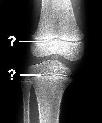
63 Epiphyseal Plate or Line? Line Plate


64 Which is a child s hand, which is the adult s hand? A Child B Adult

65 Osteoporosis Low bone mass and deterioration of the bone tissue The number of osteoblasts is decreased and the number of osteoclasts stay the same Leads to bone fragility Increased susceptibility to bone fractures Preventative measures include: Balanced diet rich in calcium and vitamin D, and a healthy lifestyle Weight-bearing exercises Bone density testing and medication when appropriate
66 Bone Injuries and Bone Disease Fractures bone breaks, normally divided into three types: simple, compound, and comminuted. Stress fractures tiny cracks caused by a rapid increase in activity or when an athlete switches training surfaces or wears footwear with improper cushioning. Shin splints a painful condition occurring on the medial or lateral side of the tibia (shin bone) are another common sports injury. Tearing of the interosseous membrane If not treated, it may lead to a stress fracture 2015 Thompson Educational Publishing, Inc. 30

67 Overuse injury Occurs on the medial or lateral side of tibia (on shaft) Tearing of the interosseous membrane or periosteum Or Inflammation of the interosseous membrane If left untreated-develop into stress fractures SHIN SPLINTS

68 Despite its mineral strength, bone may crack or even break if subjected to extreme loads, sudden impacts, or stresses from unusual directions. The damage produced constitutes a fracture. Fractures Thompson Educational Publishing, Inc All material is copyright protected. It is illegal to copy any of this material. This material may be used only in a course of study in which Exercise Science: An Introduction to Health and Physical Education (Temertzoglou/Challen) is the
 fracture no separation; bone ends")
 fracture bone breaks into separate pieces; bone ends penetrate the skin.")


69 Types of Fractures Stress fracture most difficult to detect; tiny crack in the bone; muscles too tired to absorb shock; overuse injury Simple (Closed) fracture no separation; bone ends don t penetrate the skin. Compound (Open) fracture bone breaks into separate pieces; bone ends penetrate the skin. Comminuted fracture bone shatters into many pieces. Common in the elderly (brittle bones). Greenstick fracture- bone breaks incompletely. One side bent, one side broken. Common in children. Simple fracture Compound fracture Comminuted fracture Spiral-ragged break caused by excessive twisting forces. Sports injury/injury of abuse Thompson Educational Publishing, Inc All material is copyright protected. It is illegal to copy any of this material. This material may be used only in a course of study in which Exercise Science: An Introduction to Health and Physical Education (Temertzoglou/Challen) is the required textbook.


70 Avulsion Fracture A closed fracture where a piece of bone is broken off by a sudden, forceful contraction of a muscle.


71 Fractures Greenstick Fracture Spiral Fractures Thompson Educational Publishing, Inc All material is copyright protected. It is illegal to copy any of this material. This material may be used only in a course of study in which Exercise Science: An Introduction to Health and Physical Education (Temertzoglou/Challen) is the


72 Stress Fractures Thompson Educational Publishing, Inc All material is copyright protected. It is illegal to copy any of this material. This material may be used only in a course of study in which Exercise Science: An Introduction to Health and Physical Education (Temertzoglou/Challen) is the

73 What kind of fracture is this? Comminuted Thompson Educational Publishing, Inc All material is copyright protected. It is illegal to copy any of this material. This material may be used only in a course of study in which Exercise Science: An Introduction to Health and Physical Education (Temertzoglou/Challen) is the

74 Thompson Educational Publishing, Inc All material is copyright protected. It is illegal to copy any of this material. This material may be used only in a course of study in which Exercise Science: An Introduction to Health and Physical Education (Temertzoglou/Challen) is the


75
76 Lesson 5.3 MAJOR BONES OF THE HUMAN BODY ~ ~ ~ TOPICS COVERED IN THIS LESSON (a) Bones of the Axial Skeleton (b) Bones of the Appendicular Skeleton 2015 Thompson Educational Publishing, Inc. 76

77 Bones of the Human Skull (Anterior View) 2015 Thompson Educational Publishing, Inc. 77

78 Bones of the Human Skull (Lateral View) Coronal Suture Sagittal Suture Squamous Suture Lambdoidal Suture Occipital Suture 2015 Thompson Educational Publishing, Inc. 78

79 Skull of a Newborn
80 The Vertebral Column (Lateral View) 2015 Thompson Educational Publishing, Inc. 80
81 Thompson Educational Publishing, Inc All material is copyright protected. It is illegal to copy any of this material. This material may be used only in a course of study in which Exercise Science: An Introduction to Health and Physical Education (Temertzoglou/Challen) is the

82 Thompson Educational Publishing, Inc All material is copyright protected. It is illegal to copy any of this material. This material may be used only in a course of study in which Exercise Science: An Introduction to Health and Physical Education (Temertzoglou/Challen) is the
83 Thompson Educational Publishing, Inc All material is copyright protected. It is illegal to copy any of this material. This material may be used only in a course of study in which Exercise Science: An Introduction to Health and Physical Education (Temertzoglou/Challen) is the

84 Thompson Educational Publishing, Inc All material is copyright protected. It is illegal to copy any of this material. This material may be used only in a course of study in which Exercise Science: An Introduction to Health and Physical Education (Temertzoglou/Challen) is the

85 Thompson Educational Publishing, Inc All material is copyright protected. It is illegal to copy any of this material. This material may be used only in a course of study in which Exercise Science: An Introduction to Health and Physical Education (Temertzoglou/Challen) is the

86 Thompson Educational Publishing, Inc All material is copyright protected. It is illegal to copy any of this material. This material may be used only in a course of study in which Exercise Science: An Introduction to Health and Physical Education (Temertzoglou/Challen) is the

87 Thompson Educational Publishing, Inc All material is copyright protected. It is illegal to copy any of this material. This material may be used only in a course of study in which Exercise Science: An Introduction to Health and Physical Education (Temertzoglou/Challen) is the

88 Thompson Educational Publishing, Inc All material is copyright protected. It is illegal to copy any of this material. This material may be used only in a course of study in which Exercise Science: An Introduction to Health and Physical Education (Temertzoglou/Challen) is the


89 Cervical Vertebrae Atlas C1 -nodding motion; yes Axis C2 -rotation; no

90 Axis Atlas

91 The Thoracic Cage (Anterior View) 2015 Thompson Educational Publishing, Inc. 91

92 Left Scapula (Anterior & Lateral Views) 2015 Thompson Educational Publishing, Inc. 92

93 Left Scapula (Posterior View) 2015 Thompson Educational Publishing, Inc. 40

94 Pelvis (Male, Anterior View) 2015 Thompson Educational Publishing, Inc. 94

95 Right Femur (Anterior & Posterior Views) 2015 Thompson Educational Publishing, Inc. 95
96 Right Fibula (Anterior & Superior Views) 2015 Thompson Educational Publishing, Inc. 96

97 Right Foot (Superior View) 2015 Thompson Educational Publishing, Inc. 97

98 Left Humerus (Anterior & Posterior Views) 2015 Thompson Educational Publishing, Inc. 98
99 Left Ulna and Radius (Anterior View) 2015 Thompson Educational Publishing, Inc. 99

100 Left Hand (Anterior View) 2015 Thompson Educational Publishing, Inc. 100
 The Different Types of Human Joints and Joint-")
101 Lesson 5.4 THE ARTICULAR SYSTEM ~ ~ ~ TOPICS COVERED IN THIS LESSON (a) The Different Types of Human Joints and Joint- Related Injuries (b) Shoulder Joint / Knee Joint / Ankle Joint 2015 Thompson Educational Publishing, Inc. 101

102 The Knee: Femur, tibia and patella

103 The Shoulder: Scapla, humerus and clavicle

104 The Ankle: Tibia, talus and fibula
 to or forms a joint with at least one other bone.")
105 Types of Joints With one exception (the hyoid bone), every bone in the body is connected (articulated) to or forms a joint with at least one other bone.
 Attach muscle to bone Vascular")
106 WHICH IS A TENDON? LIGAMENT? Tendons: Composed of collagen (bundles of white, fibrous protein) Attach muscle to bone Vascular Ligaments: Tough bands of white, fibrous tissue Attach bone to bone Avascular

107 Ligaments Less rigid than bone Do not stretch as much as tendons Tissue that attaches one or more bones together Made up of tough bands of white, fibrous tissue When they reach their threshold-stretch minimally, usually tear

108 Tendons Large bundles of white, fibrous protein (collagen) that attaches muscle to bone Greater stretching ability Will tear with great force


109 1 0 9
 The more vascular a tissue, the less time it takes to recover from an injury Bone and muscle are vascular and will take less time to heal than a")
110 Vascularity Amount of supplied blood a tissue has or requires Ligaments & cartilage are avascular -nutritional needs are not met through blood (take a long time to heal) The more vascular a tissue, the less time it takes to recover from an injury Bone and muscle are vascular and will take less time to heal than a ligament
111 Classification of Joints Joints are classified according to their structure (what they are made of) or their function (the type and extent of movement they permit). The structural classification recognizes three main types of joints: Fibrous joints, Cartilaginous joints, and Synovial joints 2015 Thompson Educational Publishing, Inc. 111
112 Structurally joints are classified as: (Their structural classification is based on the nature of the material comprising them.) 1. Fibrous joints joints held together by fibrous connective tissue, allow no movement, lack joint cavity eg. sutures 2. Cartilaginous joints (held together by cartilage, lacking a joint cavity, slight movement is possible) eg. intervertebral discs of the vertebral column 3. Synovial joints allow the most movement the joint contains a synovial cavity eg. Knee, shoulder, and the ankle

113 1.FIBROUS JOINTS In fibrous joints, the bones are united by dense connective tissue consisting of collagen fibres which run between the bones. There is NO JOINT CAVITY.

114 FIBROUS JOINTS (con t) The degree of movement permitted depends on the length of the collagen fibers, and on the shape and extent of the bone surface at the joint. Examples: Sutures-connecting fibres are short Syndesmosis- tib/fib jt.-a fibrous membrane connects the shafts of two long bones (connecting fibres are long) Gomphosis-peg-in-socket fibrous joint, where a tooth joins its bony socket
115 Gomphosis


116 FIBROUS JOINTS Suture Syndesmosis
117 CARTILAGINOUS JOINTS The bones are united with each other by cartilage. NO JOINT CAVITY. Ex. -the cartilaginous epiphyseal plate which separates the epiphysis from the diaphysis in long bones during growth. Obliterated by bone in adults


118 CARTILAGINOUS JOINTS Ex. -the joint between the first rib and the sternum. (hyaline cartilage) Synchondrosis. Ex.- the pubic symphysis and the intervertebral discs.(contains both hyaline cartilage and fibrocartilage) Bone is covered with hyaline cartilage Fibrocartilage joins bones together

119 CARTILAGINOUS JOINTS

120 CARTILAGINOUS JOINTS
121 The Characteristics of Synovial Joints Synovial joints permit movement between bones and are distinguished by the following: -Articular cartilage is located on the ends of bones that come in contact with one another. -The joint capsule consists of the synovial membrane and fibrous capsule. -The joint cavity is filled with synovial fluid, which acts as a lubricant for the joint. -The bursae are the small fluid sacs found at the friction points ( bursa is the singular). -Intrinsic ligaments are thick bands of fibrous connective tissue that help thicken and reinforce the joint capsule. -Extrinsic ligaments separate from the joint capsule and help to reinforce the joint Thompson Educational Publishing, Inc. 121

122 The Synovial Joint 2015 Thompson Educational Publishing, Inc. 122
 joints. This type connects flat or slightly curved bone surfaces that glide against one another (e.g., between the tarsals and among the carpals).")
123 Ball & Socket and Gliding Joints Ball-and-socket (spheroidal) joints. The ball at one bone fits into the socket of another, allowing movement around three axes (e.g., the humerus rests in the glenoid cavity). Gliding (or plane or arthrodial) joints. This type connects flat or slightly curved bone surfaces that glide against one another (e.g., between the tarsals and among the carpals) Thompson Educational Publishing, Inc. 123
 joints. A rounded point of one bone fits into a groove of another (e.g., the joint between the first two vertebrae in the neck, which allows the rotation of the head).")
124 Hinge and Pivot Joints Hinge (ginglymus) joints. A convex portion of one bone fits into a concave portion of another (movement in one plane). The joint between the ulna and the humerus is an example. Pivot (or trochoid) joints. A rounded point of one bone fits into a groove of another (e.g., the joint between the first two vertebrae in the neck, which allows the rotation of the head) Thompson Educational Publishing, Inc. 124

125 Saddle and Ellipsoid Joints Saddle joints. Saddle joints allow movement in two planes (but not rotation like a ball-and-socket joint). A key saddle joint is found at the carpo- metacarpal articulation of the thumb. Elipsoid joints. This type of synovial joint also allows movement in two planes. The wrist is an example of an ellipsoid joint Thompson Educational Publishing, Inc. 125
126 Joint-Related Injuries and Disease Dislocations. A dislocation occurs when a bone is displaced from its joint. Dislocations are often caused by collisions or falls, and are common in finger and shoulder joints. Separations. A separation is more serious than a dislocation. In a shoulder separation, the ligaments attaching the collarbone (clavicle) and shoulder blade (scapula) are disrupted. Osteoarthritis is a condition involving loss of cartilage at joints. Osteoarthritis (a joint disease) is often confused with osteoporosis, which is a disease characterized by low bone mass and bone deterioration Thompson Educational Publishing, Inc. 126

127

128


129

130 SEPARATIONS occurs when bones held together by fibrous ligaments tear and separate from each other 130

131 THE SHOULDER JOINT Also called the glenohumeral joint Extremely versatile Ball and socket joint Made up of the scapula, humerus and indirectly with the clavicle 131

132 Rotator Cuff Tears Rotator cuff tears usually involve one or all four muscles that make up the rotator cuff at the shoulder joint: supraspinatus, infraspinatus, teres minor, and subscapularis. These muscles share a common tendinous insertion on the greater tubercle of the humerus. Thus, when a part of the tendon is torn, all three muscles around the joint are affected. The severity of a rotator cuff tear must be diagnosed by a doctor Thompson Educational Publishing, Inc. 132

133 Left Shoulder Joint (Anterior View) 2015 Thompson Educational Publishing, Inc. 133

134

135 -Modified hinge joint (some rotation) -Consists of articulation b/w: femur, tibia, and patella -Largest joint in body THE KNEE

136 Right Knee (Anterior & Anterior Deep) 2015 Thompson Educational Publishing, Inc. 60

137 THE KNEE Anterior Cruciate Ligament (ACL) Comprised of 2 (sometimes 3) bands Prevents anterior translation of tibia on femur and resists internal rotation of the tibia 137
 Hyperextension of knee Direct blow to knee More often in noncontact")
138 ACL INJURY Extensive research Mechanism of injury Deceleration of knee rotation (cutting) Hyperextension of knee Direct blow to knee More often in noncontact situations
 Decrease ROM (due to swelling) Pain at time of")
139 ACL INJURY Signs and symptoms Hear or feel a pop Rapid swelling (within hours) giving way (feeling of instability) Locking (portion of ACL lig sometimes get caught in the joint) Decrease ROM (due to swelling) Pain at time of impact
 & Left")
140 140 Right Knee (Posterior) & Left Knee (Deep)

141 THE ANKLE JOINT modified hinge joint that comprises the distal ends of the tibia and fibula resting on the talus to form the ankle joint the joint is responsible for plantar flexion and dorsiflexion Ankle sprain-most common injuries seen in sports medicine Common in sports involving jumping and changes in direction 141

142 Right Ankle Joint (Medial View) 2015 Thompson Educational Publishing, Inc. 142

143 Right Ankle Joint (Lateral View) 2015 Thompson Educational Publishing, Inc. 143

144 INVERSION ANKLE SPRAINS Approx. 85% 144

145 ANKLE SPRAINS Inversion Sprains 80-85% of all ankle sprains are to the lateral ligaments -- inversion sprains Commonly referred to as rolling over your ankle or twisted ankle Can affect one or all of the lateral ligaments Less bony stability on medial side of ankle 145
146 ANKLE SPRAINS Eversion Sprains (<15% of all ankle sprains) Eversion sprains, while less frequent, are often severe. Mech. Of Injury-comb. of eversion, dorsiflexion, & foot abduction Rare because of the strength of the deltoid ligament The deltoid ligament attaches the medial malleolus to three bones of the foot and is so strong that, instead of tearing, it tears off the tip of the medial malleolus Pott s Fracture-break in the medial malleolus and a break in the fibula (15 % of cases are avulsion fracture) 146
Anatomy. Anatomy deals with the structure of the human body, and includes a precise language on body positions and relationships between body parts.
 Anatomy deals with the structure of the human body, and includes a precise language on body positions and relationships between body parts. Proper instruction on safe and efficient exercise technique requires
CHAPTER 3 What Is Anatomy?
 CHAPTER 3 What Is Anatomy? Kinesiology Books Publisher 1 TABLE OF CONTENTS The Language of Anatomy Anatomical Position Directional Terms Body Planes Movements Musculoskeletal System Human Skeleton Types
Assignment 2: Human Anatomy
 Assignment 2: Human Anatomy Chapter 2 Quiz: How Much Do You Know About Anatomy? 1. Which of the following is not a feature of the anatomical position: A) The body stands erect. B) The body is facing forward.
PRELIMINARY HSC PDHPE. CQ1 How do the musculoskeletal and cardiorespiratory systems of the body influence and respond to movement?
 PRELIMINARY HSC PDHPE CQ1 How do the musculoskeletal and cardiorespiratory systems of the body influence and respond to movement? How do the musculoskeletal and cardiorespiratory systems of the body influence
PowerPoint Lecture Slides prepared by Janice Meeking, Mount Royal College C H A P T E R. Joints: Part A. Copyright 2010 Pearson Education, Inc.
 PowerPoint Lecture Slides prepared by Janice Meeking, Mount Royal College C H A P T E R 8 Joints: Part A Warm Up 11/28/16 Happy Thanksgiving welcome back! J (be ready to share something fun you did over
Chapter 5 The Skeletal System
 Chapter 5 The Skeletal System The Skeletal System Parts of the skeletal system Bones (skeleton) Joints Cartilages Ligaments (bone to bone)(tendon=bone to muscle) Divided into two divisions Axial skeleton:
Human Skeletal System Glossary
 Acromegaly Apatite Acromegaly - is a condition which involves excessive growth of the jaw, hands, and feet. It results from overproduction of somatotropin in adults (after fusion of the ossification centres
UNIT 2 - CHAPTER 8: JOINTS OF THE SKELETAL SYSTEM LEARNING OUTCOMES:
 LEARNING OUTCOMES: 8.1 Types of Joints 1. Explain how joints can be classified according to the type of tissue that binds the bones together and the degree of movement possible at the joint. (p. 268) 2.
UNIT 2 - CHAPTER 8: JOINTS OF THE SKELETAL SYSTEM LEARNING OUTCOMES:
 LEARNING OUTCOMES: 8.1 Introduction 1. List the functions of joints. 2. Explain how joints can be classified according to the type of tissue that binds the bones together and the degree of movement possible
I. Introduction. Unit Two. of the Skeletal System. II. Classification of Joints. URLs for this chapter:
 8 URLs for this chapter: http://www.vh.org/adult/provider/radiology/joint Fluoro/JointFluoroHP.html of the Skeletal System Karen Webb Smith Unit Two http://www.science.ubc.ca/~biomania/tutorial/bonejt/
Introduction. Physiology. Classification of Bones. Anatomy of a Long Bone. Anatomy of a Long Bone. Skeletal System and Joint Movements.
 Chapter 13 Skeletal System and Joint Movements Susan G. Salvo Introduction Skeletal system is composed of bones, cartilage, ligaments, and joints 206 bones in the body Bone is living tissue Skeletal system
Answers to Pre-Lab Quiz (p. 171) Answers to Activity Questions
 Answers to Activity Questions") Answers to Pre-Lab Quiz (p. 171) 1. Holds bones together; allows the rigid skeleton some flexibility so that gross body movements can occur 2. c, amount of movement allowed by the joint 3. synovial 4.
Answers to Pre-Lab Quiz (p. 171) 1. Holds bones together; allows the rigid skeleton some flexibility so that gross body movements can occur 2. c, amount of movement allowed by the joint 3. synovial 4.
Microanatomy, Physiology of Bone & Joints
 Microanatomy, Physiology of Bone & Joints The Skeleton There are 206 bones in the human body. The bones that are required in this syllabus are the cranium, mandible, clavicle, sternum, scapula, ribs, humerous,
Exercise 13. Articulations and Body Movements
 Exercise 13 Articulations and Body Movements Articulations Articulations, or joints, are points where a bone is connected to one or more other bones. Articulations hold the skeleton together. Articulations
Ch. 5 - Skeletal System
 Ch. 5 - Skeletal System Bones are living, ever-changing structures. This allows them grow and adapt to new situations that the body encounters. The functions of the skeletal system: 1) support bones are
Definition: A joint or articulation is a place in the body where two bones come together.
 Definition: A joint or articulation is a place in the body where two bones come together. CLASSES OF JOINTS. 1. Joints are classified according to how the bones are held together. 2. The three types of
Skeletal System. Chapter 7.1. Objective- Read 7.1 and understand that bones are alive and multifunctional. Introduction:
 Chapter 7.1 Skeletal System Objective- Read 7.1 and understand that bones are alive and multifunctional. Introduction: A. Bones are very active tissues B. Each bone is made up of several types of tissues
Student Objectives. When you have completed the exercises in this chapter, you will have accomplished the following objectives:
 Student Objectives When you have completed the exercises in this chapter, you will have accomplished the following objectives: Classification of Joints 1. Define joint or articulation. 2. Classify joints
9.1 Joints. Objectives Describe the structural and functional classifications of joints
 Joints 9.1 Joints Describe the structural and functional classifications of joints Joints have both structural and functional classifications: The criteria for classifying joints structurally are anatomical
Skeletal System. Supplementary Information
 Skeletal System Supplementary Information COMMON ANATOMICAL TERMS Planes run through the body side to side and front to back eg. median plane Surfaces of the body are also named eg. anterior surface This
True / False Question 4. During the process of bone remodeling osteoblasts resorb existing bone and osteoclasts form new bone.
 Page 1 of 5 This chapter has 50 questions. Scroll down to see and select individual questions or narrow the list using the checkboxes below. 0 questions at random and keep in order s - (23) Odd Numbered
The skeletal system is the framework for the muscular system to attach to so we can move.
 Skeletal System The skeletal system is the framework for the muscular system to attach to so we can move. BONE: A rigid connective tissue Helps to move & support the body Protect the organs (skull, ribs)
Anatomy and Physiology 1 Chapter 9 self quiz Pro, Dima Darwish,MD.
 Anatomy and Physiology 1 Chapter 9 self quiz Pro, Dima Darwish,MD. 1) Joints can be classified structurally as A) bony. B) fibrous. C) cartilaginous. D) synovial. E) All of the answers are correct. 2)
Chapter 5-Skeletal System
 Chapter 5-Skeletal System The Skeletal System Bones Function in Support, Movement, Protection, Storage, and Blood Cell Production (p. 83) Bones Have a Hard Outer Layer Surrounding Spongy Bone (pp. 83-84)
The Skeletal System. Mosby items and derived items 2010, 2006, 2002, 1997, 1992 by Mosby, Inc., an affiliate of Elsevier Inc.
 The Skeletal System Functions of Skeletal System Provides internal framework that supports the body Protects internal organs Helps fight disease by producing white blood cells 2 Functions of Skeletal System
Articulations Chapter 9
 Articulations Chapter 9 Biology 210 Instructor: John McGill Original PowerPoint: Jack Bagwell Supplemental Notes: Beth Wyatt Last updated: October 2, 2007 INTRODUCTION TO ARTICULATIONS DEFINITION Articulations
Parts of the skeletal system. Bones (skeleton) Joints Cartilages Ligaments (bone to bone)(tendon=bone to muscle)
 Joints Cartilages Ligaments (bone to bone)(tendon=bone to muscle)") The Skeletal System The Skeletal System Parts of the skeletal system Bones (skeleton) Joints Cartilages Ligaments (bone to bone)(tendon=bone to muscle) Divided into two divisions Axial skeleton Appendicular
The Skeletal System The Skeletal System Parts of the skeletal system Bones (skeleton) Joints Cartilages Ligaments (bone to bone)(tendon=bone to muscle) Divided into two divisions Axial skeleton Appendicular
Copyright 2004 Lippincott Williams & Wilkins. 2. Bone Structure. Copyright 2004 Lippincott Williams & Wilkins
 Chapter 7 The Skeleton: Bones and Joints The Skeleton Skeletal system is made up of bones and joints and supporting connective tissue. 1. Bone Functions 1. To store calcium salts 2. To protect delicate
The Skeletal System. Dr. Naim Kittana. Faculty of Medicine & Health Sciences An-Najah National University
 The Skeletal System Dr. Naim Kittana Faculty of Medicine & Health Sciences An-Najah National University 1 Declaration The content and the figures of this seminar were directly adopted from the text book
LEVEL 3 DIPLOMA IN AROMATHERAPY MODULE 10 KNOWLEDGE OF ANATOMY, PHYSIOLOGY & PATHOLOGY FOR COMPLEMENTARY THERAPIES THE ARTICULAR SYSTEM COURSE MANUAL
 LEVEL 3 DIPLOMA IN AROMATHERAPY MODULE 10 KNOWLEDGE OF ANATOMY, PHYSIOLOGY & PATHOLOGY FOR COMPLEMENTARY THERAPIES THE ARTICULAR SYSTEM COURSE MANUAL CHRISTINA LYNE christina@aromalyne.com 1 THE ARTICULAR
Biology 218 Human Anatomy
 Chapter 9 Adapted form Tortora 10 th ed. LECTURE OUTLINE A. Introduction (p. 229) 1. A joint or articulation or arthrosis is a point of contact between neighboring bones, between cartilage and bones, or
Joints. Judi Laprade. Illustrations from: Essential Clinical Anatomy 3 rd ed. (ECA3) Moore, K. and Agur, A. Lippincott Williams and Wilkins, 2007
 Moore, K. and Agur, A. Lippincott Williams and Wilkins, 2007") Slide 1 Joints Judi Laprade Illustrations from: Essential Clinical Anatomy 3 rd ed. (ECA3) Moore, K. and Agur, A. Lippincott Williams and Wilkins, 2007 Grant s Atlas of Anatomy 12 th ed. (GA12) Agur, A.
Slide 1 Joints Judi Laprade Illustrations from: Essential Clinical Anatomy 3 rd ed. (ECA3) Moore, K. and Agur, A. Lippincott Williams and Wilkins, 2007 Grant s Atlas of Anatomy 12 th ed. (GA12) Agur, A.
Introduction. Fibrous Joints. 8.1: Types of Joints. Cartilaginous Joints. Fibrous Joints 12/14/2016. Chapter 08 Lecture Outline
 Introduction Chapter 08 Lecture Outline See separate PowerPoint slides for all figures and tables preinserted into PowerPoint without notes. Joints (Articulations): Functional junctions between bones Bind
CLASSIFICATION OF JOINTS STRUCTURAL VS FUNCTIONAL
 CHAPTER 8 JOINTS CLASSIFICATION OF JOINTS STRUCTURAL VS FUNCTIONAL The most moveable type of joint is a 1) Synarthrosis 2) Amphiarthrosis 3) Diarthrosis FIBROUS JOINTS Figure 8.1 Fibrous joints. (a) Suture
The Skeletal System. Dr. Naim Kittana Dr. Suhaib Hattab. Faculty of Medicine & Health Sciences An-Najah National University
 The Skeletal System Dr. Naim Kittana Dr. Suhaib Hattab Faculty of Medicine & Health Sciences An-Najah National University 1 Declaration The content and the figures of this seminar were directly adopted
Yoga Anatomy & Physiology
 Yoga Anatomy & Physiology Anatomy & Physiology Anatomy- One of the basic essential sciences of medicine that studies the structure of an organism. Physiology- The biological study of the functions of living
Bones of Thorax (Rib Cage)
") Musculoskeletal System (Part A-2) Module 7 -Chapter 10 Overview Muscles Attachments Bones Bone types Surface features of bones Divisions of the skeletal system Joints or Articulations Susie Turner, M.D.
Musculoskeletal System (Part A-2) Module 7 -Chapter 10 Overview Muscles Attachments Bones Bone types Surface features of bones Divisions of the skeletal system Joints or Articulations Susie Turner, M.D.
The Skeletal System in Action!! The Skeletal System in Action!
 Skeletal System The Skeletal System in Action!! The Skeletal System in Action! 5 Functions of the Skeletal System 1. Movement: Skeletal system provides points of attachment for muscles. Your legs and arms
Joints Dr. Ali Ebneshahidi
 Joints Dr. Ali Ebneshahidi Function of Joints 1. Serve as functional junctions between bones. 2. Bind bones, strokes, and other related tissues together. 3. Allow bone growth to occur. 4. Permit certain
Chapter 6 & 7 The Skeleton
 Chapter 6 & 7 The Skeleton Try this Make clockwise circles with your RIGHT foot, while doing this, draw the number 6 in the air with you RIGHT hand what happens to your foot???? Bony Background Adult body
Bio 103 Skeletal System 45
 45 Lecture Outline: SKELETAL SYSTEM [Chapters 7, 8] Introduction A. Components B. Functions 1. 2. 3. 4. Classification and Parts A. Bone Shapes 1. Long: 2. Short: 3. Flat: 4. Irregular: 5. Sesamoid: B.
The Skeletal System ESSENTIALS OF HUMAN ANATOMY & PHYSIOLOGY PART A ELAINE N. MARIEB EIGHTH EDITION
 5 The Skeletal System PART A PowerPoint Lecture Slide Presentation by Jerry L. Cook, Sam Houston University ESSENTIALS OF HUMAN ANATOMY & PHYSIOLOGY EIGHTH EDITION ELAINE N. MARIEB The Skeletal System
The study of the internal workings of the human body and how it moves. A user s guide
 DEFINITION The study of the internal workings of the human body and how it moves. A user s guide OUR FOCUS Bones: structure, protection, levers Joints: allow for movement Muscles: cause movement Anatomical
CHAPTER 2: MUSCULOSKELETAL SYSTEM: FRAMEWORK AND MOVEMENTS
 CHAPTER 2: MUSCULOSKELETAL SYSTEM: FRAMEWORK AND MOVEMENTS KINESIOLOGY Scientific Basis of Human Motion, 12 th edition Hamilton, Weimar & Luttgens Presentation Created by TK Koesterer, Ph.D., ATC Humboldt
Skeletal System. Std. VIII
 Skeletal System Std. VIII The skeleton in our body serves following functions : 1. Support and shape : The skeleton provides a support or framework to all the soft parts and gives the body and its parts
The Skeletal System. Chapter 4
 The Skeletal System Chapter 4 FUNCTIONS OF THE SKELETAL SYSTEM Support o Provides shape Protection o Internal organs Movement o Provides structure for muscle to act upon Storage o Minerals & fat Blood
The scapula is located on the back side of the ribcage and helps provide part of the shoulder joint and movement for the arms.
 The scapula is located on the back side of the ribcage and helps provide part of the shoulder joint and movement for the arms. Scapula Humerus (Upper Arm Bone) Radius and Ulna Radius on Top Ulna on Bottom
Skeletal System Joints, Relationship with other systems
 Skeletal System Joints, Relationship with other systems Review the Types of Bones Articulations Classification of Joints (Articulations) Joint Where two bones interact Three functional classes of joint
11/25/2012. Chapter 7 Part 2: Bones! Skeletal Organization. The Skull. Skull Bones to Know Cranium
 Chapter 7 Part 2: Bones! 5) Distinguish between the axial and appendicular skeletons and name the major parts of each 6) Locate and identify the bones and the major features of the bones that compose the
Dr.Israa H. Mohsen. Lecture 5. The vertebral column
 Anatomy Lecture 5 Dr.Israa H. Mohsen The vertebral column The vertebral column a flexible structure consisting of 33 vertebrae holds the head and torso upright, serves as an attachment point for the legs,
The Articular System OBJECTIVES ACTIVITIES. A. Completion
 C H A P T E R 8 The Articular System OBJECTIVES After studying this chapter, you should be able to: 1. Name and describe the three types of joints. 2. Name the two types of synarthroses joints. 3. Name
Biology 325 Fall 2003
 Name: pre-lab exercise due at beginning of your lab session Matching a. fibrous joints b. cartilaginous joints c. synovial joints 1. exhibit a joint cavity 2. types are sutures and syndesmoses 3. bones
Types of Body Movements
 Types of Body Movements Bởi: OpenStaxCollege Synovial joints allow the body a tremendous range of movements. Each movement at a synovial joint results from the contraction or relaxation of the muscles
Chapter 8 Joints & Skeletal Movement
 Chapter 8 Joints & Skeletal Movement Classification of joints is by functional group (the amount of movement possible), and structural group (how the bones are held together). Functional Group Structural
Skeletal System. Skeleton. Support. Function of Bones. Movement. Protection 10/15/12
 Skeleton Skeletal System 1 Axial Skeleton-Bones that form the longitudinal axis of the body (skull and spinal column). Appendicular Skeleton-Bones of the limbs and girdles. Also include joints, ligaments
Chapter 7 Skeletal System. Skeletal System: Bone Functions: Describe the role the skeletal system plays in each of the following functions.
 Chapter 7 Skeletal System Skeletal System: Bone Functions: Describe the role the skeletal system plays in each of the following functions. support protection muscle attachment - movement blood production
PowerPoint Lecture Slides. Prepared by Patty Bostwick-Taylor, Florence-Darlington Technical College. The Skeletal System Pearson Education, Inc.
 PowerPoint Lecture Slides Prepared by Patty Bostwick-Taylor, Florence-Darlington Technical College CHAPTER 5 The Skeletal System 2012 Pearson Education, Inc. Title Classification of Bones and Gross Anatomy
Chapter 9 Articulations Articulations joints where two bones interconnect. Two classification methods are used to categorize joints:
 Chapter 9 Articulations Articulations joints where two bones interconnect Two classification methods are used to categorize joints: Functional classification Structural classification Functional classification
Muscle Tissue. Isometric Contraction. Isotonic Contractions 11/22/2016. Muscles. Anatomy Two Joints And Movements
 Muscles Anatomy Two Joints And Movements Structure of a Muscle Organ Copyright 2008 by Saunders Muscle Tissue Highly elastic and vascularized, produces movement through elongation and contraction Types
Joints (Ar5cula5ons) Func5onal Classifica5on of Joints. Structural Classifica5on of Joints 10/26/14
 Func5onal Classifica5on of Joints. Structural Classifica5on of Joints 10/26/14") Joints (Ar5cula5ons) 8 Joints: Part A site where two or more bones meet Func5ons of joints: Give skeleton Hold skeleton together Func5onal Classifica5on of Joints Based on amount of allowed by the joint
Joints (Ar5cula5ons) 8 Joints: Part A site where two or more bones meet Func5ons of joints: Give skeleton Hold skeleton together Func5onal Classifica5on of Joints Based on amount of allowed by the joint
NOTES SKELETAL SYSTEM
 NOTES for the SKELETAL SYSTEM Anatomy & Physiology 2016 Johnson The Skeletal System I. System includes 4 basic parts: A. Bones (206 of em) B. Joints C. Cartilages D. Ligaments II. Bones have 5 basic functions:
The Skeletal System THE APPENDICULAR SKELETON
 The Skeletal System THE APPENDICULAR SKELETON The appendicular skeleton consists of the girdles and the skeleton of the limbs. The upper (anterior) limbs are attached to the pectoral (shoulder) girdle
Joints (Ar5cula5ons) Func5onal Classifica5on of Joints. Structural Classifica5on of Joints. Fibrous Joints. Fibrous Joints: Sutures 10/26/14
 Func5onal Classifica5on of Joints. Structural Classifica5on of Joints. Fibrous Joints. Fibrous Joints: Sutures 10/26/14") Joints (Ar5cula5ons) 8 Joints: Part A site where two or more bones meet Func5ons of joints: Give skeleton Hold skeleton together Func5onal Classifica5on of Joints Based on amount of allowed by the joint
Joints (Ar5cula5ons) 8 Joints: Part A site where two or more bones meet Func5ons of joints: Give skeleton Hold skeleton together Func5onal Classifica5on of Joints Based on amount of allowed by the joint
Exercise Science Section 4: Joint Mechanics and Joint Injuries
 Exercise Science Section 4: Joint Mechanics and Joint Injuries An Introduction to Health and Physical Education Ted Temertzoglou Paul Challen ISBN 1-55077-132-9 Types of Joints Fibrous joint Cartilaginous
Arthrology the study of joint structure, function and dysfunction. Sentenced to Life in the Joint
 Arthrology Arthrology the study of joint structure, function and dysfunction Sentenced to Life in the Joint Kinesiology study of musculo-skeletal movement Articulations any point where two bones meet (joint)
inquiry question How do bones and joints assist movement in sprinting? UNCORRECTED PAGE PROOFS
 inquiry question How do bones and joints assist movement in sprinting? chapter 2 Structure and functions of the skeletal system The skeletal and muscular systems work together to produce movement in physical
Figure ) The area that causes the lengthwise growth of a long bone is indicated by letter. Diff: 2 Page Ref:
 The area that causes the lengthwise growth of a long bone is indicated by letter. Diff: 2 Page Ref:") Essentials of Anatomy and Physiology, 9e (Marieb) Chapter 5 The Skeletal System Short Answer Figure 5.1 Using Figure 5.1, identify the following: 1) Spongy bone is indicated by letter. Diff: 1 Page Ref:
Essentials of Anatomy and Physiology, 9e (Marieb) Chapter 5 The Skeletal System Short Answer Figure 5.1 Using Figure 5.1, identify the following: 1) Spongy bone is indicated by letter. Diff: 1 Page Ref:
Joints Outline 8.1 Joints are classified into three structural and three functional categories (p. 251; Table 8.1) A. Joints are classified by
 A. Joints are classified by") Joints Outline 8.1 Joints are classified into three structural and three functional categories (p. 251; Table 8.1) A. Joints are classified by structure and by function: Structural classification focuses
Joints Outline 8.1 Joints are classified into three structural and three functional categories (p. 251; Table 8.1) A. Joints are classified by structure and by function: Structural classification focuses
Chapter 09 Articulations Pearson Education, Inc.
 Chapter 09 Articulations An Introduction to Articulations Articulations Body movement occurs at joints (articulations) where two bones connect Joint Structure Determines direction and distance of movement
Notes: The Skeletal System
 Date: Notes: The Skeletal System Humans have an endoskeleton that develops from of the mesoderm. Composed of o o o : connects bone-to-bone o : connects muscle-to-bone Functions: o o o o Divided into two
Ch. 8 Joints of the Skeletal System
 Ch. 8 Joints of the Skeletal System Part 1: Classifying Joints & Joint Movements Interactive pages 269-278 Types of Joints (AKA: Articulations) Structural Classification (type of tissue that binds the
CHAPTER 7, PART II (BONES)
") Anatomy Name: CHAPTER 7, PART II (BONES) Entry #: INSTRUCTIONS: 1) READ Chapter 7, pg. 140-161. 2) Using the outline, make a note card for each underlined bone name or phrase. 3) On each note card, put
Anatomy Name: CHAPTER 7, PART II (BONES) Entry #: INSTRUCTIONS: 1) READ Chapter 7, pg. 140-161. 2) Using the outline, make a note card for each underlined bone name or phrase. 3) On each note card, put
Skeletal Considerations for Movement. Kinesiology RHS 341 Lecture 2 Dr. Einas Al-Eisa
 Skeletal Considerations for Movement Kinesiology RHS 341 Lecture 2 Dr. Einas Al-Eisa The Skeletal System Bones, cartilage, ligaments, & joints Consists of approximately 20% of total body weight Bone constitutes
Human Body. Bones, Joints and Muscles
 Human Body Bones, Joints and Muscles 1 Bone cranium mandible clavicle sternum humerus ribs pelvis ulna femur patella phalanges phalanges vertebrae Also known as skull lower jaw collarbone breastplate upper
36 1 The Skeletal System Slide 1 of 40
 1 of 40 The Skeleton All organisms need structural support. Unicellular organisms have a cytoskeleton. Multicellular animals have either an exoskeleton (arthropods) or an endoskeleton (vertebrates). 2
Phase II Health Sciences as Applied to Coaching.
 Phase II Health Sciences as Applied to Coaching www.topform.us Overview What is going to be covered today is.. Skeletal System Muscular System Most common injuries to know about in your sport Part One:
The formation of blood cells is called. hemopoiesis. What does our bone store? Where do our bones store fat? yellow marrow.
 What are the 5/6 functions of the skeletal system? support, protection, movement, blood cell formation, storage, homeostasis The formation of blood cells is called hemopoiesis What does our bone store?
BIO 137 AXIAL SKELETON BONE STUDY THE HUMAN SKELETON
 BIO 137 THE AXIAL SKELETON MARY CATHERINE FLATH, Ph.D. THE HUMAN SKELETON AXIAL SKULL HYOID THORACIC CAGE VERTEBRAL COLUMN APPENDICULAR PECTORAL GIRDLE UPPER LIMBS PELVIC GIRDLE LOWER LIMBS AXIAL SKELETON
10/4/18. Skeletal System. 1 Copyright 2016 by Elsevier Inc. All rights reserved. Introduction. Physiology. Anatomy. Bone Cells.
 Introduction Skeletal System Chapter 19 206 bones in the body Bones are living tissue Mammals have remarkably similar bone structure - Humans, elephants, giraffes, bats, and whales all have 7 cervical
Body Planes & Positions
 Learning Objectives Objective 1: Identify and utilize anatomical positions, planes, and directional terms. Demonstrate what anatomical position is and how it is used to reference the body. Distinguish
36.3 The Integumentary System The Skin. KEY CONCEPT The integumentary system has many tissues that protect the body.
 36.3 The Integumentary System The Skin KEY CONCEPT The integumentary system has many tissues that protect the body. 36.3 The Integumentary System The Skin The integument is the body system that surrounds
Bone Composition. Bone is very strong for its relatively light weight The major components of bone are:
 Human Bones Bone Composition Bone is very strong for its relatively light weight The major components of bone are: Calcium carbonate Calcium phosphate Collagen Water Cortical Bone Spongy Bone Medullary
Skeletal system. Prof. Abdulameer Al-Nuaimi. E. mail:
 Skeletal system Prof. Abdulameer Al-Nuaimi E-mail: a.al-nuaimi@sheffield.ac.uk E. mail: abdulameerh@yahoo.com Functions of Bone and The Skeletal System Support: The skeleton serves as the structural framework
Bellwork: Copy the vocabulary.
 Bellwork: Copy the vocabulary. Arthr- joint Burs- sac Carp- wrist Chondr- cartilage Costo- ribs Duc- move Flex- bend Meta- beyond Myelo- bone marrow Osteo- bone Peri- around Pod- foot Poro- pores in the
B DAYS SKELETAL SYSTEM UNIT GUIDE DUE THURSDAY 11/17
 B DAYS SKELETAL SYSTEM UNIT GUIDE DUE THURSDAY 11/17 MONDAY TUESDAY WEDNESDAY THURSDAY FRIDAY 10/10 10/11 - B 10/12 - A 10/13 - B 10/14 - A NO SCHOOL Unit quiz Presentations 10/17 - B 10/18 - A 10/19 -
7/10/18. Introduction. Skeletal System. Physiology. Anatomy. Bone Cells. Bone Remodeling. 1 Copyright 2016 by Elsevier Inc. All rights reserved.
 Introduction Skeletal System Chapter 19 206 bones in the body Bones are living tissue Mammals have remarkably similar bone structure - Humans, elephants, giraffes, bats, and whales all have 7 cervical
NHS Training for Physiotherapy Support Workers. Workbook 11 The articular system
 NHS Training for Physiotherapy Support Workers Workbook 11 The articular system Contents Workbook 11 The articular system 1 11.1 Aim 3 11.2 Learning outcomes 3 11.3 The articular system 4 11.4 Individual
Articulations. Articulation. Joint between bones. Does not mean movement! Some joints are immovable; sutures.
 Articulations Joint between bones Articulation Does not mean movement Some joints are immovable; sutures. Classification of joints Two questions about joints: 1- How does it move? - functional 2- How is
Chapter 7: Skeletal System: Gross Anatomy
 Chapter 7: Skeletal System: Gross Anatomy I. General Considerations A. How many bones in an average adult skeleton? B. Anatomic features of bones are based on II. Axial Skeleton A. Skull 1. Functionally
EPIPHYSEAL PLATE IN FEMUR
 Reviewing: Epiphyseal Plates (younger skeletons) eventually will disappear. Bones grow lengthwise up and down from each plate, and in a circular collar like fashion around the diaphysis. These plates will
Anatomy & Physiology Skeletal System Worksheet
 1. Name the five functions of the skeleton. c) d) e) Anatomy & Physiology Skeletal System Worksheet 2. The term for the shaft of a bone is:. 3. The bony struts found in spongy bone are called. 4. In ossification,
The Skeletal System. Chapter 7a. Skeletal System Introduction Functions of the skeleton Framework of bones The skeleton through life
 The Skeletal System Skeletal System Introduction Functions of the skeleton Framework of bones The skeleton through life Chapter 7a Support Protection Movement Storage areas Minerals Lipids Hemopoiesis
2/28/18. Skeletal System. Introduction. Lesson 19.1 Function and Classification of Bones and Divisions of the Skeleton. Chapter 19
 Skeletal System Chapter 19 1 Lesson 19.1 Function and Classification of Bones and Divisions of the Skeleton 1. List the anatomy and physiology of the skeletal system and discuss bone cells and tissues.
2/28/18. Skeletal System. 1 Copyright 2016 by Elsevier Inc. All rights reserved. Anatomy. Introduction. Physiology. Bone Cells
 Skeletal System Chapter 19 Lesson 19.1 Function and Classification of Bones and Divisions of the Skeleton 1. List the anatomy and physiology of the skeletal system and discuss bone cells and tissues. 2.
CKSS 2012 Exercise Science Section 1: The Anatomical Position An Introduction to Health and Physical Education
 CKSS 2012 Exercise Science Section 1: The Anatomical Position An Introduction to Health and Physical Education Ted Temertzoglou Paul Challen ISBN 1-55077-132-9 Text Books, Work Book and Reading List Introductions
Why do we need the skeletal system?
 EQ Why do we need the skeletal system? The Skeletal System Parts of the skeletal system Bones (skeleton) Joints Cartilages Ligaments Divided into two divisions Axial skeleton- bones of the skull, vertebral
Chapter 5 The Skeletal System. Word skeleton comes from the Greek word meaning dried-up body
 Chapter 5 The Skeletal System Word skeleton comes from the Greek word meaning dried-up body The Skeletal System Parts of the skeletal system: Bones (Skeleton) Osseous tissue, connective type of tissue
Lab Exercise #04 The Skeletal System Student Performance Objectives
 Lab Exercise #04 The Skeletal System Student Performance Objectives The material that you are required to learn in this exercise can be found in either the lecture text or the supplemental materials provided
Principles of Anatomy and Physiology
 Principles of Anatomy and Physiology 14 th Edition CHAPTER 8 The Skeletal System: The Appendicular Skeleton The Appendicular Skeleton The 126 bones of the appendicular skeleton are primarily concerned