Cerebral Cortex. Gross Divisions of the Brain. Cerebrum. Diencephalon. Cerebellum. Brainstem. (cerebral hemisphere)
|
|
|
- Amanda Dean
- 6 years ago
- Views:
Transcription
1 Cerebral Cortex Gross Divisions of the Brain Cerebrum (cerebral hemisphere) Diencephalon Cerebellum Brainstem 1

2 2

3 Embryonic (developmental) divisions of the Brain Primary vesicle Secondary vesicle Derivatives Prosencephalon telencephalon diencephalon Cerebral cortex Cerebral white matter Basal ganglia Thalamus Hypothalamus Subthalamus Epithalamus Mesencephalon mesencephalon Midbrain Rhombencephalo n metencephalon myelencephalon Cerebellum Pons Medulla oblongata 3
4 Cerebrum Cerebral Hemisphere Telencephalic structure lateral ventricles 1. Cerebral Cortex -lobes, gyrus& sulcus 2. Cerebral White Matter (medullary center) 3. Basal Ganglia Cerebral Cortex Numerical Data Total surface area: : 2200 cm 2 (2.5 ft 2 ) about 1/ surface area about 2/ hidden in the sulci Thickness: : 1.5 mm (V I) mm (M I) Generally, thickest over the crest of the convolution and, thinnest in the depth of sulci Weight: : 600 gm (40 % of total brain weight) 180 gm neurons 420 gm glial cells 4
5 Cerebral Cortex Numerical Data Number of neuronal cells in cerebral cortex neurons billion glial cells billion Estimation of number of cortical neurons von Economo and Koskinas (1925) 14.0 billion Shariff (1953) 6.9 billion Sholl (1956) 5.0 billion Pakkenberg (1966) 2.6 billion Subdivision of Cerebral Cortex Allocortex Archicortex (Archipallium) Palaeocortex (Paleopallium) Isocortex Neocortex (Neopallium) cf. mesocortex, juxtallocortex, mesallocortex 5
6 Isocortex typical 6 layered cortex I. Molecular Layer II. External Granular Layer III. External Pyramidal Layer IV. Internal Granular Layer V. Internal Pyramidal Layer VI. Polymorphic Layer Histological Organization Cellular Elements 1. Pyramidal Cell - output neuron giant pyramidal cell of Betz 2. Fusiform Cell --- modified pyramidal cell 3. Granular (Stellate( Stellate) ) Cell basket cell, double bouquet cell, bipolar cell, chandlier cell, neurogliform cell 4. Horizontal Cell of Cajal (Retzius( Retzius-Cajal cell) 5. Cells of Martinotti Cerebral Cortex 6

7 Cerebral Cortex 1. Pyramidal Cell 2. Fusiform Cell 3. Granular (Stellate( Stellate) ) Cell 4. basket cell 5. double bouquet cell 6. chandlier cell 7. neurogliform cell 8. Horizontal Cell of Cajal 9. Cells of Martinotti a: axon I. Molecular Layer II. External Granular Layer III. External Pyramidal Laye Line of Kaes-Bechterew IV. Internal Granular Layer Outer band of Baillarger - Line of Gennari in area 17 V. Internal Pyramidal Layer Giant pyramidal cell of Bet Inner Band of Baillarger Golgi Nissl Weigert VI. Polymorphic Layer 7
8 Cerebral White Matter Projection fiber Corona radiata Commissural fiber Corpus callosum rostrum, genu, trunk, splenium Anterior commissure Commissure of fornix Association fiber Short association fiber Long association fiber Projection Fibers 8


9 Commissural Fibers 1. Corpus callosum 3. Forceps minor 2. Anterior commissure 4. Forceps major Association Fibers (lateral surface) 1. Superior longitudinal fasciculus 3. Uncinate fasciculus 2. Inferior occipitotemporal fasciculus 4. Perpendicular occipital fasciculus 9


10 Association Fibers (medial surface) 1. Cingulum 4. Superior occipitofrontal fasciculus 2. Uncinate fasciculus 5. Perpendicular occipital fasciculus 3. Inferior longitudinal fasciculus Fibers of Cerebral White Matter (coronal section) 1. Corpus callosum 2. Internal capsule 3. Superior occipitofrontal fasciculus 4. Superior longitudinal fasciculus 5. Inferior occipitofrontal fasciculus 6. Cingulum 7. Uncinate fasciculus 8. Inferior longitudinal fasciculus 10
11 Cortical Afferent Fiber 1. corticocortical fiber association fiber commissural fiber 2. thalamocortical fiber - specific and non-specific 3. extrathalamic subcortical fiber cholinergic fiber - acetylcholine basal nucleus of Meynert mesolimbic dopaminergic fiber - dopamine ventral tegmental area serotonergic fiber serotonine - raphe nuclei norepinephrinergic fiber - norepinephrine nucleus locus ceruleus Cortical Afferent Fiber 1. association fiber 2. commissural fiber 3. specific thalamocortical fiber 4. non-specific thalamocortical fiber 11
12 Cortical Efferent Fiber 1. Corticofugal Fiber - Projection Fiber corticostriate fiber corticothalamic fiber corticorubral fiber corticotectal fiber corticopontine fiber cortico-olivary olivary fiber corticobulbar fiber corticospinal fiber 2. Corticocortical Fiber Association fiber Commissural fiber Cortical Efferent Fiber 5. association fiber 6. commissural fiber 7. corticostriate fiber 8. corticorubral fiber corticopontine fiber corticobulbar fiber 9. corticospinal fiber corticotectal fiber 10. corticothalamic fiber 12
 1. Frontal lobe 2. Parietal lobe 3. Temporal Lobe 4. Occipital lobe 5.")
13 Columnar Cortical Unit and Cortical Circuitary A. pyramidal neuron B. excitatory granular cell C. inhibitory granular cell 1. afferent fiber 2. efferent fiber 3. corticothalamic fiber Lobes of the Cerebral Cortex (lateral surface) 1. Frontal lobe 2. Parietal lobe 3. Temporal Lobe 4. Occipital lobe 5. Insula lobe 13
14 Lobes of the Cerebral Cortex (medial surface) 1. Frontal lobe 2. Parietal lobe 3. Temporal Lobe 4. Occipital lobe 5. Insula Lobes of the Cerebral Cortex (basal surface) 1. Frontal lobe 2. Parietal lobe 3. Temporal Lobe 4. Occipital lobe 5. Insula lobe 14
15 Gyri of the Frontal Lobe Lateral Surface Precentral Gyrus --- primary motor area Superior Frontal Gyrus Middle Frontal Gyrus Inferior Frontal Gyrus Pars Opercularis Pars Triangularis ] Broca s area (dominant hemisphere) Pars Orbitalis Medial Surface Medial Frontal Gyrus Paracentral Lobule Basal Surface Rectus Gyrus Orbital Gyrus Inferior Frontal Gyrus Gyri of the Parietal Lobe Lateral Surface Postcentral Gyrus ---- primary somesthetic area Superior Parietal Lobule Inferior Parietal Lobule ---- Wernicke s area Supramarginal Gyrus Angular Gyrus Medial Surface Paracentral Lobule Precuneus 15
16 Gyri of the Occipital Lobe Lateral Surface Lateral Occipital Gyrus Superior Occipital Gyrus Inferior Occipital Gyrus Medial Surface Cuneus Lingual Gyrus Basal Surface Lingual Gyrus Occipitotemporal Gyrus Gyri of the Temporal Lobe Lateral Surface Superior Temporal Gyrus Middle Temporal Gyrus Inferior Temporal Gyrus Basal Surface Lingual Gyrus Occipitotemporal Gyrus Medial Occipitotemporal Gyrus Lateral Occipitotemporal Gyrus 16
17 Gyri of the Cerebral Cortex (Lateral Surface) Gyri of the Cerebral Cortex (Medial Surface) 17
 Insula of Reil")
18 Gyri of the Cerebral Cortex (basal surface) Insula of Reil 18
 1832) Morphological Classification of Cortical Areas based on")
 - over 200 areas von Economo")
19 Albertus Magnus ( ) 1280) Phrenology of Gall ( ) 1828) and Spurzheim ( ) 1832) Morphological Classification of Cortical Areas based on cytoarchitectonic studies Campbell (1905) about 20 areas Brodmann (1909) areas - most popular Vogt and Vogt (1919) - over 200 areas von Economo (1929) areas 19


20 20


21 Brodmann s cytoarchitectorial map 21


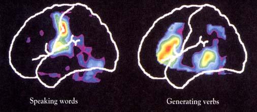
22 PET (positron emission tomography) scan 22
23 Functional Localization of Cerebral Cortex Sensory area primary sensory area secondary sensory area Motor area primary motor area secondary motor area supplementary motor area Association area parietal, occipital and temporal cortex - conceptual elaboration of sensory data prefrontal (frontal) cortex - judgement,, foresight Sensory Areas Somesthetic Area (Somesthesia( Somesthesia) Visual Area (vision) Auditory Area (Hearing) Vestibular Area (Equilibrium) Gustatory Area (Taste) Olfactory Area (Smell) S I, S II V I, V II A I, A II 23
24 Somesthetic Area S I , 1, 2 (postcentral( gyrus) afferernts: ventrobasal complex (VPLc( VPLc,, VPM) discrimination of position and intensity of sensation S II ---- superior bank of lateral fissure no clinical disorders Somesthetic Association Cortex , 7 (parietal lobule, precuneus) afferents: S I, LP of thalamus integration of geneal sensation with past experience tactile agnosia, astereognosis Sensory Homunculus 24
 Central region --- cutaneous (3b, 1)")
 superior bank of lateral")
25 Primary Somesthetic Area Thalmocortical connection (VPLc S I) Central region --- cutaneous (3b, 1) Peripheral region --- deep (3a, 2) Secondary Somesthetic Area (SII) superior bank of lateral fissure 25
 afferents: V I, pulvinar of thalamus integration of vision with past experience visual agnosia cf.")
26 Visual Cortex V I (striate cortex - line of Gennari) greatly thickened outer band of Baillarger heterotypical isocortex afferent: LGd of thalamus visual field defect: homonymous quadranopsia and macular sparing V II , 19 (visual association area) afferents: V I, pulvinar of thalamus integration of vision with past experience visual agnosia cf. occipital eye field Visual Areas 26
 heterotypical isocortex afferents: MGv of thalamus - core")
 belt projection A I auditory agnosia - sensory")
27 Visual association areas V4 (color) Face recognition Perceive Facial Expression Auditory Cortex A I , 42 (trannsverse( temporal gyrus of Heschl) heterotypical isocortex afferents: MGv of thalamus - core projection slight diminution in auditory acuity A II (Wernike's( area of original connotaion) not well-defined afferents: non-laminar part (MGm( MGm, MGd) belt projection A I auditory agnosia - sensory aphasia 27


28 Auditory Areas Planum temporale Auditory Areas A I , 42 A II
29 Other Primary Sensory Areas Vestibular Area Area 3a and 2v of S I afferents: VPLo [superior temporal gyrus anterior to A I] Gustatory Area Area 43 (inferior end of postcentral gyrus) afferents: VPMpc Olfactory Area Piriform Lobe - Limbic System Motor Areas primary Motor Area (M I) Premotor Area (PM) Supplementary Motor Area (SMA) Frontal Eye Field 29
30 Motor Homunculus Primary Motor Area M I precentral gyrus of lateral surface anterior part of paracentral lobule heterotypical agranular cortex giant pyramidal cell of Betz afferents: premotor area, SMA, S I VLc, VPLo of thalamus Motor Homunculus Upper Motor Neuron (UMN) syndrome 30
31 Other Motor Areas Premotor Area (PM) lateral surface of 6 afferents: VLc, VPLo of thalamus from cerebellum Supplementary Motor Area (SMA) medial surface of 6 afferents: VLo, Vapc of thalamus from basal ganglia Frontal Eye Field voluntary tracking movement Motor Systems IV Sensory & motor areas: post-central gyrus, parietal lobe pre-central gyrus, frontal lobe corpus striatum (basal ganglia), thalamus 31
32 Motor Systems IV Sensory & motor areas: post-central gyrus, parietal lobe pre-central gyrus, frontal lobe corpus striatum (basal ganglia), thalamus Motor circuits: putamen-globus pallidus-thalamus-premotor cortex caudate-globus pallidus-thalamus-supplementary motor cortex Motor Systems IV Sensory & motor areas: post-central gyrus, parietal lobe pre-central gyrus, frontal lobe corpus striatum (basal ganglia), thalamus Motor circuits: putamen-globus pallidus-thalamus-premotor cortex caudate-globus pallidus-thalamus-supplementary motor cortex Eye-hand co-ordination: frontal eye fields 32
33 Motor Systems IV Sensory & motor areas: post-central gyrus, parietal lobe pre-central gyrus, frontal lobe corpus striatum (basal ganglia), thalamus Motor circuits: putamen-globus pallidus-thalamus-premotor cortex caudate-globus pallidus-thalamus-supplementary motor cortex Eye-hand co-ordination: frontal eye fields Basal ganglia & cerebellum: convergence on descending pathways motor learning Premotor cortex 6 4 Motor cortex Frontal eye field 8 44 Supplementary motor cortex Motor cortex Motor areas of the cortex 33
34 Sensorimotor cortex Putamen Globus pallidus Thalamus Premotor cortex Basal ganglia Sensorimotor cortex Putamen Globus pallidus Thalamus Descending pathways Premotor cortex Basal ganglia 34
35 Sensorimotor cortex Pons Cerebellum Thalamus Cerebellum Cerebellum Thalamus Motor cortex Cerebellum 35
36 Cerebellum Descending pathways Thalamus Motor cortex Cerebellum Sensorimotor cortex Pons Cerebellum Descending pathways Thalamus Motor cortex Cerebellum 36
37 Sensorimotor cortex Sensorimotor cortex Putamen Pons Globus pallidus Cerebellum Thalamus Premotor cortex Descending pathways Thalamus Motor cortex Basal ganglia & cerebellum Basal ganglia via premotor cortex Motor cortex Cerebellum via thalamus Basal ganglia & cerebellum 37
38 Basal ganglia via premotor cortex No plasticity Motor cortex Cerebellum via thalamus Basal ganglia & cerebellum Basal ganglia via premotor cortex Synchronous Motor cortex Cerebellum via thalamus Basal ganglia & cerebellum 38
39 Basal ganglia via premotor cortex Plasticity Motor cortex Cerebellum via thalamus Basal ganglia & cerebellum Sensorimotor cortex Sensorimotor cortex Putamen Pons Globus pallidus Cerebellum Thalamus Premotor cortex Descending pathways Thalamus Motor cortex Basal ganglia & cerebellum 39
40 Association Areas Language Areas , 39, 40, 44, 45 Posterior Parietal Association Area , 7 (39, 40) body image Temporal Association Area , 21, 37, 38 (22) multisensory integration, conceptual ideation Prefrontal Association Area , 10, 11, 12, 46, 47 (44, 45) judgement,, foresight, personality Disorders of Association Cortex Agnosia Tactile agnosia Visual agnosia Alexia Auditory agnosia Apraxia Aphasia Wernicke s (receptive) aphasia Broca s (Motor) aphasia conduction aphasia global aphasia 40
 ---- 22, 39, 40 Receptive Aphasia - area 22 defect in comprehension, good spontaneous speech Anomic Aphasia - word finding difficulty Jargon")
41 Apraxia The inability to execute a voluntary motor movement despite being able to demonstrate normal muscle function. Language Areas Sensory Language Area (Wernike's( area) , 39, 40 Receptive Aphasia - area 22 defect in comprehension, good spontaneous speech Anomic Aphasia - word finding difficulty Jargon aphasia - fluent, but unintelligiable jargon 39 (supramarginal( gyrus), 40 (angular gyrus) Superior Longitudinal Fasciculus Conduction Aphasia good comprehension, good spontaneous speech poor repetition, poor response Motor Language Area (Broca( Broca s area) , 45 Motor Apahsia good comprehension, no speech 41


42 Language Areas (Geschwind( Model) Broca s Area Pars triangularis and pars opercularis of the inferior frontal gyrus of dominant hemisphere. Photograph of the brain of Paul Broca s patient called Tan (real name is Leborgne). 42


43 Paul Broca ( ) 1880) Carl Wernicke ( ) 1905) PET (positron emission tomography) scan 43


44 Prefrontal Association Areas Frontal Granular Cortex Lateral Prefrontal Association Area , 10, 46 judgement,, foresight, problem solving Orbitofrontal Cortex , 12, 47 emotion, olfaction, personality Case of Phineas Gage Prefrontal Leucotomy of Moniz and Freeman Phineas Gage ( , 1861, accident in 1848) 44
 Conceptual Framework of Cerebral Function")
45 Phineas Gage s s lesion reconstructed (H. Damasio and R. Frank, 1992) Conceptual Framework of Cerebral Function 45


46 Cerebral Dominance (Lateralization, Asymmetry) Dominant Hemisphere Language speech, writing Calculation Non-dominant Hemisphere Spatial Perception (3D subject) Singing Playing musical instrument Language Speech Writing Calculati on 3D perception Singing Playing Musical instrument 46
CEREBRUM Dr. Jamila Elmedany Dr. Essam Eldin Salama
 CEREBRUM Dr. Jamila Elmedany Dr. Essam Eldin Salama Objectives At the end of the lecture, the student should be able to: List the parts of the cerebral hemisphere (cortex, medulla, basal nuclei, lateral
Telencephalon (Cerebral Hemisphere)
") Telencephalon (Cerebral Hemisphere) OUTLINE The Cortex - Lobes, Sulci & Gyri - Functional Subdivisions - Limbic Lobe & Limbic System The Subcortex - Basal Ganglia - White Matter (Internal Capsule) - Relations
Telencephalon (Cerebral Hemisphere) OUTLINE The Cortex - Lobes, Sulci & Gyri - Functional Subdivisions - Limbic Lobe & Limbic System The Subcortex - Basal Ganglia - White Matter (Internal Capsule) - Relations
Neuroanatomy lecture (1)
") Neuroanatomy lecture (1) Introduction: Neuroanatomy has two parts: the central and peripheral nervous system. The central nervous system is composed of brain and spinal cord. The brain has the following
Neuroanatomy lecture (1) Introduction: Neuroanatomy has two parts: the central and peripheral nervous system. The central nervous system is composed of brain and spinal cord. The brain has the following
CEREBRUM & CEREBRAL CORTEX
 CEREBRUM & CEREBRAL CORTEX Seonghan Kim Dept. of Anatomy Inje University, College of Medicine THE BRAIN ANATOMICAL REGIONS A. Cerebrum B. Diencephalon Thalamus Hypothalamus C. Brain Stem Midbrain Pons
P. Hitchcock, Ph.D. Department of Cell and Developmental Biology Kellogg Eye Center. Wednesday, 16 March 2009, 1:00p.m. 2:00p.m.
 Normal CNS, Special Senses, Head and Neck TOPIC: CEREBRAL HEMISPHERES FACULTY: LECTURE: READING: P. Hitchcock, Ph.D. Department of Cell and Developmental Biology Kellogg Eye Center Wednesday, 16 March
CEREBRUM. Dr. Jamila EL Medany
 CEREBRUM Dr. Jamila EL Medany Objectives At the end of the lecture, the student should be able to: List the parts of the cerebral hemisphere (cortex, medulla, basal nuclei, lateral ventricle). Describe
Neuroanatomy. Dr. Maha ELBeltagy. Assistant Professor of Anatomy Faculty of Medicine The University of Jordan 2018
 Neuroanatomy Dr. Maha ELBeltagy Assistant Professor of Anatomy Faculty of Medicine The University of Jordan 2018 THE NERVOUS SYSTEM (NS) It is divided into 2 major divisions: 1) Central Nervous System
Homework Week 2. PreLab 2 HW #2 Synapses (Page 1 in the HW Section)
") Homework Week 2 Due in Lab PreLab 2 HW #2 Synapses (Page 1 in the HW Section) Reminders No class next Monday Quiz 1 is @ 5:30pm on Tuesday, 1/22/13 Study guide posted under Study Aids section of website
Homework Week 2 Due in Lab PreLab 2 HW #2 Synapses (Page 1 in the HW Section) Reminders No class next Monday Quiz 1 is @ 5:30pm on Tuesday, 1/22/13 Study guide posted under Study Aids section of website
Gross Morphology of the Brain
 Gross Morphology of the Brain Done by : Marah Marahleh & Razan Krishan *slides in bold Principal Parts of the Brain Cerebrum : largest part of the brain Diencephalon Thalamus & hypothalamus Cerebellum
Announcement. Danny to schedule a time if you are interested.
 Announcement If you need more experiments to participate in, contact Danny Sanchez (dsanchez@ucsd.edu) make sure to tell him that you are from LIGN171, so he will let me know about your credit (1 point).
Parts of the Brain. Hindbrain. Controls autonomic functions Breathing, Heartbeat, Blood pressure, Swallowing, Vomiting, etc. Upper part of hindbrain
 Parts of the Brain The human brain is made up of three main parts: 1) Hindbrain (or brainstem) Which is made up of: Myelencephalon Metencephalon 2) Midbrain Which is made up of: Mesencephalon 3) Forebrain
Anatomy and Physiology (Bio 220) The Brain Chapter 14 and select portions of Chapter 16
 The Brain Chapter 14 and select portions of Chapter 16") Anatomy and Physiology (Bio 220) The Brain Chapter 14 and select portions of Chapter 16 I. Introduction A. Appearance 1. physical 2. weight 3. relative weight B. Major parts of the brain 1. cerebrum 2.
Anatomy and Physiology (Bio 220) The Brain Chapter 14 and select portions of Chapter 16 I. Introduction A. Appearance 1. physical 2. weight 3. relative weight B. Major parts of the brain 1. cerebrum 2.
Cerebral Cortex 1. Sarah Heilbronner
 Cerebral Cortex 1 Sarah Heilbronner heilb028@umn.edu Want to meet? Coffee hour 10-11am Tuesday 11/27 Surdyk s Overview and organization of the cerebral cortex What is the cerebral cortex? Where is each
Leah Militello, class of 2018
 Leah Militello, class of 2018 Objectives 1. Describe the general organization of cerebral hemispheres. 2. Describe the locations and features of the different functional areas of cortex. 3. Understand
-Zeina Assaf. -Omar Odeh. - Maha Beltagy
 -3 -Zeina Assaf -Omar Odeh - Maha Beltagy 1 P a g e The Inferior Surface Of The Brain The inferior surface of the brain is divide by the stem of the lateral fissure into 2 parts : The orbital surface and
Outline of the next three lectures
 Outline of the next three lectures Lecture 35 Anatomy of the human cerebral cortex gross and microscopic cell types connections Vascular supply of the cerebral cortex Disorders involving the cerebral cortex
The Nervous system is divided into 2 major divisions: 1) Central Nervous System (CNS): found within bones & consists of:
 Central Nervous System (CNS): found within bones & consists of:") The Nervous system is divided into 2 major divisions: 1) Central Nervous System (CNS): found within bones & consists of: - The Brain: within the skull, composed of cerebrum, cerebellum and brain stem.
The Nervous system is divided into 2 major divisions: 1) Central Nervous System (CNS): found within bones & consists of: - The Brain: within the skull, composed of cerebrum, cerebellum and brain stem.
14 - Central Nervous System. The Brain Taft College Human Physiology
 14 - Central Nervous System The Brain Taft College Human Physiology Development of the Brain The brain begins as a simple tube, a neural tube. The tube or chamber (ventricle) is filled with cerebrospinal
Cerebrum-Cerebral Hemispheres. Cuneyt Mirzanli Istanbul Gelisim University
 Cerebrum-Cerebral Hemispheres Cuneyt Mirzanli Istanbul Gelisim University The largest part of the brain. Ovoid shape. Two incompletely separated cerebral hemispheres. The outer surface of the cerebral
BASIC ANATOMICAL STRUCTURES. The Cerebrum
 BASIC ANATOMICAL STRUCTURES The Cerebrum Development of the Cerebral Vesicles Primary Vesicle secondary Vesicles CNS structures Ventricle telencephalon Cerebral Hemispheres Lateral Ventricles Prosencephalon
Motor Functions of Cerebral Cortex
 Motor Functions of Cerebral Cortex I: To list the functions of different cortical laminae II: To describe the four motor areas of the cerebral cortex. III: To discuss the functions and dysfunctions of
Inferior Surface of the Brain:-
 The following sheet s sources: -Recording Section 2 -Slides All slides not mentioned by the doctor will also be included in the sheet. -Wikipedia Inferior Surface of the Brain:- The inferior surface of
Anatomy Lab (1) Theoretical Part. Page (2 A) Page (2B)
 Theoretical Part. Page (2 A) Page (2B)") Anatomy Lab (1) This sheet only includes the extra notes for the lab handout regarding the theoretical part, as for the practical part it includes everything the doctor mentioned. Theoretical Part Page
Anatomy Lab (1) This sheet only includes the extra notes for the lab handout regarding the theoretical part, as for the practical part it includes everything the doctor mentioned. Theoretical Part Page
The Central Nervous System I. Chapter 12
 The Central Nervous System I Chapter 12 The Central Nervous System The Brain and Spinal Cord Contained within the Axial Skeleton Brain Regions and Organization Medical Scheme (4 regions) 1. Cerebral Hemispheres
Nervous System: Part IV The Central Nervous System The Brain
 Nervous System: Part IV The Central Nervous System The Brain Can you survive when part of your brain is destroyed? 2 Essential Knowledge 3.D.2 2. Cells communicate with each other through direct contact
Human Anatomy. Brain and Cranial Nerves
 Human Anatomy Brain and Cranial Nerves 1 Brain and Cranial Nerves An adult brain weighs between 1.35 and 1.4 kilograms (kg) (around 3 pounds) and has a volume of about 1200 cubic centimeters (cc). Brain
Chapter 14: The Brain and Cranial Nerves. Copyright 2009, John Wiley & Sons, Inc.
 Chapter 14: The Brain and Cranial Nerves Development of the Brain Three to four-week embryo: prosencephalon, mesencephalon and rhombencephalon. Five-week embryo: telencephalon (cerebrum), diencephalon
Systems Neuroscience Dan Kiper. Today: Wolfger von der Behrens
 Systems Neuroscience Dan Kiper Today: Wolfger von der Behrens wolfger@ini.ethz.ch 18.9.2018 Neurons Pyramidal neuron by Santiago Ramón y Cajal (1852-1934, Nobel prize with Camillo Golgi in 1906) Neurons
Psyc 311A, fall 2008 Conference week 3 TA: Jürgen Germann
 Psyc 311A, fall 2008 Conference week 3 TA: Jürgen Germann e-mail: jurgen.germann@mcgill.ca Overview: 1. Meninges 2. Cerebral cortex-cytoarchitecture 3. Diencephalon (thalamus/hypothalamus) (this replaces
FRONTAL LOBE. Central Sulcus. Ascending ramus of the Cingulate Sulcus. Cingulate Sulcus. Lateral Sulcus
 FRONTAL LOBE Central Ascending ramus of the Cingulate Cingulate Lateral Lateral View Medial View Motor execution and higher cognitive functions (e.g., language production, impulse inhibition, reasoning
Chapter 18: The Brain & Cranial Nerves. Origin of the Brain
 Chapter 18: The Brain & Cranial Nerves BIO 218 Fall 2015 Origin of the Brain The brain originates from a structure called the neural tube, which arises during a developmental stage called neurulation.
Stanley Pruisinger 1980's
 Neuroanatomy Prion disease cerebellum chapter b/c cerebellar ataxia here as a warning for obvious reasons. Creutzfeldt - Jakob Disease (CJD) "Spongiform" (brain turns to sponge) Jews in Lybia who ate
Cortical Organization. Functionally, cortex is classically divided into 3 general types: 1. Primary cortex:. - receptive field:.
 Cortical Organization Functionally, cortex is classically divided into 3 general types: 1. Primary cortex:. - receptive field:. 2. Secondary cortex: located immediately adjacent to primary cortical areas,
GENERALITIES OF THE ADULT BRAIN
 GENERALITIES OF THE ADULT BRAIN Average weight = 1.3-1.4 kg o 2-3% of body weight Total intracranial volume = 1,700 ml o Brain = 1,400 ml (80%) o Blood = 150 ml (10%) o CSF = 150 ml (10%) Number of neurons
PROPERTY OF ELSEVIER SAMPLE CONTENT - NOT FINAL. Gross Anatomy and General Organization of the Central Nervous System
 3 Gross Anatomy and General Organization of the Central Nervous System C h a p t e r O u t l i n e The Long Axis of the CNS Bends at the Cephalic Flexure Hemisecting a Brain Reveals Parts of the Diencephalon,
Exam 1 PSYC Fall 1998
 Exam 1 PSYC 2022 Fall 1998 (2 points) Briefly describe the difference between a dualistic and a materialistic explanation of brain-mind relationships. (1 point) True or False. George Berkely was a monist.
PSY 302: CHAPTER 3 NOTES THE BRAIN (PART II) - 9/5/17. By: Joseline
 - 9/5/17. By: Joseline") PSY 302: CHAPTER 3 NOTES THE BRAIN (PART II) - 9/5/17 By: Joseline Left 3 MAJOR FISSURES : 2HEMISPHERES Right Lateral Ventricle Central Fissure Third Ventricle Sulcus Lateral Fissure Gyros Fissure- Fissures
PSY 302: CHAPTER 3 NOTES THE BRAIN (PART II) - 9/5/17 By: Joseline Left 3 MAJOR FISSURES : 2HEMISPHERES Right Lateral Ventricle Central Fissure Third Ventricle Sulcus Lateral Fissure Gyros Fissure- Fissures
Gives few collaterals, it is mainly a single process surrounded by a myelin sheath
 Lecture 1 - Nerve fiber refers to both axons and dendrites, the dendrites are the afferent fibers (sensory); they receive impulses from neighbouring neurons, and the axon is the efferent fiber (motor);
Chapter 14, Part 2! Chapter 14 Part 2 Brain/Cranial Nerves! The Cerebrum and Cranial Nerves! pp !
 Chapter 14, Part 2! The Cerebrum and Cranial pp. 482 505! SECTION 14-9! The cerebrum, the largest region of the brain, contains motor, sensory, and association areas! 2! White Matter of the Cerebrum! 1.
I: To describe the pyramidal and extrapyramidal tracts. II: To discuss the functions of the descending tracts.
 Descending Tracts I: To describe the pyramidal and extrapyramidal tracts. II: To discuss the functions of the descending tracts. III: To define the upper and the lower motor neurons. 1. The corticonuclear
神經解剖學 NEUROANATOMY TELENCEPHALON 盧家鋒助理教授 臺北醫學大學醫學系解剖學暨細胞生物學科 臺北醫學大學醫學院轉譯影像研究中心.
 神經解剖學 NEUROANATOMY TELENCEPHALON 盧家鋒助理教授 臺北醫學大學醫學系解剖學暨細胞生物學科 臺北醫學大學醫學院轉譯影像研究中心 http://www.ym.edu.tw/~cflu REGIONAL NEUROBIOLOGY Telencephalon (Cerebrum) Diencephalon (Thalamus) Cerebellum Brain stem Midbrain
For more information about how to cite these materials visit
 Author(s): Peter Hitchcock, PH.D., 2009 License: Unless otherwise noted, this material is made available under the terms of the Creative Commons Attribution Non-commercial Share Alike 3.0 License: http://creativecommons.org/licenses/by-nc-sa/3.0/
Chapter 14, Part 2! The Cerebrum and Cranial Nerves! pp !
 Chapter 14, Part 2! The Cerebrum and Cranial pp. 482 505! SECTION 14-9! The cerebrum, the largest region of the brain, contains motor, sensory, and association areas! 2! 1! ! Chapter 14 Part 2 Brain/Cranial
Supplementary Material S3 Further Seed Regions
 Supplementary Material S3 Further Seed Regions Figure I. Changes in connectivity with the right anterior insular cortex. (A) wake > mild sedation, showing a reduction in connectivity between the anterior
Biological Bases of Behavior. 3: Structure of the Nervous System
 Biological Bases of Behavior 3: Structure of the Nervous System Neuroanatomy Terms The neuraxis is an imaginary line drawn through the spinal cord up to the front of the brain Anatomical directions are
The Neuroscience of Music in Therapy
 Course Objectives The Neuroscience of Music in Therapy Unit I. Learn Basic Brain Information Unit II. Music in the Brain; Why Music Works Unit III. Considerations for Populations a. Rehabilitation b. Habilitation
Overview of Brain Structures
 First Overview of Brain Structures Psychology 470 Introduction to Chemical Additions Steven E. Meier, Ph.D. All parts are interrelated. You need all parts to function normally. Neurons = Nerve cells Listen
25/09/2012. Capgras Syndrome. Chapter 2. Capgras Syndrome - 2. The Neural Basis of Cognition
 Chapter 2 The Neural Basis of Cognition Capgras Syndrome Alzheimer s patients & others delusion that significant others are robots or impersonators - paranoia Two brain systems for facial recognition -
b. The groove between the two crests is called 2. The neural folds move toward each other & the fuse to create a
 Chapter 13: Brain and Cranial Nerves I. Development of the CNS A. The CNS begins as a flat plate called the B. The process proceeds as: 1. The lateral sides of the become elevated as waves called a. The
CNS consists of brain and spinal cord Cephalization Evolutionary development of rostral (anterior) portion of CNS Increased number of neurons in head
 portion of CNS Increased number of neurons in head") CNS consists of brain and spinal cord Cephalization Evolutionary development of rostral (anterior) portion of CNS Increased number of neurons in head Highest level reached in human brain 1 Mostly to orient
CNS consists of brain and spinal cord Cephalization Evolutionary development of rostral (anterior) portion of CNS Increased number of neurons in head Highest level reached in human brain 1 Mostly to orient
Copy Right- Hongqi ZHANG-Department of Anatomy-Fudan University. Systematic Anatomy. Nervous system Telencephalon. Dr.Hongqi Zhang ( 张红旗 )
") Systematic Anatomy Nervous system Telencephalon Dr.Hongqi Zhang ( 张红旗 ) Email: zhanghq58@126.com 1 The Telencephalon Gray matter Cortex Basilar nuclei White matter-medulla Lateral ventricles General introduction
Systematic Anatomy Nervous system Telencephalon Dr.Hongqi Zhang ( 张红旗 ) Email: zhanghq58@126.com 1 The Telencephalon Gray matter Cortex Basilar nuclei White matter-medulla Lateral ventricles General introduction
Chapter 3. Structure and Function of the Nervous System. Copyright (c) Allyn and Bacon 2004
 Allyn and Bacon 2004") Chapter 3 Structure and Function of the Nervous System 1 Basic Features of the Nervous System Neuraxis: An imaginary line drawn through the center of the length of the central nervous system, from the
Chapter 3 Structure and Function of the Nervous System 1 Basic Features of the Nervous System Neuraxis: An imaginary line drawn through the center of the length of the central nervous system, from the
The Central Nervous System
 The Central Nervous System 1 The Central Nervous System Brain Spinal Cord Components 2 Protection of the Brain The brain is protected from injury by The skull enough said! You already know this. What else
Neocortex. Hemispheres 9/22/2010. Psychology 472 Pharmacology of Psychoactive Drugs. Structures are divided into several section or lobes.
 Neocortex Psychology 472 Pharmacology of Psychoactive Drugs 1 Is the most developed in Humans Has many folds and fissures The folds of tissue are called gyri or a gyrus (single) The fissures or valleys
I. Anatomy of the Brain A. Cranial Meninges and Ventricles of the Brain 1. Meninges a. Dura mater 1) Endosteal/Periosteal Layer - Outer 2) Meningeal
 Endosteal/Periosteal Layer - Outer 2) Meningeal") I. Anatomy of the Brain A. Cranial Meninges and Ventricles of the Brain 1. Meninges a. Dura mater 1) Endosteal/Periosteal Layer - Outer 2) Meningeal Layer - Inner 3) Falx cerebri a) Superior sagittal sinus
I. Anatomy of the Brain A. Cranial Meninges and Ventricles of the Brain 1. Meninges a. Dura mater 1) Endosteal/Periosteal Layer - Outer 2) Meningeal Layer - Inner 3) Falx cerebri a) Superior sagittal sinus
The One Who Falls And Gets Up Is So Much Stronger Than The One Who Never Fell
 Text Important Formulas Numbers Doctor notes Notes and explanation Lecture No.19 The One Who Falls And Gets Up Is So Much Stronger Than The One Who Never Fell 1 Function of Cerebral hemisphere Objectives:
Telencephalon part 2
 Telencephalon part 2 1. Olfactory system, rhinencephalon 2. Limbic system: hippocampal formation amygdala 3. Main cortical areas: sensory areas of the cortex motor areas of the cortex 4. Functional localization
Learning Objectives.
 Emilie O Neill, class of 2016 Learning Objectives 1. Describe the types of deficits that occur with lesions in association areas including: prosopagnosia, neglect, aphasias, agnosia, apraxia 2. Discuss
Auditory and Vestibular Systems
 Auditory and Vestibular Systems Objective To learn the functional organization of the auditory and vestibular systems To understand how one can use changes in auditory function following injury to localize
49a A&P: Nervous System -! Synaptic Transmission and Central Nervous System
 49a A&P: Nervous System -! Synaptic Transmission and Central Nervous System 49a A&P: Nervous System -! Synaptic Transmission and Central Nervous System! Class Outline" 5 minutes" "Attendance, Breath of
Neuroanatomy. Dr. Maha ELBeltagy. Assistant Professor of Anatomy Faculty of Medicine The University of Jordan
 Neuroanatomy Dr. Maha ELBeltagy Assistant Professor of Anatomy Faculty of Medicine The University of Jordan 2018 Prof Yousry 10/15/17 Types of brain fibers THE WHITE MATTER OF THE BRAIN The white matter
The Nervous System PART B
 7 The Nervous System PART B PowerPoint Lecture Slide Presentation by Jerry L. Cook, Sam Houston University ESSENTIALS OF HUMAN ANATOMY & PHYSIOLOGY EIGHTH EDITION ELAINE N. MARIEB The Reflex Arc Reflex
Nervous System, Neuroanatomy, Neurotransmitters
 Nervous System, Neuroanatomy, Neurotransmitters Neurons Structure of neurons Soma Dendrites Spines Axon Myelin Nodes of Ranvier Neurons Structure of neurons Axon collaterals 1 Neurons Structure of neurons
Effects of Cerebellar Damage
 Effects of Cerebellar Damage Impaired rapid alternation movements Directed movements overshoot their mark (past-pointing on finger-tonose). Diencephalon COMPOSED OF THREE THALAMIC STRUCTURES: Thalamus
biological psychology, p. 40 The study of the nervous system, especially the brain. neuroscience, p. 40
 biological psychology, p. 40 The specialized branch of psychology that studies the relationship between behavior and bodily processes and system; also called biopsychology or psychobiology. neuroscience,
Gross Organization I The Brain. Reading: BCP Chapter 7
 Gross Organization I The Brain Reading: BCP Chapter 7 Layout of the Nervous System Central Nervous System (CNS) Located inside of bone Includes the brain (in the skull) and the spinal cord (in the backbone)
Prof. Saeed Abuel Makarem & Dr.Sanaa Alshaarawy
 Prof. Saeed Abuel Makarem & Dr.Sanaa Alshaarawy 1 Objectives By the end of the lecture, you should be able to: Describe the anatomy and main functions of the thalamus. Name and identify different nuclei
Central Nervous System. January 7, 2016
 Central Nervous System January 7, 2016 Anatomy of a neuron Cell Body (soma) Receives information from the soma s extensions (dendrites) Passes on information away from the soma towards extensions (axons)
Central Nervous System
 Central Nervous System January 7, 2016 Anatomy of a neuron Cell Body (soma) Receives information from the soma s extensions (dendrites) Passes on information away from the soma towards extensions (axons)
The neurvous system senses, interprets, and responds to changes in the environment. Two types of cells makes this possible:
 NERVOUS SYSTEM The neurvous system senses, interprets, and responds to changes in the environment. Two types of cells makes this possible: the neuron and the supporting cells ("glial cells"). Neuron Neurons
Introduction to the Central Nervous System: Internal Structure
 Introduction to the Central Nervous System: Internal Structure Objective To understand, in general terms, the internal organization of the brain and spinal cord. To understand the 3-dimensional organization
BRAIN PART I (A & B): VENTRICLES & MENINGES
: VENTRICLES & MENINGES") BRAIN PART I (A & B): VENTRICLES & MENINGES Cranial Meninges Cranial meninges are continuous with spinal meninges Dura mater: inner layer (meningeal layer) outer layer (endosteal layer) fused to periosteum
BRAIN PART I (A & B): VENTRICLES & MENINGES Cranial Meninges Cranial meninges are continuous with spinal meninges Dura mater: inner layer (meningeal layer) outer layer (endosteal layer) fused to periosteum
Fig.1: A, Sagittal 110x110 mm subimage close to the midline, passing through the cingulum. Note that the fibers of the corpus callosum run at a
 Fig.1 E Fig.1:, Sagittal 110x110 mm subimage close to the midline, passing through the cingulum. Note that the fibers of the corpus callosum run at a slight angle are through the plane (blue dots with
Blood supply to the brain Blood brain barrier isolates neural tissue from general circulation
 The Brain and Cranial Nerves Objectives Name the major regions of the brain and describe their functions. Discuss the formation, circulation, and functions of the CSF. List the main components of the medulla
Telencephalon part 1 ( endbrain )
") part 1 ( endbrain ) 1. (cerebrum) general overview 2. Embryonic and postnatal development 3. Cerebral hemispheres: surface anatomy major sulci, gyri and lobes cerebral cortex microscopic structure, cyto-
part 1 ( endbrain ) 1. (cerebrum) general overview 2. Embryonic and postnatal development 3. Cerebral hemispheres: surface anatomy major sulci, gyri and lobes cerebral cortex microscopic structure, cyto-
Layered organization of cortex: Paleocortex 3 layers hippocampal formation / ventral & medial cortex closest to brainstem
 Layered organization of cortex: Paleocortex 3 layers hippocampal formation / ventral & medial cortex closest to brainstem Archicortex 3-4 layers hippocampal formation / amygdala Neocortex 6 layers more
skilled pathways: distal somatic muscles (fingers, hands) (brainstem, cortex) are giving excitatory signals to the descending pathway
 (brainstem, cortex) are giving excitatory signals to the descending pathway") L15 - Motor Cortex General - descending pathways: how we control our body - motor = somatic muscles and movement (it is a descending motor output pathway) - two types of movement: goal-driven/voluntary
L15 - Motor Cortex General - descending pathways: how we control our body - motor = somatic muscles and movement (it is a descending motor output pathway) - two types of movement: goal-driven/voluntary
Neuroanatomy. Cerebral Cortex: Movement and Speech
 Neuroanatomy Cerebral Cortex: Movement and Speech Functional Neuroanatomy Phrenology: Pseudoscience Functional neuroanatomy is the study of how different parts of the brain control different aspects of
THE CENTRAL NERVOUS SYSTEM. The Brain & Spinal Cord
 THE CENTRAL NERVOUS SYSTEM The Brain & Spinal Cord Review: Nervous System Parallel Distributed Processing Composition of the CNS Nuclei: Clusters of neurons in the CNS ( neighborhoods ) Fiber Tracts/Pathways:
1. The basic anatomy of the Central Nervous System (CNS)
") Psyc 311A, fall 2008 Conference week 1 Sept 9 th to 11 th TA: Jürgen Germann; e-mail: jurgen.germann@mcgill.ca Overview: 1. The basic anatomy of the Central Nervous System (CNS) 2. Cells of the CNS 3.
Psyc 311A, fall 2008 Conference week 1 Sept 9 th to 11 th TA: Jürgen Germann; e-mail: jurgen.germann@mcgill.ca Overview: 1. The basic anatomy of the Central Nervous System (CNS) 2. Cells of the CNS 3.
Central Nervous System
 Anatomy of a neuron Cell Body (soma) Receives information from the soma s extensions (dendrites) Central Nervous System January 7, 2016 Passes on information away from the soma towards extensions (axons)
The Nervous System 7PART B. PowerPoint Lecture Slide Presentation by Patty Bostwick-Taylor, Florence-Darlington Technical College
 PowerPoint Lecture Slide Presentation by Patty Bostwick-Taylor, Florence-Darlington Technical College The Nervous System 7PART B What is a reflex? What is a reflex? What is meant by the statement that
Histology of the CNS
 Histology of the CNS Lecture Objectives Describe the histology of the cerebral cortex layers. Describe the histological features of the cerebellum; layers and cells of cerebellar cortex. Describe the elements
M555 Medical Neuroscience Lab 1: Gross Anatomy of Brain, Crainal Nerves and Cerebral Blood Vessels
 M555 Medical Neuroscience Lab 1: Gross Anatomy of Brain, Crainal Nerves and Cerebral Blood Vessels Anatomical Directions Terms like dorsal, ventral, and posterior provide a means of locating structures
BASAL GANGLIA. Dr JAMILA EL MEDANY
 BASAL GANGLIA Dr JAMILA EL MEDANY OBJECTIVES At the end of the lecture, the student should be able to: Define basal ganglia and enumerate its components. Enumerate parts of Corpus Striatum and their important
From Brodal, 2 nd ed. fig 20.1; 3 rd ed. Fig 21.1
 From Brodal, 2 nd ed. fig 20.1; 3 rd ed. Fig 21.1 Note the names of the layers-- named for cell types rather than for axonal stratification. Figure removed due to copyright restrictions. Please see figure
Medical Neuroscience Tutorial Notes
 Medical Neuroscience Tutorial Notes Lateral Surface of the Brain MAP TO NEUROSCIENCE CORE CONCEPTS 1 NCC1. The brain is the body's most complex organ. LEARNING OBJECTIVES After study of the assigned learning
Forebrain Brain Structures Limbic System. Brain Stem Midbrain Basil Ganglia. Cerebellum Reticular Formation Medulla oblongata
 Brain structures (1) Cut out the following cards (2) Identify the three major divisions of the brain (as defined by your book). Initially, try this without any form of aid such as your textbook. (3) Organize
Ch 13: Central Nervous System Part 1: The Brain p 374
 Ch 13: Central Nervous System Part 1: The Brain p 374 Discuss the organization of the brain, including the major structures and how they relate to one another! Review the meninges of the spinal cord and
Announcement. Overview. Words Describing Sectional Planes. Words Describing Spatial Orientation. Explore! Basic neuroscience terminology
 Announcement Explore! The reading list is a good place to start, especially the Perspectives section. Overview Basic neuroscience terminology A Roadmap to the course Also, check out short articles in the
Brain anatomy and artificial intelligence. L. Andrew Coward Australian National University, Canberra, ACT 0200, Australia
 Brain anatomy and artificial intelligence L. Andrew Coward Australian National University, Canberra, ACT 0200, Australia The Fourth Conference on Artificial General Intelligence August 2011 Architectures
Myers Psychology for AP*
 Myers Psychology for AP* David G. Myers PowerPoint Presentation Slides by Kent Korek Germantown High School Worth Publishers, 2010 *AP is a trademark registered and/or owned by the College Board, which
Group D: Central nervous system yellow
 Group D: Central nervous system yellow Central nervous system 1. General structure of nervous system (neuron, glia, synapsis, mediators, receptors) Main points: types of neurons and glial cells, synapses,
Orientation, Development, Gross Anatomy, Blood Supply and Meninges References... 3
 Section I Orientation, Development, Gross Anatomy, Blood Supply and Meninges... 1 1 Orientation... 3 References... 3 2 Development... 7 Early Morphogenesis... 7 FormationoftheBrainRegions... 9 Histogenesis...
The human brain. of cognition need to make sense gives the structure of the brain (duh). ! What is the basic physiology of this organ?
. ! What is the basic physiology of this organ?") The human brain The human brain! What is the basic physiology of this organ?! Understanding the parts of this organ provides a hypothesis space for its function perhaps different parts perform different
The human brain The human brain! What is the basic physiology of this organ?! Understanding the parts of this organ provides a hypothesis space for its function perhaps different parts perform different
DIENCEPHALON. ..Central core of the forebrain..consists of three paired structures
 DIENCEPHALON..Central core of the forebrain..consists of three paired structures ---------- --thalamus, --------hypothalamus, -------epithalamus..encloses the third ventricle Diencephalon between cerebral
Anatomy of Cerebral Hemispheres
 Anatomy of Cerebral Hemispheres Please check our Editing File هذا العمل مب ي ن بشكل أسا ي س عىل عمل دفعة 436 مع المراجعة والتدقيق وإضافة المالحظات وال يغ ي ن عن المصدر األسا ي س للمذاكرة Lecture (15) Important
2401 : Anatomy/Physiology
 Dr. Chris Doumen Week 7 2401 : Anatomy/Physiology The Cerebrum Central Nervous System TextBook Readings Pages 434-456 and 460-461 Make use of the figures in your textbook ; a picture is worth a thousand
PETER PAZMANY CATHOLIC UNIVERSITY Consortium members SEMMELWEIS UNIVERSITY, DIALOG CAMPUS PUBLISHER
 PETER PAZMANY CATHOLIC UNIVERSITY SEMMELWEIS UNIVERSITY Development of Complex Curricula for Molecular Bionics and Infobionics Programs within a consortial* framework** Consortium leader PETER PAZMANY
Lecture - Chapter 13: Central Nervous System
 Lecture - Chapter 13: Central Nervous System 1. Describe the following structures of the brain, what is the general function of each: a. Cerebrum b. Diencephalon c. Brain Stem d. Cerebellum 2. What structures