Copy Right- Hongqi ZHANG-Department of Anatomy-Fudan University. Systematic Anatomy. Nervous system Telencephalon. Dr.Hongqi Zhang ( 张红旗 )
|
|
|
- Arthur Bennett
- 5 years ago
- Views:
Transcription


 Email:")
1 Systematic Anatomy Nervous system Telencephalon Dr.Hongqi Zhang ( 张红旗 ) zhanghq58@126.com 1
2 The Telencephalon
3 Gray matter Cortex Basilar nuclei White matter-medulla Lateral ventricles

4 General introduction of the telencephalon From the development of forebrain vesicle, Younger but with important function. Situated at the sup.extremity of the brain Formation of many sulci and gyri because of different growth speed of brain. Sulci and gyri increase the surface area of cortex
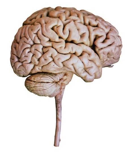
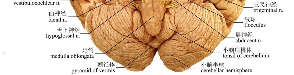
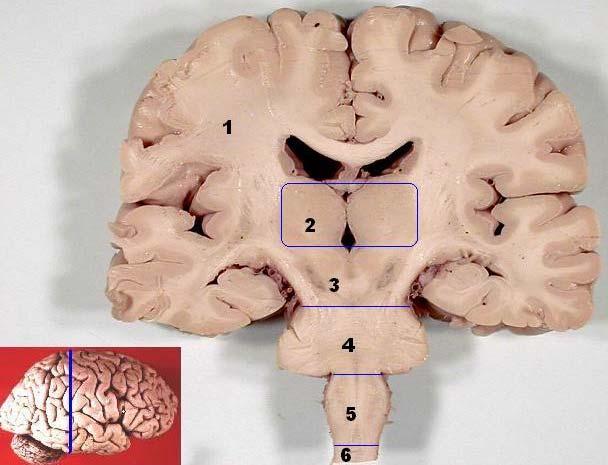
5 Frontal lobe Olfactory tract Optic n. Optic chiasma Temporal lobe Ponse Tonsile of cerebellum Pyramid Olive Cerebellum Spinal cord

6 Precentral gyrus Sup.frontal sulcus Central sulcus Inf.frontal sulcus postcentral gyrus Posstcentral sulcus Olfactory tract Insula Fornix Cerebellum
7 Cingulate gyrus Collosal sulcus Corpus collasum Cerebral aqueduct Parietoccipital sulcus Calcarine sulcus Transparent septum Optic n. Third ventricle Transverse Cerebral fissure Midbrain Fourth ventricle Pons Medulla Copy Right- Hongqi ZHANG-Department oblongata of Anatomy-Fudan University

8 Superior view Anterior view Lateral view inferior view Posterior view sagittal view

9 Frontal lobe Olfactory tract Optic n. Optic chiasma Temporal lobe Ponse Tonsile of cerebellum Pyramid Olive Cerebellum Spinal cord
connects the hemispheres across the midline 2 1")
10 General Appearance of Cerebrum right & left cerebral hemisphere, separated by cerebral longitudinal fissure(1)- cerebral falx In the depths of the fissure, the corpus callosum(2)connects the hemispheres across the midline 2 1 3

11 General Appearance of Cerebrum The cerebral transverse fissure(1) intervenes between the hemispheres and the cerebellum- cerebellar tentorium Each hemisphere has three surfaces: superolateral,medial and inferior one. 1 1 Cerebellar tentorium 1


12 Key points of the appearance of the telencephalon Three sulci Three surfaces Five lobes Three poles Two ventricles


13 1 Three sulci 3 Lateral view 2 1-Central sulcus 2-Lateral sulcus 3-Parietooccipital sulcus medial view


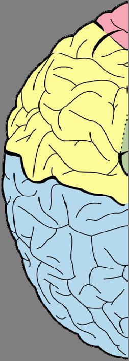
14 Five lobes of Cerebral Hemisphere Frontal lobe Parietal lobe Temporal lobe Occipital lobe Insular lobe Insular lobe


15 Three Surfaces of telencephalon 1- Dorsolateral surface 2- Medial surface 3- Inferior surface 1 2 3

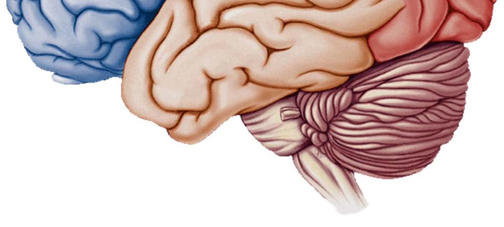
16 Three poles of telencephalon 1-Frontal pole 2-Occipital pole 3-Temporal pole 1 4-Preoccipital notch 3 Boundary between temporal lobe and occipital lobe 4 2
17 Main sulci & gyri of dorsolateral surface 1-Central sulcus 2-Lateral sulcus Precentral sulcus 9 4-Postcentral sulcus 5-sup.frontal sulcus 6-Inf.frontal sulcus 6 7- Inf.temporal sulcus 2 8- Sup.temporal sulcus 8 9-Intraparietal sulcus 7

18 Main gyri of dorsolateral surface Sup.frontal g. Mid.frontal g. Inf.frontal g. Sup.temporal g. Mid.temporal g. Inf.temporal g. Precentral g. Sup.parietal lobule Inf. parietal lobule Supramarginal g. Angular g. Postcentral g. Trans.temporal g. g.-gyrus

19 Copy Right- Hongqi Brain ZHANG-Department lateral aspect of Anatomy-Fudan University


20 Sulci and gyri of Superolateral surface Precentral gyrus Precentral sulcus Postcentral gyrus Superior frontal sulcus Inferior frontal sulcus Sup,mid & inf. frontal gyri Postcentral sulcus Intraparietal sulcus Superior parietal lobule Supramarginal gyrus Angular gyrus Superior temporal gyrus Middle temporal gyrus Inferior temporal gyrus Superior temporal sulcus Inferior temporal sulcus Central sulcus


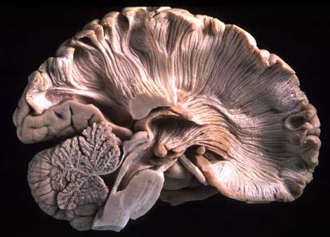
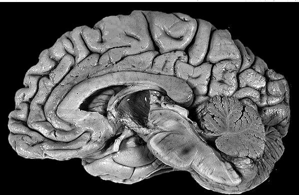
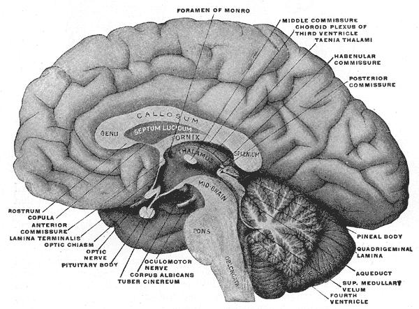
21 Brain median sagittal plane

22 Medial surface of hemisphere

23 Sulci and Gyri of Medial Surface Callosal sulcus cingulate gyrus Cingulate sulcus Corpus callosum Paracentral lobule Marginal ramus Parietooccipital sulcus Cuneus Calcarine sulcus Lingual gyrus

24 Inferior surface of hemisphere

25 Brain and roots of cranial nerve(basilar aspect)

26 Limbic lobe and limbic system Cingulate.parahippocampal gyri,uncus & the hippocampal formation surrounding the upper brain stem,constitutes the limbic lobe. the limbic system is used to include the limbic lobe as well as associated subcortical structures including amygdaloid body, septal nuclei, mammillary body,ant. nucleus of thalamus,olfactory bulb.
27 The function of limbic system The limbic system is also known as visceral brain, its function is concerned with: Emotional expression and genesis together with visceral response to the emotions. Survival of individual and species. Cognitive processes involved in memory.


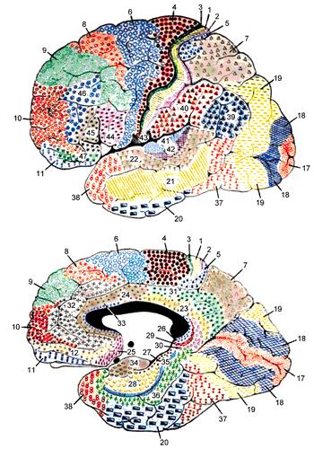
28 Functional Location of Cerebral Cortex Brodmann 52 areas

29 Some structures of temporal lobe Fornix Hippocampus Dentate gyrus Hippocampal formation Commissure of fornix
30 The internal structures of telencephalon Gray matter Cortex Basilar ganglion cortex medulla nuclear white matter-medulla Lateral ventricles
31 Key functional principal of the cerebral cortex Very important Somatic motor area Somatic sensory area Visual area Auditory area Motor speech area Writing area Auditory speech area Visual speech area Olfactory area Gustatory area sensory area Visceral activity area The rest area except the functional areas are called association area.
32 Precentral gyrus Somatic motor area location Precentral gyrus ant.part of the paracentral lobule (B4.6) Ant.part of the paracentral lobule

33 Motor cortex The main motor cortical area is located on the anterior wall of the central sulcus and the adjacent portion of the precentral gyrus. Brodmann Area 4 - rich in pyramidal neurons, which provide the anatomical substrates for the motor output function

34 Characters Representative area of the body is inverted,but head is upright Representative area of various part of body in cortex have relations with its function,rather than its size Left and right decussation: one somatic motor center innervate the movement of skeletal m.of contralateral limbs & both trunk skeleton mm.


35 First Somatic Motor Area

 post.")
36 Copy Right- Hongqi ZHANG-Department of Anatomy-Fudan postcentral University gyrus Somatic sensory area Location Postcentral gyrus post.part of the paracentral lobule (B3.1.2) post.part of the paracentral lobule

37 Characteristics First Somatic Motor Area Representative area of the body is inverted,but it is upright Represented area of different part in cortex have relations with its function,rather than its size Left and right decussation: One somatic sensory area receive contralateral sensory impulse
38 Visual area Calcarine sulcus and adjacent portions of the cuneus and the lingual gyrus (B17) Visual cortex of one hemisphere receives impression from temporal part of retina of same side & nasal part of opposite side retina Lesions of visual cortex produce contralateral homonymous visual field defections Visual area

.")


39 Auditory area Transverse temporal gyri (B41). Each auditory area receive auditory information from both ears. Auditory area


40 Language Area It is dominant in left hemisphere in right-handed person Motor speech area Located in post. portion of inferior frontal gyrus Damage: motor aphasia Writing area Located in post. portion of middle frontal gyrus Damage: agraphia Auditory speech area Located in post. portion of superior temporal gyrus Lesion: sensory aphasia Visual speech area Located in angular gyrus Lesion: alexia writing area Motor speech area visual speech area auditory speech area
41 Vestibular,olfactory and taste areas Vestibular area located in the front of superior temporal gyrus Olfactory area area near the uncus Taste area - area at frontal operculum Visceral area - limbic system

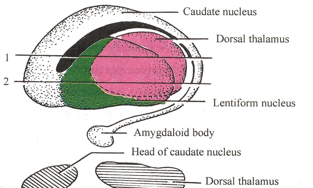
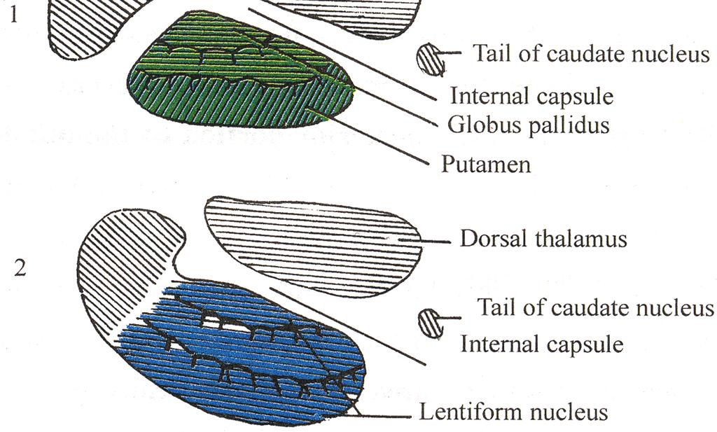
42 Basal ganglion (nuclei) 1- Lentiform nucleus 2- Caudate nucleus 3- Claustrum 4- Amygdaloid body 3 T T- dorsal thalamus

43 Basal Nuclei Corpus striatum Lentiform nucleus Caudate nucleus Claustrum Amygdaloid body Caudate nucleus Globus pallidus -paleostriatum Putamen Neostriatum Putamen Globus pallidus Claustrum lentiform nucleus thalamus Amygdaloid body Left view
44
45 White matter of the cerebrum Association fiber within one side hemisphere Commissure fiber connect two hemisphere Projection fiber connect other brain & spinal cord, at different level. Association fiber The Copy three Right- fibers Hongqi of cerebral ZHANG-Department meduallary substance(coronary of Anatomy-Fudan of hemisphere) University


46 The association fibers of cerebral hemisphere Run between gyri within the same hemisphere Cerebral arcuate fibers Sup. longitudinal fasciculi Inf.longitudinal fasciculi Uncinate fasciculus


47 The commissural fiber Corpus callosum Anterior commissure Commissure of fornix Corpus callosum Commissure of fornix Anterior Commissure Copy Right- The commissural Hongqi ZHANG-Department fibers in cerebral of medullary Anatomy-Fudan substance University

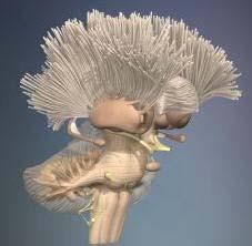
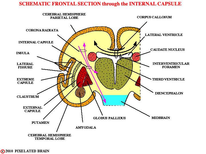
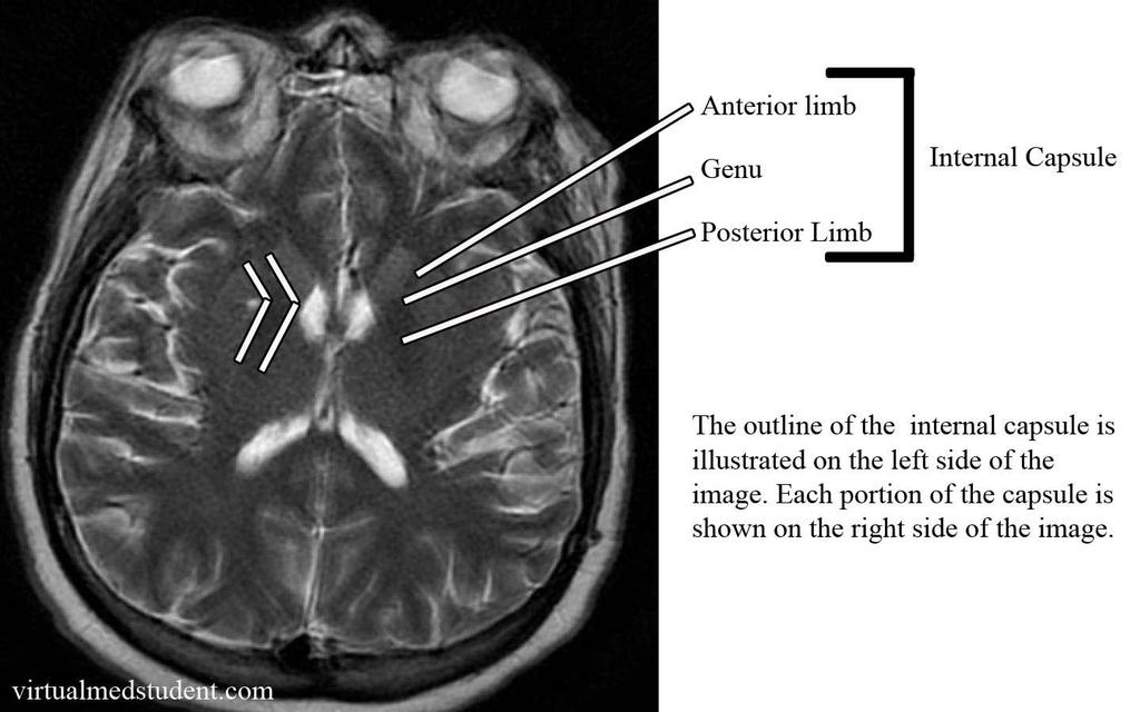
48 White Matter- projection fiber Projection fibers Connect cortex with lower part of brain & spinal cord and they include both ascending & descending fibers Internal capsule Position: a thick lamina of white matter lying between caudate nucleus, thalamus and lentiform nucleus


49 Internal Capsule Head of Caudate nucleus Ant. Body of Caudate nucleus Internal capsule Lentiform nucleus Post. Tain of Caudate nucleus Dorsal thalamus
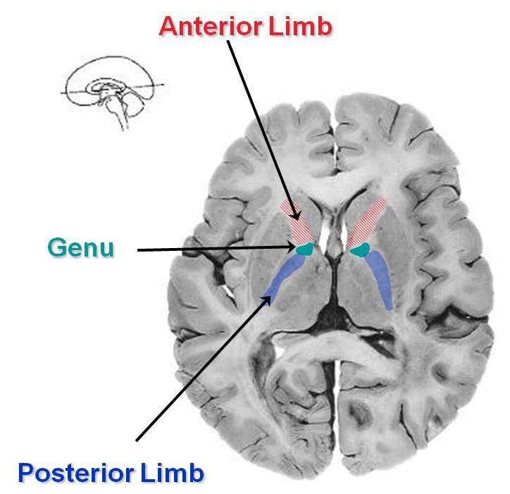
50 Internal Capsule Three parts Ant. limb of internal capsule Lies between caudate nucleus and lentiform nucleus Containing frontopontine tract and anterior thalamic radiation Genu of internal capsule Is angle at which ant. and post limbs meet Containing corticonuclear tract Post.limb of internal capsule Lies between thalamus & lentiform nucleus Contain corticospinal tract, corticorubral tract, central thalamic radiation, parieto- occipito-temporo-pontine tract, acoustic radiation and optic radiation genu Post.limb Ant.limb
51 Copy Right- Hongqi ZHANG-Department ofcerebrum Anatomy-Fudan University Longitudinal fissure of Head of caudate nucleus corpus callosum Lateral ventricle Ant.limb Lentiform nucleus Genu insula Post.limb claustrum thalamus Post.angle Of lat.ventricle Cerebral cortex medulla Horizontal section of the cerebrum
52 Longitudinal fissure of cerebrum Head of caudate nucleus Ant.limb of IC Lentiform nucleus Claustrum Post.limb of IC Medulla Corpus callosum Lateral ventricle Genu of IC Insula Thalamus Cerebral cortex Horizontal section of the cerebrum


53 Internal capsule Head of caudate nucleus Ant. thalamic radiation Frontopontine tract Corticonuclear tract Corticospinal tract Dorsal thalamus Central thalamic radiation Lentiform nucleus Corticorubral tract Parieto-occipitotemporo-pontine tract Acoustic radiation Med. geniculate body Lat. geniculate body Optic radiation

54 Fiber tracts pass through internal capsule Ant.limb Frontopontine tract Ant. thalamic radiation Three portion Genu Post.limb Corticonuclear tract Corticospinal tract Corticorubral tract Central thalamic radiation Parieto-occipito-temporo-pontine tract Acoustic radiation Optic radiation


55 Sympotom after Injury of right internal capsule
56 Injury of the whole internal capsule If Infarction,hemorrhage appear in internal capsule,it will block the important sensory and motor conducting tracts.it will occur: Blindness in the opposite visual field of both eye Impairment of general sensation on the opposite side of the body Opposite skeletal muscle paralysis of upper and lower limb

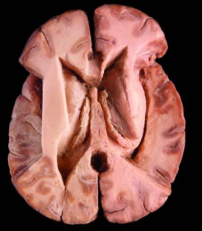
57 Horizental section of the cerebrum

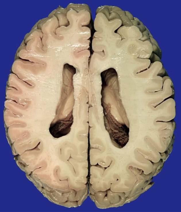
58 Lateral Ventricle Position: located in cerebral hemispheres Four parts 2 1-Central part: lies in parietal lobe 2-Anterior horn: extends into frontal lobe 3-Posterior horn: extend into occipital lobe 4-Inferior horn: extend into temporal lobe 4 1 Mesencephalic aquduct 3rd ventricle 4th ventricle 3

59 Projective diagram of the ventricular system of brain
60 The superior view of lateral ventricle

61 Lateral Ventricle Transverse MRI scan, at the level of the anterior horn of the lateral ventricle.


62 Lateral Ventricle Communication lateral ventricle interventricular foramen third ventricle
63

64
65
 MR images show ROIs (white areas) at which ADC, FA, and RA values were")

66 Transverse (1570/3.9/800) MR images show ROIs (white areas) at which ADC, FA, and RA values were determined in the, A, parietal lobe, B, frontal lobe, C, corpus callosum and pericallosal areas, D, internal capsule, E, occipital lobe, Copy RightHongqi ZHANG-Department of Anatomy-Fudan University and, F, temporal lobe.
67
68
69
70

71 FIGURE This view is one we will use repeatedly as the starting point in describing neural pathways

72 FIGURE This view is a modified version of the one shown above. On the right, four "tan colored" thalamic nuclei have been added and numbered arrows show how fibers radiate laterally from the thalamus, passing "under" the caudate, and entering the internal capsule. On the left, a horizontal cut has been made through the brain, revealing the classic "V" shape of the internal capsule. The "1" on both sides shows the position of fibers traveling in the anterior limb of the capsule; The "2, 3, 4" show the position of fibers traveling in the posterior limb.

73 FIGURE This view simply draws in the DC-ML pathway on our standard dorsal view of the brainstem and thalamus. Note that the pathway retains a somatotopic plan of organization all the way up to the cortex. VP is an abbreviation for ventralis posterior, the name of the thalamic nucleus where the cell bodies of the third order neurons are found. Note that on the left, the letters L and A show the position of the Copy "arm" Rightand "leg" Hongqi fibers ZHANG-Department in the posterior limb of of Anatomy-Fudan the capsule University
74

75 answer thalamus 2 - pineal body 3 - parieto-0ccipital sulcus 4 - calcarine sulcus 5 - inferior colliculus 6 - anterior medullary velum 7 - fourth ventricle 8 - basilar pons 9 - oculomotor nerve 10 - mammillary body 11 - optic chiasm 12 - anterior commissure 13 - cingulate gyrus 14 - corpus callosum

76 1 - lateral ventricle, ant horn 2 - septum pellucidum 3 - fornix 4 - massa intermedia 5 - thalamus 6 - pineal body 7 - superior colliculus 8 - inferior colliculus 9 - cerebellar hemisphere 10 - fourth ventricle 11 - cerebellar peduncles 12 - medial geniculate nucleus 13 - caudate nucleus, tail 14 - lateral geniculate nucleus 15 - habenular nucleus 16 - globus pallidus 17 - caudate nucleus, head 18 - corpus callosum

77

78 answer cingulate gyrus 2 - corpus callosum 3 - fornix 4 - thalamus 5 - superior &inferior colliculi 6 - superior cerebellar peduncle 7 - fourth ventricle 8 - inferior olive 9 - mammillary body 10 - fornix 11 - lateral ventricle, ant horn

79 answer fornix 2 - septum pellucidum 3 - corpus callosum 4 - caudate nucleus, head 5 - putamen 6 - globus pallidus 7 - olfactory tract 8 - anterior commissure 9 - middle cerebral artery 10 - lateral fissure

80 answer cingulate gyrus 2 - fornix 3 - thalamus 4 - putamen 5 - subthalamic nucleus 6 - hippocampal formation 7 - lateral ventricle, inf horn 8 - caudate nucleus

81 answer optic radiation 2 - lateral ventricle, post horn 3 - superior cerebellar peduncle 4 - corticospinal tract 5 - middle cerebellar peduncle 6 - cerebellum

82 answer insula 2 - internal capsule, ant limb 3 - fornix 4 - lateral ventricle, ant horn 5 - caudate nucleus, head 6 - putamen 7 - globus pallidus 8 - thalamus 9 - caudate nucleus, tail 10 - lateral ventricle, post horn 11 - optic radiation 12 - internal capsule, post limb 13 - internal capsule, genu


83 answer olfactory tract 2 - optic chiasm 3 - amygdala 4 - lateral ventricle, inf horn 5 - cerebral peduncle 6 - red nucleus 7 - substantia nigra 8 - hippocampus
84
85

86 ^_^ 哈哈! 下课了!
Telencephalon (Cerebral Hemisphere)
") Telencephalon (Cerebral Hemisphere) OUTLINE The Cortex - Lobes, Sulci & Gyri - Functional Subdivisions - Limbic Lobe & Limbic System The Subcortex - Basal Ganglia - White Matter (Internal Capsule) - Relations
Telencephalon (Cerebral Hemisphere) OUTLINE The Cortex - Lobes, Sulci & Gyri - Functional Subdivisions - Limbic Lobe & Limbic System The Subcortex - Basal Ganglia - White Matter (Internal Capsule) - Relations
Introduction to the Central Nervous System: Internal Structure
 Introduction to the Central Nervous System: Internal Structure Objective To understand, in general terms, the internal organization of the brain and spinal cord. To understand the 3-dimensional organization
CEREBRUM Dr. Jamila Elmedany Dr. Essam Eldin Salama
 CEREBRUM Dr. Jamila Elmedany Dr. Essam Eldin Salama Objectives At the end of the lecture, the student should be able to: List the parts of the cerebral hemisphere (cortex, medulla, basal nuclei, lateral
Anatomy and Physiology (Bio 220) The Brain Chapter 14 and select portions of Chapter 16
 The Brain Chapter 14 and select portions of Chapter 16") Anatomy and Physiology (Bio 220) The Brain Chapter 14 and select portions of Chapter 16 I. Introduction A. Appearance 1. physical 2. weight 3. relative weight B. Major parts of the brain 1. cerebrum 2.
Anatomy and Physiology (Bio 220) The Brain Chapter 14 and select portions of Chapter 16 I. Introduction A. Appearance 1. physical 2. weight 3. relative weight B. Major parts of the brain 1. cerebrum 2.
M555 Medical Neuroscience Lab 1: Gross Anatomy of Brain, Crainal Nerves and Cerebral Blood Vessels
 M555 Medical Neuroscience Lab 1: Gross Anatomy of Brain, Crainal Nerves and Cerebral Blood Vessels Anatomical Directions Terms like dorsal, ventral, and posterior provide a means of locating structures
Gross Morphology of the Brain
 Gross Morphology of the Brain Done by : Marah Marahleh & Razan Krishan *slides in bold Principal Parts of the Brain Cerebrum : largest part of the brain Diencephalon Thalamus & hypothalamus Cerebellum
The Nervous system is divided into 2 major divisions: 1) Central Nervous System (CNS): found within bones & consists of:
 Central Nervous System (CNS): found within bones & consists of:") The Nervous system is divided into 2 major divisions: 1) Central Nervous System (CNS): found within bones & consists of: - The Brain: within the skull, composed of cerebrum, cerebellum and brain stem.
The Nervous system is divided into 2 major divisions: 1) Central Nervous System (CNS): found within bones & consists of: - The Brain: within the skull, composed of cerebrum, cerebellum and brain stem.
PROPERTY OF ELSEVIER SAMPLE CONTENT - NOT FINAL. Gross Anatomy and General Organization of the Central Nervous System
 3 Gross Anatomy and General Organization of the Central Nervous System C h a p t e r O u t l i n e The Long Axis of the CNS Bends at the Cephalic Flexure Hemisecting a Brain Reveals Parts of the Diencephalon,
The Central Nervous System I. Chapter 12
 The Central Nervous System I Chapter 12 The Central Nervous System The Brain and Spinal Cord Contained within the Axial Skeleton Brain Regions and Organization Medical Scheme (4 regions) 1. Cerebral Hemispheres
I. Anatomy of the Brain A. Cranial Meninges and Ventricles of the Brain 1. Meninges a. Dura mater 1) Endosteal/Periosteal Layer - Outer 2) Meningeal
 Endosteal/Periosteal Layer - Outer 2) Meningeal") I. Anatomy of the Brain A. Cranial Meninges and Ventricles of the Brain 1. Meninges a. Dura mater 1) Endosteal/Periosteal Layer - Outer 2) Meningeal Layer - Inner 3) Falx cerebri a) Superior sagittal sinus
I. Anatomy of the Brain A. Cranial Meninges and Ventricles of the Brain 1. Meninges a. Dura mater 1) Endosteal/Periosteal Layer - Outer 2) Meningeal Layer - Inner 3) Falx cerebri a) Superior sagittal sinus
DISSECTION OF THE SHEEP'S BRAIN
 Sheep Brain Dissection Guide Page 1 DISSECTION OF THE SHEEP'S BRAIN Introduction The purpose of the sheep brain dissection is to familiarize you with the threedimensional structure of the brain and teach
Blood supply to the brain Blood brain barrier isolates neural tissue from general circulation
 The Brain and Cranial Nerves Objectives Name the major regions of the brain and describe their functions. Discuss the formation, circulation, and functions of the CSF. List the main components of the medulla
CEREBRUM. Dr. Jamila EL Medany
 CEREBRUM Dr. Jamila EL Medany Objectives At the end of the lecture, the student should be able to: List the parts of the cerebral hemisphere (cortex, medulla, basal nuclei, lateral ventricle). Describe
Neuroanatomy lecture (1)
") Neuroanatomy lecture (1) Introduction: Neuroanatomy has two parts: the central and peripheral nervous system. The central nervous system is composed of brain and spinal cord. The brain has the following
Neuroanatomy lecture (1) Introduction: Neuroanatomy has two parts: the central and peripheral nervous system. The central nervous system is composed of brain and spinal cord. The brain has the following
Regional and Lobe Parcellation Rhesus Monkey Brain Atlas. Manual Tracing for Parcellation Template
 Regional and Lobe Parcellation Rhesus Monkey Brain Atlas Manual Tracing for Parcellation Template Overview of Tracing Guidelines A) Traces are performed in a systematic order they, allowing the more easily
Lecture 4 The BRAINSTEM Medulla Oblongata
 Lecture 4 The BRAINSTEM Medulla Oblongata Introduction to brainstem 1- Medulla oblongata 2- Pons 3- Midbrain - - - occupies the posterior cranial fossa of the skull. connects the narrow spinal cord
Developmental sequence of brain
 Cerebellum Developmental sequence of brain Fourth week Fifth week Location of cerebellum Lies above and behind the medullar and pons and occupies posterior cranial fossa Location of cerebellum External
b. The groove between the two crests is called 2. The neural folds move toward each other & the fuse to create a
 Chapter 13: Brain and Cranial Nerves I. Development of the CNS A. The CNS begins as a flat plate called the B. The process proceeds as: 1. The lateral sides of the become elevated as waves called a. The
Fig.1: A, Sagittal 110x110 mm subimage close to the midline, passing through the cingulum. Note that the fibers of the corpus callosum run at a
 Fig.1 E Fig.1:, Sagittal 110x110 mm subimage close to the midline, passing through the cingulum. Note that the fibers of the corpus callosum run at a slight angle are through the plane (blue dots with
Biological Bases of Behavior. 3: Structure of the Nervous System
 Biological Bases of Behavior 3: Structure of the Nervous System Neuroanatomy Terms The neuraxis is an imaginary line drawn through the spinal cord up to the front of the brain Anatomical directions are
Human Anatomy. Brain and Cranial Nerves
 Human Anatomy Brain and Cranial Nerves 1 Brain and Cranial Nerves An adult brain weighs between 1.35 and 1.4 kilograms (kg) (around 3 pounds) and has a volume of about 1200 cubic centimeters (cc). Brain
TRANSVERSE SECTION PLANE Scalp 2. Cranium. 13. Superior sagittal sinus
 TRANSVERSE SECTION PLANE 1 1. Scalp 2. Cranium 3. Superior sagittal sinus 4. Dura mater 5. Falx cerebri 6. Frontal lobes of the cerebrum 7. Middle meningeal artery 8. Cortex, grey matter 9. Cerebral vessels
Chapter 3. Structure and Function of the Nervous System. Copyright (c) Allyn and Bacon 2004
 Allyn and Bacon 2004") Chapter 3 Structure and Function of the Nervous System 1 Basic Features of the Nervous System Neuraxis: An imaginary line drawn through the center of the length of the central nervous system, from the
Chapter 3 Structure and Function of the Nervous System 1 Basic Features of the Nervous System Neuraxis: An imaginary line drawn through the center of the length of the central nervous system, from the
Chapter 14: The Brain and Cranial Nerves. Copyright 2009, John Wiley & Sons, Inc.
 Chapter 14: The Brain and Cranial Nerves Development of the Brain Three to four-week embryo: prosencephalon, mesencephalon and rhombencephalon. Five-week embryo: telencephalon (cerebrum), diencephalon
DIENCEPHALON. ..Central core of the forebrain..consists of three paired structures
 DIENCEPHALON..Central core of the forebrain..consists of three paired structures ---------- --thalamus, --------hypothalamus, -------epithalamus..encloses the third ventricle Diencephalon between cerebral
CEREBRUM & CEREBRAL CORTEX
 CEREBRUM & CEREBRAL CORTEX Seonghan Kim Dept. of Anatomy Inje University, College of Medicine THE BRAIN ANATOMICAL REGIONS A. Cerebrum B. Diencephalon Thalamus Hypothalamus C. Brain Stem Midbrain Pons
-Zeina Assaf. -Omar Odeh. - Maha Beltagy
 -3 -Zeina Assaf -Omar Odeh - Maha Beltagy 1 P a g e The Inferior Surface Of The Brain The inferior surface of the brain is divide by the stem of the lateral fissure into 2 parts : The orbital surface and
Stanley Pruisinger 1980's
 Neuroanatomy Prion disease cerebellum chapter b/c cerebellar ataxia here as a warning for obvious reasons. Creutzfeldt - Jakob Disease (CJD) "Spongiform" (brain turns to sponge) Jews in Lybia who ate
Model 3-50B or 3-88 III VIII. Olfactory Nerve. Optic Nerve. Oculomotor Nerve. Trochlear Nerve. Trigeminal Nerve. Abducens Nerve.
 Model 3-50B or 3-88 I Olfactory Nerve II Optic Nerve Oculomotor Nerve III IV Trochlear Nerve Trigeminal Nerve V VI Abducens Nerve Glossopharyngeal Nerve IX VII Facial Nerve VIII Vestibocochlear Nerve or
Embryonic Brain Development
 Chapter 14 The Brain and Cranial Nerves Largest organ in the body? Brain functions in sensations, memory, emotions, decision making, behavior 19-1 19-2 Embryonic Brain Development Principal Parts of the
DEVELOPMENT OF BRAIN
 Ahmed Fathalla OBJECTIVES At the end of the lecture, students should: List the components of brain stem. Describe the site of brain stem. Describe the relations between components of brain stem & their
Brainstem. By Dr. Bhushan R. Kavimandan
 Brainstem By Dr. Bhushan R. Kavimandan Development Ventricles in brainstem Mesencephalon cerebral aqueduct Metencephalon 4 th ventricle Mylencephalon 4 th ventricle Corpus callosum Posterior commissure
Announcement. Danny to schedule a time if you are interested.
 Announcement If you need more experiments to participate in, contact Danny Sanchez (dsanchez@ucsd.edu) make sure to tell him that you are from LIGN171, so he will let me know about your credit (1 point).
Prof. Saeed Abuel Makarem & Dr.Sanaa Alshaarawy
 Prof. Saeed Abuel Makarem & Dr.Sanaa Alshaarawy 1 Objectives By the end of the lecture, you should be able to: Describe the anatomy and main functions of the thalamus. Name and identify different nuclei
ANATOMY & PHYSIOLOGY DISSECTION OF THE SHEEP BRAIN LAB GROUP:
 ANATOMY & PHYSIOLOGY DISSECTION OF THE SHEEP BRAIN LAB GROUP: Introduction The purpose of the sheep brain dissection is to familiarize you with the three dimensional structure of the brain and teach you
P. Hitchcock, Ph.D. Department of Cell and Developmental Biology Kellogg Eye Center. Wednesday, 16 March 2009, 1:00p.m. 2:00p.m.
 Normal CNS, Special Senses, Head and Neck TOPIC: CEREBRAL HEMISPHERES FACULTY: LECTURE: READING: P. Hitchcock, Ph.D. Department of Cell and Developmental Biology Kellogg Eye Center Wednesday, 16 March
Auditory and Vestibular Systems
 Auditory and Vestibular Systems Objective To learn the functional organization of the auditory and vestibular systems To understand how one can use changes in auditory function following injury to localize
PSY 302: CHAPTER 3 NOTES THE BRAIN (PART II) - 9/5/17. By: Joseline
 - 9/5/17. By: Joseline") PSY 302: CHAPTER 3 NOTES THE BRAIN (PART II) - 9/5/17 By: Joseline Left 3 MAJOR FISSURES : 2HEMISPHERES Right Lateral Ventricle Central Fissure Third Ventricle Sulcus Lateral Fissure Gyros Fissure- Fissures
PSY 302: CHAPTER 3 NOTES THE BRAIN (PART II) - 9/5/17 By: Joseline Left 3 MAJOR FISSURES : 2HEMISPHERES Right Lateral Ventricle Central Fissure Third Ventricle Sulcus Lateral Fissure Gyros Fissure- Fissures
Neuroanatomy. Dr. Maha ELBeltagy. Assistant Professor of Anatomy Faculty of Medicine The University of Jordan
 Neuroanatomy Dr. Maha ELBeltagy Assistant Professor of Anatomy Faculty of Medicine The University of Jordan 2018 Prof Yousry 10/15/17 Types of brain fibers THE WHITE MATTER OF THE BRAIN The white matter
ACTIVITY 7: NERVOUS SYSTEM HISTOLOGY, BRAIN, CRANIAL NERVES
 ACTIVITY 7: NERVOUS SYSTEM HISTOLOGY, BRAIN, CRANIAL NERVES LABORATORY OBJECTIVES: 1. Histology: Identify structures indicated on three different slides or images of nervous system tissue. These images
Chapter 18: The Brain & Cranial Nerves. Origin of the Brain
 Chapter 18: The Brain & Cranial Nerves BIO 218 Fall 2015 Origin of the Brain The brain originates from a structure called the neural tube, which arises during a developmental stage called neurulation.
Human Brain and Senses October 13, 2008 Page 1. Examination of the Human Brain
 Human Brain and Senses October 13, 2008 Page 1 Examination of the Human Brain With only a few hours today we can only begin to scratch the surface of a complex subject like neuroanatomy. The purpose of
Chapter 13 Brain and Cranial Nerves
 Chapter 13 Brain and Cranial Nerves 13-1 Brain and Cranial Nerves Brain Part of CNS contained in cranial cavity Control center for many of body s functions Much like a complex computer but more Parts of
The neurvous system senses, interprets, and responds to changes in the environment. Two types of cells makes this possible:
 NERVOUS SYSTEM The neurvous system senses, interprets, and responds to changes in the environment. Two types of cells makes this possible: the neuron and the supporting cells ("glial cells"). Neuron Neurons
Anatomy Lab (1) Theoretical Part. Page (2 A) Page (2B)
 Theoretical Part. Page (2 A) Page (2B)") Anatomy Lab (1) This sheet only includes the extra notes for the lab handout regarding the theoretical part, as for the practical part it includes everything the doctor mentioned. Theoretical Part Page
Anatomy Lab (1) This sheet only includes the extra notes for the lab handout regarding the theoretical part, as for the practical part it includes everything the doctor mentioned. Theoretical Part Page
Brainstem. Amadi O. Ihunwo, PhD School of Anatomical Sciences
 Brainstem Amadi O. Ihunwo, PhD School of Anatomical Sciences Lecture Outline Constituents Basic general internal features of brainstem External and Internal features of Midbrain Pons Medulla Constituents
Psyc 311A, fall 2008 Conference week 3 TA: Jürgen Germann
 Psyc 311A, fall 2008 Conference week 3 TA: Jürgen Germann e-mail: jurgen.germann@mcgill.ca Overview: 1. Meninges 2. Cerebral cortex-cytoarchitecture 3. Diencephalon (thalamus/hypothalamus) (this replaces
The Nervous System: Sensory and Motor Tracts of the Spinal Cord
 15 The Nervous System: Sensory and Motor Tracts of the Spinal Cord PowerPoint Lecture Presentations prepared by Steven Bassett Southeast Community College Lincoln, Nebraska Introduction Millions of sensory
Lab 2. we will look into several angled horizontal sections ( orbitomeatal plane ) i.e passing from the orbit into the ear
 i.e passing from the orbit into the ear") we will look into several angled horizontal sections ( orbitomeatal plane ) i.e passing from the orbit into the ear Figure I page 76 : looking at the key on the left side this section passed through the
we will look into several angled horizontal sections ( orbitomeatal plane ) i.e passing from the orbit into the ear Figure I page 76 : looking at the key on the left side this section passed through the
LIMBIC SYSTEM. Dr. Amani A. Elfaki Associate Professor Department of Anatomy
 LIMBIC SYSTEM Dr. Amani A. Elfaki Associate Professor Department of Anatomy Learning Objectives Define the limbic system Identify the parts of the limbic system Describe the circulation of the limbic system
Sectional Anatomy Head Practice Problems
 1. Which of the following is illustrated by #3? (Fig. 5-42) A) maxillary sinus B) vomer C) septal cartilage D) perpendicular plate of ethmoid bone 2. What number illustrates the cornea? (Fig. 5-42) A)
Homework Week 2. PreLab 2 HW #2 Synapses (Page 1 in the HW Section)
") Homework Week 2 Due in Lab PreLab 2 HW #2 Synapses (Page 1 in the HW Section) Reminders No class next Monday Quiz 1 is @ 5:30pm on Tuesday, 1/22/13 Study guide posted under Study Aids section of website
Homework Week 2 Due in Lab PreLab 2 HW #2 Synapses (Page 1 in the HW Section) Reminders No class next Monday Quiz 1 is @ 5:30pm on Tuesday, 1/22/13 Study guide posted under Study Aids section of website
BASAL GANGLIA. Dr JAMILA EL MEDANY
 BASAL GANGLIA Dr JAMILA EL MEDANY OBJECTIVES At the end of the lecture, the student should be able to: Define basal ganglia and enumerate its components. Enumerate parts of Corpus Striatum and their important
Outline of the next three lectures
 Outline of the next three lectures Lecture 35 Anatomy of the human cerebral cortex gross and microscopic cell types connections Vascular supply of the cerebral cortex Disorders involving the cerebral cortex
Dissection of the Sheep Brain
 Dissection of the Sheep Brain Laboratory Objectives After completing this lab, you should be able to: 1. Identify the main structures in the sheep brain and to compare them with those of the human brain.
I: To describe the pyramidal and extrapyramidal tracts. II: To discuss the functions of the descending tracts.
 Descending Tracts I: To describe the pyramidal and extrapyramidal tracts. II: To discuss the functions of the descending tracts. III: To define the upper and the lower motor neurons. 1. The corticonuclear
Chapter 13 Lecture Outline *
 Anatomy and Physiology, Seventh Edition Rod R. Seeley Idaho State University Trent D. Stephens Idaho State University Philip Tate Phoenix College Chapter 13 Lecture Outline * *See PowerPoint Image Slides
Slide 1. Slide 2. Slide 3. Tomography vs Topography. Computed Tomography (CT): A simplified Topographical review of the Brain. Learning Objective
: A simplified Topographical review of the Brain. Learning Objective") Slide 1 Computed Tomography (CT): A simplified Topographical review of the Brain Jon Wheiler, ACNP-BC Slide 2 Tomography vs Topography Tomography: A technique for displaying a representation of a cross
Slide 1 Computed Tomography (CT): A simplified Topographical review of the Brain Jon Wheiler, ACNP-BC Slide 2 Tomography vs Topography Tomography: A technique for displaying a representation of a cross
By Dr. Saeed Vohra & Dr. Sanaa Alshaarawy
 By Dr. Saeed Vohra & Dr. Sanaa Alshaarawy 1 By the end of the lecture, students will be able to : Distinguish the internal structure of the components of the brain stem in different levels and the specific
 Spinal Cord Tracts DESCENDING SPINAL TRACTS: Are concerned with somatic motor function, modification of ms. tone, visceral innervation, segmental reflexes. Main tracts arise form cerebral cortex and others
BIO 210 CHAPTER 13. The Central Nervous System SUPPLEMENT 2. PowerPoint by John McGill Supplemental Notes by Beth Wyatt CEREBELLUM
 BIO 210 CHAPTER 13 The Central Nervous System SUPPLEMENT 2 PowerPoint by John McGill Supplemental Notes by Beth Wyatt CEREBELLUM Second Largest Division of the Brain Lies Below the Posterior Portion of
The Central Nervous System
 The Central Nervous System 1 The Central Nervous System Brain Spinal Cord Components 2 Protection of the Brain The brain is protected from injury by The skull enough said! You already know this. What else
Medical Neuroscience Tutorial
 Pain Pathways Medical Neuroscience Tutorial Pain Pathways MAP TO NEUROSCIENCE CORE CONCEPTS 1 NCC1. The brain is the body's most complex organ. NCC3. Genetically determined circuits are the foundation
Ch 13: Central Nervous System Part 1: The Brain p 374
 Ch 13: Central Nervous System Part 1: The Brain p 374 Discuss the organization of the brain, including the major structures and how they relate to one another! Review the meninges of the spinal cord and
Unit 12a: The Nervous System The Brain. MDL231 Principle of Anatomy
 Unit 12a: The Nervous System The Brain MDL231 Principle of Anatomy The Brain - Overview Cerebrum T PP H midbrain Cerebellum pons m.o. Brain stem medulla oblongata (M.O.) pons midbrain (mesencephalon) Diencephalon
Nsci 2100: Human Neuroanatomy 2017 Examination 3
 Name KEY Lab Section Nsci 2100: Human Neuroanatomy 2017 Examination 3 On this page, write your name and lab section. On your bubble answer sheet, enter your name (last name, space, first name), internet
PARIETAL LOBE. Vasilios A. Zerris MD, MPH, MSc, FAANS
 PARIETAL LOBE Vasilios A. Zerris MD, MPH, MSc, FAANS Diplomate of the American Board of Neurological Surgery Fellow of the American Association of Neurological Surgeons Professor of Neurosurgery, European
Medical Neuroscience Tutorial Notes
 Medical Neuroscience Tutorial Notes Blood Supply to the Brain MAP TO NEUROSCIENCE CORE CONCEPTS 1 NCC1. The brain is the body's most complex organ. LEARNING OBJECTIVES After study of the assigned learning
Sheep Brain Dissection
 Sheep Brain Dissection Mammalian brains have many features in common. Human brains may not be available, so sheep brains often are dissected as an aid to understanding the mammalian brain since he general
Gross Organization I The Brain. Reading: BCP Chapter 7
 Gross Organization I The Brain Reading: BCP Chapter 7 Layout of the Nervous System Central Nervous System (CNS) Located inside of bone Includes the brain (in the skull) and the spinal cord (in the backbone)
1. The basic anatomy of the Central Nervous System (CNS)
") Psyc 311A, fall 2008 Conference week 1 Sept 9 th to 11 th TA: Jürgen Germann; e-mail: jurgen.germann@mcgill.ca Overview: 1. The basic anatomy of the Central Nervous System (CNS) 2. Cells of the CNS 3.
Psyc 311A, fall 2008 Conference week 1 Sept 9 th to 11 th TA: Jürgen Germann; e-mail: jurgen.germann@mcgill.ca Overview: 1. The basic anatomy of the Central Nervous System (CNS) 2. Cells of the CNS 3.
14 - Central Nervous System. The Brain Taft College Human Physiology
 14 - Central Nervous System The Brain Taft College Human Physiology Development of the Brain The brain begins as a simple tube, a neural tube. The tube or chamber (ventricle) is filled with cerebrospinal
Lecture - Chapter 13: Central Nervous System
 Lecture - Chapter 13: Central Nervous System 1. Describe the following structures of the brain, what is the general function of each: a. Cerebrum b. Diencephalon c. Brain Stem d. Cerebellum 2. What structures
BASIC ANATOMICAL STRUCTURES. The Cerebrum
 BASIC ANATOMICAL STRUCTURES The Cerebrum Development of the Cerebral Vesicles Primary Vesicle secondary Vesicles CNS structures Ventricle telencephalon Cerebral Hemispheres Lateral Ventricles Prosencephalon
Inferior Surface of the Brain:-
 The following sheet s sources: -Recording Section 2 -Slides All slides not mentioned by the doctor will also be included in the sheet. -Wikipedia Inferior Surface of the Brain:- The inferior surface of
If I Only Had a Brain
 If I Only Had a Brain A Heart. (The Nerve!) Regions of the Brain Cerebral hemisphere Diencephalon Cerebellum (b) Adult brain Brain stem Regions of the Brain: Cerebrum Precentral gyrus Frontal lobe Central
Laboratory Manual for Comparative Anatomy and Physiology Figure 15.1 Transparency Master 114
 Neuron Capillary Astrocyte Microglial cell Neuron Fluid-filled cavity Process of oligodendrocyte Ependymal cells Brain or spinal cord tissue Myelin sheath Nerve fibers Figure 15.1 Transparency Master 114
Copy Right- Hongqi ZHANG-Department of Anatomy-Fudan University. Systematic Anatomy. Nervous system Cerebellum. Dr.Hongqi Zhang ( 张红旗 )
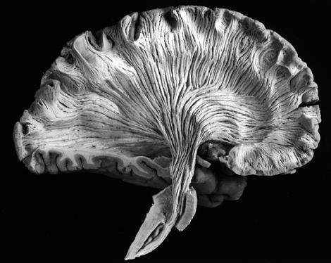
") Systematic Anatomy Nervous system Cerebellum Dr.Hongqi Zhang ( 张红旗 ) Email: zhanghq58@126.com 1 The Cerebellum Cerebellum evolved and developed with the complication of animal movement. Key points about
Systematic Anatomy Nervous system Cerebellum Dr.Hongqi Zhang ( 张红旗 ) Email: zhanghq58@126.com 1 The Cerebellum Cerebellum evolved and developed with the complication of animal movement. Key points about
Done by : Areej Al-Hadidi
 Brainstem &diencephalon Done by : Areej Al-Hadidi Brainstem Functions Ascending and descending tracts Reflex centers Cardiovascular and respiratory centers Coughing, sneezing, swallowing Nuclei of the
Department of Human Anatomy GUIDELINES. nuclei. The lateral ventricles. White substance of cerebral hemispheres. course 1
 Department of Human Anatomy GUIDELINES Academic discipline Human Anatomy Module 2 Content module 11 Study subject The olfactory brain. Basal nuclei. The lateral ventricles. White substance of cerebral
Systems Neuroscience Dan Kiper. Today: Wolfger von der Behrens
 Systems Neuroscience Dan Kiper Today: Wolfger von der Behrens wolfger@ini.ethz.ch 18.9.2018 Neurons Pyramidal neuron by Santiago Ramón y Cajal (1852-1934, Nobel prize with Camillo Golgi in 1906) Neurons
Anatomy Lecture Notes Chapter 13
 I. embryonic development of the CNS A. neurulation is the formation of the CNS in the embryo invagination of dorsal ectoderm (outer layer of embryo cells) this process is induced (caused) by the notochord
BIOL Dissection of the Sheep and Human Brain
 BIOL 2401 Dissection of the Sheep and Human Brain Laboratory Objectives After completing this lab, you should be able to: Identify the main structures in the sheep brain and to compare them with those
Anatomy & Physiology Central Nervous System Worksheet
 1. What are the two parts of the CNS? 2. What are the four functions of the CNS Anatomy & Physiology Central Nervous System Worksheet 3. What are the four functions of the meninges? (p430) 4. Starting
Development of Brain Stem, Cerebellum and Cerebrum
 Development of Brain Stem, Cerebellum and Cerebrum The neural tube cranial to the 4th pair of somites develop into the brain. 3 dilatations and 2 flexures form at the cephalic end of the neural tube during
The Nervous System PART B
 7 The Nervous System PART B PowerPoint Lecture Slide Presentation by Jerry L. Cook, Sam Houston University ESSENTIALS OF HUMAN ANATOMY & PHYSIOLOGY EIGHTH EDITION ELAINE N. MARIEB The Reflex Arc Reflex
ACTIVITY 7: NERVOUS SYSTEM HISTOLOGY, BRAIN, CRANIAL NERVES NERVOUS SYSTEM TISSUES: HISTOLOGY SLIDES
 ACTIVITY 7: NERVOUS SYSTEM HISTOLOGY, BRAIN, CRANIAL NERVES OBJECTIVES: 1) How to get ready: Read Chapter 14 & 15 McKinley et al., Human Anatomy, 4e. All text references are for this textbook. Read dissection
CNS consists of brain and spinal cord Cephalization Evolutionary development of rostral (anterior) portion of CNS Increased number of neurons in head
 portion of CNS Increased number of neurons in head") CNS consists of brain and spinal cord Cephalization Evolutionary development of rostral (anterior) portion of CNS Increased number of neurons in head Highest level reached in human brain 1 Mostly to orient
CNS consists of brain and spinal cord Cephalization Evolutionary development of rostral (anterior) portion of CNS Increased number of neurons in head Highest level reached in human brain 1 Mostly to orient
Cerebral hemisphere. Parietal Frontal Occipital Temporal
 Cerebral hemisphere Sulcus / Fissure Central Precental gyrus Postcentral gyrus Lateral (cerebral) Parieto-occipital Cerebral cortex Frontal lobe Parietal lobe Temporal lobe Insula Amygdala Hippocampus
Brain ميهاربا لض اف دمح ا د The Meninges 1- Dura Mater of the Brain endosteal layer does not extend meningeal layer falx cerebri tentorium cerebelli
 .احمد د فاضل ابراهيم Lecture 15 Brain The Meninges Three protective membranes or meninges surround the brain in the skull: the dura mater, the arachnoid mater, and the pia mater 1- Dura Mater of the Brain
Brain and Cranial Nerves (Ch. 15) Human Anatomy lecture. caudal = toward the spinal cord)
 Human Anatomy lecture. caudal = toward the spinal cord)") Insight: Some cranial nerve disorders Brain and Cranial Nerves (Ch. 15) Human Anatomy lecture I. Overview (Directional terms: rostral = toward the forehead caudal = toward the spinal cord) A. 3 Major parts
Insight: Some cranial nerve disorders Brain and Cranial Nerves (Ch. 15) Human Anatomy lecture I. Overview (Directional terms: rostral = toward the forehead caudal = toward the spinal cord) A. 3 Major parts
神經解剖學 NEUROANATOMY TELENCEPHALON 盧家鋒助理教授 臺北醫學大學醫學系解剖學暨細胞生物學科 臺北醫學大學醫學院轉譯影像研究中心.
 神經解剖學 NEUROANATOMY TELENCEPHALON 盧家鋒助理教授 臺北醫學大學醫學系解剖學暨細胞生物學科 臺北醫學大學醫學院轉譯影像研究中心 http://www.ym.edu.tw/~cflu REGIONAL NEUROBIOLOGY Telencephalon (Cerebrum) Diencephalon (Thalamus) Cerebellum Brain stem Midbrain
Brain, Cranial Nerves, and Spinal Cord
 Bio101 Laboratory 13 Neuron/Spinal Cord Histology Brain Anatomy Ear & Eye Anatomy 1 Brain, Cranial Nerves, and Spinal Cord Objectives for today s lab Become familiar with the gross anatomy of the brain
Department of Cognitive Science UCSD
 Department of Cognitive Science UCSD Verse 1: Neocortex, frontal lobe, Brain stem, brain stem, Hippocampus, neural node, Right hemisphere, Pons and cortex visual, Brain stem, brain stem, Sylvian fissure,
Brain anatomy and artificial intelligence. L. Andrew Coward Australian National University, Canberra, ACT 0200, Australia
 Brain anatomy and artificial intelligence L. Andrew Coward Australian National University, Canberra, ACT 0200, Australia The Fourth Conference on Artificial General Intelligence August 2011 Architectures
Read Chapter 14 & 15 McKinley et al
 ACTIVITY 7: NERVOUS SYSTEM HISTOLOGY, BRAIN, CRANIAL NERVES OBJECTIVES: 1) How to get ready: Read Chapter 14 & 15 McKinley et al., Human Anatomy, 5e. All text references are for this textbook. Read dissection
Longitudinal fissure separates right and left hemispheres.
 L 10 A B O R A T O R Y Brain/Skull CEREBRAL CORTEX (telencephalon) Longitudinal fissure separates right and left hemispheres. Identify the following structures of the frontal lobe: lateral sulcus central
Blood Supply of the CNS
 Blood Supply of the CNS Lecture Objectives Describe the four arteries supplying the CNS. Follow up each artery to its destination. Describe the circle of Willis and its branches. Discuss the principle
The Brain. Brain. Spinal Cord. Cauda Equina
 The Brain Brain Spinal Cord Cauda Equina The Brain Ventricles- cavities in the brain filled with cerebrospinal fluid connected to the subarachnoid space- fluid filled space surrounding the brain Brain
Brainstem. Steven McLoon Department of Neuroscience University of Minnesota
 Brainstem Steven McLoon Department of Neuroscience University of Minnesota 1 Course News Change in Lab Sequence Week of Oct 2 Lab 5 Week of Oct 9 Lab 4 2 Goal Today Know the regions of the brainstem. Know