Lecture #2. Respiratory system. Development
|
|
|
- Rafe Preston
- 6 years ago
- Views:
Transcription

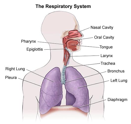
1 Lecture #2 Respiratory system. Development


2 Respiratory system - is a biological system consisting of specific organs and structures used for the process of respiration in an organism

3 Breathing and Respiration BREATHING is the mechanical action of getting air in and out of the lungs. RESPIRATION is the chemical reaction that provides the energy that makes the organism function. It occurs in the cells, more precisely in the mitochondria (the powerplant of the cell).
4 Systema respiratoria
5
 Ectoderm Mesoderm")
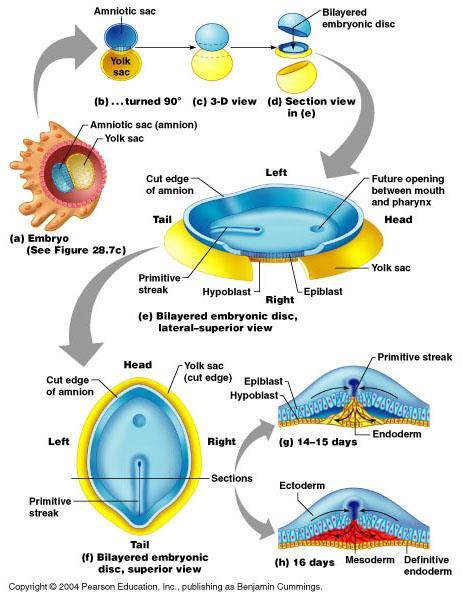
6 Gastrulation formation of germ layers (4 th week) Ectoderm Mesoderm Endoderm
 appendicular skeleton and")
7 Intraembryonic mesoderm plates: Paraxial (dorsal) mesoderm axial skeleton (somites) Intermediate mesoderm urogenital apparatus Lateral mesoderm (somatic and splanchnic) appendicular skeleton and internal organs
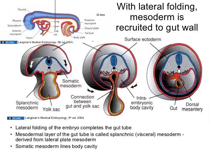
8 Coelom
9 Primary gut (foregut) Intraembryonic cavity Hollow organs (trachea, bronchi) Pleural cavity

10

11 From foregut develop: - Esophagus - Stomach - Duodenum (proximal part) - Liver, pancreas, gall bladder - Respiratory tube Blood supply truncus coeliacus Sympathetic innervation n. splanchnicus major Parasympathetic innervation n.vagus


12 Tubular organ layers development - Mucosa Epithelial lining and glands Lamina propria Muscularis mucosae - Derived from endoderm - Submucosa Derived from visceral mesoderm - Muscularis externa/cartilages - Adventitia/Serosa
13 Development of the upper respiratory system: - nose, nasopharynx, oropharynx
14 Development of the face (from 4 th to 8 th weeks) 1. Development of the primitive mouth stomodeum (beginning of the 4 th week) 2. Rupture of oropharyngeal membrane (the 24 th day) 3. Development of the nasal cavity (from the end of the 4 th week) 4. Rupture of oronasal membrane (the 6 th week) 5. Development of paranasal air sinuses from diverticuli of nasal walls during late fetal life & after birth

15 Cranial end of the foregut Ratke`s pouch
16 Development of the face (from 4 th to 8 th weeks) 1. Development of the primitive mouth stomodeum (beginning of the 4 th week) 2. Rupture of oropharyngeal membrane (the 24 th day) 3. Development of the nasal cavity (from the end of the 4 th week) 4. Rupture of oronasal membrane (the 6 th week) 5. Development of paranasal air sinuses from diverticuli of nasal walls during late fetal life & after birth

 Ratke`s pouch Epithelial lining of pharynx Sublingual salivary")
17 Rupture of buccopharyngeal membrane Fauces Stomodeum Oral cavity Buccopharyngeal/oropharyngeal membrane Fauces Ectoderm Epithelial lining of oral cavity, teeth Ectoderm Parotid and submandibular salivary glands Endoderm (foregut) Endoderm (foregut) Ratke`s pouch Epithelial lining of pharynx Sublingual salivary gland Adenohypophysis
18 Development of the face (from 4 th to 8 th weeks) 1. Development of the primitive mouth stomodeum (beginning of the 4 th week) 2. Rupture of oropharyngeal membrane (the 24 th day) 3. Development of the nasal cavity (from the end of the 4 th week) 4. Rupture of oronasal membrane (the 6 th week) 5. Development of paranasal air sinuses from diverticuli of nasal walls during late fetal life & after birth
 Frontonasal prominence")

19 Development of the primitive mouth It develops from five facial primordia: (Stomodeum) Frontonasal prominence Paired maxillary prominences Paired mandibular prominences Medial and Lateral Nasal Prominences form a boundary of Naris


20 Stomodeum and nasal placodes Nasal placodes -bilateral right and left oval thickenings of surface ectoderm, the end of the 4 th week Stomodeum Nasal placodes Nasal pits Nasal sacs
 - Development of choana - Development of primary and secondary")
21 Development of nasal cavity and palate 6 th week - Rupture of oronasal membrane (6th week) - Development of choana - Development of primary and secondary palate


22 Development of nasal cavity and palate Nasal septum, incisive bone & central part of upper lip develop from merged medial nasal prominences
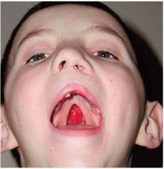
23 Development of the palate and palate cleft
 It connect")


24 Congenital anomalies of middle face 1. Oblique cleft of the face (persistent nasolacrimal groove) It connect mouth to medial palpebral angle of the orbit Nasolacrimal duct is present as open grove It results from failure of fusion of lateral nasal and maxillary prominences 2. Cleft of upper lip, superior alveolar arch and palate It results from failure of fusion of medial nasal and maxillary prominences They could be unilateral or bilateral
25 Development of the face (from 4 th to 8 th weeks) 1. Development of the primitive mouth stomodeum (beginning of the 4 th week) 2. Rupture of oropharyngeal membrane (the 24 th day) 3. Development of the nasal cavity (from the end of the 4 th week) 4. Rupture of oronasal membrane (the 6 th week) 5. Development of paranasal air sinuses from diverticuli of nasal walls during late fetal life & after birth


26 Paranasal air sinuses -facial skull bones which contain air spaces lined with mucous membrane -make the skull light -impart resonance to voice -act as conditioning chambers for inspired air

27 - Sinuses appear on 12 th week of gestation - Rapid growth till 12-years after birth - Growth together with facial skull bones - Growth of maxillary sinus stops with eruption of last molars

28 Development of branchial apparatus (arches, pouches, grooves) The 4 th week - neural crest cells migrate through the mesenchyme to the future head and neck region, forming elevations of mesoderm on each side of the primitive pharynx

29 Pharyngeal arches - a series of mesodermal outpouchings on both sides of the developing pharynx - covered with: Ectoderm outside formation of pharyngeal clefts Endoderm inside formation of pharyngeal pouches
30 The 1 st Pharyngeal arch (mandibular) Develop: - masticatory muscles - m. mylohyoideus, m. digastricus (venter anterior) - m. tensor veli palatini, m. tensor tympani - Lips - Jaws - Palate - Anterior 2/3 of the tongue - Porus accusticus externus - Membrana tympanica - Malleus and incus - External ear (anterior part) - Innervated by trigeminal nerve (V)

31 Meckel`s cartilage mandible NB! Mandible buds are paired!
32 The 2 nd Pharyngeal arch (hyoid) - Innervated by facial nerve (VII) - mimic muscles - m. digastricus (venter posterior) - m. stylohyoideus - Stapes and m.stapedius - Processus styloideus - Hyoid bone - Tonsilla palatina - External ear (posterior part)
33 The 3 rd Pharyngeal arch - Innervated by glossopharyngeal (IX) nerve - muscles of the palate - m. stylopharyngeus - Hyoid bone - Thymus
34 The 4 th and the 5 th Pharyngeal arches - Innervated by vagal (X) nerve - muscles of the palate and pharynx - Cartilages and muscles of the larynx - Thymus
35 Development of the tongue Tongue parts Tongue buds Origin of the bud Fusion Anterior 2/3 of the tongue Posterior 1/3 of the tongue -Median bud -2 distal buds - Copula - Hypobranchial prominence Pharyngeal arch 1 Pharyngeal arches 2,3,4 Median sulcus Sulcus terminalis Foramen cecum

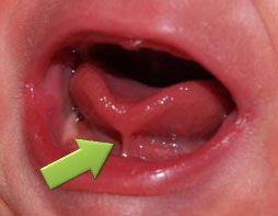
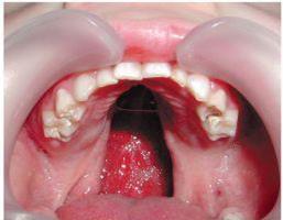
36 Pathology of the tongue development

37 Development of the lower airway, lungs and pleura
38 Endoderm Mesoderm Epithelium and glands of the lower airway Connective tissue, cartilage, muscle, vessels and pleurae
 nerve - muscles of the palate and pharynx - Cartilages and muscles of the larynx -")
39 Development of the larynx - the 4 th and the 5 th Pharyngeal arches - Innervated by vagal (X) nerve - muscles of the palate and pharynx - Cartilages and muscles of the larynx - Thymus

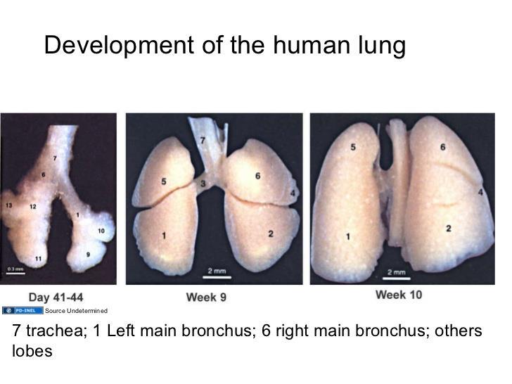
40 4 th week 5 th week 6 th week 10 th week

41 Development of the trachea Endoderm Mesoderm Epithelium and glands of the lower airway Connective tissue, cartilage, muscle, vessels and adventitia

42 Congenital anomalies of trachea 1. Tracheoesophageal fistula communication between trachea and esophagus
 and Atresia")
43 Congenital anomalies of trachea 2. Tracheal Stenosis (narrowing) and Atresia (closure)

44 Congenital anomalies of trachea 3. Tracheal diverticulum
45 Development of the bronchi - Lung buds divide into two bronchial buds - The 5 th week bronchial buds enlarge to form primary bronchi - Primary bronchi further subdivide into secondary bronchi - Secondary bronchi further subdivide into tertiary (=segmental) bronchi
46 Development of the bronchi


47 Bronchopulmonary segments - is a segment of lung tissue supplied by a tertiary (segmental) bronchus
48 Development of human lung Periods of Lung Development: 1. Pseudoglandular period (5 17 weeks) 2. Canalicular period (16 25 weeks) 3. Terminal sac period (24 weeks to birth) 4. Alveolar period (late fetal period to 8 years after birth)
49

50 Type I pneumocytes thin, flat cells that make up part of the blood-air barrier Type II pneumocytes cells, that produce surfactant
 at the")
51 Pulmonary surfactant - a surface-active lipoprotein complex (phospholipoprotein) formed by type II alveolar cells. By adsorbing to the air-water interface of alveoli it reduces surface tension and prevents collapse of the lung (atelectasis) at the end of expiration.

52 Premature fetuses born between week 25 and 28 can survive with intensive care! This is the earliest point at which fetus can survive. Adequate vascularization and surfactant levels are the most important factors for the survival of premature infants!
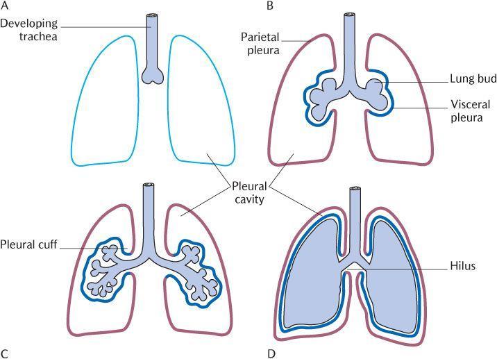
53 Development of the pleura and pleural cavities Pleura serose membrane lining lungs and walls of the thoracic cavity - is derived from intraembryonic mesoderm
 Pleural cavity Somatic mesoderm Parietal pleura Splanchnic mesoderm Visceral pleura")
54 Lateral mesoderm forms two plates: somatic and splanchnic EEM, extraembryonic mesoderm; YS, Yolk sac; NP, neural plate. Intraembryonic body cavity(coelom) Pleural cavity Somatic mesoderm Parietal pleura Splanchnic mesoderm Visceral pleura


55 Development of the pleura and pleural cavities Expansion of the lung buds into the pericadioperitoneal canals. At this stage, the canals are in communication with the peritoneal and pericardial cavities.
56 Development of pleura

57 Development of the pleura and pleural cavities
) Also called")


58 Congenital abnormalities (Infant respiratory distress Syndrome (IRDS)) Also called Hyaline Membrane Disease: Congenital Lung Cysts Agenesis of Lungs or one Lung Lung Hypoplasia Accessory Lung Lobe of Azygos Vein Agenesis of the right lung Lung hypoplasia Accessory lobe
 3) Dorsal Mesentery of Esophagus")
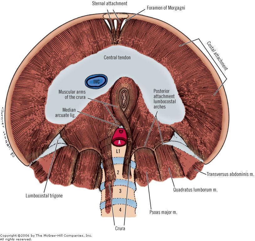
59 Diaphragm develops from 4 sources: 1) Septum Transversum 2) Pleuroperitoneal Membranes (folds) 3) Dorsal Mesentery of Esophagus 4) Body Wall
60
61 The trachea is lined with pseudostratified ciliated columnar epithelium with goblet cells. This epithelium is derived from 1)Neuroectoderm 2)Endoderm 3)Ectoderm 4)Visceral mesoderm 5)Mesoderm of 4 th and 6 th pharyngeal arches 2
62 Smooth muscle, connective tissue, and cartilage of primary bronchi are derived from which of the following sources? 1)Neuroectoderm 2)Endoderm 3)Ectoderm 4)Visceral mesoderm 5)Mesoderm of 4 th and 6 th pharyngeal arches 4
63 Components of blood-air barrier in the lungs are derived from which of the following sources? 1)Ectoderm only 2)Visceral mesoderm only 3)Visceral mesoderm and ectoderm 4)Endoderm and ectoderm 5)Visceral mesoderm and endoderm 5
64 The laryngotracheal tube initially is an open communication with the primitive foregut. Which of the following embryonic structures is responsible for separating these two structures? 1)Laryngotracheal tube 2)Posterior esophageal folds 3)Laryngoesophageal diverticulum 4)Tracheoesophageal septum 5)Bronchopulmonary segment 4

65 Changes that occur after birth After birth, the circulation of fetal blood through the placenta ceases: Delivery of oxygenated blood to fetus via umbilical vein ceases Hypoxia of all tissues is increasing Respiratory centers of the brain stem are stimulated by carbon dioxide Inspiratory muscles contract, thoracic cage is expanded Expansion of the lungs and First Breath takes place Inspired air enters respiratory passageways, pushes the contained fluids out of the way and inflates the bronchial and respiratory trees Infant s lungs begin to function and newborn infant utters a loud cry
66


67 Absence of nasal breathing Consequences: - Retarded development of the nose - Inflammation of the nasal cavity - Small chin - Wrong position of the tongue - Problems with speech and phonation - Increased salivation - Anterior position of the head problems with cervical part of the vertebral column - Retarded physical development, passive hypoxia - Retarded mental development hypoxia of the brain - Weak muscles of the lips changes in teeth position and malocclusion - Temporomandibular joint disorders, etc.
68 Thank you for your attention!
Development of Respiratory System. Dr. Sanaa Alshaarawy& Dr. Saeed Vohra
 Development of Respiratory System Dr. Sanaa Alshaarawy& Dr. Saeed Vohra OBJECTIVES At the end of the lecture the students should be able to: Identify the development of the laryngeotracheal (respiratory)
Pharyngeal Apparatus. Pouches Endoderm Grooves Ectoderm Arch Neural Crest Somitomeres Aortic Arch - Vessel
 Pharyngeal Apparatus Pouches Endoderm Grooves Ectoderm Arch Neural Crest Somitomeres Aortic Arch - Vessel Segmental Organization Humans: Arch 1-4 prominent Arch 5 absent Arch 6 - transient First Arch Face
Development of the nasal cavity :
 Development of the nasal cavity : several processes contribute to the development of the nose, the nose consists of 2 cavities separated by a septum, and the nasal cavity is separated from the oral cavity
04 Development of the Face and Neck. Development of the Face Development of the neck
 04 Development of the Face and Neck Development of the Face Development of the neck Development of the face Overview of facial development The fourth week ~ the twelfth week of prenatal development Between
Respiratory System. Functional Anatomy of the Respiratory System
 Respiratory System Overview of the Respiratory System s Job Major Duty Respiration Other important aspects ph control Vocalization Processing incoming air Protection Metabolism (ACE) What structures allow
REVIEW OF CLINICAL EMBRYOLOGY OF HEAD AND NECK
 REVIEW OF CLINICAL EMBRYOLOGY OF HEAD AND NECK OUTLINE - EMBRYOLOGY UNDERLYING CLINICAL CONDITIONS I. EARLY DEVELOPMENT OF FACE: CLEFT LIP, CLEFT PALATE, OBSTRUCTED NASOLACRIMAL DUCT II. BRANCHIAL ARCHES
Remember from the first year embryology Trilaminar disc has 3 layers: ectoderm, mesoderm, and endoderm
 Development of face Remember from the first year embryology Trilaminar disc has 3 layers: ectoderm, mesoderm, and endoderm The ectoderm forms the neural groove, then tube The neural tube lies in the mesoderm
 Drawings illustrating the human pharyngeal apparatus. Drawings illustrating the human pharyngeal apparatus. Drawings illustrating the human pharyngeal apparatus. Drawings illustrating the human pharyngeal
The embryonic endoderm initially is widely connected with the yolk sac. As a consequence of cephalocaudal and lateral folding, a portion of the
 DIGESTIVE SYSTEM The embryonic endoderm initially is widely connected with the yolk sac. As a consequence of cephalocaudal and lateral folding, a portion of the endoderm-lined yolk sac cavity is incorporated
2/2/2011. Primitive Gut Tube Proctodeum and Stomodeum Stomach Duodenum Pancreas Liver and Biliary Apparatus Spleen Midgut
 DEVELOPMENT OF THE DIGESTIVE SYSTEM Development of Endodermal Organs Primitive Gut Tube Proctodeum and Stomodeum Stomach Duodenum Pancreas Liver and Biliary Apparatus Spleen Midgut Wednesday, February
Pharyngeal apparatus. - At the third week, it is a 3 layered structure: ectoderm, mesoderm and endoderm. This is called trilaminar disc
 Pharyngeal apparatus Remember from the first year embryology - The embryo was disc shaped in the second week of development (this is called embryonic disc) and it is a 2 layered disc (composed of two layers)---bilaminar
Lecture 21Development of respiratory system Dr. Rehan Asad At the end of session students should able to Describe formation of lung buds Describe
 Lecture 21Development of respiratory system Dr. Rehan Asad At the end of session students should able to Describe formation of lung buds Describe development of larynx, trachea and bronchi. Describe the
NEUROCRANIUM VISCEROCRANIUM VISCEROCRANIUM VISCEROCRANIUM
 LECTURE 4 SKULL NEUROCRANIUM VISCEROCRANIUM VISCEROCRANIUM VISCEROCRANIUM CRANIUM NEUROCRANIUM (protective case around brain) VISCEROCRANIUM (skeleton of face) NASOMAXILLARY COMPLEX MANDIBLE (DESMOCRANIUM)
NURSE-UP RESPIRATORY SYSTEM
 NURSE-UP RESPIRATORY SYSTEM FUNCTIONS OF THE RESPIRATORY SYSTEM Pulmonary Ventilation - Breathing Gas exchanger External Respiration between lungs and bloodstream Internal Respiration between bloodstream
Essentials in Head and Neck Embryology. Part 3 Development of the head, face, and oral cavity
 Essentials in Head and Neck Embryology Part 3 Development of the head, face, and oral cavity Outline General overview of prenatal development Embryonic period phase 1 Formation of bilaminar disk Formation
The Respiratory System
 PowerPoint Lecture Slide Presentation by Vince Austin Human Anatomy & Physiology FIFTH EDITION Elaine N. Marieb The Respiratory System Dr Nabil Khouri. MD, Ph.D Respiratory System Consists of a conducting
The Respiratory System
 13 PART A The Respiratory System PowerPoint Lecture Slide Presentation by Jerry L. Cook, Sam Houston University ESSENTIALS OF HUMAN ANATOMY & PHYSIOLOGY EIGHTH EDITION ELAINE N. MARIEB Organs of the Respiratory
B. Correct! As air travels through the nasal cavities, it is warmed and humidified.
 Human Anatomy - Problem Drill 20: The Respiratory System Question No. 1 of 10 1. Which of the following statements about the portion of the respiratory system labeled in the image below is correct? Question
Histology and development of the respiratory system
 Histology and development of the respiratory system Árpád Dobolyi Semmelweis University, Department of Anatomy, Histology and Embryology Outline of the lecture 1. Structure of the trachea 2. Histology
Chapter 16. Respiratory System
 Chapter 16 Respiratory System Introduction Respiration = the entire process of exchanging gases between the atmosphere and body cells 1. Ventilation 2. Gas exchange 3. Gas transport : 4. Cellular respiration
Ch16: Respiratory System
 Ch16: Respiratory System Function: - O2 in and CO2 out of the blood vessels in the lungs - O2 out and CO2 into the blood vessels around the cells - Gas exchange happens in - Other organs purify, humidify,
Respiratory System Embryology
 Respiratory System Embryology Development of the nose and Palate Development of the nose At the end of the fourth week, facial prominences consisting primarily of neural crest-derived mesenchyme and formed
Bronchioles. Alveoli. Type I alveolar cells are very thin simple squamous epithelial cells and form most of the lining of an alveolus.
 276 Bronchioles Bronchioles continue on to form bronchi. The primary identifying feature is the loss of hyaline cartilage. The epithelium has become simple ciliated columnar, and there is a complete ring
The Respiratory System
 The Respiratory System Cells continually use O2 & release CO2 Respiratory system designed for gas exchange Cardiovascular system transports gases in blood Failure of either system rapid cell death from
THE RESPIRATORY SYSTEM
 THE RESPIRATORY SYSTEM Functions of the Respiratory System Provides extensive gas exchange surface area between air and circulating blood Moves air to and from exchange surfaces of lungs Protects respiratory
Development of the Pharyngeal Arches
 Development of the Pharyngeal Arches Thomas A. Marino, Ph.D. Temple University School of Medicine Competencies: Upon completion of this section of the course, the student must be able to: 1. Recall the
Lab Activity 27. Anatomy of the Respiratory System. Portland Community College BI 233
 Lab Activity 27 Anatomy of the Respiratory System Portland Community College BI 233 1 Terminology Pulmonary Ventilation: aka breathing, is the movement of air into and out of the lungs External Respiration:
Organs of the Respiratory System Laboratory Exercise 52
 Organs of the Respiratory System Laboratory Exercise 52 Background The organs of the respiratory system include the nose, nasal cavity, sinuses, pharynx, larynx, trachea, bronchial tree, and lungs. They
Head and Neck I. PHARYNGEAL APPARATUS (FIGURE 12.1; TABLE 12.1)
") chapter 12 Head and Neck I. PHARYNGEAL APPARATUS (FIGURE 12.1; TABLE 12.1) The pharyngeal apparatus consists of the pharyngeal arches, pharyngeal pouches, pharyngeal grooves, and pharyngeal membranes,
chapter 12 Head and Neck I. PHARYNGEAL APPARATUS (FIGURE 12.1; TABLE 12.1) The pharyngeal apparatus consists of the pharyngeal arches, pharyngeal pouches, pharyngeal grooves, and pharyngeal membranes,
The Respiratory System. Dr. Ali Ebneshahidi
 The Respiratory System Dr. Ali Ebneshahidi Functions of The Respiratory System To allow gases from the environment to enter the bronchial tree through inspiration by expanding the thoracic volume. To allow
Embryo#1. Mohammad Hisham Al-Mohtaseb باشق جهاد. 0 P a g e
 Embryo#1 Mohammad Hisham Al-Mohtaseb باشق جهاد 0 P a g e Before you start, it is important to link what you learn in gross anatomy with developmental stages discussed in embryology. Cells that form organs
I. Anatomy of the Respiratory System A. Upper Respiratory System Structures 1. Nose a. External Nares (Nostrils) 1) Vestibule Stratified Squamous
 1) Vestibule Stratified Squamous") I. Anatomy of the Respiratory System A. Upper Respiratory System Structures 1. Nose a. External Nares (Nostrils) 1) Vestibule Stratified Squamous Epithelium b. Nasal Cartilages 1) Nasal Cavity Pseudostratified
I. Anatomy of the Respiratory System A. Upper Respiratory System Structures 1. Nose a. External Nares (Nostrils) 1) Vestibule Stratified Squamous Epithelium b. Nasal Cartilages 1) Nasal Cavity Pseudostratified
RESPIRATORY SYSTEM. described: pp. 744,746 fig. 25.1, described: p. 746 fig described: p. 776 fig. 26.3
 ACTIVITY 11: RESPIRATORY AND DIGESTIVE SYSTEMS OBJECTIVES: 1) How to get ready: Read Chapters 25 and 26, McKinley et al., Human Anatomy, 5e. All text references are for this textbook. 2) Identify structures
The Respiratory System:
 The Respiratory System: Respiration Involves both the respiratory and the circulatory systems Four processes that supply the body with O 2 and dispose of CO 2 Respiration Pulmonary ventilation (breathing):
Organs Histology D. Sahar AL-Sharqi. Respiratory system
 Respiratory system The respiratory system provides for exchange of O2 and CO2 to and from the blood. Respiratory organs include the lungs and a branching system of bronchial tubes that link the sites of
ACTIVITY 11: RESPIRATORY AND DIGESTIVE SYSTEMS RESPIRATORY SYSTEM
 ACTIVITY 11: RESPIRATORY AND DIGESTIVE SYSTEMS OBJECTIVES: 1) How to get ready: Read Chapters 25 and 26, McKinley et al., Human Anatomy, 4e. All text references are for this textbook. 2) Identify structures
The Respiratory System. Supplies body with oxygen Disposes of carbon dioxide Four processes in respiration
 C H A P T E R 22 The Respiratory System The Respiratory System Supplies body with oxygen Disposes of carbon dioxide Four processes in respiration Pulmonary ventilation External respiration Transport of
CHAPTER 22 RESPIRATORY
 pulmonary ventilation move air external respiration exchange gases transportation of gases internal respiration exchange gases CHAPTER 22 RESPIRATORY in / out lungs air - blood blood - cells cell respiration
-Tamara Wahbeh. -Razan Abu Rumman. Dr. Mohammed Al-Muhtaseb
 -2 -Tamara Wahbeh -Razan Abu Rumman Dr. Mohammed Al-Muhtaseb I tried to include everything the doctor mentioned in both the lecture and his slides in the simplest way possible, so hopefully there would
Anatomical Considerations for Lab Practical II
 Anatomical Considerations for Lab Practical II For each of the following please be prepared to provide: Identification System Organ(s) or ducts to Function(s) location which it is attached Use your lecture
Chapter 23 The Respiratory System
 Chapter 23 The Respiratory System Cells continually use O 2 & release CO 2 Respiratory System designed for gas exchange Cardiovascular system transports gases in blood Failure of either system rapid cell
Endeavour College of Natural Health endeavour.edu.au
 Endeavour College of Natural Health endeavour.edu.au BIOH122 Human Biological Science 2 Session 10 Respiratory System 1 Anatomy & Physiology Bioscience Department Endeavour College of Natural Health endeavour.edu.au
RESPIRATORY SYSTEM. A. Upper respiratory tract (Fig. 23.1) Use the half-head models.
 Use the half-head models.") RESPIRATORY SYSTEM I. OVERVIEW OF THE RESPIRATORY SYSTEM AND THORAX A. Upper respiratory tract (Fig. 23.1) Use the half-head models. Nasal cavity Pharynx (fare-rinks) B. Lower respiratory tract (Fig. 23.1)
RESPIRATORY SYSTEM I. OVERVIEW OF THE RESPIRATORY SYSTEM AND THORAX A. Upper respiratory tract (Fig. 23.1) Use the half-head models. Nasal cavity Pharynx (fare-rinks) B. Lower respiratory tract (Fig. 23.1)
The Anatomy and Physiology of the Respiratory System
 CHAPTER 1 The Anatomy and Physiology of the Respiratory System Sagittal Section of Upper Airway Fig. 1-1. Sagittal section of upper airway. Structure of the Nose Fig. 1-2. Structure of the nose. Sagittal
The Respiratory System
 C h a p t e r 24 The Respiratory System PowerPoint Lecture Slides prepared by Jason LaPres North Harris College Houston, Texas Copyright 2009 Pearson Education, Inc., publishing as Pearson Benjamin Cummings
The Respiratory System
 Essentials of Human Anatomy & Physiology Elaine N. Marieb Seventh Edition Chapter 13 The Respiratory System Slides 13.1 13.30 Lecture Slides in PowerPoint by Jerry L. Cook Copyright 2003 Pearson Education,
Karachi King s College of Nursing
 Karachi King s College of Nursing Badil Dass Lecturer Respiratory system Respiratory System Respiratory system consist of: Nose Pharynx (Throat) Larynx (Voice Box) Trachea (Wind Pipe) Bronchi Bronchioles
THYROID & PARATHYROID. By Prof. Saeed Abuel Makarem & Dr. Sanaa Al-Sharawy
 THYROID & PARATHYROID By Prof. Saeed Abuel Makarem & Dr. Sanaa Al-Sharawy 1 OBJECTIVES By the end of the lecture, the student should be able to: Describe the shape, position, relations and structure of
Embryology of the Heart
 *Page 1A: Embryology of the Heart Human embryonic disc is divided into three layers: ectoderm, intraembryonic mesoderm, and endoderm. The embryonic disc lies between the amniotic cavity and the primary
The Respiratory System
 The Respiratory System If you have not done so already, please print and bring to class the Laboratory Practical II Preparation Guide. We will begin using this shortly in preparation of your second laboratory
Lecture Overview. Respiratory System. Martini s Visual Anatomy and Physiology First Edition. Chapter 20 - Respiratory System Lecture 11
 Martini s Visual Anatomy and Physiology First Edition Martini Ober Chapter 20 - Respiratory System Lecture 11 1 Lecture Overview Overview of respiration Functions of breathing Organs of the respiratory
The RESPIRATORY System
 The RESPIRATORY System Respira5on The exchange of gases between the atmosphere, blood, and cells Pulmonary Ven5la5on - the exchange of air between the atmosphere and lungs External (Pulmonary) Respira5on
The respiratory system has multiple organs, we will begin with the nose and nasal cavity.
 Respiratory System (Peer reviewed and edited) Slide 1: Respiratory System Slide 2: Functions Functions of respiratory system include gas exchange, communication, olfaction, and ph regulation. Gas exchange
SYLLABUS BDS I PROFESSIONAL GENERAL HUMAN ANATOMY INCLUDING EMBRYOLOGY AND HISTOLOGY
 GENERAL HUMAN ANATOMY INCLUDING EMBRYOLOGY AND HISTOLOGY I. General Anatomy 1. Anatomical terms 2. Skin, superficial fascia & deep fascia 3. Cardiovascular system, portal system, collateral circulation
Head and Neck Development and Malformations
 Head and Neck Development and Malformations Yang Chai, DDS, PhD Professor George and MaryLou Boone Chair Ostrow School of Dentistry of USC ychai@usc.edu C D E A. B Learning Objectives - Learn cranial neural
Al s 202 study guide answers Answers Respiratory System 1 External nares (nostrils) 33 Carina 2 Vestibule 34 Left primary bronchus 3 Nasal cavity 35
 33 Carina 2 Vestibule 34 Left primary bronchus 3 Nasal cavity 35") Trachea & Respiratory Histology 1 Epiglottis 26 Capillary 2 Larynx 27 Alveolar sac 3 Thyroid cartilage 28 Alveoli/Alveolus 4 Cricoid cartilage 29 Basement membrane 5 Vocal folds (True vocal cords) 30 Cilia
Trachea & Respiratory Histology 1 Epiglottis 26 Capillary 2 Larynx 27 Alveolar sac 3 Thyroid cartilage 28 Alveoli/Alveolus 4 Cricoid cartilage 29 Basement membrane 5 Vocal folds (True vocal cords) 30 Cilia
HISTOLOGY OF THE RESPIRATORY SYSTEM I. Introduction A. The respiratory system provides for gas exchange between the environment and the blood. B.
 HISTOLOGY OF THE RESPIRATORY SYSTEM I. Introduction A. The respiratory system provides for gas exchange between the environment and the blood. B. The human respiratory system may be subdivided into two
Cranial nerves.
 Cranial nerves eaglezhyxzy@163.com Key Points of Learning Name Components Passing through Peripheral distribution Central connection Function Cranial nerves Ⅰ olfactory Ⅱ optic Ⅲ occulomotor Ⅳ trochlear
Function of Breathing. Jeanine D Armiento, M.D., Ph.D. Respiratory Portion. Conducting Portion. Critical to the Development of the Lung
 Function of Breathing Jeanine D Armiento, M.D., Ph.D. Associate Professor Department of Medicine P&S 9-449 5-3745 jmd12@columbia.edu Air Sacs (alveoli) Ventilation-air conduction Moving gas in and out
Embryology: Development of digestive system
 Embryology: Development of digestive system Embryo folding incorporation of endoderm to form primitive gut. Outside of embryo yolk sac and allantois. Vitelline duct Stomodeum (primitive mouth) the oral
Development of the Digestive System. W.S. O The University of Hong Kong
 Development of the Digestive System W.S. O The University of Hong Kong Plan for the GI system Then GI system in the abdomen first develops as a tube suspended by dorsal and ventral mesenteries. Blood
When you see this diagram, remember that you are looking at the embryo from above, through the amniotic cavity, where the epiblast appears as an oval
 When you see this diagram, remember that you are looking at the embryo from above, through the amniotic cavity, where the epiblast appears as an oval disc 2 Why the embryo needs the vascular system? When
LECTURE 2 THE RESPIRATORY SYSTEM
 LECTURE 2 THE RESPIRATORY SYSTEM Respiratory system - a complex of organs and anatomical structures exercising function of external respiration. Functions of the respiratory system: - Provides the organism
Introduction to Head and Neck Anatomy
 Introduction to Head and Neck Anatomy Nervous Tissue Controls and integrates all body activities within limits that maintain life Three basic functions 1. sensing changes with sensory receptors 2. interpreting
SESSION 2: THE MOUTH AND PHARYNX
 SESSION 2: THE MOUTH AND PHARYNX 9 In the pig s digestive tract, food flows in only one direction from mouth to anus.this allows for greatly specialized sections that can act independently of each other.
The Respiratory System
 The Respiratory System Respiration Includes Pulmonary ventilation Air moves in and out of lungs Continuous replacement of gases in alveoli (air sacs) External respiration Gas exchange between blood and
General Anatomy p. 1 Organization of the Human Body p. 1 Skeleton of the Human Body p. 4 Ossification of the Bones p. 6 Bone Structure p. 8 Joints p.
 General Anatomy p. 1 Organization of the Human Body p. 1 Skeleton of the Human Body p. 4 Ossification of the Bones p. 6 Bone Structure p. 8 Joints p. 10 Principal Joints (Immovable) p. 12 Synovial Joints
Digestive system (Systema digestorium/ alimentarium) Lecture #1
 Lecture #1") Digestive system (Systema digestorium/ alimentarium) Lecture #1 Internal organs are grouped into 1. System - Have the same functions and development Digestive system Respiratory system 2. Apparatus - Have
Digestive system (Systema digestorium/ alimentarium) Lecture #1 Internal organs are grouped into 1. System - Have the same functions and development Digestive system Respiratory system 2. Apparatus - Have
Chapter 11. The respiratory system. Glossary. Anthony Wheeldon
 Chapter 11 The respiratory system Anthony Wheeldon Glossary Accessory muscles Muscles not normally involved in respiration that can be utilised to increase inspiration. Acid base balance The mechanisms
PTERYGOPALATINE FOSSA
 PTERYGOPALATINE FOSSA Outline Anatomical Structure and Boundaries Foramina and Communications with other spaces and cavities Contents Pterygopalatine Ganglion Especial emphasis on certain arteries and
A. Incorrect! Think of a therapy that reduces prostaglandin synthesis. B. Incorrect! Think of a therapy that reduces prostaglandin synthesis.
 USMLE Step 1 - Problem Drill 02: Embryology Question No. 1 of 10 1. A premature infant is born with a patent ductus arteriosis. Which of the following treatments may be used as part of the treatment regimen?
The Respiratory System
 PowerPoint Lecture Slides prepared by Leslie Hendon University of Alabama, Birmingham C H A P T E R 22 Part 1 The Respiratory System The Respiratory System Basic functions of the respiratory system Supplies
Respiratory System Functions. Respiratory System Organization. Respiratory System Organization
 Respiratory System Functions Functions of Respiratory System Gas exchange between blood and air Move air to and from exchange surfaces Protect exchange surfaces from environmental variations and pathogens
-Ibrahim Al-Naser. -Dr Al- Muhtaseb. 1 P a g e
 -1 -Ibrahim Al-Naser - -Dr Al- Muhtaseb 1 P a g e The Digestive System The doctor started the lecture by talking about the class rules. The GI system is an organ system, it is divided into: The Alimentary
The Respiratory System
 The Respiratory System Function of the Respiratory System Oversees gas exchanges (oxygen and carbon dioxide) between the blood and external environment Exchange of gasses takes place within the lungs in
Read Me. We are the Learning Lab. to look
 Respiratory Tract Anatomy Lab In-Lab Exercises Read Me We are going to look at models and slides. Much of this can be done in the Learning Lab on your own time. The steps do not have to be done in order,
Trigeminal Nerve (V)
") Trigeminal Nerve (V) Lecture Objectives Discuss briefly how the face is developed. Follow up the course of trigeminal nerve from its point of central connections, exit and down to its target areas. Describe
Trigeminal Nerve (V) Lecture Objectives Discuss briefly how the face is developed. Follow up the course of trigeminal nerve from its point of central connections, exit and down to its target areas. Describe
The RESPIRATORY System. Unit 9
 The RESPIRATORY System Unit 9 Respiration The exchange of gases between the atmosphere, blood, and cells Pulmonary Ventilation - the exchange of air between the atmosphere and lungs External (Pulmonary)
RESPIRATORY LAB. Introduction: trachea, extrapulmonary bronchi, and lungs b) passage for and conditioning of air (moisten, warm, and filtering)
 passage for and conditioning of air (moisten, warm, and filtering)") RESPIRATORY LAB Danil Hammoudi.MD Introduction: a) system includes nasal cavity, pharynx, larynx, trachea, extrapulmonary bronchi, and lungs b) passage for and conditioning of air (moisten, warm, and filtering)
RESPIRATORY LAB Danil Hammoudi.MD Introduction: a) system includes nasal cavity, pharynx, larynx, trachea, extrapulmonary bronchi, and lungs b) passage for and conditioning of air (moisten, warm, and filtering)
Group B: Organ systems (digestive, respiratory, urinary, genital system, heart, glands and skin) green
 green") Group B: Organ systems (digestive, respiratory, urinary, genital system, heart, glands and skin) green Digestive system 1. Teeth Main points: external and internal structure of a tooth, fixation of a tooth
Group B: Organ systems (digestive, respiratory, urinary, genital system, heart, glands and skin) green Digestive system 1. Teeth Main points: external and internal structure of a tooth, fixation of a tooth
Omran Saeed. Luma Taweel. Mohammad Almohtaseb. 1 P a g e
 2 Omran Saeed Luma Taweel Mohammad Almohtaseb 1 P a g e I didn t include all the photos in this sheet in order to keep it as small as possible so if you need more clarification please refer to slides In
UNIVERSITY OF NAIROBI
 UNIVERSITY OF NAIROBI UNIVERSITY EXAMINATIONS 2013/2014 LEVEL I MID-SEMESTER II EXAMINATION FOR THE DEGREE OF BACHELOR OF SCIENCE IN NURSING (BScN) AND BACHELOR OF PHARMACY (B.PHARM) MARKING SCHEME HNS101/UPC106:
Bio 322 Human Anatomy Objectives for the laboratory exercise Respiratory System
 Bio 322 Human Anatomy Objectives for the laboratory exercise Respiratory System Required reading before beginning this lab: Saladin, KS: Human Anatomy 5 th ed (2017) Chapter 23 For this lab you will use
CHAPTER 24. Respiratory System
 CHAPTER 24 Respiratory System RESPIRATION INCLUDES Air moves in and out of lungs Continuous replacement of gases in alveoli (air sacs) Gas exchange between blood and air at alveoli Transport of respiratory
Objectives. Module A2: Upper Airway Anatomy & Physiology. Function of the Lungs/Heart. The lung is for gas exchange. Failure of the Lungs/Heart
 Module A2: Upper Airway Anatomy & Physiology Objectives Classify epithelial tissue based on cell type and tissue layers. Identify location of tissue epithelium in the respiratory system. Describe the major
Nose & Mouth OUTLINE. Nose. - Nasal Cavity & Its Walls. - Paranasal Sinuses. - Neurovascular Structures. Mouth. - Oral Cavity & Its Contents
 Dept. of Human Anatomy, Si Chuan University Zhou hongying eaglezhyxzy@163.com Nose & Mouth OUTLINE Nose - Nasal Cavity & Its Walls - Paranasal Sinuses - Neurovascular Structures Mouth - Oral Cavity & Its
Unit Nine - The Respiratory System
 Unit Nine - The Respiratory System I. Introduction A. Definition: the respiratory system consists of the nose, nasal cavity, (throat), (voice box), (windpipe), bronchi and lungs (which contain the alveoli).
Thyroid gland. importance. relations and connections. external laryngeal nerves. malformations.
 Thyroid gland 1. Recognize and understand the coverings of the thyroid gland and their clinical importance. 2. Recognize and understand the main parts of the thyroid gland and their locations, relations
Brain and spinal nerve. By: shirin Kashfi
 Brain and spinal nerve By: shirin Kashfi Nervous system: central nervous system (CNS) peripheral nervous system (PNS) Brain (cranial) nerves Spinal nerves Ganglions (dorsal root ganglions, sympathetic
Prevertebral Region, Pharynx and Soft Palate
 Unit 20: Prevertebral Region, Pharynx and Soft Palate Dissection Instructions: Step1 Step 2 Step 1: Insert your fingers posterior to the sternocleidomastoid muscle, vagus nerve, internal jugular vein,
Phases of Respiration. Chapter 18: The Respiratory System. Structures of the Respiratory System. Structures of the Respiratory System
 Phases of Respiration Chapter 18: The Respiratory System Respiration Process of obtaining oxygen from environment and delivering it to cells Phases of Respiration 1. Pulmonary ventilation between air and
Development of the Digestive System. W.S. O School of Biomedical Sciences, University of Hong Kong.
 Development of the Digestive System W.S. O School of Biomedical Sciences, University of Hong Kong. Organization of the GI tract: Foregut (abdominal part) supplied by coeliac trunk; derivatives include
Basic Anatomy and Physiology of the Lips and Oral Cavity. Dr. Faghih
 Basic Anatomy and Physiology of the Lips and Oral Cavity Dr. Faghih It is divided into seven specific subsites : 1. Lips 2. dentoalveolar ridges 3. oral tongue 4. retromolar trigone 5. floor of mouth 6.
2. List seven functions performed by the respiratory system?
 The Respiratory System C23 Study Guide Tortora and Derrickson 1. In physiology we recognize that the word respiration has three meanings. What are the three different meanings of the word respiration as
Organogenesis Part 2. V. Lateral Plate Mesoderm VI. Endoderm VII. Development of the Tetrapod Limb VIII. Sex Determination. V. Lateral Plate Mesoderm
 Organogenesis Part 2 V. Lateral Plate Mesoderm VI. Endoderm VII. Development of the Tetrapod Limb VIII. Sex Determination V. Lateral Plate Mesoderm chordamesoderm paraxial mesoderm intermediate mesoderm
Lec [8]: Mandibular nerve:
![Lec [8]: Mandibular nerve:](/thumbs/94/121295776.jpg "Lec [8]: Mandibular nerve:") Lec [8]: Mandibular nerve: The mandibular branch from the trigeminal ganglion lies in the middle cranial fossa lateral to the cavernous sinus. With the motor root of the trigeminal nerve [motor roots lies
Lec [8]: Mandibular nerve: The mandibular branch from the trigeminal ganglion lies in the middle cranial fossa lateral to the cavernous sinus. With the motor root of the trigeminal nerve [motor roots lies
Lecturer: Ms DS Pillay ROOM 2P24 25 February 2013
 Lecturer: Ms DS Pillay ROOM 2P24 25 February 2013 Thoracic Wall Consists of thoracic cage Muscle Fascia Thoracic Cavity 3 Compartments of the Thorax (Great Vessels) (Heart) Superior thoracic aperture