WS 206: US Guidance for Lower Limb Chemodenervation Procedures AAPM&R 2015
|
|
|
- Gwendolyn Walker
- 5 years ago
- Views:
Transcription
1 WS 206: US Guidance for Lower Limb Chemodenervation Procedures AAPM&R 2015 Katharine E Alter Rita Ayyangar Elie Elovic Troy Henning John Lesher John McGuire Heakyung Kim Michael Munin Jeff Strakowski
2 Disclosures Katharine Alter: Consultant fees Allergan, Royalties Demos Medical Publishing Elie Elovic: Speaker fees Allergan John McGuire: speaker fee Allergan Jeff Strakowski: speaker fees, Allergan, Royalties Demos Medical Publishing
3 Disclosures: Off Label Use of Botulinum Toxins (BoNT) OnabotulinumtoxinA (OBTA) is approved in the USA for the treatment of upper limb spasticity in adult patients All 4 of the FDA approved BoNT products (OBTA, abobotulinumtoxina, incobotulinumtoxina and rimabotulinumtoxinb ) are approve for the treatment of muscle overactivity in cervical dystonia Currently the use of all of the FDA approved BoNT products is off label for the treatment of spasticity in the lower limb and for the treament of children In other countries BoNTs are approved for the treatment of spasticity in other muscles and in pediatric patients This course will mention off label or unapproved uses of BoNTsCurrently the use of all of the FDA approved BoNT products is off label applications
4 Objectives Provide hands on US Training for Chemodenervation Procedures At the conclusion of the Workshop participants will Be familiar with ultrasound appearance of key muscles and nerves Gain skills in operation and handling of ultrasound transducers Be familiar with various scanning techniques used for procedural guidance
5 Agenda Course agenda Review of US guidance 15 minutes To provide time for hands on scanning an abbreviated review of this information will be presented Hands on US Guidance for lower limb muscles 75 minutes Additional handouts provided online Comparison of Guidance Techniques and Review of Evidence Supporting US Guidance for BoNT* Review of US Guidance/Physics
6 Course Agenda Hands On Scanning 40 minutes: Proximal muscles Iliopsoas/iliacus, adductors, Obturator Nerve, piriformis: 35 minutes Calf muscles Posterior/Medial Gastrocnemius, Soleus, FDL, Tibialis Posterior Posterior tibial nerve Procedural Guidance Techniques 1 station will be set up for procedural guidance practice Rotate to this station when you are waiting to scan


7 Why use US for Chemodenervation & BoNT Procedures? Correct target isolation is important for Efficacy Minimizing risk/adverse events Potentially to reduce Required dose Side effects Traditional localization techniques have recognized limitations B-Mode US is increasingly recognized as a more accurate guidance technique for chemodenervation procedures
8 Why you should consider using US for BoNT Injections? ADVANTAGES OF US GUIDANCE FOR CHEMODENERVATION PROCEDURES

9 US for BoNT Injections: Advantages Improved accuracy Complex/overlapping anatomy obscures muscle identification Small/large patients or children Provides direct assessment of target Location Depth Structures to be avoided Transverse View, Posterior Calf Transverse Scan at groin


10 US for BoNT Injections: Advantages Visualize/isolate target structures Quickly Easily Accurately Less painful Smaller needles Pediatric patients often require no sedation Distract patients during procedure Abdominal Muscles


11 BoNT Injections: Why Use US? Focal dystonia Identify and target individual muscle fascicles Ex: FDS digit 3 vs. 4 US increases accuracy and decreases time to isolate correct muscle fascicles Reduces pain Longitudinal View, FDS FDS longitudinal view, mid forearm Short axis view of needle


12 US for BoNT Injections: Advantages High risk targets Avoid untargeted muscles or structures Vessels/nerves/lung High stakes muscles SCM/Scalenes Oromandibular muscles Pterygoids Psoas/iliopsoas Others SCM Transverse Scan, Out of Plane Injection Transverse View Posterior Calf


13 US for BoNT Injections: Advantages Improved accuracy When localization limited by: Involuntary muscle activity Co-contraction Motor control, patient cooperation US does not require AROM to isolate muscle Muscle identification is based on pattern recognition


14 BoNT Injections: Why use US? Advantages Non-muscle targets: Salivary Glands Correctly isolating gland is critical to reduce the risk of dysphagia Parotid EMG and E-Stim are of no help Submandibular

15 Ultrasound and Procedural Guidance Disadvantages Equipment factors Availability Cost Clinician related factors Lack of experience Steep learning curve Limited access to training specific for BoNT injections Transverse view, proximal forearm

16 Ultrasound for BoNT Injection: Summary Localization techniques Palpation EMG Nerve stimulators Ultrasound All have advantages & disadvantages Best Strategy: Be skilled in multiple techniques Be aware of The limitations of each technique Evidence supporting/refuting the accuracy of the various techniques


17 Comparison of Injection Techniques Palpation EMG Stimulation Sonography Accuracy +/- +/ Practicability + - +/- ++ Availability +/- +/- +/- + Pain + - +/- +++ Speed +/- - +/- ++ Evaluation +/- - +/- +++ Future research
18 See online handout for review ULTRASOUND BASICS

19 Ultrasound Pulse Generation and Piezoelectric Crystals Reception Convert electrical pulses into vibrations Converts returning vibrations back into electrical pulses A linear array of crystals is used to create planar images Returning echoes are processed to create grey scale 2D/3D/4D images

 Frequency of US λ determines Depth of penetration")
20 Ultrasound Equipment Basics: Soundwaves are produced by piezoelectric crystals Cystal arrays are placed into transducers Transducers Determine the frequency of US waveform ( λ) Frequency of US λ determines Depth of penetration Resolution of the image

21 Appearance of tissues is determined by their acoustic impedance/densisty Speckle from scattering in tissue. L~ λ Weak scattering from blood and fluids with low impedance to US λ Tissues will appears dark or hypoechoic Strong echoes from mirror-like interfaces will appear bright or hyperechoic


22 Ultrasound: Transducer Selection Select size and shape to match the clinical application Size/Shape of transducer Linear: Best for flat surfaces Curvilinear: Best for abdomen/pelvic/gyn Hockey stick: Hand Small irregular surfaces
23 US Basics: Transducer Frequency MHz Depth/Penetration Application cm OB/GYN cm Deep muscles cm Leg 10 5cm Forearm cm Hand, face Select transducer to match required penetration depth MHz for superficial structure Hand, forearm 3-5 MHz for deep muscles Piriformis, iliacus, quadratus lumborum Most transducers have mixed frequencies 3-5, 7-12 etc


24 Transducer Handling/Orientation To correctly orient the transducer on the patient Look for a manufacturer s mark on one end of the transducer The marked end = screen left on display To confirm this orientation: Tap the end of the transducer Observe the location of movement on the display screen to confirm the orientation Notched end
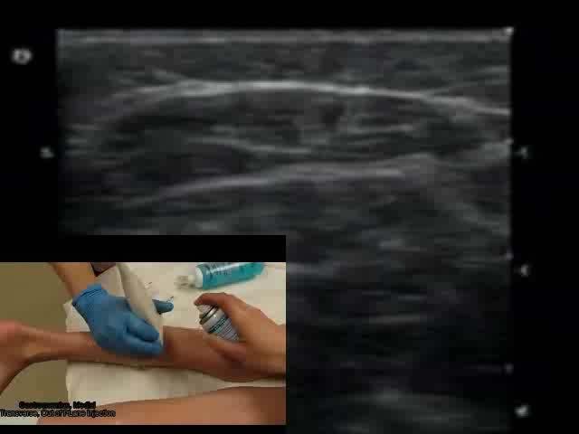
25 US Basics: View convention Top of image is superficial i.e. skin Bottom deeper structures Superficial left Right Deep Transverse view, posterior calf
26 US Basics: View convention Transverse scans How do you place the transducer on the patient? Conventions vary Standard cross sectional imaging Screen left = patient right Simplified cross sectional imaging Screen left = medial left Superficial Deep Right Transverse view, posterior calf

27 US Basics: View convention Longitudinal view Convention Place the transducer on the patient so that Proximal = screen left Distal = screen right Qadriceps tendon and patella Superficial Proximal Distal Deep

28 Holding the transducer Grasp the transducer lightly using your Thumb + index or Thumb + index+ middle finger Do not over grip Keep hand in contact with the patient at all times to avoid slipping Using heel of hand or 4 th and 5 th finger Correct : Maintaining contact with patient Incorrect : No contact with patient

29 Anatomic Plane/Transducer Orientation Longitudinal/Long Axis Transverse/Short Axis


30 US Basics: Tissue Properties Muscle Hypoechoic background (contractile elements/fascicles) Interspersed hyperechoic bands of fibroadipose tissue Long axis CT appears as parallel hyperechoic lines, less uniform than in tendon Short Axis CT intramuscular tendons, aponeurosis appear as bands and streaks Longitudidal view Transverse view Transverse view

31 US Muscle identification Identification of muscles is based on pattern recognition of Contour lines Adjacent structures Bones Vessels Other muscles Real-time Use AROM/PROM to assist muscle identification Pronator teres FCR

32 US Basics: Transducer Orientation Long Axis of Transducer Short Axis of Transducer


33 Scanning Tips/Techniques: Injection Techniques In Plane: Needle Inserted Along Long Axis of Transducer In plane/long Axis needle view: Keep needle parallel to transducer Insert needle at flat angle Poor needle visualization Oblique position Steep angle needle


34 Scanning Tips/Techniques: Injection Techniques Out of plane/short axis needle view: Needle View out of plane Keep needle tip under US beam If needle tip is outside of US beam, visualization is lost May be in untargeted structure or muscle Walk down technique Follow movement of needle tip passing through tissues planes to target
35 Interventional MS Ultrasound: Clinical Pearls Real time injection Whatever technique is used: Keep needle within the ultrasound beam If needle tip is outside of the narrow US beam visualization is lost Tip may not be in target structure
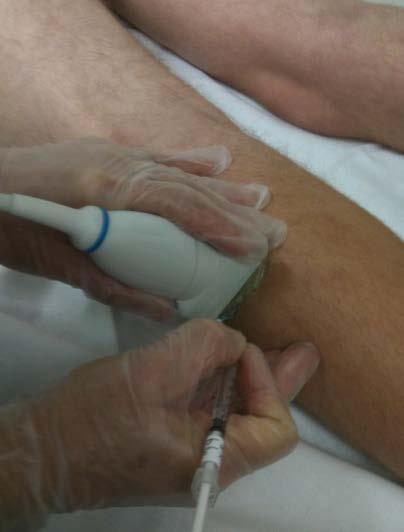
36 Interventional MS Ultrasound: Pearls of Wisdom Larger needles are easier to see than small needles Larger needles hurt more 27g needles are easily seen particularly in an in plane view Non-insulated needles are visualized better than insulated. Etched Needles are also available Small amount of air (.2-.3 ml) helps define needle location Agitate injectate: increases reflection from bubbles Agitating may denature the toxin Billing: In the USA, to charge/bill for US, a picture or cineloop must be saved to document the procedure Billing Code: 76942: Ultrasound for Needle guidance, aspiration
37 MUSCLE REVIEW

38 Iliopsoas Illustration from Ultrasound Guided Chemodenervation, Text and Atlas Demos Medical Publishing. Used with Permission


39 Iliopsoas, Distal Transverse, Proximal Thigh/Distal to Inguinal Crease Transverse Ultrasound Image Psoas Distal Palpation RC.JPG Psoas Distal Tv RC1.JPG Psoas Distal R RC

40 Iliopsoas; Distal Transverse Scan


41 Adductors Adductors, Transverse, Out of plane injection, Add Longus Video Link: adductor s Tv inject.wmv


42 Semimembranosis, Longitudinal Scan Injection, In Plane Picture Link: Semimembranosis Longitudinal BB.png Picture Link: HS longit long axis AS.jpg Semi M LA Inj IPBBt.wmv


43 Semimembranosis, Transverse Scan Injection, Out of Plane Injection HS Transverse, short axis AS.jpg Video Link: SemiTTV Inj OP DF.mpg


44 Medial, Lateral Gastrocnemius Soleus, Tibialis Post
45 Gastrocnemius lateral, Soleus Tibialis Posterior Doppler Doppler US Jpg, 4 y.o. Soleus, Tibialis Posterior, Transverse Junction of Middle 1/3:Distal 1/3 calf Picture Link: gastsoleus tib doppler edit.jpg Picture Link: Gastroc soleus tib post edit.jpg


46 Soleus/Tibialis Posterior 4 y.o. Child Transverse Lower-Middle 1/3 of Posterior Calf Out of Plane Injection, Soleus Picture Link: Soleus Tv OP 4 yo KM Labeled Soleus Tv OP 4yo KM.wmv


47 FDL Tibialis Post Picture Link: FDL LA OP RB Picture Link: Gastroc Tv Scan Localiz IMG_0509.jpg

48 Flexor Hallicus Brevis, Flexor Digitorum Brevis Transverse view, Plantar surface Foot
WS 434 US Guidance for Upper Limb Chemodenervation Procedures AAPM&R 2015
 WS 434 US Guidance for Upper Limb Chemodenervation Procedures AAPM&R 2015 Katharine E Alter MD Zach Bohart MD Robert Cooper MD Elie Elovic MD Heakyung Kim MD John McGuire MD Michael Munin MD Jeff Strakowski
Terason. AACPDM 2014 Pre-Course 4: B Mode Ultrasound for Muscle Hypertonia and Neuromuscular Disorders: Review of Scanning
 AACPDM 2014 Pre-Course 4: B Mode Ultrasound for Muscle Hypertonia and Neuromuscular Disorders: Review of Scanning Moderators Katharine Alter MD Florian Heinen MD Faculty, Lecture Session: Katharine Alter
Terason. AACPDM 2015 Pre-Course 4 Hands-On Ultrasound Course: Muscle Localizationeview of Scanning Techniques and Hands On. Training.
 AACPDM 2015 Pre-Course 4 Hands-On Ultrasound Course: Muscle Localizationeview of Scanning Techniques and Hands On Moderators Katharine Alter MD Florian Heinen MD Sebastian Schroeder MD Faculty, Lecture
Principles of Ultrasound. Cara C. Prideaux, M.D. University of Utah PM&R Sports Medicine Fellow March 14, 2012
 Principles of Ultrasound Cara C. Prideaux, M.D. University of Utah PM&R Sports Medicine Fellow March 14, 2012 None Disclosures Outline Introduction Benefits and Limitations of US Ultrasound (US) Physics
Terminology Tissue Appearance
 By Marc Nielsen, MD Advantages/Disadvantages Generation of Image Ultrasound Machine/Transducer selection Modes of Ultrasound Terminology Tissue Appearance Scanning Technique Real-time Portable No ionizing
3/20/2017. Disclosures. Ultrasound Fundamentals. Ultrasound Fundamentals. Bone Anatomy. Tissue Characteristics
 Disclosures Images of ultrasound equipment in this presentation are not an endorsement Fundamentals of Musculoskeletal Ultrasound Physics and Knobology Shane A. Shapiro, M.D. Assistant Professor Orthopedic
Basic Physics of Ultrasound and Knobology
 WELCOME TO UTMB Basic Physics of Ultrasound and Knobology By Daneshvari Solanki, FRCA Laura B. McDaniel Distinguished Professor Anesthesiology and Pain Medicine University of Texas Medical Branch Galveston,
Background & Indications Probe Selection
 Teresa S. Wu, MD, FACEP Director, EM Ultrasound Program & Fellowship Co-Director, Simulation Based Training Program & Fellowship Associate Program Director, EM Residency Program Maricopa Medical Center
Ultrasound Guided Lower Extremity Blocks
 Ultrasound Guided Lower Extremity Blocks CONTENTS: 1. Femoral Nerve Block 2. Popliteal Nerve Block Updated December 2017 1 1. Femoral Nerve Block Indications Surgery involving the knee, anterior thigh,
Ultrasound Physics and Knobology Alan Macfarlane. Consultant Anaesthetist Glasgow Royal Infirmary
 Ultrasound Physics and Knobology Alan Macfarlane Consultant Anaesthetist Glasgow Royal Infirmary RAPM 2009; 34: 40-46 Ultrasound Proficiency Understanding US image generation and device operation Image
Diagnostic Ultrasound. Sutiporn Khampunnip, M.D.
 Diagnostic Ultrasound Sutiporn Khampunnip, M.D. Definition of Ultrasound Ultrasound is simply sound waves, like audible sound. High-frequency sound and refers to mechanical vibrations above 20 khz. Human
INTRODUCTION. Getting the best scan. Choosing a probe. Choosing the frequency
 Getting the best scan Choosing a probe Select the most appropriate probe for the particular scan required. s vary in their: operating frequency range higher ultrasound frequencies provide better discrimination
The Essentials Tissue Characterization and Knobology
 The Essentials Tissue Characterization and Knobology Randy E. Moore, DC, RDMS RMSK No relevant financial relationships Ultrasound The New Standard of Care Musculoskeletal sonography has become the standard
Musculoskeletal Ultrasound: Basics, Utility, and Clinical Applications
 Musculoskeletal Ultrasound: Basics, Utility, and Clinical Applications Andrew Lavigne, MD, FRCPC Physical Medicine and Rehabilitation CSCN Diplomat (EMG) Dip Sport Medicine Eugene Maida, MD, PGY-4 Resident
Basic of Ultrasound Physics E FAST & Renal Examination. Dr Muhammad Umer Ihsan MBBS,MD, DCH CCPU,DDU1,FACEM
 Basic of Ultrasound Physics E FAST & Renal Examination Dr Muhammad Umer Ihsan MBBS,MD, DCH CCPU,DDU1,FACEM What is Sound? Sound is Mechanical pressure waves What is Ultrasound? Ultrasounds are sound waves
Ultrasound in Peripheral Nerve Interventions
 Ultrasound in Peripheral Nerve Interventions John L. Lin, M.D. Shepherd Center Assistant Clinical Professor Emory University, School of Medicine Outline Ultrasound basics Nerve blocks in physiatric setting
Ultrasonography of the Neck as an Adjunct to FNA. Nicole Massoll M.D.
 Ultrasonography of the Neck as an Adjunct to FNA Nicole Massoll M.D. Basic Features of Head and Neck Ultrasound and Anatomy Nicole Massoll M.D. University of Arkansas for Medical Sciences, Little Rock
inerve Guide to Nerves 2009
 inerve Guide to Nerves 2009 A guide to self learning and self assessment Context: The following guide is intended to help interpret the sono-anatomy and follow a systematic stepwise approach to the practice
Introduction to Ultrasound Guided Region Anesthesia
 Introduction to Ultrasound Guided Region Anesthesia Brian D. Sites, MD Dept of Anesthesiology Dartmouth-Hitchcock Medical Center INTRODUCTION Welcome to Introduction to Ultrasound Guided Regional Anesthesia.
Ultrasonography of Peripheral Nerve -upper extremity
 Ultrasonography of Peripheral Nerve -upper extremity Department of Physical Medicine and Rehabilitation Korea University Guro Hospital Korea University College of Medicine Yoon Joon Shik Normal median
Ultrasound Principles cycle Frequency Wavelength Period Velocity
 ! Teresa S. Wu, MD, FACEP Director, EM Ultrasound Program & Fellowship Co-Director, Simulation Based Training Program & Fellowship Associate Program Director, EM Residency Program Maricopa Medical Center
Human Anatomy Lab #7: Muscles of the Cadaver
 Human Anatomy Lab #7: Muscles of the Cadaver Table of Contents: Expected Learning Outcomes.... 1 Introduction...... 1 Identifying Muscles on Yourself.... 2 Muscles of the Anterior Trunk and Arm.. 2 Muscles
The Muscular System. Chapter 10 Part D. PowerPoint Lecture Slides prepared by Karen Dunbar Kareiva Ivy Tech Community College
 Chapter 10 Part D The Muscular System Annie Leibovitz/Contact Press Images PowerPoint Lecture Slides prepared by Karen Dunbar Kareiva Ivy Tech Community College Table 10.14: Muscles Crossing the Hip and
Basics of US Regional Anaesthesia. November 2008
 Basics of US Regional Anaesthesia November 2008 Essential Physics HIGH frequency = great resolution but poor penetration LOW frequency = poor resolution but great penetration Potential Advantages of US
lesser trochanter of femur lesser trochanter of femur iliotibial tract (connective tissue) medial surface of proximal tibia
 medial surface of proximal tibia") LOWER LIMB MUSCLES OF THE APPENDICULAR SKELETON The muscles that act on the lower limb fall into three groups: those that move the thigh, those that move the lower leg, and those that move the ankle, foot,
LOWER LIMB MUSCLES OF THE APPENDICULAR SKELETON The muscles that act on the lower limb fall into three groups: those that move the thigh, those that move the lower leg, and those that move the ankle, foot,
The Human Muscular System Required reading before beginning this lab: Saladin, KS: Human Anatomy 5th ed (2017) Chapters 10, 11, 12 INTRODUCTION
 Chapters 10, 11, 12 INTRODUCTION") Biology 322: Human Anatomy The Human Muscular System Required reading before beginning this lab: Saladin, KS: Human Anatomy 5 th ed (2017) Chapters 10, 11, 12 INTRODUCTION We will use a number of lab periods
Biology 322: Human Anatomy The Human Muscular System Required reading before beginning this lab: Saladin, KS: Human Anatomy 5 th ed (2017) Chapters 10, 11, 12 INTRODUCTION We will use a number of lab periods
Where should you palpate the pulse of different arteries in the lower limb?
 Where should you palpate the pulse of different arteries in the lower limb? The femoral artery In the femoral triangle, its pulse is easily felt just inferior to the inguinal ligament midway between the
The Foot. Dr. Wegdan Moh.Mustafa Medicine Faculty Assistant Professor Mob:
 The Foot Dr. Wegdan Moh.Mustafa Medicine Faculty Assistant Professor Mob: 0127155717 The skeleton of the foot Cutaneous innervations Sole of foot layers of muscles First layer -Abductor hallucis -Flexor
WELCOME! Introduction to Bedside Ultrasound
 WELCOME! Introduction to Bedside Ultrasound TEACHERS University of California-Irvine School of Medicine Nathan Molina nathan.d.molina@gmail.com Trevor Plescia taplescia90@gmail.com Jack Silva jpsilva42@gmail.com
Chapter 9. The Muscular System
 1 Chapter 9 The Muscular System 2 Introduction Skeletal muscles: movement in environment Smooth muscles: intestines, ureters, veins and arteries Cardiac muscle: pumps blood through heart and blood vessels
NCVH. Ultrasongraphy: State of the Art Vein Forum 2015 A Multidisciplinary Approach to Otptimizing Venous Circulation From Wounds to WOW
 Ultrasongraphy: State of the Art 2015 NCVH New Cardiovascular Horizons Vein Forum 2015 A Multidisciplinary Approach to Otptimizing Venous Circulation From Wounds to WOW Anil K. Chagarlamudi, M.D. Cardiovascular
Surface Anatomy and Sonoanatomy for the Occasional Regional Anesthesiologist
 Surface Anatomy and Sonoanatomy for the Occasional Regional Anesthesiologist Edward R. Mariano, M.D., M.A.S. Professor of Anesthesiology, Perioperative & Pain Medicine Stanford University School of Medicine
1 Fundamentals. Basic Definitions and Physics Principles. Fundamentals
 1 To become versed in the language of ultrasonography, it is necessary to review some of the basic principles of physics. The wave physics principles of ordinary (i.e., audible) sound apply to ultrasound
musculoskeletal system anatomy muscles of foot sheet done by: dina sawadha & mohammad abukabeer
 musculoskeletal system anatomy muscles of foot sheet done by: dina sawadha & mohammad abukabeer Extensor retinaculum : A- superior extensor retinaculum (SER) : originates from the distal ends of the tibia
Ultrasound Knobology
 Ultrasound Knobology Raj Dasgupta MD, FACP, FCCP, FASSM Assistant Professor of Clinical Medicine Pulmonary / Critical Care / Sleep Medicine University of Southern California (USC) Objectives Physics of
Botulinum Toxin Therapy Billing & Coding Update 2016
 Botulinum Toxin Therapy Billing & Coding Update 2016 Martin Taylor, DO, PhD Neurology / OrthoNeuro Clinical Associate Professor, Neurology Ohio University College of Osteopathic Medicine Columbus, OH Disclosures
Definition of Anatomy. Anatomy is the science of the structure of the body and the relation of its parts.
 Definition of Anatomy Anatomy is the science of the structure of the body and the relation of its parts. Basic Anatomical Terms Anatomical terms for describing positions: Anatomical position: Supine position:
Lower Limb Dr. Robin Paudel
 Lower Limb n What is a limb? n Skeleton n Joints n Pelvis or limb girdle n Hip/Hip Muscles n Lumber and sacral plexus getting spinal nerves out onto limb n Muscles anterior and posterior compartments n
Preamble (disclaimer)
") Preamble (disclaimer) PHYSICS AND PRINCIPLES OF HEAD/NECK ULTRASOUND Joseph C. Sniezek, MD FACS LTC, MC, USA Otolaryngology/H&N Surgery Tripler Army Medical Center 1. I am not a physicist 2. ACS has recommended
Preamble (disclaimer) PHYSICS AND PRINCIPLES OF HEAD/NECK ULTRASOUND Joseph C. Sniezek, MD FACS LTC, MC, USA Otolaryngology/H&N Surgery Tripler Army Medical Center 1. I am not a physicist 2. ACS has recommended
How do you use the AiM Vascular Mapping Kit?
 How do you use the AiM Vascular Mapping Kit? Protocol for Successful Skin Mapping Evaluate vessel anatomy, perform measurements, etc. according to your laboratory s protocol prior to applying the AiM device
Muscle. Dr. Carmen E. Rexach Anatomy 35 Mt San Antonio College
 Muscle Dr. Carmen E. Rexach Anatomy 35 Mt San Antonio College Functions Movements of bones and soft body parts Movements of fluids through a tube (blood, digestive) Functions Maintain posture Support soft
Introduction to Ultrasound Examination of the Hand and upper
 Introduction to Ultrasound Examination of the Hand and upper Emil Dionysian, M.D. Ultrasound of upper ext. Upside Convenient Opens another exam dimension Can be like a stethoscope Helps 3-D D visualization
The Lower Limb VI: The Leg. Anatomy RHS 241 Lecture 6 Dr. Einas Al-Eisa
 The Lower Limb VI: The Leg Anatomy RHS 241 Lecture 6 Dr. Einas Al-Eisa Muscles of the leg Posterior compartment (superficial & deep): primary plantar flexors of the foot flexors of the toes Anterior compartment:
Foot. Dr. Heba Kalbouneh Associate Professor of Anatomy and Histology
 Foot Dr. Heba Kalbouneh Associate Professor of Anatomy and Histology Dorsum of the Foot Sole of the Foot Plantar aponeurosis It is a triangular thickening of deep fascia in the sole of the foot Attachments:
Practical 1 Worksheet
 Practical 1 Worksheet ANATOMICAL TERMS 1. Use the word bank to fill in the missing words. reference side stand body arms palms anatomical forward All anatomical terms have a(n) point which is called the
Ultrasound Guided Injections
 Ultrasound Guided Injection Technique More accurate injections Better Results! 1 Benefits: Increased Level of Certainty ie : really know how accurate PRP/Prolotherapy Avoid damage to articular cartilage
Biceps Brachii. Muscles of the Arm and Hand 4/4/2017 MR. S. KELLY
 Muscles of the Arm and Hand PSK 4U MR. S. KELLY NORTH GRENVILLE DHS Biceps Brachii Origin: scapula Insertion: radius, fascia of forearm (bicipital aponeurosis) Action: supination and elbow flexion Innervation:
USRA OF THE LOWER EXTREMITY
 USRA OF THE LOWER EXTREMITY Christian R. Falyar, CRNA, DNAP Department of Nurse Anesthesia Virginia Commonwealth University Disclosure Statement of Financial Interest I, Christian Falyar, DO NOT have a
Muscles of the lower extremities. Dr. Nabil khouri MD, MSc, Ph.D
 Muscles of the lower extremities Dr. Nabil khouri MD, MSc, Ph.D Posterior leg Popliteal fossa Boundaries Biceps femoris (superior-lateral) Semitendinosis and semimembranosis (superior-medial) Gastrocnemius
Lower Limb Nerves. Clinical Anatomy
 Lower Limb Nerves Clinical Anatomy Lumbar Plexus Ventral rami L1 L4 Supplies: Abdominal wall External genitalia Anteromedial thigh Major nerves.. Lumbar Plexus Nerves relation to psoas m. : Obturator n.
ULTRASOUND IMAGING EE 472 F2018. Prof. Yasser Mostafa Kadah
 ULTRASOUND IMAGING EE 472 F2018 Prof. Yasser Mostafa Kadah www.k-space.org Recommended Textbook Diagnostic Ultrasound: Physics and Equipment, 2nd ed., by Peter R. Hoskins (Editor), Kevin Martin (Editor),
Lumbar Plexus. Ventral rami L1 L4 Supplies: Major nerves.. Abdominal wall External genitalia Anteromedial thigh
 Lower Limb Nerves Lectures Objectives Describe the structure and relationships of the plexuses of the lower limb. Describe the course, relationships and structures supplied for the major nerves of the
Ultrasound Guided Peripheral Intravenous Access
 Ultrasound Guided Peripheral Intravenous Access J. Christian Fox, MD, RDMS, FACEP, FAAEM, FAIUM Professor and Interim Chair of Emergency Medicine Director of Instructional Ultrasound University of California,
Disclosure. Pre-Procedural Considerations. Transducer Selection. Sterile Procedure. Sterile Procedure. Ultrasound Guided Foot and Ankle Injections
 Ultrasound Guided Foot and Ankle Injections Disclosure No relevant financial relationships exist Shane A. Shapiro, M.D. Assistant Professor, Orthopedic Surgery Mayo Clinic Florida @ShaneShapiroMD 2012
The Physics of Ultrasound. The Physics of Ultrasound. Claus G. Roehrborn. Professor and Chairman. Ultrasound Physics
 The Physics of Ultrasound Pipe Organ 10-8000 Emission Dog 452-1080 Man 85-1100 Spectrum Bat 10,000-120,000 Porpoise 7000-120,000 Claus G. Roehrborn Professor and Chairman 10 20 Cycles per second Reception
The Elbow 3/5/2015. The Elbow Scanning Sequence. * Anterior Joint (The anterior Pyramid ) * Lateral Epicondyle * Medial Epicondyle * Posterior Joint
 * Lateral Epicondyle * Medial Epicondyle * Posterior Joint") Scanning Sequence * Anterior Joint (The anterior Pyramid ) * Lateral Epicondyle * Medial Epicondyle * Posterior Joint Anterior Elbow Pyramid Courtesy of Jay Smith, MD. Vice chair PMR Mayo Clinic Rochester,
Scanning Sequence * Anterior Joint (The anterior Pyramid ) * Lateral Epicondyle * Medial Epicondyle * Posterior Joint Anterior Elbow Pyramid Courtesy of Jay Smith, MD. Vice chair PMR Mayo Clinic Rochester,
The plantar aponeurosis
 Anatomy of the foot The plantar aponeurosis Is a triangular thickening of the deep fascia Its apex is attached to the medial and lateral tubercles of the calcaneum. The base of the aponeurosis divides
Peripheral Nervous System: Lower Body
 Peripheral Nervous System: Lower Body MSTN121 - Neurophysiology Session 11 Department of Myotherapy Lumbar Plexus Iliohypogastric nerve (T12-L1) Motor: Transverse abdominis and internal obliques Sensory:
Muscles of the Hip 1. Tensor Fasciae Latae O: iliac crest I: lateral femoral condyle Action: abducts the thigh Nerve: gluteal nerve
 Muscles of the Hip 1. Tensor Fasciae Latae O: iliac crest I: lateral femoral condyle Action: abducts the thigh Nerve: gluteal nerve 2. Gluteus Maximus O: ilium I: femur Action: abduct the thigh Nerve:
High resolution ultrasound scanner for skin imaging
 High resolution ultrasound scanner for skin imaging Christine Turlat Sales Director Atys medical 17 Parc d Arbora 69510 SOUCIEU EN JARREST Atys company Principle of ultrasound imaging DERMCUP Normal image
HUMAN BODY COURSE LOWER LIMB NERVES AND VESSELS
 HUMAN BODY COURSE LOWER LIMB NERVES AND VESSELS October 22, 2010 D. LOWER LIMB MUSCLES 2. Lower limb compartments ANTERIOR THIGH COMPARTMENT General lfunction: Hip flexion, knee extension, other motions
Ultrasound. Principles of Medical Imaging. Contents. Prof. Dr. Philippe Cattin. MIAC, University of Basel. Oct 17th, 2016
 Ultrasound Principles of Medical Imaging Prof. Dr. Philippe Cattin MIAC, University of Basel Contents Abstract 1 Image Generation Echography A-Mode B-Mode M-Mode 2.5D Ultrasound 3D Ultrasound 4D Ultrasound
Prime movers provide the major force for producing a specific movement Antagonists oppose or reverse a particular movement Synergists
 Dr. Gary Mumaugh Prime movers provide the major force for producing a specific movement Antagonists oppose or reverse a particular movement Synergists Add force to a movement Reduce undesirable or unnecessary
In which arm muscle are intramuscular injections most often given? (not in text)
") AP1 Lab 9 - Muscles of the Arms and Legs Locate the following muscles on the models and on yourself. Recall anatomical position. Directional terms such as anterior, posterior, lateral, etc. all assume
AP1 Lab 9 - Muscles of the Arms and Legs Locate the following muscles on the models and on yourself. Recall anatomical position. Directional terms such as anterior, posterior, lateral, etc. all assume
Year 2004 Paper one: Questions supplied by Megan
 QUESTION 47 A 58yo man is noted to have a right foot drop three days following a right total hip replacement. On examination there is weakness of right ankle dorsiflexion and toe extension (grade 4/5).
Copyright 2004, Yoshiyuki Shiratori. All right reserved.
 Ankle and Leg Evaluation 1. History Chief Complaint: A. What happened? B. Is it a sharp or dull pain? C. How long have you had the pain? D. Can you pinpoint the pain? E. Do you have any numbness or tingling?
Ultrasound Physics & Terminology
 Ultrasound Physics & Terminology This module includes the following: Basic physics terms Basic principles of ultrasound Ultrasound terminology and terms Common artifacts seen Doppler principles Terms for
Arterial Line Insertion Pre Reading
 PROCEDURE ACCREDITATION THE CANBERRA HOSPITAL EMERGENCY DEPARTMENT Arterial Line Insertion Pre Reading Indications Requirement for continuous blood pressure monitoring (all patients on pressors, inotropes,
Abdominal Ultrasound
 Abdominal Ultrasound Imaging Control Buttons Depth The organ imaged should take up 3/4 of the screen Frequency = Penetration Use high frequencies (harmonics) for fluid filled and superficial structures
Lower limb summary. Anterior compartment of the thigh. Done By: Laith Qashou. Doctor_2016
 Lower limb summary Done By: Laith Qashou Doctor_2016 Anterior compartment of the thigh Sartorius Anterior superior iliac spine Upper medial surface of shaft of tibia 1. Flexes, abducts, laterally rotates
LOWER EXTREMITY VENOUS COMPRESSION ULTRASOUND. CPT Stacey Good, DO Emergency Medicine Ultrasound Fellow Madigan Army Medical Center
 LOWER EXTREMITY VENOUS COMPRESSION ULTRASOUND CPT Stacey Good, DO Emergency Medicine Ultrasound Fellow Madigan Army Medical Center Learning Objectives Setup and patient positioning for optimizing success
MUSCLES OF THE LOWER LIMBS
 MUSCLES OF THE LOWER LIMBS Naming, location and general function Dr. Nabil khouri ROLES THAT SHOULD NOT BE FORGOTTEN Most anterior compartment muscles of the hip and thigh Flexor of the femur at the hip
Sound in medicine. CH.12. Dr.Rajaa أ.م.د. رجاء سهيل جنم جامعة تكريت كلية طب االسنان. General Properties of Sound
 CH.12. Dr.Rajaa Sound in medicine أ.م.د. رجاء سهيل جنم جامعة تكريت كلية Sound : It is the audible waves of frequency between 20 Hz and 20 khz. Infrasound : refers to the sound of frequency below the normal
Human Anatomy Biology 351
 Human Anatomy Biology 351 Lower Limb Please place your name on the back of the last page of this exam. You must answer all questions on this exam. Because statistics demonstrate that, on average, between
Treatment Framework patient tracker
 Treatment Framework patient tracker Utilize the BOTOX Treatment Framework when evaluating your Focal Spasticity patients to establish The right goals The right muscles/dose The right plan Use the following
Point-of-Care Ultrasound: An Introduction
 Point-of-Care Ultrasound: An Introduction Delegation Teaching Package for Registered Respiratory Therapists and Anesthesia Assistants Developed by: Rob Bryan RRT, AA Edited by: Kelly Hassall RRT, FCSRT,
Muscle fiber (cell) Blood vessel. Perimysium. Epimysium. Fascicle (wrapped by perimysium) Endomysium (between fibers) Tendon. Bone
 Blood vessel. Perimysium. Epimysium. Fascicle (wrapped by perimysium) Endomysium (between fibers) Tendon. Bone") Figure 6.1 Connective tissue wrappings of skeletal muscle. Blood vessel Muscle fiber (cell) Perimysium Epimysium Fascicle (wrapped by perimysium) Tendon Endomysium (between fibers) Bone Figure 6.15 Superficial
Figure 6.1 Connective tissue wrappings of skeletal muscle. Blood vessel Muscle fiber (cell) Perimysium Epimysium Fascicle (wrapped by perimysium) Tendon Endomysium (between fibers) Bone Figure 6.15 Superficial
Dr Nabil khouri MD. MSc. Ph.D
 Dr Nabil khouri MD. MSc. Ph.D Foot Anatomy The foot consists of 26 bones: 14 phalangeal, 5 metatarsal, and 7 tarsal. Toes are used to balance the body. Metatarsal Bones gives elasticity to the foot in
The Leg. Prof. Oluwadiya KS
 The Leg Prof. Oluwadiya KS www.oluwadiya.sitesled.com Compartments of the leg 4 Four Compartments: 1. Anterior compartment Deep fibular nerve Dorsiflexes the foot and toes 2. Lateral Compartment Superficial
Musculoskeletal Ultrasound of the Knee, Foot and ankle
 Musculoskeletal Ultrasound of the Knee, Foot and ankle ADVANCED TEAM PHYSICIAN COURSE SAN DIEGO, CALIFORNIA DECEMBER 11TH 2016 Jonathan S. Halperin MD Learning objec-ves: Understand the basics of knee,
Introduction to Anatomy. Dr. Maher Hadidi. Laith Al-Hawajreh. Mar/25 th /2013
 Introduction to Anatomy Dr. Maher Hadidi Laith Al-Hawajreh 22 Mar/25 th /2013 Lower limb - The leg The skeleton of the leg is formed by two bones: 1) Medial: Tibia 2) Lateral: Fibula The two bones are
EDL EHL. Extensor Hallucis Longus L5 Extensor Digitorum longus L5,1 Peroneus Tertius L5 1 Extensor Digitorum Brevis S1,2 [like intrinsic muscle]
![EDL EHL. Extensor Hallucis Longus L5 Extensor Digitorum longus L5,1 Peroneus Tertius L5 1 Extensor Digitorum Brevis S1,2 [like intrinsic muscle]](/thumbs/78/77875930.jpg "EDL EHL. Extensor Hallucis Longus L5 Extensor Digitorum longus L5,1 Peroneus Tertius L5 1 Extensor Digitorum Brevis S1,2 [like intrinsic muscle]") ANATOMY OF ANKLE AND FOOT Lateral aspect: [Dorsal medial to lateral= dorsal under extensor retinaculum] Tibialis Anterior EHL Artery [Dorsal pedal A] and Anterior tibial N EDL Peroneus Tertius Behind the
ANATOMY OF ANKLE AND FOOT Lateral aspect: [Dorsal medial to lateral= dorsal under extensor retinaculum] Tibialis Anterior EHL Artery [Dorsal pedal A] and Anterior tibial N EDL Peroneus Tertius Behind the
CONTENTS. Test Number cpd Tanya Reynolds (Nat. Dip. Diag. Rad., B. Tech. Diag. Rad., B. Tech. Ultrasound)
") CONTENTS page 1-15 page 16 BASIC 2-DIMENSIONAL ULTRASOUND PRINCIPLES Multiple Choice Test Test Number cpd 41640 Tanya Reynolds (Nat. Dip. Diag. Rad., B. Tech. Diag. Rad., B. Tech. Ultrasound) Tanya is
CONTENTS page 1-15 page 16 BASIC 2-DIMENSIONAL ULTRASOUND PRINCIPLES Multiple Choice Test Test Number cpd 41640 Tanya Reynolds (Nat. Dip. Diag. Rad., B. Tech. Diag. Rad., B. Tech. Ultrasound) Tanya is
Due in Lab weeks because of Thanksgiving Prelab #10. Homework #8. Both sides! Both sides!
 Lab 8 MUSCLES Due in Lab 10 2 weeks because of Thanksgiving Prelab #10 Both sides! Homework #8 Both sides! Refer to Muscles 22-23 Naming of muscles Origin Site of muscle attachment that doesn t move during
BIOL 4260 Human Evolu3onary Anatomy Lecture 12: Limb Development. Lecture 2: Fossil Record
 BIOL 4260 Human Evolu3onary Anatomy Lecture 12: Limb Development Lecture 2: Fossil Record Outline Limb Evolution Limb Development Limb Function A Few Definitions Appendicular skeleton girdles & limbs Pectoral
The Muscular System PART C. PowerPoint Lecture Slide Presentation by Patty Bostwick-Taylor, Florence-Darlington Technical College
 PowerPoint Lecture Slide Presentation by Patty Bostwick-Taylor, Florence-Darlington Technical College The Muscular System 6 PART C Five Golden Rules of Skeletal Muscle Activity Table 6.2 Muscles and Body
In-Depth Foundations: Anatomy Terms to Know
 Be familiar with / able to identify and define all the following parts. The Spine Cranium Vertebrae Cervical, Thoracic, Lumbar Sacrum Coccyx Bones of Upper Body Cranium Mastoid process; Occipital condyle,
Ultrasound Physics & Doppler
 Ultrasound Physics & Doppler Endocrine University 2018 Mark Lupo, MD, FACE, ECNU Objectives Review the essential components of ultrasound physics in neck sonography Demonstrate the importance of ultrasound
Muscles of Lesson Five. Muscular Nomenclature and Kinesiology - Two. Muscles of Lesson Five, cont. Chapter 16
 Chapter 16 Muscular Nomenclature and Kinesiology - Two Lessons 5-6 Muscles of Lesson Five Iliopsoas (psoas major, iliacus) Hip outward rotators (piriformis, gemellus superior, gemellus inferior, obturator
Dystonias Peter McAllister, MD
 Dystonias Peter McAllister, MD Medical Director, New England Institute for Neurology and Headache Chief Medical Officer, New England Institute for Clinical Research Clinical Assistant Professor, Neurology,
Anatomy MCQs Week 13
 Anatomy MCQs Week 13 1. Posterior to the medial malleolus of the ankle: The neurovascular bundle lies between Tibialis Posterior and Flexor Digitorum Longus The tendon of Tibialis Posterior inserts into
Anatomy & Physiology. Muscles of the Lower Limbs.
 Anatomy & Physiology Muscles of the Lower Limbs http://www.ishapeup.com/musclecharts.html Muscles of the Lower Limbs Among the strongest muscles in the body. Because pelvic girdle is composed of heavy,
Chapter 2 Pitfalls in Musculoskeletal Ultrasound
 Chapter 2 Pitfalls in Musculoskeletal Ultrasound Violeta Maria Vlad MD, PhD Introduction Taking a good ultrasound (US) picture is an art. Interpreting it is a science. This is in fact everything US is
BLUE SKY SCHOOL OF PROFESSIONAL MASSAGE AND THERAPEUTIC BODYWORK. Musculoskeletal Anatomy & Kinesiology MUSCLES, MOVEMENTS & BIOMECHANICS
 BLUE SKY SCHOOL OF PROFESSIONAL MASSAGE AND THERAPEUTIC BODYWORK Musculoskeletal Anatomy & Kinesiology MUSCLES, MOVEMENTS & BIOMECHANICS MSAK101-I Session 7 Learning Objectives: 1. List the three types
11/15/2018. Temporalis Elevates & retracts mandible. Masseter = Prime mover of jaw closure. Levator scapulae Supraspinatus Clavicle.
 Due in Lab 10 Lab 8 MUSCLES 2 weeks because of Thanksgiving Prelab #10 Both sides! Homework #8 Both sides! Refer to Muscles 22-23 Examples of Origin & Insertion Naming of muscles Origin Site of muscle
Physical Principles of Ultrasound
 Physical Principles of Ultrasound Grateful appreciation to Richard A. Lopchinsky, MD, FACS and Nancy H. Van Name, RDMS, RTR, and MarleneKattaron, RDMS 2000 UIC All Rights Reserved. Course Objectives Identify
~, /' ~::'~ EXTENSOR HALLUCIS LONGUS. Leg-anterolateral :.:~ / ~\,
 TIBIALIS ANTERIOR Lateral condyle of tibia, upper half of lateral surface of tibia, interosseous membrane Medial side and plantar surface of medial cuneiform bone, and base of first metatarsal bone Dorsiflexes
ARM Brachium Musculature
 ARM Brachium Musculature Coracobrachialis coracoid process of the scapula medial shaft of the humerus at about its middle 1. flexes the humerus 2. assists to adduct the humerus Blood: muscular branches
The thigh. Prof. Oluwadiya KS
 The thigh Prof. Oluwadiya KS www.oluwadiya.com The Thigh: Boundaries The thigh is the region of the lower limb that is approximately between the hip and knee joints Anteriorly, it is separated from the
Figure 11-1: The lever-fulcrum principle is illustrated by flexion of the forearm.
 Chapter 11: The Muscular System Read pages 325 to 399 NAME Topic Outline And Objectives: A. How skeletal muscles produce movement, and naming muscles 1. Describe the relationship between bones and skeletal