Micronodular Lung Disease an algorithm
|
|
|
- Alexina Jennings
- 5 years ago
- Views:
Transcription
1 Micronodular Lung Disease an algorithm H. Page McAdams, MD Department of Radiology Duke University Medical Center Durham, NC USA
2 Question Which of the following lung diseases is MOST likely to show a perilymphatic distribution of micronodules on thinsection CT? 1. Langerhans cell histiocytosis 2. Airway-disseminated tuberculosis 3. Hypersensitivity pneumonitis 4. Sarcoidosis 5. Respiratory bronchiolitis
3 Definition Rounded opacities < 3- to 5-mm Numerous

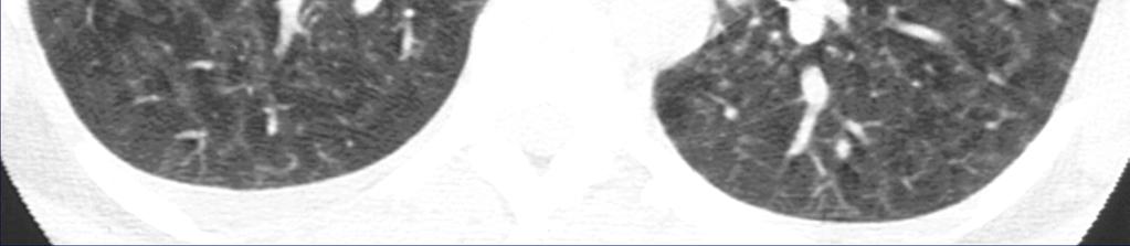
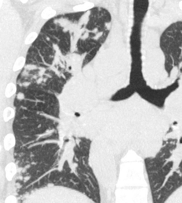
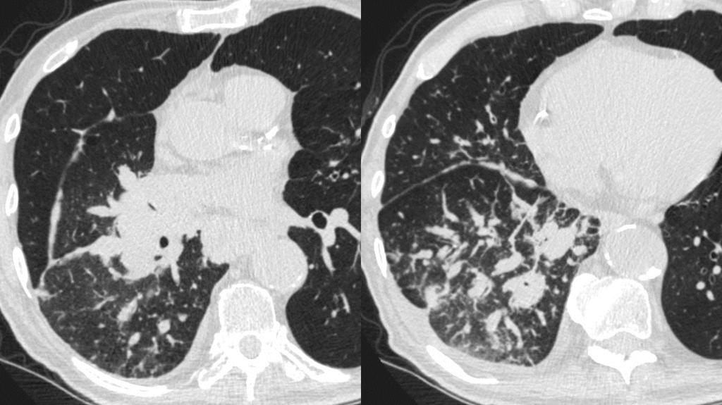
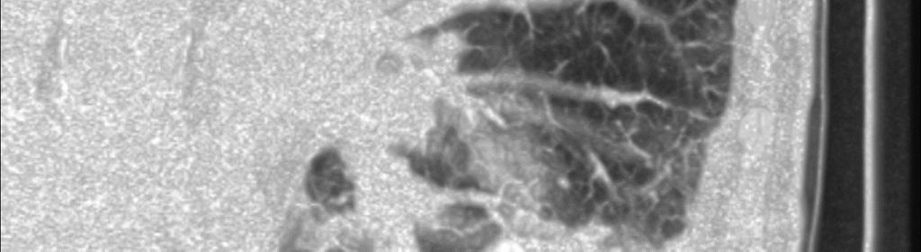
4 CT Detection Sliding MIPS improve detection of subtle disease 5-mm thick 1-mm increment
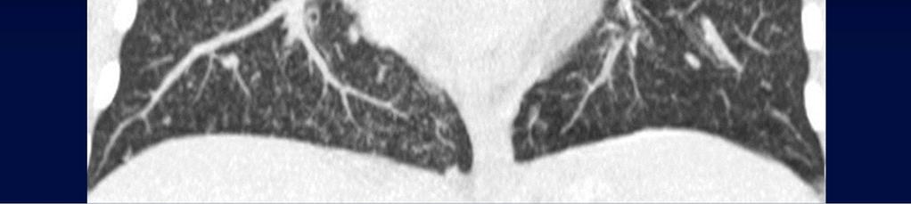
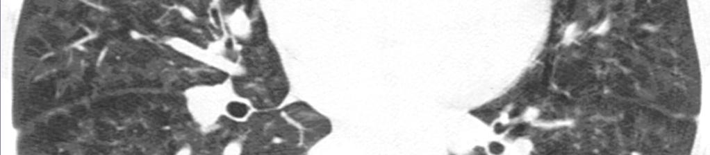
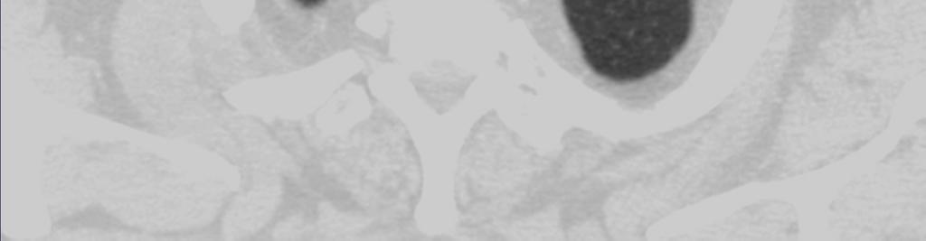
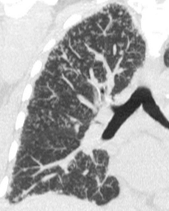
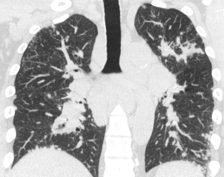
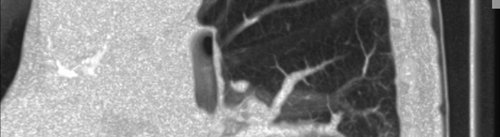
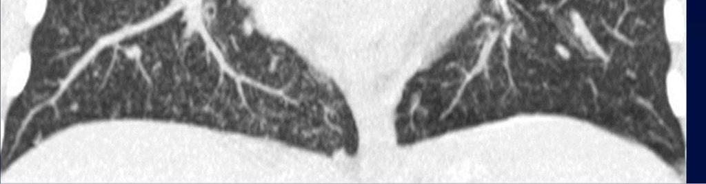
5 CT Detection Coronal
6 CT Detection Coronal disease distribution
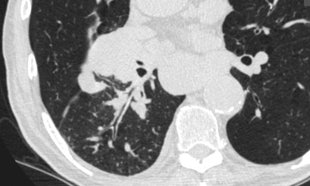
7

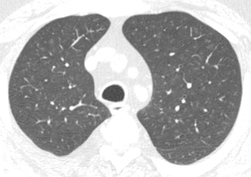
8 Micronodules

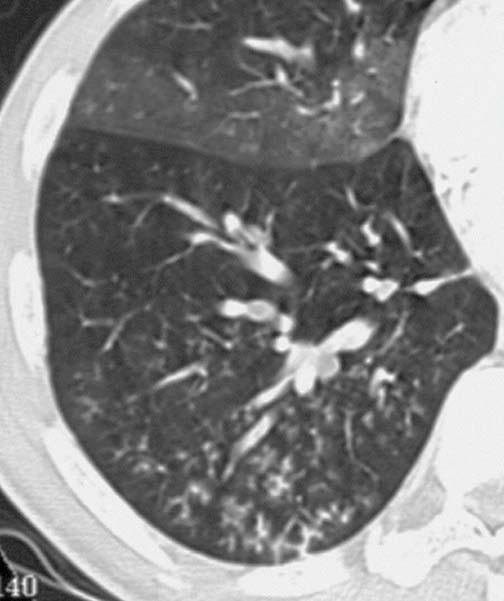
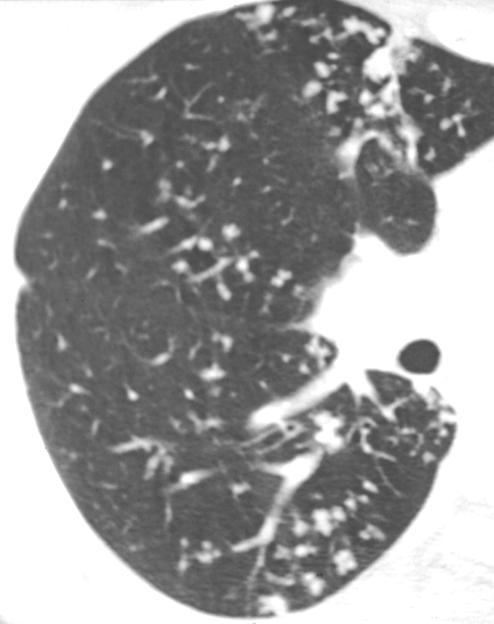
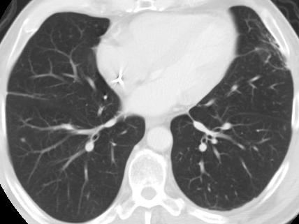
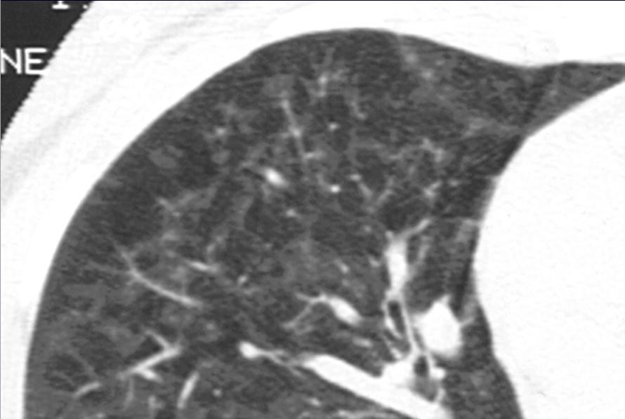
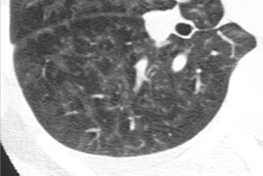
9 Micronodules Distribution


10 Micronodules Distribution Random Perilymphatic Centrilobular
11 Micronodules Distribution Random Perilymphatic Centrilobular
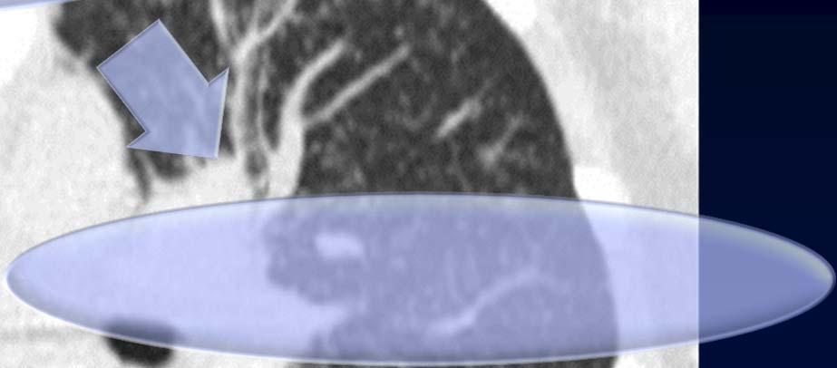
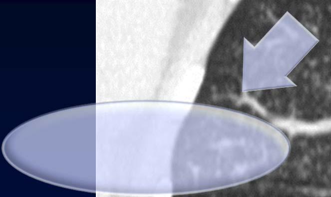
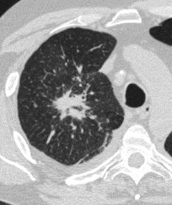
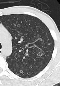
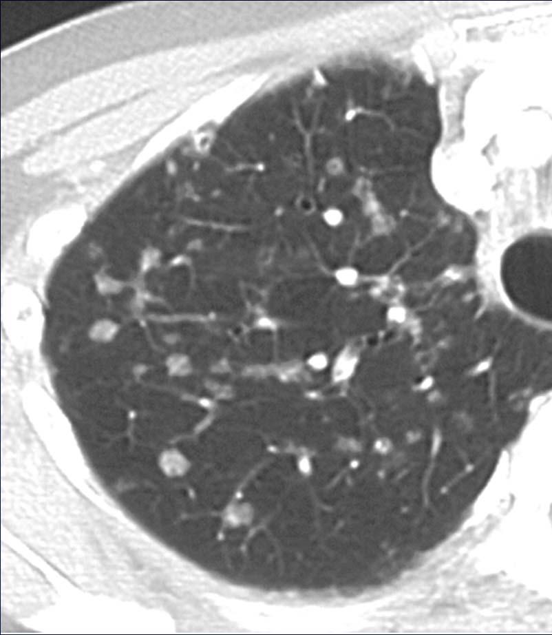
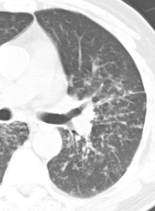
12 Centrilobular Nodules
13 Centrilobular Nodules
14 Micronodules Distribution Random Perilymphatic Centrilobular Ground Glass Solid Tree-in-Bud


15 Micronodules Distribution Random Perilymphatic Centrilobular Ground Glass Solid Tree-in-Bud


16 Solid CL Nodules
17 Solid CL Nodules Silicosis/CWP Pulmonary LCH Infection Aspiration Metastases (rare)
18 Silicosis
19 Silicosis


20 Pulmonary LCH

21 Pulmonary LCH
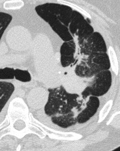
22 Tree-in-Bud

23 Tree-in-Bud
24 Solid CL Nodules Silicosis/CWP Pulmonary LCH Infection TB, NTMB tree-in-bud viral Aspiration
25 Solid CL Nodules Silicosis/CWP Pulmonary LCH Infection TB, NTMB viral tree-in-bud Aspiration

26 Tree-in-Bud Tuberculosis Aspiration

27 Esophageal Stricture Courtesy Laura Heyneman, MD

28 Esophageal Stricture 6 months s/p PEG

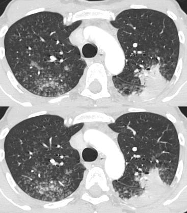
29 Micronodules Distribution Random Perilymphatic Centrilobular Ground Glass Solid Tree-in-Bud


30 CL GG Nodules
31 CL GG Nodules Hypersensitivity pneumonitis Bronchiolitis respiratory infection (viral) Adenocarcinoma (BAC) OP/NSIP Edema/vasculitis
32 Hypersensitivity Pn

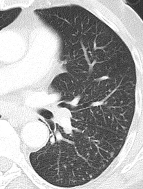
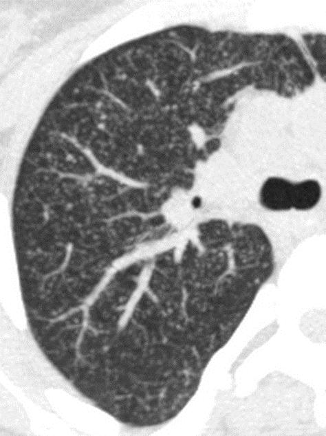
33 Bird Fancier s Lung
34 Hypersensitivity Pn Insp Exp

35 Hot Tub Lung Insp Exp
36 Resp Bronchiolitis


37 Resp Bronchiolitis Sliding MIPS
38 Adenocarcinoma


39 Micronodules Distribution Random Perilymphatic Centrilobular Ground Glass Solid Tree-in-Bud
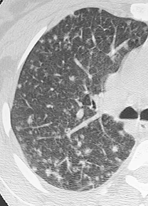
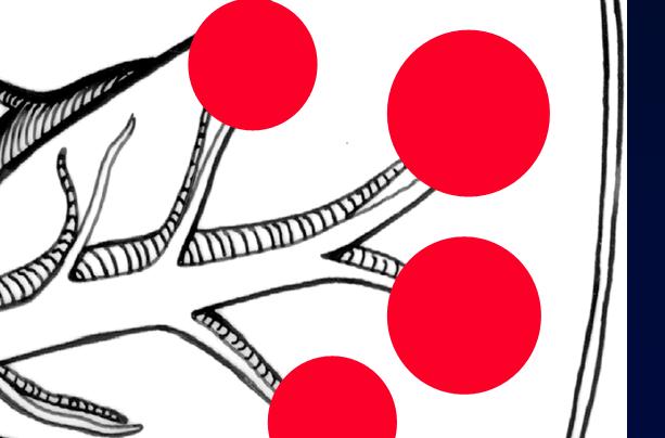
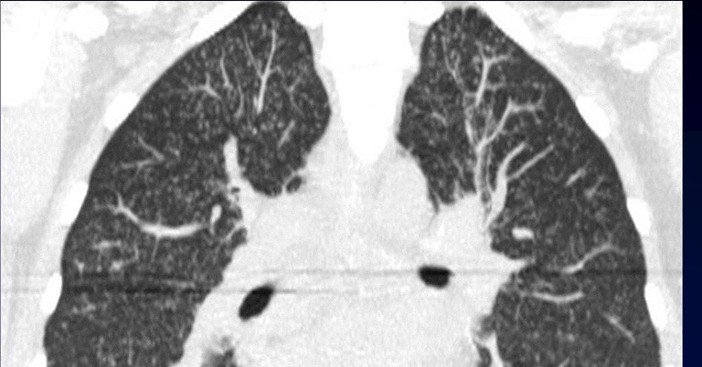
40 Perilymphatic Nodules
41 Perilymphatic Nodules


42 Perilymphatic Nodules


43 Perilymphatic Nodules
44 Perilymphatic Nodules
45 Perilymphatic Nodules Sarcoidosis Lymphangitic carcinomatosis Silicosis/CWP majority Lymphocytic IP (rare) vast majority Amyloidosis (rare)
46 Sarcoidosis
47 Lymphangitic Carc * *
48 Lymphangitic Carc * *


49 Lymphangitic Carc PV MPR


50 Lymphangitic Carc PV Volume Rendered

51 Micronodules Distribution Random Perilymphatic Centrilobular Ground Glass Solid Tree-in-Bud


52 Random Nodules


53 Random Nodules
54 Random Nodules Disseminated infection (miliary) Sarcoidosis Metastatic disease Silicosis, CWP majority Langerhans cell histiocytosis vast majority
55 Histoplasmosis
56 Micronodules Distribution Random Perilymphatic Centrilobular Ground Glass Solid Tree-in-Bud
57 References Beigelman-Aubry C, et al. MDCT and postprocessing techniques in the assessment of diffuse lung disease. Radiographics 2005; 25: Devakonda A, et al. Bronchiolar disorders: a clinical-radiological diagnostic algorithm. Chest 2010; 137: Raoof S, et al. Pictorial essay: Multinodular disease: an HRCT diagnostic algorithm. Chest 2006; 129: Remy-Jardin M, et al. Diffuse infiltrative lung disease: clinical value of sliding-thin-slab maximum intensity projection CT scans in the detection of mild micronodular patterns. Radiology 1996; 200: Sakai M, et al. Can maximum intensity projection images with MDCT help to differentiate between the micronodular distribution of focal and diffuse infiltrative lung diseases? JCAT 2005; 29:
58 Question Which of the following lung diseases is MOST likely to show a perilymphatic distribution of micronodules on thinsection CT? 1. Langerhans cell histiocytosis 2. Airway-disseminated tuberculosis 3. Hypersensitivity pneumonitis 4. Sarcoidosis 5. Respiratory bronchiolitis
59 Micronodular Lung Disease an algorithm H. Page McAdams, MD Department of Radiology Duke University Medical Center Durham, NC USA
Case 1: Question. 1.1 What is the main pattern of this HRCT? 1. Intralobular line 2. Groundglass opacity 3. Perilymphatic nodule
 HRCT WORK SHOP Case 1 Case 1: Question 1.1 What is the main pattern of this HRCT? 1. Intralobular line 2. Groundglass opacity 3. Perilymphatic nodule Case 1: Question 1.2 What is the diagnosis? 1. Hypersensitivity
HRCT in Diffuse Interstitial Lung Disease Steps in High Resolution CT Diagnosis. Where are the lymphatics? Anatomic distribution
 Steps in High Resolution CT Diagnosis Pattern of abnormality Distribution of disease Associated findings Clinical history Tomás Franquet MD What is the diagnosis? Hospital de Sant Pau. Barcelona Secondary
Case 1 : Question. 1.1 What is the intralobular distribution? 1. Centrilobular 2. Perilymphatic 3. Random
 Interesting case Case 1 Case 1 : Question 1.1 What is the intralobular distribution? 1. Centrilobular 2. Perilymphatic 3. Random Case 1: Answer 1.1 What is the intralobular distribution? 1. Centrilobular
Acute and Chronic Lung Disease
 KATHOLIEKE UNIVERSITEIT LEUVEN Faculty of Medicine Acute and Chronic Lung Disease W De Wever, JA Verschakelen Department of Radiology, University Hospitals Leuven, Belgium Clinical utility of HRCT To detect
11/10/2014. Multi-disciplinary Approach to Diffuse Lung Disease: The Imager s Perspective. Radiology
 Multi-disciplinary Approach to Diffuse Lung Disease: The Imager s Perspective Radiology Pathology Clinical 1 Role of HRCT Diagnosis Fibrosis vs. inflammation Next step in management Response to treatment
Micronodular lung pattern - Differential diagnosis
 Micronodular lung pattern - Differential diagnosis Poster No.: P-0074 Congress: ESTI 2015 Type: Educational Poster Authors: P. Ninitas, F. Marinho, P. Campos, I. Távora ; Lisbon/PT, 1 2 2 3 1 1 3 Funchal/PT,
Radiologic-pathologic correlation of pulmonary diseases
 The 1578 th Chest Conference/ 3 rd Biennial Clinical- Radiologic-Pathologic Correlation Radiologic-pathologic correlation of pulmonary diseases Harumi Itoh, M.D. University of Fukui, Japan Centriacinar
Financial disclosure COMMON DIAGNOSES IN HRCT. High Res Chest HRCT. HRCT Pre test. I have no financial relationships to disclose. Anatomy Nomenclature
 Financial disclosure I have no financial relationships to disclose. Douglas Johnson D.O. Cardiothoracic Imaging Gaston Radiology COMMON DIAGNOSES IN HRCT High Res Chest Anatomy Nomenclature HRCT Sampling
Interstitial Syndrome Ground glass attenuation miliary and nodular images Linear images
 Interstitial Syndrome Ground glass attenuation miliary and nodular images Linear images Dr Etienne Leroy-Terquem Centre hospitalier de Meulan les Mureaux. France French-cambodian association for pneumology
Interstitial syndrome
 Interstitial syndrome Ground-glass attenuation Miliary and nodular images linear images Etienne Leroy Terquem Pierre L Her SPI / ISP Soutien Pneumologique International / International Support for Pulmonology
HRCT Versus Volume Rendering (Three Colors, Three Densities Lung Images) in Diagnosis of Small Airway Disease: A Comparative Study
 in Diagnosis of Small Airway Disease: A Comparative Study") Med. J. Cairo Univ., Vol. 84, No. 1, March: 359-364, 2016 www.medicaljournalofcairouniversity.net HRCT Versus Volume Rendering (Three Colors, Three Densities Lung Images) in Diagnosis of Small Airway Disease:
Med. J. Cairo Univ., Vol. 84, No. 1, March: 359-364, 2016 www.medicaljournalofcairouniversity.net HRCT Versus Volume Rendering (Three Colors, Three Densities Lung Images) in Diagnosis of Small Airway Disease:
Resident Case Review CHEST. Daria Manos CAR 2016
 Resident Case Review CHEST CAR 2016 Daria Manos Disclosure Speakers bureau, Roche CAR 2016 Daria Manos 1. Recognize common and critical chest radiograph and computed tomography signs and use these clues
Cystic Lung Disease. Cristopher A. Meyer, MD
 Cystic Lung Disease Cristopher A. Meyer, MD Air filled structure with definable wall typically less than 1 mm thick Cris A. Meyer, M.D. Professor of Radiology University of Wisconsin School of Medicine
Pictorial essay of unusual radiologic manifestations of pulmonary and airway metastasis at initial presentation of lung cancer
 Pictorial essay of unusual radiologic manifestations of pulmonary and airway metastasis at initial presentation of lung cancer Poster No.: C-2297 Congress: ECR 2012 Type: Educational Exhibit Authors: Y.
CT findings in multifocal or diffuse non-mucinous bronchioloalveolar carcinoma (BAC)
") CT findings in multifocal or diffuse non-mucinous bronchioloalveolar carcinoma (BAC) Poster No.: C-2192 Congress: ECR 2014 Type: Educational Exhibit Authors: I. Sandu, A. R. Popita, I.-A. Brumboiu; Cluj-Napoca/RO
CT findings in multifocal or diffuse non-mucinous bronchioloalveolar carcinoma (BAC) Poster No.: C-2192 Congress: ECR 2014 Type: Educational Exhibit Authors: I. Sandu, A. R. Popita, I.-A. Brumboiu; Cluj-Napoca/RO
Pictorial Essay: Multinodular Disease* A High-Resolution CT Scan Diagnostic Algorithm
 CHEST Chest Imaging for Clinicians Pictorial Essay: Multinodular Disease* A High-Resolution CT Scan Diagnostic Algorithm Suhail Raoof, MD, FCCP; Alexey Amchentsev, MD; Ioannis Vlahos, MD; Ajay Goud, MD;
Typical and atypical findings of pulmonary sarcoidosis at high resolution CT
 Typical and atypical findings of pulmonary sarcoidosis at high resolution CT Poster No.: C-0169 Congress: ECR 2013 Type: Educational Exhibit Authors: L. Raposo Rodríguez, C. Mejía, B. Escobar Mallada,
Immunocompromised patients. Immunocompromised patients. Immunocompromised patients
 Value of CT in Early Pneumonia in Immunocompromised Patients Nantaka Kiranantawat, PSU Preventative Factors Phagocyts Cellular immunity Humoral immunity Predisposing Factors Infection, Stress, Poor nutrition,
Radiologists toolbox to differentiate alveolar versus interstitial lung diseases
 Radiologists toolbox to differentiate alveolar versus interstitial lung diseases Dr Sumer Shikhare, Dr Trishna Shimpi, Dr Ashish Chawla Khoo Teck Puat Hospital Singapore. Relevant financial disclosures
How to Analyse Difficult Chest CT
 How to Analyse Difficult Chest CT Complex diseases are:- - Large lesion - Unusual or atypical pattern - Multiple discordant findings Diffuse diseases are:- - Numerous findings in both sides 3 basic steps
Hypersensitivity Pneumonitis: Spectrum of High-Resolution CT and Pathologic Findings
 CT of Hypersensitivity Pneumonitis Chest Imaging Pictorial Essay C. Isabela S. Silva 1 ndrew Churg 2 Nestor L. Müller 1 Silva CIS, Churg, Müller NL Keywords: high-resolution CT, hypersensitivity pneumonitis,
Imaging Small Airways Diseases: Not Just Air trapping. Eric J. Stern MD University of Washington
 Imaging Small Airways Diseases: Not Just Air trapping Eric J. Stern MD University of Washington What we are discussing SAD classification SAD imaging with MDCT emphasis What is a small airway? Airway with
5/9/2015. Multi-disciplinary Approach to Diffuse Lung Disease: The Imager s Perspective. No, I am not a pulmonologist! Radiology
 Multi-disciplinary Approach to Diffuse Lung Disease: The Imager s Perspective No, I am not a pulmonologist! Radiology Pathology Clinical 1 Everyone needs a CT Confidence in diagnosis Definitive HRCT +
RADIOLOGIC EVALUATION OF PULMONARY NTM INFECTION. Tilman Koelsch, MD National Jewish Health - Department of Radiology
 Pr N op ot er fo ty r R of ep Pr ro es du en ct te io r n RADIOLOGIC EVALUATION OF PULMONARY NTM INFECTION Tilman Koelsch, MD National Jewish Health - Department of Radiology Disclosures No relevant financial
Manish Powari Regional Training Day 10/12/2014
 Manish Powari Regional Training Day 10/12/2014 Large number of different types of Interstitial Lung Disease (ILD). Most are very rare Most patients present with one of a smaller number of commoner diseases
Chest imaging III: Nodular pulmonary disease. Ádám Domonkos Tárnoki, MD, PhD Assistant professor Department of Radiology, Semmelweis University 1
 Chest imaging III: Nodular pulmonary disease Ádám Domonkos Tárnoki, MD, PhD Assistant professor Department of Radiology, Semmelweis University 1 Pattern 2 Nodular pattern Several round opacity, typically
I have no relevant conflicts of interest to disclose
 I have no relevant conflicts of interest to disclose Diffuse parenchymal lung disease (DPLD) and its associations Secondary lobular anatomy DPLD History, clinical findings, temporal evolution, and exposures
New Horizons in the Imaging of the Lung
 New Horizons in the Imaging of the Lung Postprocessing. How to do it and when do we need it? Peter M.A. van Ooijen, MSc, PhD Principal Investigator, Radiology, UMCG Discipline Leader Medical Imaging Informatics
Uses, limitations and interpretation of CT in pulmonary infections: A practical approach
 Uses, limitations and interpretation of CT in pulmonary infections: A practical approach Canadian Association of Radiologists 2013 DISCLOSURES Speakers honorarium, Siemens Canada Objectives 1. Recognize
An Image Repository for Chest CT
 An Image Repository for Chest CT Francesco Frajoli for the Chest CT in Antibody Deficiency Group An Image Repository for Chest CT he Chest CT in Antibody Deficiency Group is an international and interdisciplinary
RADIOLOGIC EVALUATION OF PULMONARY NTM INFECTION. Tilman Koelsch, MD National Jewish Health - Department of Radiology
 Pr N op ot e fo rty rr o f ep Pr ro es du en ct te io r n RADIOLOGIC EVALUATION OF PULMONARY NTM INFECTION Tilman Koelsch, MD National Jewish Health - Department of Radiology Disclosures None Goals Identify
Differential diagnosis
 Differential diagnosis Idiopathic pulmonary fibrosis (IPF) is part of a large family of idiopathic interstitial pneumonias (IIP), one of four subgroups of interstitial lung disease (ILD). Differential
Outline Definition of Terms: Lexicon. Traction Bronchiectasis
 HRCT OF IDIOPATHIC INTERSTITIAL PNEUMONIAS Disclosures Genentech, Inc. Speakers Bureau Tadashi Allen, MD University of Minnesota Assistant Professor Diagnostic Radiology 10/29/2016 Outline Definition of
Eun-Young Kang, M.D., Jae Wook Lee, M.D., Ji Yung Choo, M.D., Hwan Seok Yong, M.D., Ki Yeol Lee, M.D., Yu-Whan Oh, M.D.
 Eun-Young Kang, M.D., Jae Wook Lee, M.D., Ji Yung Choo, M.D., Hwan Seok Yong, M.D., Ki Yeol Lee, M.D., Yu-Whan Oh, M.D. Department of Radiology, Korea University Guro Hospital, College of Medicine, Korea
HYPERSENSITIVITY PNEUMONITIS
 HYPERSENSITIVITY PNEUMONITIS A preventable fibrosis MOSAVIR ANSARIE MB., FCCP INTERSTITIAL LUNG DISEASES A heterogeneous group of non infectious, non malignant diffuse parenchymal disorders of the lower
Mimics in chest disease: interstitial opacities
 Insights Imaging (2013) 4:9 27 DOI 10.1007/s13244-012-0207-7 PICTORIAL REVIEW Mimics in chest disease: interstitial opacities Anastasia Oikonomou & Panos Prassopoulos Received: 19 June 2012 / Revised:
PULMONARY TUBERCULOSIS RADIOLOGY
 PULMONARY TUBERCULOSIS RADIOLOGY RADIOLOGICAL MODALITIES Medical radiophotography Radiography Fluoroscopy Linear (conventional) tomography Computed tomography Pulmonary angiography, bronchography Ultrasonography,
10/17/2016. Nuts and Bolts of Thoracic Radiology. Objectives. Techniques
 Nuts and Bolts of Thoracic Radiology October 20, 2016 Carleen Risaliti Objectives Understand the basics of chest radiograph Develop a system for interpreting chest radiographs Correctly identify thoracic
Excavated pulmonary nodule: steps to diagnosis?
 Excavated pulmonary nodule: steps to diagnosis? Poster No.: C-1044 Congress: ECR 2014 Type: Authors: Keywords: DOI: Educational Exhibit W. Mnari, M. MAATOUK, A. Zrig, B. Hmida, M. GOLLI; Monastir/ TN Metastases,
Pulmonary Sarcoidosis - Radiological Evaluation
 Original Research Article Pulmonary Sarcoidosis - Radiological Evaluation Jayesh Shah 1, Darshan Shah 2*, C. Raychaudhuri 3 1 Associate Professor, 2 1 st Year Resident, 3 Professor and HOD Radiology Department,
CT findings of high-attenuation pulmonary abnormalities
 Insights Imaging (2010) 1:287 292 DOI 10.1007/s13244-010-0039-2 PICTORIAL REVIEW CT findings of high-attenuation pulmonary abnormalities Naim Ceylan & Selen Bayraktaroglu & Recep Savaş & Hudaver Alper
Nontuberculous Mycobacterial Lung Disease
 Non-TB Mycobacterial Disease Jeffrey P. Kanne, MD Nontuberculous Mycobacterial Lung Disease Jeffrey P. Kanne, M.D. Consultant Disclosures Perceptive Informatics Royalties (book author) Amirsys, Inc. Wolters
Pediatric High-Resolution Chest CT
 Pediatric High-Resolution Chest CT Alan S. Brody, MD Professor of Radiology and Pediatrics Chief, Thoracic Imaging Cincinnati Children s s Hospital Cincinnati, Ohio, USA Pediatric High-Resolution CT Short
Daria Manos RSNA 2016 RC 401. https://medicine.dal.ca/departments/depar tment-sites/radiology/contact/faculty/dariamanos.html
 Daria Manos RSNA 2016 RC 401 https://medicine.dal.ca/departments/depar tment-sites/radiology/contact/faculty/dariamanos.html STEP1: Is this fibrotic lung disease? STEP 2: Is this a UIP pattern? If yes:
Low Grade Coal Worker's Pneumoconiosis
 Acta Radiologica ISSN: 0284-181 (Print) 1600-04 (Online) Journal homepage: https://www.tandfonline.com/loi/iard20 Low Grade Coal Worker's Pneumoconiosis P. A. Gevenois, E. Pichot, F. Dargent, S. Dedeire,
Lung Cancer Imaging. Terence Z. Wong, MD,PhD. Department of Radiology Duke University Medical Center Durham, NC 9/9/09
 Lung Cancer Imaging Terence Z. Wong, MD,PhD Department of Radiology Duke University Medical Center Durham, NC 9/9/09 Acknowledgements Edward F. Patz, Jr., MD Jenny Hoang, MD Ellen L. Jones, MD, PhD Lung
ESOR COURSES FOR EDiR 2018 CHEST IMAGING
 ESOR COURSES FOR EDiR 2018 Education in partnership myesr.org/esor myesr.org/esor Course information In 2018, ESOR is continuing to organise foundation courses to prepare/assist those entering examinations
Radiologic Approach to Smoking Related Interstitial Lung Disease
 Radiologic Approach to Smoking Related Interstitial Lung Disease Poster No.: C-1854 Congress: ECR 2013 Type: Educational Exhibit Authors: K.-N. Lee, J.-Y. Han, E.-J. Kang, J. Kang; Busan/KR Keywords: Toxicity,
Diffuse Cystic Lung Disease at High-Resolution CT
 Cardiopulmonary Imaging Pictorial Essay Seaman et al. HRCT of Diffuse Cystic Lung Disease Cardiopulmonary Imaging Pictorial Essay Downloaded from www.ajronline.org by 37.44.193.85 on 01/05/18 from IP address
Bronchiolitis: A Schematic Diagnostic Approach with Radiologic-pathologic Correlation
 Bronchiolitis: A Schematic Diagnostic Approach with Radiologic-pathologic Correlation Mariana Benegas Urteaga 1, MD; M Sanchez 1, MD; J Ramirez 2, MD; D Barnes 1, MD; T de Caralt 1, MD; R J Perea 1, MD
Smoking-related Interstitial Lung Diseases: High-Resolution CT Findings
 Smoking-related Interstitial Lung Diseases: High-Resolution CT Findings Poster No.: C-2358 Congress: ECR 2013 Type: Educational Exhibit Authors: V. Cuartero Revilla, M. Nogueras Carrasco, P. Olmedilla
INTERSTITIAL LUNG DISEASE Dr. Zulqarnain Ashraf
 Indep Rev Jul-Dec 2018;20(7-12) Dr. Zulqarnain Ashraf IR-653 Abstract: ILD is a group of diseases affect interstitium of the lung. Repeated insult to the lung cause the interstitium to be damaged. Similarly
Case of the Day Chest
 Case of the Day Chest Darin White MDCM FRCPC Department of Radiology, Mayo Clinic 76 th Annual Scientific Meeting Canadian Association of Radiologists Montreal, QC April 26, 2013 2013 MFMER slide-1 Disclosures
Comparison of High-resolution CT Findings between Miliary Metastases and Miliary Tuberculosis 1
 Comparison of High-resolution CT Findings between Miliary Metastases and Miliary Tuberculosis 1 Chan Sung Kim, M.D., Ki-Nam Lee, M.D., Jin Hwa Lee, M.D. Purpose: To compare the findings of high-resolution
Hypothesis on the Evolution of Cavitary Lesions in Nontuberculous Mycobacterial Pulmonary Infection: Thin-Section CT and Histopathologic Correlation
 CT of Nontuberculous Mycobacterial Pulmonary Infection Tae Sung Kim 1 Won-Jung Koh 2 Joungho Han 3 Myung Jin Chung 1 Ju Hyun Lee 1 Kyung Soo Lee 1 O Jung Kwon 2 Kim TS, Koh W-J, Han J, et al. Received
Chronic Thromboembolic Pulmonary Hypertension (CTEPH): A Primer
: A Primer") Chronic Thromboembolic Pulmonary Hypertension (CTEPH): A Primer H. Page McAdams, MD Duke University Medical Center Durham, NC 27710 page.mcadams@duke.edu Question Which of the following imaging tests is
Chronic Thromboembolic Pulmonary Hypertension (CTEPH): A Primer H. Page McAdams, MD Duke University Medical Center Durham, NC 27710 page.mcadams@duke.edu Question Which of the following imaging tests is
and localized ground glass opacities, or bronchiolar focal or multifocal micronodules;
 E1 Chest CT scan and Pneumoniae_YE Claessens et al- Supplementary methods Level of CAP probability according to CT scan - definite CAP: systematic alveolar condensation, or alveolar condensation with peripheral
I appreciate the courtesy of Kusumoto at NCC for this presentation. What is Early Lung Cancers. Early Lung Cancers. Early Lung Cancers 18/10/55
 I appreciate the courtesy of Kusumoto at NCC for this presentation. Dr. What is Early Lung Cancers DEATH Early period in its lifetime Curative period in its lifetime Early Lung Cancers Early Lung Cancers
The role of high-resolution computed tomography in the follow-up of diffuse lung disease
 SERIES RADIOLOGY The role of high-resolution computed tomography in the follow-up of diffuse lung disease Brett M. Elicker, Kimberly G. Kallianos and Travis S. Henry Number 2 in the Series Radiology Edited
NONE OVERVIEW FINANCIAL DISCLOSURES UPDATE ON IDIOPATHIC PULMONARY FIBROSIS/IPF (UIP) FOR PATHOLOGISTS. IPF = Idiopathic UIP Radiologic UIP Path UIP
 FOR PATHOLOGISTS. IPF = Idiopathic UIP Radiologic UIP Path UIP") UPDATE ON IDIOPATHIC PULMONARY FIBROSIS/IPF () FOR PATHOLOGISTS Thomas V. Colby, M.D. Professor of Pathology (Emeritus) Mayo Clinic Arizona FINANCIAL DISCLOSURES NONE OVERVIEW IPF Radiologic Dx Pathologic
UPDATE ON IDIOPATHIC PULMONARY FIBROSIS/IPF () FOR PATHOLOGISTS Thomas V. Colby, M.D. Professor of Pathology (Emeritus) Mayo Clinic Arizona FINANCIAL DISCLOSURES NONE OVERVIEW IPF Radiologic Dx Pathologic
Progress in Idiopathic Pulmonary Fibrosis
 Progress in Idiopathic Pulmonary Fibrosis David A. Lynch, MB Disclosures Progress in Idiopathic Pulmonary Fibrosis David A Lynch, MB Consultant: t Research support: Perceptive Imaging Boehringer Ingelheim
August 2018 Imaging Case of the Month: Dyspnea in a 55-Year-Old Smoker. Michael B. Gotway, MD
 August 2018 Imaging Case of the Month: Dyspnea in a 55-Year-Old Smoker Michael B. Gotway, MD Department of Radiology Mayo Clinic Arizona Scottsdale, AZ USA Clinical History: A 55 year old woman presented
Subpleural micronodular pattern at CT and radiopathologic correlation
 Subpleural micronodular pattern at CT and radiopathologic correlation Poster No.: C-0876 Congress: ECR 2012 Type: Educational Exhibit Authors: G. Cabrera, E. Romá de Villegas, M. L. Domingo, C. P. 1 2
Diffuse Pulmonary Nodules
 Residents Section Pattern of the Month oitsios et al. Diffuse Pulmonary Nodules Residents Section Pattern of the Month Downloaded from www.ajronline.org by 37.44.196.156 on 12/07/17 from IP address 37.44.196.156.
Low-dose CT Lung Cancer Screening Guidelines for Pulmonary Nodules Management Version 2
 Low-dose CT Lung Cancer Screening Guidelines for Pulmonary Nodules Management Version 2 The Committee for Management of CT-screening-detected Pulmonary Nodules 2009-2011 The Japanese Society of CT Screening
Computerized Medical Imaging and Graphics
 Computerized Medical Imaging and Graphics 36 (2012) 72 84 Contents lists available at ScienceDirect Computerized Medical Imaging and Graphics journa l h o me pa g e: www.elsevier.com/locate/compmedimag
Diagnostic Imaging of Diffuse Infiltrative Disease of the Lung
 Thematic Review Series Respiration 2004;71:4 19 DOI: 10.1159/000075642 Diagnostic Imaging of Diffuse Infiltrative Disease of the Lung Maurizio Zompatori a Claudio Bnà a Venerino Poletti c Enrica Spaggiari
Epidemiology and classification of smoking related interstitial lung diseases
 Epidemiology and classification of smoking related interstitial lung diseases Šterclová M. Department of Respiratory Diseases, Thomayer Hospital, Prague, Czech Republic Supported by an IGA Grant No G 1207
Diagnostic Evaluation of NTM and Bronchiectasis
 Division of Pulmonary, Critical Care and Sleep Medicine Diagnostic Evaluation of NTM and Bronchiectasis Ashwin Basavaraj, MD, FCCP NTM patient education program November 9, 2016 Involves a combination
Pneumocystis jirovecci pneumonia: from mild disease to a real disaster. A pictorial review of the different radiologic patterns in acute settings
 Pneumocystis jirovecci pneumonia: from mild disease to a real disaster. A pictorial review of the different radiologic patterns in acute settings Poster No.: C-1425 Congress: ECR 2017 Type: Educational
Liebow and Carrington's original classification of IIP
 Liebow and Carrington's original classification of IIP-- 1969 Eric J. Stern MD University of Washington UIP Usual interstitial pneumonia DIP Desquamative interstitial pneumonia BIP Bronchiolitis obliterans
Thoracic Sarcoidosis Imaging Updated: Jul 19, 2013
 Thoracic Sarcoidosis Imaging Updated: Jul 19, 2013 Overview Radiography Computed Tomography Magnetic Resonance Imaging Nuclear Imaging Show All Multimedia Library References Overview For patients with
September 2014 Imaging Case of the Month. Michael B. Gotway, MD. Department of Radiology Mayo Clinic Arizona Scottsdale, AZ
 September 2014 Imaging Case of the Month Michael B. Gotway, MD Department of Radiology Mayo Clinic Arizona Scottsdale, AZ Clinical History: A 57-year-old non-smoking woman presented to her physician as
Workshop Cyst & Lucency. How to Approach
 Workshop Cyst & Lucency How to Approach To Approach Cystic Lung Disease True cysts? Cavitary disease Cystic bronchiectasis Mosaic attenuation Subpleural cysts Bullae Paraseptal emphysema Honeycombing Birt
Pulmonary fibrosis on the lateral chest radiograph: Kerley D lines revisited
 Insights Imaging (2017) 8:483 489 DOI 10.1007/s13244-017-0565-2 PICTORIAL REVIEW Pulmonary fibrosis on the lateral chest radiograph: Kerley D lines revisited Daniel B. Green 1 & Alan C. Legasto 1 & Ian
Thin-Section CT Findings of Arc-Welders Pneumoconiosis
 Thin-Section CT Findings of Arc-Welders Pneumoconiosis Daehee Han, MD 1 Jin Mo Goo, MD 1 Jung-Gi Im, MD 1 Kyung Soo Lee, MD 2 Do Myung Paek, MD 3 Seong Ho Park, MD 1 Index words: Lungs, diseases Lungs,
Diagnosis of TB: Radiology David Finlay, MD
 TB Intensive Tyler, Texas June 2-4, 2010 Diagnosis of TB: Radiology David Finlay, MD June 3, 2010 2stages stages- Tuberculosis 1. primary infection 2. reactivation, or post primary disease 2 1 Primary
Chest Radiology LYMPHANGITIC CARCINOMATOSIS CERTAIN CANCERS SPREAD BY PLUGGING THE LYMPHATICS
 2 Chest Radiology Includes plain film diagnosis, CT, MRI, and interventional techniques used in the diagnosis of diseases of the lungs, pleura, and mediastinum including the heart and great vessels. LYMPHANGITIC
Cryptogenic Organizing Pneumonia Diagnosis Approach Based on a Clinical-Radiologic-Pathologic Consensus
 Cryptogenic Organizing Pneumonia Diagnosis Approach Based on a Clinical-Radiologic-Pathologic Consensus Poster No.: C-1622 Congress: ECR 2012 Type: Scientific Exhibit Authors: C. Cordero Lares, E. Zorita
Bronchiectasis: An Imaging Approach
 Bronchiectasis: An Imaging Approach Travis S Henry, MD Associate Professor of Clinical Radiology Cardiac and Pulmonary Imaging Section University of California, San Francisco Large Middle Small 1 Bronchiectasis
Diffuse Interstitial Lung Diseases: Is There Really Anything New?
 : Is There Really Anything New? Sujal R. Desai, MBBS, MD ESTI SPEAKER SUNDAY Society of Thoracic Radiology San Antonio, Texas March 2014 Diffuse Interstitial Lung Disease The State of Play DILDs Is There
An Introduction to Radiology for TB Nurses
 An Introduction to Radiology for TB Nurses Garold O. Minns, MD September 14, 2017 TB Nurse Case Management September 12 14, 2017 EXCELLENCE EXPERTISE INNOVATION Garold O. Minns, MD has the following disclosures
Atopic Pulmonary Disease: Findings on Thoracic Imaging
 July 2003 Atopic Pulmonary Disease: Findings on Thoracic Imaging Rebecca G. Breslow Harvard Medical School Year IV Churg-Strauss Syndrome Hypersensitivity Pneumonitis Asthma Atopic Pulmonary Disease Allergic
Case Presentations in ILD. Harold R. Collard, MD Department of Medicine University of California San Francisco
 Case Presentations in ILD Harold R. Collard, MD Department of Medicine University of California San Francisco Outline Overview of diagnosis in ILD Definition/Classification High-resolution CT scan Multidisciplinary
Post treatment contribution in semiological analysis of thoracic helical CT
 Post treatment contribution in semiological analysis of thoracic helical CT Poster No.: C-0996 Congress: ECR 2010 Type: Educational Exhibit Topic: Chest - Miscellaneous Authors: M. Gaha, N. Mama, N. Arifa,
Pulmonary TB: HRCT findings
 Pulmonary TB: HRCT findings Tuberculosis: History BC 5000: Evidence of TB in neolithic man Jung-Gi Im, MD Department of Radiology Seoul National University Hospital BC 2900: Pyramid builders BC 1000 :
Clinical significance of respiratory bronchiolitis on open lung biopsy and its relationship to smoking related interstitial lung disease
 Thorax 1999;54:1009 1014 1009 James Moon, Ronald M du Bois, Thomas V Colby, David M Hansell, Andrew G Nicholson Interstitial Lung Disease Unit J Moon R M du Bois Department of Radiology D M Hansell Department
Lung Allograft Dysfunction
 Lung Allograft Dysfunction Carlos S. Restrepo M.D. Ameya Baxi M.D. Department of Radiology University of Texas Health San Antonio Disclaimer: We do not have any conflict of interest or financial gain to
Bronkhorst colloquium Interstitiële longziekten. Katrien Grünberg, klinisch patholoog
 Bronkhorst colloquium 2013-2014 Interstitiële longziekten De pathologie achter de CT Katrien Grünberg, klinisch patholoog K.grunberg@vumc.nl Preparing: introduction and 3 cases The introduction on microscopic
The crazy-paving pattern: A radiological-pathological correlated and illustrated overview
 The crazy-paving pattern: A radiological-pathological correlated and illustrated overview Poster No.: C-0827 Congress: ECR 2010 Type: Educational Exhibit Topic: Chest Authors: W. F. M. De Wever, J. Coolen,
Non-neoplastic Lung Disease II
 Pathobasic Non-neoplastic Lung Disease II Spasenija Savic Prince Pathology Program Systematic approach to surgical lung biopsies with ILD Examples (chronic ILD): Idiopathic interstitial pneumonias: UIP,
Diagnosis of Respiratory Bronchiolitis associated interstitial lung disease
 Monaldi Arch Chest Dis 2006; 65: 2, 96-101 ORIGINAL ARTICLE Diagnosis of Respiratory Bronchiolitis associated interstitial lung disease C. Robalo Cordeiro 1, S. Freitas 1, B. Rodrigues 1, A. Catarino 1,
Case 1. Background. Presenting Symptoms. Schecter Case1 Differential Diagnosis of TB 1
 TB or Not TB? Case 1 Gisela Schecter, M.D., M.P.H. California Department of Public Health Background 26 year old African American male Born and raised in Bay Area of California Convicted of cocaine trafficking
Extraordinary Patterns of Tuberculosis
 Extraordinary Patterns of Tuberculosis E. Kadakovska Infectology Center of Latvia, Clinic of Tuberculosis and Lung Diseases, Diagnostics and Radiology Department 1 Target Importance of recognizing of tuberculosis
Diagnosing ILD. What is important in 2016? Chris Grainge
 Diagnosing ILD What is important in 2016? Chris Grainge Senior Staff Specialist Respiratory Medicine John Hunter Hospital Conjoint A/Prof University of Newcastle Conflict of interest I have acted as a