THE JOURNAL OF COMPARATIVE NEUROLOGY 250~ (1986)
|
|
|
- Irma Curtis
- 5 years ago
- Views:
Transcription
1 THE JOURNAL OF COMPARATIVE NEUROLOGY 250~ (1986) Microelectrode Maps, Myeloarchitecture, and Cortical Connections of Three Somatotopically Organized Representations of the Body Surface in the Parietal Cortex of Squirrels L.A. KRUBITZER, M.A. SESMA, AND J.H. KAAS Department of Psychology, Vanderbilt University, Nashville, Tennessee ABSTRACT Microelectrode mapping methods and anatomical procedures were combined in the same animals to reveal the cortical connections of three architectonically distinct representations of the body surface in the somatosensory cortex of grey squirrels. In individual experiments, microelectrode multiunit recordings were used to determine the somatotopic organization of regions of the cortex and to identify sites for injections of the anatomical tracer, wheat germ agglutinin conjugated to horseradish peroxidase. After the brains were perfused, the cortex was separated from the brainstem, flattened, and cut parallel to the flattened surface to facilitate comparisons of areal connection patterns, physiological data, and architectonic subdivisions. Recordings in the primary (S-I) and secondary (S-11) somatosensory fields confirmed earlier descriptions of the somatotopic organization of these fields (Sur et al.: J. Comp. NeuroL 179: , 78; Nelson et al.: J. Comp. NeuroL 184:473490, 79). In addition, recordings in the cortex caudal to S-I and ventral to S-I1 revealed a third representation of the body surface in parietal cortex, the parietal ventral area (PV). Neurons in PV were responsive to light tactile stimulation of skin and hairs. Multiple unit receptive fields of neurons in PV were larger than those for neurons in S-I but similar in size to those for neurons in S-11. PV represented the contralateral body surface in a somatotopic manner that can be roughly characterized as an inverted homunculus with the limbs directed medially, the trunk located ventrally, and the face congruent with the representations of the upper lip and nose in S-I. Neurons in some electrode penetrations in PV were also responsive to auditory clicks. Microlesions placed at physiologically determined borders allowed all three somatic representations to be related to myeloarchitectonically defined fields. S-I was architectonically distinct as a densely myelinated region. Within S-I, a lightly myelinated oval of the cortex between the representation of the hand and face, the unresponsive zone (Sur et al.: J. Comp. NeuroL 179: , 78), was an easily recognized landmark. S-I1 and PV corresponded to less densely myelinated fields. Other subdivisions such as motor cortex, primary auditory cortex, and visual areas 17 and 18 were distinguished. Accepted March 11, M.A. Sesma s present address is School of Optometry, University of Missouri-St. Louis, 8001 Natural Bridge Road, St. Louis, MO ALAN R. LISS, INC.
 zone along the caudal border of S-I, and a parietal rhinal (PR) zone ventral")
2 404 L.A. KRUBITZER, M.A. SESMA, AND J.H. KAAS Connections were revealed by placing injections within S-I, S-11, or PV. Injections in S-I demonstrated reciprocal ipsilateral connections with the motor cortex, S-11, PV, a parietal medial (PM) zone along the caudal border of S-I, and a parietal rhinal (PR) zone ventral to PV and dorsal to the rhinal fissure. Injections placed in different parts of S-I suggested that connections with S-11, PV, PM, and the motor cortex are somatotopic. Callosal connections were with somatotopically equivalent parts of S-I. In one case where the unresponsive zone (UZ) was involved in the injection site, more extensive callosal connections included the motor cortex, S-11, PV, PR, PM, and UZ. Injections placed in S-I1 revealed ipsilateral interconnections with S-I, PM, PV, PR, and the cortex caudal to S-11. There were no significant connections with the motor cortex. Callosal connections were with S-11. Injections in PV demonstrated ipsilateral connections with S-I, S-11, PR, PM, and the motor cortex and callosal connections with PV, S-I, S-11, PR, and PM. The results led to two major conclusions. First, the parietal cortex in squirrels and possibly other rodents has at least three, and possibly as many as five, interconnected representations of the body. Second, PV and S-I1 are so similar to each other in size, responsiveness, and location relative to S-I that if both S-I1 and PV exist in other rodents or in other mammals, the two fields could be confused with each other from study to study. Key words: primary somatosensory cortex, motor cortex, corpus callosum, architectonics The brain is intriguing, in part, because it is so variable in structure and function across vertebrate species. Brain diversity is highly apparent in mammals, where, as an obvious example, the amount of neocortex varies from being a small cap on the forebrain in an insectivore such as the hedgehog to the expanded bulk engulfing most of forebrain in humans. In addition to such vast differences in relative size of the neocortex, there are almost certainly profound differences in organization. A traditional and currently supportable view of forebrain organization is that mammals with little neocortex, such as the hedgehog, have few subdivisions of functional significance, while species with considerable neocortex have many subdivisions (see Merzenich and Kaas, '80; Kaas, '82). Since mammals with expanded neocortex evolved independently in a number of lines of descent, it follows that increases in the complexity of cortical organization occurred independently a number of times. However, our understanding of the evolution of advanced brains and the corresponding wondrous changes in mental abilities has been limited by a lack of accurate information on how the cortex is organized in different mammals. Most studies of cortical organization have been on cats, several species of monkeys, and rats, and even in these mammais only a few subdivisions of the cortex have been defined with certainty. However, modern methods of microrecording, microstimulation, histochemistry, and the tracing of connections allow subdivisions of the brain and their connections to be reliably defined, and great progress in understanding cortical organization is now possible. The present study is part of a larger effort to determine further the basic organization of the neocortex in mammals and how this organization has been modified in the major lines of descent. The grey squirrel was chosen as an easily obtained rodent with the advantages of a larger brain and generally more distinct architectonic subdivisions than the commonly used rats and mice. Our investigation of the somatosensory cortex is paralleled by similar studies of the visual cortex (Hall et al., '71; Kaas et al., '72; Cusick et al., '80) and the auditory cortex (Merzenich et al., '76; Luethke et al., '85) in squirrels. Previous studies of the somatosensory cortex in squirrels have used microelectrode mapping methods to define the primary (Sur et al., '78) and secondary (Nelson et al., '79) somatosensory representations. In addition, the overall pattern of callosal projections of these two fields has been described (Gould and Kaas, '81). The initial goal of the present study was to use the sensitive tracer wheat germ agglutinin conjugated to horseradish peroxidase (WGA-HRP) to determine the cortical connections of S-I, and S-II. Because a major projection zone of S-I, and S-I1 was demonstrated in the ventral parietal cortex, we extended the original studies to map and further determine additional connections of a previously unknown rep resentation of the body surface. An abstract of some of the results has appeared elsewhere (Krubitzer et al., '85). METHODS Multiunit recording methods were used to investigate subdivisions of the somatosensory cortex in 17 adult grey squirrels (Sciurus carolinensis). In the same animals, cortical connections were determined by injecting the anatomical tracer wheat germ agglutinin conjugated with horseradish peroxidase (WGA-HRP) into electrophysiologically defined locations. Physiological and anatomical results were later related to the cortical architecture in sections cut parallel to the cortical surface of artificially flattened cortex. Microelectrode recording techniques were adopted from Sur et al. ('81), the anatomical procedures correspond to those outlined in Sesma et al. ('84), and the methods of flattening cortex are described in Gould and Kaas ('81)and Sesma et al. ('84). At the start of each experiment, the squirrel was initially anesthetized with 0.3 cc ketamine hydrochloride (130 mg/ kg; see White et al., '82) and 0.1 cc of acepromazine (4.3 mg/ kg). In addition, 0.1 cc of 2% xylocaine hydrochloride, a local anesthetic, was injected subcutaneously where ear bars entered the auditory meatus and where the scalp was
, were given as needed to maintain a surgical level of anesthesia.")
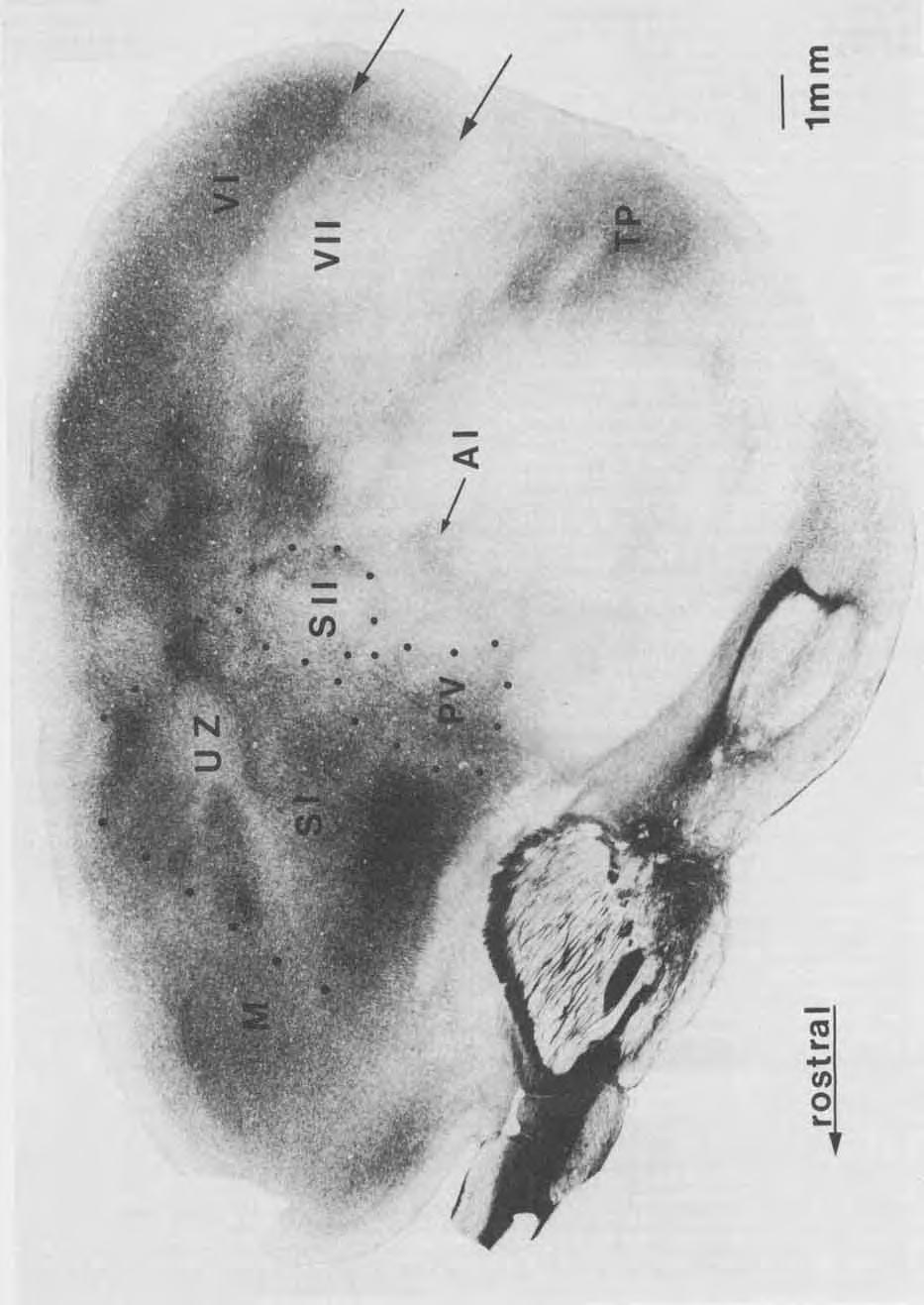
3 SUBDIVISIONS OF PAIUETAL CORTEX 405 to be cut. Maintenance doses of 0.15 cc of ketamine, supplemented in some cases with cc of nembutal (5.4 mg/ kg), were given as needed to maintain a surgical level of anesthesia. After the animal was deeply anesthetized, a portion of the skull was removed, and the dura was retracted to expose most or all of the somatosensory cortex. An acrylic dam was built around the opening and filled with silicone fluid, which protected the brain from desiccation. The head was tilted in a plane that allowed the electrode to penetrate the cortex perpendicular to the surface of the cortical area of interest. Prior to recording, the exposed cortex was photographed so that the blood vessel pattern could be used to mark the location of electrode penetrations. Recordings were made with low-impedance tungsten microelectrodes ( MQ at 1,000 Hz) with tip exposures designed to record from small clusters of neurons. Electrodes were advanced with a stepping microdrive. Recording depths were approximately 500-1,000 pm from the pial surface, although better recordings of auditory responses from neurons in the parietal ventral area (PV) were obtained at deeper depths (see Results). To obtain the receptive fields for neurons in a given penetration, the body surface was stimulated with fine wires and glass probes to find the location where stimuli maximally activated the neurons. Stimulation consisted of light tapping on the glabrous surface of the skin, displacement of hairs and vibrissae on the hairy surface of the skin, or lightly scratching the hairy or glabrous surface of the skin. The somatotopic organization of a region of cortex was determined from receptive fields for a number of closely spaced recording sites. After detailed mapping of a given area (the primary, S-I, and secondary somatosensory areas, S-11, or PV) was completed, marker lesions were made at electrophysiologically defined borders in S-I (Sur et al., '78), S-I1 (Nelson et al., '79) and PV so that physiological boundaries could be subsequently related to brain sections reacted for HRP or stained for myelin. Next, an anatomical tracer was injected at one or more electrophysiologically identified sites. Over a period of 20 minutes, p1 of 0.5% WGA-HRP was injected through a calibrated glass pipette into the representations of various body parts in S-I, S-II, or PV. Upon completion of the injection, the exposed cortex was covered with an acrylic cap, and the skin opening was closed. After 2 days, the animal was deeply anesthetized and perfused through the heart with 0.9% saline followed by 1.5% glutaraldehyde and 1% paraformaldahyde in phosphate buffer (ph 7.4). The fixed brain was removed, and the cortex was dissected from the brainstem, flattened between glass plates, and left to soak for approximately 24 hours in 30% sucrose phosphate buffer. The flattened cortices were frozen and cut into 40-pm sections in a plane parallel to the cortical surface. Two in four sections were reacted for HRP with tetramethylbenzidine (TMB) (Mesulam, '78). The remaining adjacent sections were stained for myelin with the Gallyas ('79) silver procedure. Sections reacted for HRP were sometimes stained with cresyl violet for cytoarchitecture, but these sections were less useful than the myelin preparations for revealing architectonic boundaries. For each case, drawings were made from several sections of tissue where the reacted anatomical tracer was most apparent. These drawings included the injection site, anterogradely labeled axons and terminals, retrogradely labeled cell bodies, and marker lesions. Drawings of individual sections were aligned by using blood vessels and tissue artifacts and compiled into one comprehensive reconstruction. Architectonic borders determined from myelin stained sections were superimposed on the reconstructions. RESULTS In the present experiments, multiunit recording procedures were used to reveal boundaries and features of SOmatotopic organization of two previously defined subdivisions of somatosensory cortex, the primary somatosensory area, S-I (Sur et al., '78) and the secondary somatosensory area, S-II RJelson et al., '79). In addition, the somatotopic organization of a third and previously unknown field, the parietal ventral area (PV), was determined. Ipsilateral and contralateral cortical connections of these three subdivisions of the brain were determined in the same animals by injecting WGA-HRP into electrophysiologically identified locations. The electrophysiological and connectional results were related to myeloarchitectonic subdivisions of cerebral cortex of the same animals. Correlation of structural and electrophysiological data was facilitated by artificially flattening the cerebral hemispheres and cutting cortical tissue parallel to the flattened surface. The myeloarchitecture of cortex Myeloarchitectonic differences reliably identified the boundaries of a number of cortical areas in brain sections cut parallel to the cortical surface. While parts of boundaries are obvious in single sections (Figs. 1-3), boundaries are better determined by examining a series of sections, since the flattened cortex may not always be cut in a plane exactly parallel to the cortical surface and the myelination pattern of cortical subdivisions changes throughout the cortical layers. Figure 1 illustrates the fact that many architectonic borders are apparent in single sections of flattened cortex. The first visual area, V-I or area 17, is most easily identified in the supragranular layers since it is more densely myelinated in these layers than the adjoining second visual area, V-I1 or area 18 (Hall et al., '71; Gould, '84). Thus, the 17/18 border is more distinct caudally in Figure 1 because it is slightly more superficial than the rostra1 portion of these visual areas. At favorable laminar levels, the outer boundary of area 18 is identified by a decrease in myelination density (caudal arrows in Fig 1). In temporal cortex, the densely myelinated temporal posterior region (TP) is distinct. This region has been reported to have darkly staining fiber bands in layers III and V (Kaas et al., '72). Since TP is surrounded by lightly myelinated cortex, all of its boundaries are pronounced. Rostrally in the temporal cortex, the temporal anterior region, TA (Kaas et al., '72), which includes the primary auditory field A-I (Merzenich et al., '76; Luethke et al., '851, is more heavily myelinated in deeper cortical layers than most adjoining cortex. Because of the level of the section, only part of A-I is evident in Figure 1. In parietal and frontal cortex, subdivisions of somatosensory cortex and motor cortex are also apparent. The architectonic characteristics of somatosensory areas and how they relate to the physiological and anatomical findings are described in more detail below. Architectonic features of neocortex in general have been described by Kaas et al. ('72), while the architecture of the visual (Hall et al., '71), auditory (Merzenich et al., '761, and somatosensory cortices (Nelson et al., '79; Sur et al., '78) has been described in more detail in other papers.

4

5 SUBDIVISIONS OF PARIETAL CORTEX Myeloarchitecture of S-I, S-11, and PV S-I is architectonically distinct from S-11, PV, and surrounding regions of cortex. It occupies a large region and is the most extensive representation of the body surface in the squirrel cortex. Its rostral extent is readily determined since S-I is more heavily myelinated than the neighboring motor cortex (Fig. 1,2A). The caudal boundary of S-I is also easily identified since S-I is more densely myelinated throughout most of the cortical layers than the parietal medial zone (PM), S-11, and PV, all of which abut S-I caudally (Fig. 2A). At its lateral border, S-I is bounded by lightly myelinated cortex. Within the densely myelinated S-I, there are regions that are lightly myelinated. A 1-mm oval of very lightly myelinated cortex is coextensive with a portion of S-I that has been described as unresponsive to light cutaneous stimulation (Sur et al., '78). This "unresponsive zone, " the UZ (Figs. 1, 2A), is also characterized by a less granular layer IV (Kaas et al., '72; Sur et al., '78). Two lightly myelinated strips of cortex extend rostrally and rostrolaterally within91 from UZ. The cortex lateral to the unresponsive zone contains a cutaneous representation of the upper and lower lips. This large portion of S-I occupies approximately a 4 x 3 mm expanse of the cortex. Caudolateral to the unresponsive zone, a moderately myelinated region is coextensive with the nose representation in S-I. Immediately caudal to the unresponsive zone, another region of moderately myelinated cortex is coextensive with the vibrissae representation. The portion of S-I just medial to the unresponsive zone tapers into a moderately myelinated region of the cortex that contains representations of the forelimb, trunk, and hindlimb in a lateral to medial cortical progression from UZ. Several fields border S-I. Motor cortex (MI, just rostral to S-I, is less myelinated than S-I. Although the motor cortex becomes more myelinated in deeper cortical layers, it remains less myelinated than S-I. Thus, the S-VM border is distinct in almost all sections (Figs. 1, 2A, 3A). A lightly myelinated strip of the cortex, the parietal medial area (PM), is immediately caudal to S-I and medial to S-11. The border between S-I and PM is most apparent in superficial layers of the cortex (Fig. 2A). In deeper cortical layers, PM is more heavily myelinated so that the S-I/PM border is less Fig. 1. The myeloarchitecture of the neocortex in the grey squirrel. A section cut parallel to the surface of an artificially flattened cerebral hemisphere has been stained for myelin. The single section provides a view of the extents and locations of cortical areas that is comparable to a reconstructed lateral view of the brain. The advantages of the plane of section are that some architectonic boundaries can be followed over much of their extents in a single section and that reconstructions can be based on few sections. For example, the border of the primary W-I) with the secondary W-ID visual cortex is obvious over much of its caudal extent (arrow, upper right), and more superficial sections clearly revealed its rostral location. The lateral border of V-I1 (area 18) is also obvious caudally (between arrows on right). Rostrally, a sudden reduction in overall density of myelination marks the border between the primary motor (M) and the primary somatosensory (S-D cortex (see the rostral row of mediolateral dots). Within S-I, the lightly myelinated unresponsive zone, the UZ, separates a medial representation of the forelimb from a lateral representation of the face. Lightly myelinated bands extend from UZ to the motor cortex. More caudally, the caudal borders of S-I, the second somatosensory area (S-II), and the parietal ventral somatosensory area (PV) are associated with sudden decreases in the density of myelination. These borders are more apparent when several brain sections are considered. The primary auditory area, A-I, is more densely myelinated than the surrounding cortex, but this is more apparent in deeper sections. Finally, the densely myelinated temporal posterior region, TP, is a useful reference. Rostra1 is right, and medial is upper in the section. 407 obvious. S-I1 is in a lateral portion of the cortex that is poorly myelinated in superficial cortical layers so that the boundary with the well-myelinated S-I is unambiguous (Fig. 2A). S-I1 is moderately myelinated in the middle cortical layers (Fig. 2B) and is bordered laterally and caudally by lightly myelinated cortex, making the caudolateral perimeter distinct. Like S-11, PV is less myelinated than S-I in more superficial cortical layers so that the boundary between S-I and PV is clearly visible (Fig. 2A). In successively deeper layers of the cortex, PV is first moderately myelinated (Fig. 3A) and then densely myelinated (Fig. 3B). PV is bounded medially and laterally by lightly myelinated cortex, and caudally by a moderately to heavily myelinated area of cortex (Fig. 3A,B) that is coextensive with the physiologically defined A-I (Merzenich et al., '76; Luethke et al., '85). PV is also apparent in brain sections cut in the frontal plane. Figure 8 of Nelson et al. ('79) shows frontal brain sections through S-I1 with the borders of S-I1 marked. The cortex just ventral to S-I1 in the photomicrographs is PV. In these frontal sections, PV can be seen to have moderately dense myelination in the middle layers, and moderately dense packing of small cells in layers I11 and IV. Representations of the body surface Multiunit recordings revealed three systematic representations of the body surface in the parietal cortex of squirrels, S-I, S-11, and PV. Our recordings from S-I were limited, since the larger size of the area (Sur et al., '78) made it easy to place restricted injection sites in the identified locations within the field. However, recording results from S-I are briefly described to show consistencies with previous findings. Because the smaller size of s-i1 made it more difficult to confine injections to that area, we mapped it more extensively in order to identify and lesion all boundaries before injecting the anatomical tracers. In two experiments, more detailed results on the internal organization of S-11 were obtained than those previously published for squirrels (Nelson et al., '79). S-I. Neurons in S-I were highly responsive to somatosensory stimuli across the middle cortical layers where most recordings were obtained. The general characteristics of the neurons in S-I were like those previously described for the squirrel (Sur et al., '78). As noted previously, neurons in S- I responded to light cutaneous stimulation and the receptive fields were small and on the contralateral body surface. Representations of restricted body parts were usually continuous, and the general orientation of the body representation was inverted. Since S-I in the grey squirrel has already been mapped in detail (Sur et al., '78), recordings were limited to only parts of S-I in any given squirrel. In cases and 84-54, where the forelimb representation was mapped, the physiological results were in good agreement with previous findings (Sur et al., '78). Thus, a double representation of the forepaw was found in the rostral portion of the forelimb representation of S-I, while the forearm and wrist were represented in the caudal portion of the forelimb representation. Another mapped portion of S-I represented the lips (cases 85-50,84-57, 84-99, Figs. 10A, 11A). As in the earlier study, the present investigation identified a large lower lip representation at the rostral boundary of the face representation and a large upper lip representation immediately caudal to that of the lower lip.
6 Figure 2

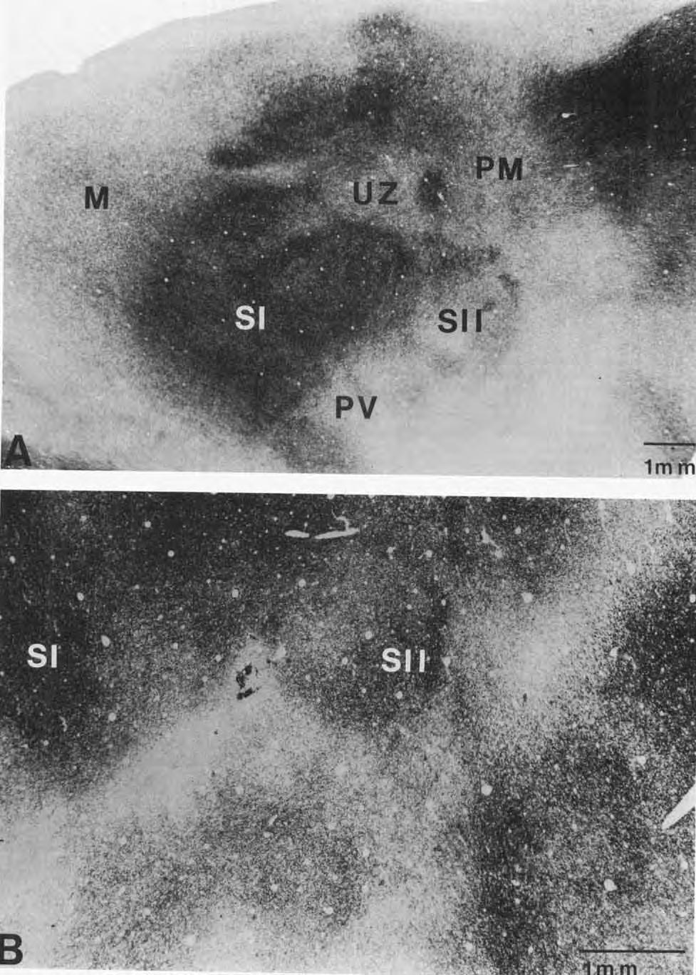
7 SUBDIVISIONS OF PARIETAL CORTEX 409 In addition to those regions of S-I that were systematically mapped, many recording sites were made near S-US- I1 and S-WV borders. Figure 4 illustrates penetrations near the S-US-I1 border in cortex devoted to the upper lip and nose in S-I. With the progression of recording sites across the S-US-I1 border, the receptive fields became large and reversed in somatic sequence to retrace the receptive field locations for neurons in S-I. Figures 6 and 7 illustrate receptive fields for neurons in penetrations extending from the representations of the lip and nose in S-I into PV. Again, receptive fields were smaller for neurons in S-I than those for neurons in PV. As for S-US-11 border, a reversal in the receptive field sequence occurred as recording sites progressed from s-i into PV. The entire physiological representation of the body surface in S-I was found to be coextensive with the myeloarchitectonically identified area described in the previous section, and some body parts could be related to subdivisions of the architectonic field. Thus, after injections of WGA-HRP, label in S-I could be assigned, not only to S-I, but to the representations of body parts within S-I on the basis of myeloarchiteture. S-ZZ The main purpose in mapping S-I1 in detail was to carefully determine boundaries so that injections of tracers could be placed and confined within the field. However, the mapping results also allowed comparisons with previously published maps of S-I1 in squirrels. As for S-I, present findings were in good agreement with previous findings. Figure 4 illustrates penetrations in representations of various body parts in S-11 and their corresponding receptive fields. The physiologically defined S-I1 occupies a small expanse of the cortex (approximately 2 mm x 2 mm) that is adjacent to the nose representation in S-I. Penetrations 1-7 in Figure 4 illustrate receptive fields for a row of recording sites across S-US-11 border. Receptive fields of neurons for progressively caudal electrode penetrations (from S-I into S-11) revealed a clear reversal sequence on the body surface (e.g., penetrations 1-7, Fig. 4).In addition, the size of the receptive fields for S-I1 were clearly larger than those for S-I. S-11 was bounded medially, caudally, and laterally by cortex unresponsive to tactile stimulation. As previously reported, the basic orientation of the representation in S-I1 in the grey squirrel was an upright (noninverted) homunculus. The head was represented rostromedially, and it occupied the greatest portion of S-II. The forelimb was represented caudolateral to the face representation. Within the forelimb representation, the upper arm was represented caudally, and the digits were represented rostrolaterally. Cortex devoted to the trunk was caudolateral to the face and forelimb representations. Within the trunk region, the Fig. 2. Lightfield photomicrographs of sections cut in a plane parallel to the cortical surface illustrating subdivisions of the somatosensory cortex. In A, the heavily myelinated primary somatosensory area, S-I, is surrounded bv less-well-mvelinated motor cortex rostrally and by PM, S-11, and PV caudally. The lightly myelinated oval in the center ofs-1 is the unresponsive zone that serves as a reliable landmark. In more superficial sections, such as in A, S-I1 and PV are less well myelinated than S-I, and borders between the three fields are easily identified. The rostral border of S-II with S-I is not readily apparent in the deeper layers of the cortex shown in B. However, the caudal and lateral borders of S-11 can be distinguished from surrounding regions of the cortex in these sections. PM = parietal medial area. For other abbreviations see Figure 1. In both A and B, rostral is right and medial is up. dorsal trunk was caudal and the ventral trunk was rostral. The representation of the hindlimb was caudolateral to the representation of the trunk. Like the representation of the digits of the forelimb, the digits of the hindlimb were represented rostrolaterally, and the upper hindlimb was represented caudally. The gluteal region and tail were represented in a small amount of cortex in the most caudolateral part of S-11. Figure 4 illustrates some of the results from an extensive map in which receptive fields were determined for 39 electrode penetrations in S-II. The observations on the internal organization of S-I1 described above were based on this and other maps of S-11. Figure 5 illustrates summary maps from two other cases for comparison. The organization of these maps was much like that in the case illustrated in Figure 4, varying somewhat in the amount of cortical space allotted to a particular body part representation. When the proportions of S-I1 devoted to the head, trunk, forelimb, and hindlimb were calculated, not only for the three maps illustrated in this paper (Figs. 4,5), but also for the three cases illustrated in the paper of Nelson et al. ( 78), the head was found to occupy an average of 47% of S-11, the trunk 23%, the forelimb 14%, and the hindlimb 16%. These average values were very close to those obtained for four of the six individual cases. In the two remaining cases, the proportions varied with the head occupying as much as 64% or as little as 38% of S-11. Undoubtedly, this variability reflects difficulties in obtaining complete maps and accurate measures of proportions of body parts represented in S-11. The physiologically defined S-11 was coextensive with the myeloarchitectonically defined S-11 region described above. Therefore, label could be assigned to S-I1 reliably on the basis of myeloarchitectonic criteria. In addition, because the general arrangement of body parts in S-I1 was consistent across cases, transported label found in different locations in S-11 could be related to the representations of body parts with some accuracy. The parietal ventral area, PI.: A complete representation of the contralateral body surface was found within the architectonically distinct PV region. Recordings were obtained from over 100 penetrations in PV in five grey squirrels, and general physiological characteristics were determined. The receptive fields were on the contralateral body surface, although some receptive fields along the midline extended slightly onto the ipsilateral body surface. Receptive field sizes were generally similar to those found for neurons in S-II. However, several differences were detected. For example, receptive fields on the forepaw frequently were restricted to single digits or just the distal portions of all four digits for sites in PV (Figs. 6, 71, while such restricted fields were not found for sites in S-II. Unlike S-I and S-11 neurons, neurons in PV periodically were activated by light stimulation of both the glabrous and hairy surfaces of the skin. The strongest responses were frequently obtained when the skin was lightly scratched rather than tapped, especially for the forepaw and hindpaw. Such enhanced responses to moving stimuli were unique to neurons in PV. Finally, the posterior portion of PV responded to low-threshold aiditog stimulation such as and tapping *gain, this was a unique feature Of pv. Overall, Pv formed an inverted representation of the body, with the digits of the forelimb and hindlimb directed medially, the trunk located ventrally, and the face con- pent with the face representation Of at the upper lip, nose, and teeth representations. The representation of the
 middle to 03) deeper")
8 Fig. 3. Lightfield photomicrographs of sections cut in a plane parallel to the cortical surface illustrating the change in density of myelination in PV as one progresses from (A) middle to 03) deeper cortical layers. In these figures, the rostra1 border of PV is not as easily identified as in more superficial cortical layers depicted in Figure 2. However, the caudal and lateral boundaries of PV are distinguishable. Microlesions (open circles) around PV and A-I allow physiologically determined subdivisions to be related to myeloarchitecture. For abbreviations see Figure 1.
9 SUBDIVISIONS OF PARIETAL CORTEX 411 Fig. 4. Receptive fields for recording sites in S-I and S-11. Upper ler: cortical areas on flattened cortex as revealed in myelin-stained sections. Solid lines mark architectonic subdivisions of the cortex. Dashed lines indicate the location of the recorded region of the cortex shown at greater magnification. Upper right In this magnified view, dots indicate penetrations where neurons responded to light cutaneous stimulation. Hyphens indicate penetrations where neurons were unresponsive to cutaneous stim- ulation. Below: Receptive fields for numbered electrode penetrations in S-I and S-11. Penetrations 1-7 progress across S-US-I1 border. Note the increase in receptive field size as well as the reversal in receptive field progression as the S-US-I1 border is crossed. Lines within S-I1 separate electrode penetrations with receptive fields on the head, neck, and face, on the forelimb, on the trunk, or on the hindlimb. PR = parietal rhinal area.

10 412 L.A. KRUBITZER, M.A. SESMA, AND J.H. KAAS 1.0 mm head n t runk hind I i m b M t c Fig. 5. The somatotopic organization of S-I1 as determined in two other mapping experiments. In each case, the architectonic border of S-II corresponds to the outer boundary of the field. Dots mark electrode penetrations. Electrode penetrations with neurons having receptive fields on the same general body region are indicated. Compare with Figure 4 and note the consistency in basic organization. Also see Nelson et al. ( 79) for three additional maps. Because limbs point ventrally or rostroventrally, the orientation of the homuncular representation in S-I1 has been characterized as erect (see text). forelimb extended mediolaterally in PV, so that mediolateral progressions of recording sites in the forelimb region produced receptive field sequences that moved from distal to proximal on the body (Fig. 6, penetrations 13-17; Fig. 7, penetrations 11-14). Selected progressions of recording sites from the rostra1 to the caudal in PV related to the receptive field sequences from the head to the hindlimb (Fig. 6, penetrations 4-8; Fig. 7, penetrations 6-10). Although similar parts of the face were represented in both PV and S-I along the common border, with cells on either side of S-I/PV border responding to stimulation of the nose, upper lip, and teeth, the physiological boundary between the two fields was clearly discernable due to the dramatic increase in receptive field size with progressions of recording sites from S-I to PV. In most instances, there was also a clear reversal in the receptive field sequence with recording progressions across S-I/PV border (Fig. 6, penetrations 1-3, and Fig 7, penetrations a-g). PV was found to be about in surface area (Fig. 8). About one half of PV appeared to be devoted to the face (45%, case ; 55%, case 85-20). The upper face, vibrissae, and top of the head were represented most medially in PV. These representations were bounded mediocaudally by cortex that was unresponsive to somatosensory or auditory stimulation and were bordered rostrally by the representation of the nose in S-I. The cortex devoted to the nose in PV was immediately lateral to the head and upper face representations. Rostrally, the nose representation was adjacent to the nose representation in S-I, and caudally, it was adjacent to the forepaw representation in PV (Fig. 8). The upper lip representation was found immediately ventral to the nose representation in PV, where it was contiguous with the upper lip representation of S-I. Further caudally, recording sites were found in PV for which receptive fields on the upper lip expanded to include part or all of the lower lip. The cortex caudal to the representation of the lips was devoted to the chin. This cortex was bounded laterally by trunk and caudally by forelimb representations. The teeth were represented in the most ventral portion of the face sector of PV. In most instances, PV was bounded ventrally by cortex unresponsive to tactile or auditory stimulation. The representation of the forelimb in PV was immediately caudal to the representation of the face. The forelimb occupied less than one third of PV (29%, case ; 25%, case 85-20), and was represented from distal to proximal in a mediolateral sequence so that the digits pointed medially in the representation and the wrist and forearm were found more laterally (Figs. 6, 7). As illustrated in Figure 8, the hand and wrist formed most of the forelimb representation. The arm representation, just ventral to the hand and wrist representations, occupied a smaller portion of the cortex. Receptive field sizes in the forelimb varied. For example, receptive fields for the digits were typically small, containing one digit or all five digit tips only, whereas receptive field sizes for the wrist were somewhat larger, and those for the lower and upper arm were larger still. A receptive field for neurons in the arm representation sometimes included part of the upper trunk, the entire arm, and part of the hand (Fig. 6, penetration 17). The trunk was represented in the most ventral tenth of PV (12% in case ; 8% in case 85-20). The trunk adjoined the face representation at the chin and the forelimb representation at the upper arm. For rostrocaudal rows of microelectrode penetrations, the corresponding receptive fields progressed from the upper trunk to the lower trunk (Figs. 6, 7). Because the trunk representation occupied a small amount of the cortex, few penetrations were placed in this part of PV. The receptive field sizes for neurons in the trunk representation were the largest observed in PV. These receptive fields often encompassed an entire upper or lower ventral trunk region and included parts of the limbs (Figs. 6, 7). The trunk representation was bounded ventrally by a region weakly responsive to moderately in-
11 SUBDIVISIONS OF PARIETAL CORTEX 413 Fig. 6. The somatotopic organization of the parietal ventral area, PV. Upper left: a section from the flattened brain with architectonic borders. The dashed box marks the region of the cortex enlarged on the upper right. Within the enlarged view, dots mark electrode penetrations where neurons responded to light cutaneous stimulation. Stars indicate penetration where neurons responded to both light cutaneous and auditory stimylation, while the letter A denotes a penetration where only auditory responses were detected. Hyphens mark penetrations where neurons were unresponsive to cutaneous or auditory stimulation. Receptive fields on the squirrel bodies below are numbered to correspond to numbered penetrations on the brain on the upper right. Receptive field 14 was on both dorsal (hairy) and ventral (glabrous) aspects of the forepaw. Note that the receptive fields for neurons in PV are larger than those for neurons in the S-I. Also, distal limbs are represented dorsally and proximal limbs ventrally in PV, so the orientation of the homuncular representation can be characterized as inverted (see text). In this and other figures, receptive fields extending past the outlines of the body surface indicate that the receptive field continues onto skin not seen in the view drawn (e.g., 6,9,10) or includes discontinuous skin surfaces (e.g., 14). Abbreviations as in previous figures.
12 414 L.A. KRUBITZER, M.A. SESMA, AND J.H. KAAS Fig. 7. The organization of the parietal ventral area, PV, in another case. Compare with Figure 6. Note the increase in receptive field sizes and the caudalward reversal of the receptive field progressions as recording sites progressed from S-I to PV (see receptive fields for sites a-g). Conventions as in Figure 6.

13 SUBDIVISIONS OF PARIETAL CORTEX 415 Fig. 8. Details of the somatotopic organizations of PV in the experiments illustrated in Figures 6 and 7. Lines in PV segregate groups of electrode penetrations whose neurons have receptive fields on the same designated body part. In each map, a region of three or four penetrations was found where neurons had no cutaneous response (NCR). Although the two maps differ in detail, the overall organization is that of an inverted homunculus with the digits and toes directed medially and the trunk located ventrally. tense auditory stimulation and dorsally by the hindlimb representation. The representation of the hindlimb, slightly over one tenth of the total area of PV (14%, case ; 11%, case 85-20), was immediately caudal to the representation of the forelimb and adjoined the lower trunklgenital representa- tion laterally. Like the representation of the digits of the forepaw, those of the hindpaw were directed medially. The distal hindlimb was represented medially, and the proximal hindlimb was represented laterally. The hindlimb representation was bounded caudally by the cortex that was responsive to low-threshold auditory stimulation (Fig. 9). Auditory responses in P K In addition to the neuronal responses to somatic stimuli in PV, responses to auditory stimuli were observed in certain parts of PV as well (see starred penetrations in Figs. 6, 7, 9). Neurons in PV responded to clicks and sounds produced by tapping objects together. However, these neurons were not highly activated by pure tones, and best frequencies were not determined. The extent of the cortex responsive to both somatosensory and auditory stimulation varied from case to case. For instance, case (Fig. 6) had 19 electrode penetrations in which both auditory and somatosensory responses were recorded. In this case, the entire hindlimb representation, half of the trunk and forelimb representations, and part of the face representation were responsive to both somatosensory and auditory stimulation. Case (Fig. 6), on the other hand, had only three somatosensory/auditory penetrations, one in the hindlimb and two in the trunk. While responses to auditory and somatosensory stimuli were recorded in the same penetrations, the best responses to either somatosensory or auditory stimuli were obtained from different cortical depths. The best somatosensory responses were from depths of pm, while the best auditory responses were from 900-1,200 pm from the pial surface. Recordings were obtained from neuron groups that were activated by both auditory and somatosensory stimuli, but it was not certain that individual neurons responded to both stimuli. Cortex lateral and caudal to PV was responsive to auditory but not to somatosensory stimuli (Fig. 9). The region that was responsive to auditory stimuli included A-I and the parietal rhinal area (PR). A-I is easily identified by a characteristic tonotopic organization and neurons selective for best frequencies (Merzenich et al., 76; Luethke et al., 85), as well as by dense myelination (Fig. 3B). The PR (parietal rhinal) region includes the rostra1 auditory area (Merzenich et al., 76), which represents frequencies in a reverse pattern from that in A-I, and adjoining auditory cortex. We found that neurons in the PR region rapidly habituated to repeated auditory stimuli. Cortical connections of S-I Injections of WGA-HRP were placed in different parts of the primary somatosensory cortex in four grey squirrels. In two cases a single injection was placed just lateral to the unresponsive zone (UZ) in the representation of the upper lip and part of the lower lip (not shown). In another case, a single injection was placed in the upper liphose representation (Fig. 11). The dense injection core (about 1.0 mm wide) also included the lateral margin of UZ and some of the representation of the vibrissae. In the final case, three small injections were placed in a mediolateral row in part of S-I just medial to UZ. This cortex was devoted to the shoulder and forearm representations (Fig. 12A). In all cases the injection was completely confined to S-I. All four cases demonstrated a local pattern of intrinsic connections within S-I. The intrinsic pattern was most extensive in case where the injection core included the lateral margin of UZ (Fig. 10A). In this case, labeled cells

14 416 L.A. KRUBITZER, M.A. SESMA, AND J.H. KAAS 0 I \ SII A* A' 9 A' A*.- A" +I 0 A' - 0.5mm Fig. 9. The physiological response properties of neurons in PV and surrounding regions of the cortex. Above: architectonic subdivisions of cortex on a section parallel to the surface of the flattened cortex. Somatic and auditory areas of interest are located, and the region of auditory and somatosensory overlap is crosshatched. Below: an enlarged view of the region enclosed by a dashed box above. Solid lines indicate architectonic borders. As mark electrode penetrations in primary auditory cortex where neurons responded consistently to repeated clicks and tones. A*'s mark sites outside of A-I where responses to repeated auditory stimuli habituated. Dots mark somatosensory sites, while circles filled with stars indicate penetrations where neurons were activated by both auditory and light tactile stimuli. Hyphens are sites that were unresponsive to somatosensory or auditory stimuli, while 0's are sites marked by electrolytic lesions. Note the good correspondence between physiological and architectonic borders (within 200 pm). Other conventions as in previous figures. and terminals surrounded the injection core and filled UZ. In addition, a rostral extension of intrinsic label was located along the lateral margin of the hand representation in lightly myelinated cortex extending from UZ. Other smaller foci of intrinsic label were in the medial cortex devoted to the forearm and forepaw representations. Both diffuse label suggesting anterograde transport and labeled cells indicating retrograde transport were found lateral to the injection site in the cortex devoted to the lips. The two cases with the injection in the representation of the upper and lower lips (not shown) also had a zone of intrinsic label around the dense injection core. Some of this adjoining label extended laterally toward the representation of the oral cavity, rostrally into the cortex representing the lower lip, and medially into UZ. A rostral locus of label was in the less myelinated cortex along the lateral margin of the representation of the forepaw. The case with injections medial to UZ (Fig. 12) had intrinsic connections around the injection sites, several small foci of label rostral to the injection sites in parts of S-I devoted to the arm, trunk, and leg, and a focus extending rostrally along the lateral margin of the forepaw representation. In this case, very little intrinsic label was found in UZ. Taken together, these four cases indicate that intrinsic connections are likely to be distributed in a fairly uniform manner along the margins of the injection sites, in several separate small foci in the cortex devoted to related body parts (other parts of the face for face injections, other parts of the body for body injections), and somewhat densely in UZ and the less myelinated cortical strips that extend from UZ (Fig. 1). However, it is important to note that the lateral injections in the representation of the face, but not the medial injections in the representation of the forelimb, resulted in label in UZ. Thus, it appears that some body parts in S-I are interconnected with UZ while others may only relate to the smaller, lightly myelinated zones, jutting out from UZ. Cases with S-I injections also demonstrated reciprocal patterns of ipsilateral and contralateral cortical connections with other fields. The ipsilateral connections were with S-11, PV, PM, PR, and motor cortex (Figs. 10, 12). In addition, the locations of the foci of label varied within these fields in a manner suggesting that most connections are somatotopically matched. In cases (Figs. 10, 11) and (not shown) injections in lateral S-I devoted to the face labeled parts of PV and S-11 that adjoin S-I and that also represent the face. Other label was in the lateral motor cortex where electrical stimulation evokes face movements (unpublished experiments). Within the parietal medial strip of the cortex (PM) immediately caudal to S-I, reciprocal connections were lateral, just medial to S-11, suggesting that the face is represented in lateral PM. The injections in medial S-I, where the forelimb is represented, produced label in caudolateral parts of S-I1 and caudomedial parts of PV known to represent the limbs. In addition, anterograde and retrograde label was found in the medial part of the motor cortex where limb and trunk movements are evoked and in a more medial part of PM. Thus, the connection patterns of S-I with M, S-II, and PV, where the somatotopic patterns of representations are known, were at least roughly homotopic. In contrast, foci in PR did not vary much according to the placement of the injection in S-I, and no somatotopic pattern was evident. Of the ipsilateral targets of S-I, the connections to PV appeared to be slightly more dense than those of S-11, PR, and motor cortex, while connections to PM were less dense. Foci of connections were distributed over large portions of motor cortex, and they varied in density. Foci in PV and S-I1 were smaller, but they occupied proportionately equivalent amounts of these representations. Callosal connections were with parts of S-I representing body parts equivalent to that of the injection site (Figs. 10B, and 12B). Thus, the lateral cortex representing one side of the face projected to the cortex representing the other side of the face in the opposite S-I, and the medial cortex repre-
15 417 A B Fig. 10. Cortical connections of S-I. An injection of WGA-HRP (black circle) was placed in the representation of the upper lip. The injection core extended slightly into the UZ. Drawings include label from several sections imposed on single brain sections from the injected (A) and contralateral (B) hemispheres. Retrogradely labeled cell bodies and presumptive anterograde label are indicated by course and fine dots, respectively. Other abbreviations are as in previous figures.
16
.")
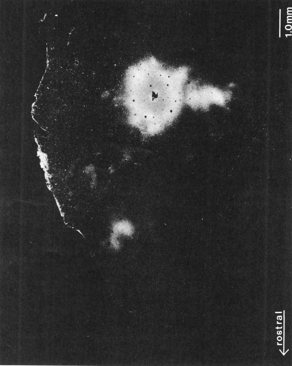
17 SUBDIVISIONS OF PARIETAL CORTEX senting the forelimb projected to the medial cortex devoted to the other forelimb. The three medial injections produced only two light foci in the contralateral S-I (Fig. 12B). This is in agreement with previous evidence that callosal connections of the forelimb in s-i are light and unevenly distributed (Gould and Kaas, 81). The injection that involved the lateral margin of UZ produced dense label in the contralateral UZ as well as in PV, PR, S-11, and PM, and motor cortex. The more extensive pattern of callosal connections involving areas in addition to S-I was not seen in the other three cases where the injection did not include part of UZ. Thus, UZ may be a specialized part of S-I with more extensive callosal connections. Cortical connections of S-I1 Two grey squirrels were used to determine the cortical connections of the second somatosensory area, S-11. One injection was placed in the shoulderheck representation of S-11 (not shown), while the other was centered on the forelimb and included parts of the face and trunk representations in S-11 (Fig. 13A). Although both injections were restricted to S-11, the injections filled much of the area so that the full extent of local connections within S-11 was not determined. However, the spread of intrinsic label immediately around the injection site was apparent in each of the two cases. In addition to the intrinsic connections, the results revealed reciprocal connections of S-11 with other somatosensory areas. Despite the proportionately large injection sites, there was evidence from these cases that connections are somatotopic. In both cases, dense anterograde label was found in more superficial sections, suggesting feedback from S-11 to S-I. Reciprocal connections were observed in both cases with representations in S-I that corresponded to those injected in S-II. For example, the single injection in Figure 13A was concentrated in the forelimb, trunk, and face representations in S-11, and the labeled regions in s-i related to the same body parts. The most medial patch of labeled cell bodies and terminals in S-I (Fig. 13A) corresponded to the trunk representation. Cortex in S-I devoted to the forelimb, lateral to the cortex devoted to the trunk, also contained a rather dense collection of labeled cells and terminals. Finally, the cortex in S-I representing face regions adjacent to the face representations in S-11 was labeled both anterogradely and retrogradely. In the case where the injection was centered on the shoulder and neck portion of S-II, label was found in the shoulder and neck portions of S-I. Connections with PV also appeared to be somatotopically matched. In Figure 13A, the rostromedial patch of label in PV corresponds to the upper face representation, the patch of label caudal to this corresponds to the hand representation, and the most lateral focus of label is in the expected Fig. 11. A darkfield photomicrograph of an injection site in S-I and transported label in a section from the flattened cortex reacted for HRP, case (see Fig. 10). The dark center of the bright circle is the damage resulting from the injection pipette. The central gray circle is the dense label of the injection, while the white fringe around the gray indicates local transport of the label. Patches of white immediately medial and rostral to the injection site consist of labeled cell bodies and axons in other parts of S- I. The large puff of white label immediately ventral to the injection site is in PV. The dense white patch of label in the rostral pole of the cortex is in the motor cortex. Compare with Figure location of the trunk representation. The interconnections of S-I1 and PM were in agreement with the hypothesis that the face is represented laterally and the body medially in PM. Thus, the most medial part of PM was not labeled, while intermediate and lateral levels were labeled after the face, forelimb, and trunk injections in S-I1 (Fig. 13A). Finally, in both cases of S-I1 injections, label was found in the band of cortex encircling S-I1 caudolaterally. In case 84-95, some additional label was located rostral to S-I on the medial wall just medial to motor cortex. Label was also found in PR. In both cases, the most dense ipsilateral connections of s-i1 were with s-i. However, these connections were only slightly more dense than those with PV, PR, or PM. In both cases with an S-11 injection, dense label was observed in the portion of S-11 of the contralateral hemisphere that closely matched the dense injection core in location. Thus, the representation of a particular body part in S-II in one hemisphere appears to be reciprocally connected to the representation of the same body part in the contralateral hemisphere (Fig. 13B). The callosal connections in the SII formed several clumps or patches. The only other contralatera1 connections observed were with the area of the cortex encircling S-11, and these were seen in only one (not shown) of the two cases. Like the ipsilateral cortical connections with this S-11 bordering region, labeled cell bodies and terminals formed small patches. Cortical connections of PV Injections of WGA-HRP were confined to PV in three cases. These cases demonstrated local intrinsic connections and reciprocal ipsilateral connections with S-11, S-I, PM, motor cortex, PR, the cortex just dorsal to the rhinal fissure, and possibly subdivisions of the auditory cortex. Callosal connections were with PV and adjoining regions of the cortex. As for S-11, the injections were large relative to the size of PV, but there was some evidence that most connections were somatotopically matched. Considering both distribution and density, the major interconnections of PV were with s-i (Figs. 14A, 15A, 16A). The anterograde label was most apparent in superficial sections, suggesting a feedback pathway (see Maunsell and Van Essen, 83). After a large injection that included portions of the face, forelimb, and hindlimb representation in PV, label was scattered over similar representations in S-I (Figs. 14A, 15A). The label in S-I partly included UZ. Other locations of label were in a broad band just medial to UZ where the forelimb is represented, and more medially where the foot and hindlimb are represented. The gap in the label in medial S-I may correspond to the trunk representation, which was probably outside the dense injection core in PV. Additional foci of label were in caudolateral S-I that is devoted to the upper lip and face, while rostrolateral S-I, devoted to the lower lip and jaw, was relatively free of label. A more restricted injection in PV, centered in the portion representing the forelimb, resulted in label largely confined to the portion of S-I representing the forelimb and the medial part of UZ (Fig. 16A). A third case with an injection in the forelimb portion of PV produced similar results. Other major connections were between PV and the motor cortex, M. The two cases with injections in the forelimb region of PV resulted in concentrations of labeled cells and terminals in the medial portion of M where the forelimb is represented, while the large injection in PV produced
18 420 L.A. KRUBITZER, M.A. SESMA, AND J.H. KAAS A Fig. 12. Ipsilateral (above) and contralateral (below) cortical connections of S-I. Three restricted injections of WGA-HRP were placed in the forelimb representation in the S-I. Solid lines indicate architectonic borders while dashed lines mark uncertain borders estimated by position. Short, irregular lines are labeled axons. Compare with Figure 10 and note the sparseness of callosal connections when UZ is not involved in the injection site. Other conventions as in Figure 10.
19 SUBDIVISIONS OF PARIETAL CORTEX 421 A Fig. 13. Ipsilateral (above) and contralateral (below) cortical connections of S-II. An injection of WGA-HRP into the forelimb and face representation. Open circles with L s mark lesions placed just within the physiological limits of S-II. Solid lines indicate architectonic boundaries. Conventions as in previous figures. See Figure 5 for the somatotopic map of S-II for this case.
20 422 L.A. KRUBITZER, M.A. SESMA, AND J.H. KAAS A Fig. 14. Ipsilateral (above) and contralateral (below) connections of PV. An injection was placed in the representation of the face and forelimb. Microlesions (circles with L s) were used to mark the physiological borders of PV, ER = entorhinal cortex; OF = the orbitofrontal cortex. Other conventions as in previous figures.
. Note the dense white label ventral to the injection site in PR. B. The left cerebral hemisphere.")
21 SUBDIVISIONS OF PARIETAL CORTEX 423 Fig. 15. Darkfield photomicrographs of brain sections reacted for HRP after WGA-HRP injection in PV (case 85-66, compare with Fig. 14). A. The right cerebral hemisphere with the injection site (dark core with white halo). Note the dense white label ventral to the injection site in PR. B. The left cerebral hemisphere. The dense label is in PV while less dense label is in the PR. Abbreviations as in previous figures.
22 424 L.A. KRUBITZER, M.A. SESMA, AND J.H. KAAS A B ANTE ROGRADE Fig. 16. Ipsilateral (above) and contralateral (below) connections of PV. An injection was placed in the forelimb representation. Conventions as in previous figures.

23 SUBDIVISIONS OF PARIETAL CORTEX clumps of label scattered over more of M. Similarly, the more restricted forelimb injections in PV resulted in more restricted label in S-I1 in a centered location (Fig. 16A) that closely matches the expected location of the forelimb representation (Fig. 5). The restricted PV injections produced label in middle mediolateral levels of PM, consistent with the proposed somatotopic organization of PM. Surprisingly, the large injection in PV resulted in relatively little label in PM. However, a dense focus of label just caudal to S-II raises the possibility that PM extends laterally to the caudal margin of S-11, where parts of the head may be represented in PM. Very dense concentrations of label were found just lateral to PV in part of PR. There was no evidence of a topographic pattern, and the concentration of label in only part of PR suggests that the field is functionally heterogeneous. The two restricted PV injections produced zones of superficial anterograde label that extended caudally to include rostral A-I. The larger PV injection resulted in both labeled cells and fine, scattered label in A-I. In addition, label was apparent in several locations in TA (Fig. 16A). In two of the three cases of PV injections (Figs. 14A, 16A), label was also found in a band along the rhinal fissure. The orbitofrontal cortex (OF) and the entorhinal cortex (ER) were sparsely labeled in one case Fig. 14A), and sparse scattered label was inconsistently observed in several other locations across the three cases. The callosal connections of PV (Figs. 14B, 15B, 16B) were more extensive than those observed for S-I or S-II. Label was scattered over much of the contralateral PV, but there was a clear suggestion that it was most concentrated in the part of the contralateral PV that matched the injection site. Dense callosal connections were also noted in the portion of PR that roughly matched the portion with the dense ipsilateral connections. Two of the three cases had sparse scattered label in s-i1 (Figs. 14B, 16B). In case with the large injection, callosal label was also apparent in PM and the cortex caudal to S-11 (Fig. 14B). In addition to these results, the present study provided information on the subcortical connections of the somatosensory cortex. These results will be described in a subsequent paper (Krubitzer and Kaas, in preparation). DISCUSSION In the present study, electrophysiological and anatomical procedures were combined in the same animals to reveal subdivisions, somatotopic patterns, and processing sequences in somatosensory cortex of squirrels. The most important discovery was of a previously unknown somatosensory cortical representation that we termed by location the parietal ventral area (PV). Thus, squirrels, and perhaps other rodents and other mammals, have at least three systematic representations of the body surface in the parietal cortex: the primary somatosensory cortex (S-I), secondary somatosensory cortex (S-II), and PV. Furthermore, similarities in the S-I1 and PV in relative size, responsiveness to somatosensory stimuli, connections, and position along the caudal border of S-I suggest that if both fields exist in other mammals, they could have been confused in individual experiments. An unusual feature of PV is that it also contains neurons that respond to auditory stimuli. Finally, connections of S-I, S-11, and PV identify two other regions of parietal cortex with dense somatosensory inputs: the parietal medial area (PM), which is medial to S-11, and the parietal rhinal area (PR), which is ventral to PV. Thus, at least five interconnected cortical fields in squirrels may be 425 predominantly somatosensory in function. These results and conclusions are discussed further below. The organization and connections of S-I Although the microelectrode mapping results on the organization of S-I in squirrels were quite limited in the present study, results were consistent with the previously reported organization (Sur et al., 78). As in S-I of other mammals (Kaas, 83), S-I in the squirrel is standing on its head so that a large lateral sector of the cortex represents the head, the more medial cortex is devoted to the forelimb, while the trunk and hindlimb are represented most medially. A striking feature of S-I in squirrels is the occurrence of a 1-mm oval of the cortex centered in S-I that is unresponsive to light cutaneous stimuli in the typical anesthetized preparation (Sur et al., 78). While most of S-I has a welldeveloped layer IV that is tightly packed with granule cells, UZ zone has a less distinct layer IV. A contribution of the present study was to show how distinctive the unresponsive zone, UZ, is in surface-view brain sections stained for myelin. These sections not only clearly demonstrate UZ as a lightly myelinated oval surrounded by a densely myelinated S-I, but also show two lightly stained bandlike rostral extensions of UZ, the most medial of which reaches the motor cortex and separates the forelimb from the face representation. In rats, a similarly located dysgranular unresponsive zone separates forelimb and face representations and has rostralward extensions to the motor cortex (C. Welker, 76; Akers and Killackey, 78; Chapin and Lin, 84; Welker et al., 84). Much earlier, Krieg ( 46) illustrated an oval of architectonically distinct cortex within S-I region of rats (distinguished as 2a within a large area 2 ) that may have been UZ. Given that squirrels and rats are on quite different branches of rodent evolution, separating perhaps 60 million years ago (e.g., Eisenberg, 81), it is likely that this UZ was present in the common ancestor, and we expect that UZ zones and bands exist in many other rodents as well. S-I of rodents appears to be homologous with area 3b of primates (Kaas, 831, but it is not certain whether UZ should be considered part of S-I, or part or all of another field within and perhaps around S-I (see Akers and Killackey, 78). Cortical connections of S-I in squirrels are summarized in Figure 17. There are widespread intrinsic connections within S-I including major connections from granular S-I to UZ. UZ, in turn, projects strongly to UZ of the other hemisphere, the motor cortex, as well as to other areas. Similar connections of the dysgranular UZ zone within S-I have been previously described by Gould ( 81) for squirrels and Akers and Killackey ( 78) for rats. Akers and Killackey ( 78) further postulated that the dysgranular UZ within S-I was part of a more extensive system including the dysgranular bands seen extending from UZ and dysgranular fringes of cortex along the rostral and caudal borders of granular S-I. Although the lightly myelinated UZ in squirrels has two lightly myelinated rostralward extensions that probably correspond to the dysgranular bands in rats, architectonic, connectional, and microstimulation (unpublished) data suggest that the moderately myelinated cortex bordering s-i rostrally in squirrels is the motor cortex, and the cortex immediately caudal to S-I contains several somatosensory areas (PV, S-11, PM) that are unlike UZ. The dense callosal connections of UZ in squirrels also have been noted in a description of the total pattern of callosal projections (Gould and Kaas, 81). In addition, a
 and mice (White and DeAmicis, 77), but there have been no architectonic or electrophysiological identifications")
24 426 L.A. KRUBITZER, M.A. SESMA, AND J.H. KAAS IPS1 - CONTRA- S-I in rats (Akers and Killackey, 78) and mice (White and LATERAL LATERAL DeAmicis, 77; Porter and White, 83) are to the motor cortex. Caudal projections of S-I also have been described for rats (Akers and Killackey, 78) and mice (White and DeAmicis, 77), but there have been no architectonic or electrophysiological identifications of projection zones. Some caudalward projections have been attributed to S-11, largely - on the basis of position, and some to a caudal dysgranular fringe. Present results suggest that the most medial of these projections are to PM, while other projections could be to any or all of S-11, PV, and PR. --I, I SI The organization and connections of S-I1 I The somatotopic features and architectonic characteristics of S-I1 in squirrels have been previously described by.-+-i Nelson et al. ( 79). We remapped the field in comparable detail in three squirrels (Figs. 4,5), and the combined reports now show head, forelimb, trunk, and hindlimb regions I of S-I1 for a total of six squirrels. In all cases, the head was represented along S-I border with the forelimb, trunk, and hindlimb following in a caudolateral cortical progression. M I The border of S-I1 with S-I was congruent so that similar CL--- I, /# I parts of the head midline were represented along the com- U mon border. The S-I1 representation was found to be more I / somatotopic and less disrupted than S-I (Sur et al., 78), so that the nose and lips, forepaw, ventral trunk and hindpaw were all rostrolateral, respectively, to the eye and cranium, forearm, dorsal trunk and thigh. When drawn relative to i, S-I on a lateral view of the brain, the body representation in S-I1 appears to be standing on its feet in a roughly normal position (Fig. 18). Such an orientation for S-I1 has been characterized as erect (Nelson et al., 79). While S-11 was first characterized as inverted in orientation (Woolsey, 52, 58; Lende and Woolsey, 56; T. Woolsey, I PR 67; Pinto Hamuy et al., 56; Zeigler, 641, S-I1 subsequently has been described as erect in cats (Haight, 72; Burton et al., 821, raccoons (Herron 78), sheep (Johnson et al., 74), and some rodents (Campos and Welker, 76; Pimentel-Souza et al., 80) including squirrels (Nelson et al., 79). Present results support the conclusion that S-11 in squirrels is erect, and also indicate that another somatosensory area, PV, is largely, but not completely, inverted in orientation. This raises the issue of whether or not S-I1 and PV have sometimes been confused. Early reports on the organization of Fig. 17. Major processing sequences in the somatosensory cortex of squirrels. Arrows indicate the probable feedforward projections. Thick arrows mark major projections and dashed lines indicate sparse projections. See text for details. MI, the primary motor cortex; S-I, the first somatosensory area; UZ, the unresponsive zone of S-I; S-11, the second somatosensory area; PM, the parietal medial area; PV, the parietal ventral area; PR, the parietal rhinal cortex; ER, the entorhinal cortex. band of cortex between UZ and the motor cortex, corresponding to the medial band of light myelination in the present study, was shown to have dense callosal connections. Granular S-I of squirrels also projects callosally, and the study of Gould and Kaas ( 81) shows that these connections are extremely variable in density according to the body region in the representation. In the same hemisphere, granular S-I of squirrels projects rostrally to the motor cortex and caudally to PM, S-11, PV, md PR. The most dense connections are with PV and the least dense are with PM. Similarly, rostra1 connections of S-I1 in rodents were based on recordings from large surface electrodes that reflected the activity of relatively large portions of the mapped fields, and it is not clear from the illustrated data if the somatotopic organization of the mapped fields had the erect orientation of PV or the inverted orientation of s-ii. However, C. Welker and Sinha ( 72) used microelectrodes to explore the somatosensory cortex in rats and described S-11 as inverted. More recently, Pimentel-Souza ( 80) also used microelectrodes to map S-11 in another rodent (aguoti), and, in contrast, found S-11 to be upright with an overall organization much like that of S-I1 in squirrels. Possibly the study of C. Welker and Sinha ( 72) involved PV, rather than S-11, since the illustrated location of S-11 in rats was in the relative position of the PV in squirrels. In addition, the orientation of S-I1 was described as inverted as in PV. Other than the present report, there have been no studies of connections with lesions or injections confined to S-I1 in rodents. However, some information is available on S-I1 connections from injections of tracers into S-I or the motor cortex in mice and rats. Our studies show that S-I1 in

25 SUBDIVISIONS OF PARIETAL CORTEX Fig. 18. A schematic portrayal of basic features of somatotopic organization of SI,S-II, and PV in the traditional homuncular format. S-I is organized with the body in the cortex dorsal to the cortex devoted to the head, so that the general orientation is of an animal standing on its head. In both S-II and PV the representations of the head adjoin S-I so that the face is rostra1 to the cranium. Thus, both representations of the head are in a roughly erect orientation. However, S-II and PV differ in that the limbs point ventrally in S-II and dorsally in PV. Thus, S-I1 can be characterized as erect in orientation, while the body in PV is inverted. Rostra1 is right, dorsal is upper. squirrels is interconnected ipsilaterally with S-I, PV, PM, and PR (Fig. 17). It was surprising that no obvious connections with the motor cortex were observed, since such connections have been described in mice (Porter and White, 83), rats (Donoghue and Parham, 83), cats (Jones and 427 Powell, 68; Kawamura and Otani, 70; Burton and Kopf, 841, and monkeys (Jones and Powell, 70; Pandya and Vignolo, 71; Jones et al., 78). However, the evidence for motor connections of S-II in monkeys can be questioned, since it is not certain that S-11, rather than a bordering field (see Robinson and Burton, 80b), is the source of the connections. This possible confusion seems less likely in cats, where S-I1 has been mapped in some detail and S-I1 can be located relative to fissure patterns (Haight, 72; Burton et al., 82). However, Burton and Kopf ( 84) noted that the density of label in the motor cortex of cats after S-II injections was much less than that suggested by earlier studies (Jones and Powell, 68; Kawamura and Otani, 70). Finally, the reported connections between the motor cortex and S-II in mice (Porter and White, 83) are based on the presumptive location of S-11 and could reflect connections of other fields. Thus, connections between S-11 and the motor cortex may be sparse or absent in other mammals as well as squirrels. Interconnections of S-I with S-11, as demonstrated in squirrels, appear to be a common feature of the mammalian cortex (see Burton and Kopf, 84, for review). Akers and Killackey ( 78) report S-I connections with the presumed location of S-I1 in rats, but it seems quite possible that the described projections include other regions of cortex. In general, many of the connections of physiologically identified S-I1 in cats (e.g., Burton and Kopf, 84) appear to be quite similar to those of S-I1 in squirrels, suggesting that shared features will be found in a range of mammalian species. Thus, S-11 in cats and squirrels projects to more medial cortex just caudal to S-I (PM in squirrels and area 5 in cats) and to two areas just lateral to S-I1 (the areas PV and PR in squirrels and S-IV and insular cortex in cats). Cases with injections that were centered in S-11 of squirrels demonstrated major interconnections with central regions of S-II of the opposite hemisphere. Furthermore, these cases show that in squirrels the major source of callosal connections to S-II is from S-II, although some input is from PV. The input from PV appeared to involve parts of S-I1 devoted to distal limbs. Previously, callosal input to S-11 in squirrels was shown to be uneven in density, but to include all major body parts in the representations, even the distal limbs (Gould and Kaas, 81), and present results are consistent with this conclusion. Investigators typically have concluded that parts of s-11 devoted to the distal limbs in rodents and other mammals are acallosal (see auld and Kaas, 81, for review), but this conclusion appears to be based on relatively insensitive procedures for revealing connections and on an assumption about the location of S- 11 and the body parts in S-II. Recently, there has been evidence that the portion of S-11 in monkeys responding to stimulation of the hand has callosal connections (Manzoni et al., 84). The organization and connections of PV The parietal ventral area forms a second systematic representation of the body surface that adjoins the larger s-i representation along the caudal border of S-I devoted to the face. PV has not been previously described as an area distinct from S-11 in rodents, although the lateral location and inverted organization described for S-11 in rats by C. Welker and Sinha ( 72) appears to more closely correspond to that of PV rather than SII.The arrangement of body parts in PV does not form a simple homunculus as in S-II. The representation of the face and upper head in PV forms
.")
26 428 L.A. KRUBITZER, M.A. SESMA, AND J.H. KAAS a congruent (see Allman and Kaas, 75) or matched border with S-I, with more rostra1 skin on the head midline represented more medially in both S-I and PV. Thus, the representation of the head in PV is in the erect orientation (Fig. 18). In contrast, the trunk is represented laterally, the proximal limbs medial to the trunk, and the distal limbs most medially in PV. Thus, the body is in the inverted orient ation. In squirrels, PV is interconnected ipsilaterally with the motor cortex, S-I, S-11, PM, PR, and the entorhinal cortex (ER). Callosal connections are relatively widespread and include PV, S-11, and PR. Since PV is a newly defined area in squirrels, connections of PV have not been described as matic and auditory fields. However, microelecrode recording studies in cats indicate that s-iv, the cortex caudal to S-IV (Clemo and Stein, 83), and the cortex caudal to S-11 (Cameras and Anderson, 63) are activated by both somatic and auditory stimuli. In monkeys, retroinsular, granular insular, and adjacent portions of area 7b are responsive to auditory as well as somatosensory stimuli (Robinson and Burton, 80b). Cortical processing of somatosensory information in rodents: A distributed system The evidence indicates that squirrels have at least three such in other rodents. However, some of the projections of systematic representations of the body surface and at least S-I that have been attributed to S-I1 in rats (Akers and five interconnected somatosensory areas in the parietal Killackev, 78) may have actually been to PV. In addition, region of the cortex. The cortical system in squirrels, al- projections to the motor cortex described as arising from S- I1 in mice (Porter and White, 83) and rats (Donoghue and Parham, 83) are more probably from PV, since in squirrels, PV, rather than S-11, is strongly connected with motor cortex. PV possibly exists in other mammals as well. In the opposum, the ventral location and the responsiveness to auditory as well as somatic stimuli suggest that S-11 (Pubols, 77) may correspond to PV. The fourth somatosensory area of cats is very similar to PV. In cats, S-IV has been described as a somatotopic representation of the body on the lateral border of the S-11 (Clemo and Stein, 82, 83; Burton and Koph, 84). Like PV, S-IV appears to contain an inverted representation, and some neurons respond to auditory stimuli. In addition, both S-IV and PV project to more lateral cortex and to the cortex along the caudal border of S-I. However, S-IV does not appear to have connections with s-i and MI, as does PV. Thus, it is not certain if PV and S-IV are the same area with somewhat different patterns of connections, or if they are different areas in the same position relative to S-11. Further comparisons of subcortical connections of s-iv (Stein et al., 83) with those of PV may help answer this question. In monkeys, several body surface representations have been described in the S-I1 region (Robinson and Burton, 80b), and it is possible that one of these fields, perhaps the retroinsular area on the border of S-I1 proper, is the homologue of PV. However, there is little information on the connections of the retroinsular area to test the validity of this hypothesis (see Friedman, 83). At least part of PV in squirrels was bimodal, containing neurons responsive to somatic stimuli and neurons responsive to auditory stimuli. The somatosensory activation dominated, so that neurons throughout PV were easily activated by light touch, while neurons in only some penetrations were driven by auditory stimuli. In general, the best auditory responses were recorded at somewhat deeper recording depths than the best somatosensory responses, and the auditory responses were most often obtained in penetrations near auditory cortex in parts of PV representing the body rather than the head. While the significance of these observations on the distribution of auditory responses in PV is uncertain, responsiveness of the cortex in the general region of PV to both somatic and auditory stimuli has been reported for a number of mammals. The overlap of S-I1 with the auditory cortex described in studies using surface recordings in rodents (Lende and Woolsey, 56; Woolsey, 58; T. Woolsey, 67) could reflect the mixed responsiveness of PV, or volume conduction from separate but adjacent so- though apparently somewhat simpler, is remarkably similar to that described for monkeys and cats where multiple somatosensory areas with complex interconnections have been described (see Merzenich and Kaas, 80, for review). Aside from S-I and S-11, which appear to exist in all eutherian mammals (see Kaas et al., 83), it is not certain which, if any, additional components of the cortical system in squirrels also exist in other mammals. Thus, there is uncertainty about the extent to which the similar systems in rodents, cats, and monkeys were already present in a common ancestor or evolved independently. Nevertheless, it appears that mammals with relatively simple brains, such as squirrels, and mammals with more complex brains, such as cats and monkeys, all process somatosensory information in a basically similar manner. In each of these mammals, a number of somatotopically organized cortical fields are so complexly interconnected that processing hierarchies can be only broadly defined. All three mammals also have more than one cortical field that receives sensory information over a rather direct route from the thalamus. These connections suggest that functions are distributed in the cortical system, and that sensory events activate neurons almost simultaneously in a number of cortical fields (see Merzenich and Kaas, 80). Yet, a semblance of a hierarchy is embedded in this distributed cortical system of squirrels (Fig. 17). Laminar patterns of connections, which have been classified into feedforward and feedback types (e.g., Maunsell and Van Essen, 83), have been useful in constructing hypothetical processing hierarchies. While our preparations were not ideal for an analysis of laminar patterns, both S-I1 and PV clearly provided widespread feedback to superficial layers of S-I. Thus, S-I may be considered the first level in a cortical hierarchy of processing, relaying information to UZ, PM, S-11, PV, and PR, although these fields may also receive more direct sensory information from the thalamus. Further steps in a hypothetical processing hierarchy are blurred by the manifold connections, but there may be reasons to consider some areas such as PR and PV as higher than S-II and PM on the basis of outputs to perirhinal and limbic structures. In monkeys, Mishkin and co-workers (e.g., Mishkin, 79; Turner et al., 80; Mishkin et al., 84; also see Pandya and Seltzer, 82) have argued that cortical fields with inputs into memory mechanisms in the limbic system form the final stages of cortical processing that relate to object identification. Thus, the somatosensory system of rodents, as well as cats and monkeys, appears to consist of a weakly hierarchical and distributed forebrain system with manifold parallel, divergent, and convergent processing channels. It is interesting

27 SUBDIVISIONS OF PARIETAL CORTEX 429 to note that fields both early (S-I and UZ) and late (PV) in the hierarchy relate strongly to motor cortex while an intermediate level (S-11) does not. ACKNOWLEDGMENTS We thank M. Huerta, L. Luethke, and J. Wall for helpful comments on the manuscript, and J. Ives for histological assistance. This work was supported by NIH grant NS LITERATURE CITED Akers R.M., and H.P. Killackey (1978) Organization of corticocortical connections in the parietal cortex of the rat. J. Comp. Neurol. 181: Allman, J.M., and J.H. Kaas (1975) The dorsomedial cortical visual area: A third tier area in the occipital lobe of the owl monkey (Aotus triuirgatus). Brain Res. loot Burton, H. (1984) Corticothalamic connections from the second somatosensory area and neighboring regions in the lateral sulcus of macaque monkeys. Brain Res. 309t Burton, H., and E.M. Kopf (1984) Connections between the thalamus and the somatosensory areas of the anterior ectosylvian gyrus in the cat. J. Comp Neurol. 224t Burton, H., G. Mitchell, and D. Brent (1982) Second somatosensory area in the cerebral cortex of cats: A somatotopic organization and cytoarchitecture. J. Comp. Neurol. 210: Campos, G.B., and W.I. Welker (1976) Comparisons between brains of a large and a small hystricomorph rodent Capybara, hydrochwrus and guinea pig, cavia; neocortical projection regions and measurements of brain subdivisions. Brain Behav. Evol Carerras, M., and S.A. Andersson (1963) Functional properties of neurons of the anterior ectosylvian gyrus of the cat. J. Neurophysiol. 26t Chapin, J.K., and C.S. Lin (1984) Mapping the body representation in the SI cortex of anesthetized and awake rats. J. Comp. Neurol. 229t Clemo, H.R., and B.E. Stein (1982) Somatosensory cortex: A new somatotopic representation. Brain Res. 235t Clemo, H.R., and B.E. Stein (1983) Organization of a fourth somatosensory area of cortex in cat. J. Neurophysiol. 50: Cusik, C.G., T.P. Pons, and J.H. Kaas (1980) Some connections of striate cortex (area 17) in the grey squirrel. Soc. Neurosci. Abstrs. 6t579. Donoghue, J.P., and C. Parham (1983) AfTerent connections of the lateral agranular field of the rat motor cortex. J. Comp. Neurol. 217: Eisenberg, J.F. (1981) The Mammalian Radiations: An Analysis of Trends in Evolution, Adaptation, and Behavior. Chicago: University of Chicago Press, 610 pages. Friedman, D.P. (1983) Laminar patterns of termination of corticocortical afferents in the somatosensory system. Brain Res. 273t Gallyas, F. (1979) Silver staining of myelin by means of physical development. Neurol. Res Gould, H.J., 111 (1981) Connections of the unresponsive zone in the grey squirrel (Sciurus carolinensis). Soc. Neurosci. Abstrs. 7t833. Gould, H.J. III (1984) Interhemispheric connections of the visual cortex in the grey squirrel (Sciurus carolinensis). J. Comp. Neurol. 196: Gould. H.J., III, and J.H. Kaas (1981) The distribution of commissural terminations in somatosensory areas I and I1 of the grey squirrel. J. Comp. Neurol. 196t Hall, W.C., J.H. Kaas, H. Killackey, and I.T. Diamond (1971) Cortical visual areas in the grey squirrel (Sciurus carolinensis): A correlation between cortical evoked potential maps and architectonic subdivisions. J. Neurophysiol. 34t Haight, J.R. (1972) The general organization of somatotopic projections to SII cerebral neocortex in the cat. Brain Res. 44t Herron, P. (1978) Somatotopic organization of mechanosensory projections to SII cerebral neocortex in the raccoon (Procyon lotor). J. Comp. Neurol. 181: Johnson, J.I., E.W. Rubel, and G.I. Hatton (1974) Mechanosensory projections to cerebral cortex of sheep. J. Comp. Neurol. 158t Jones, E.G., and T.P.S. Powell (1968) The ipsilateral cortical connections of the somatic sensory areas in the cat. Brain Res. 9: Jones, E.G., and T.P.S. Powell (1970) An experimental study of converging sensory pathways in the cerebral cortex of the monkey. Brain 93t Jones, E.G., J.D. Coulter, and S.H.C. Hendry (1978) Intracortical connectivity of architectonic fields in the somatic sensory motor and parietal cortex of monkeys. J. Comp. Neurol. 181: Kaas, J.H. (1982) The segregation of function in the nervous system: Why do sensory systems have so many subdivisions? Contrib. Sen. Physiol Kaas, J.H. (1983) What, if anything, is S-I? Organization of first somatosenwry area of cortex. Physiol. Rev Kaas, J.H., W.C. Hall, and I.T. Diamond (1972) Visual cortex of the grey squirrel (Sciureus carolinensis): Architectonic subdivisions and connections from the visual thalamus. J. Comp. Neurol Kawamura, K., and K. Otani (1970) Corticocortical fiber connections in the cat cerebrum: The frontal region. J. Comp. Neurol. 139r Krieg, J.S. (1946) Connections of the cerebral cortex. I. The albino rat. A. Topography of the cortical areas. J. Comp. Neurol Krubitzer, L.A., M.A. Sesma, and J.H. Kaas (1985) The somatotopic organization of a third area of somatosensory cortex in rodents. Soe. Neurosci. Abstr. 11:754. Lende, R.A., and C.N. Woolsey (1956) Sensory and motor localization in cerebral cortex of porcupine (Erethigon Dorsatum). J. Neurophysiol Luethke, L.E., L.A. Krubitzer, and J.H. Kaas (1985) Connections of auditory cortex in squirrels. Soc. Neurosci. Abstr. 11:33. Manzoni, T., P. Barbaresi, and F. Conti (1984) Callosal mechanism for the interhemispheric transfer of hand somatosensory information in the monkey. Behav. Brain Res. llt Maunsell, J.H.R., and D.C. Van Essen (1983) The connections of the middle temporal visual area (MT) and their relationship to a cortical hierarchy in the macaque monkey. J. Neurosci Merzenich, M.J., and J.H. Kaas (1980) Principles of organization of sensoryperceptual systems in mammals. Progr. Psychobiol. Physiol. Psychol. 9:l-420. Merzenich, M.M., J.H. Kaas, and G. Lihn Roth (1976) Auditory cortex in the grey squirrel: Tonotopic organization and architectonic fields. J. Comp. Neurol. 166t Mesulam, M.M. (1978) Tetramethylbenzidine for horseradish peroxidase neurohistochemistry: A noncarcinogenic blue reaction-product with superior sensitivity for visualizing neural afferents and efferents. J. Histochem. Cytochem. 26t Mishkin, M. (1979) Analogous neural models for tactual and visual learning. Neuropsychologica 17t Mishkin, M., B. Malamut, and J. Bachevalier (1984) Memories and habits: Two neural systems. In G. Lynch, J.L. McGaugh, and N.M. Weinberger (eds): Neurobiology of Learning and Memory. New York Guilford Press, pp Nelson, R.J., M. Sur, and J.H. Kaas (1979) The organization of the second somatosensory area (SII) of the grey squirrel. J. Comp. Neurol. 184: Pandya, D.N., and B. Seltzer (1982) Association areas of the cerebral cortex. Trends Neurosci. 5: Pandya, D.N., and L.A. Vignolo (1971) Intra- and interhemispheric projections of the precentral, premotor and arcuate areas of the rhesus monkey. Brain Rev Pimentel-Sousa, F., R.M. Cosenza, G.B. Campos, and J.I. Johnson (1980) Somatic sensory cortical regions of the agouti, Dasyproctaaguti. Brain Behav. Evol. 27~ Pinto Humuy, T., R.B. Bromiley, and C.N. Woolsey (1956) Somatic afferent areas I and II of dog s cerebral cortex. J. Neurophysiol. 19t Porter, L.L., and E.L. White (1983) Afferent and efferent pathways of the vibrissal region of primary motor cortex in the mouse. J. Comp. Neurol. 214: Pubols, B.H., Jr. (1977) The second somatic sensory area (Sm-II) of opossum neocortex. J. Comp. Neurol. 174~ Robinson, C.J., and H. Burton (198Oa) Somatotopographic organization in the second somatosensory area of M. Fascicularis. J. Comp. Neurol Robinson, C.J., and H. Burton (1980b) Organization of somatosensory receptive fields in cortical areas 7b, retroinsular, postauditory, and granular insula of M. Fascicularis. J. Comp. Neurol. 192t Sanes, D.H., and M. Constantine-Paton (1985) The sharpening of frequency tuning curves requires patterned activity during development in the mouse, Mus. musculus. J. Neurosci. 5t Sesma, M.A., V.A. Casagrande, and J.H. Kaas (1984) Cortical connections of area 17 in tree shrews. J. Comp. Neurol. 23Ot
 The representation of the body surface in somatosensory area 1 of the grey squirrel. J. Comp. Neurol. 179:425-450. Sur, M. R.E. Weller, and J.H. Kaas (1981) The organization of somatosenwry area II in tree shrews.")
28 430 L.A. KRUBITZER, M.A. SESMA, AND J.H. KAAS Stein, B.E., R.F. Spencer, and S.B. Edwards (1983) The origin of somatosensory corticotectal projections in cat. J. Neurophysiol. 50: Sur, M., R.J. Nelson, and J.H. Kaas (1978) The representation of the body surface in somatosensory area 1 of the grey squirrel. J. Comp. Neurol. 179: Sur, M. R.E. Weller, and J.H. Kaas (1981) The organization of somatosenwry area II in tree shrews. J. Comp. Neurol. 210: Turner, B.H., M. Mishkin, and M. Knapp (1980) Organization of the amygdalopetal projections from modality-specific cortical association areas in the monkey. J. Comp. Neurol. 191: Welker, C. (1976) Receptive fields of barrels in the somatosensory neocortex of the rat. J. Comp. Neurol. 166: Welker, C, and M.M. Sinha (1972) Somatotopic organization of Sm-11 cerebral neocortex in albino rat. Brain Res. 37: Welker, W., K.J. Sanderson, and G.M. Shambes (1984) Patterns of afferent projections to transitional zones in the somatic sensorimotor cerebral cortex of albino rats. Brain Res. 292: White, E.L., and R.A. DeAmicis (1977) Afferent and efferent projections of the region in mouse Sm-I cortex which contains the posteromedial barrel subfield. J. Comp. Neurol. 175: White, P.F., W.L. Way, and A.J. Trevor (1982) Ketamine-its pharmacology and therapeutic uses. Anesthesiology 56: Woolsey, C.N. (1952) Patterns of localization in sensory and motor areas of the cerebral cortex. In: The Biology of Mental Health and Disease. New York: Milbank Memorial Fund, Hoeber, pp Woolsey, C.N. (1958) Organization of somatic sensory and motor areas of the cerebral cortex. In H.F. Harlow, and C.N. Woolsey (eds): Biological and Biochemical Bases of Behavior. Madison: Univ. of Wisconsin Press, pp Woolsey, T.A. (1967) Somatosensory, auditory and visual cortical areas of the mouse. Johns Hopkins Med. J. 121: Zeigler, H.P. (1964) Cortical sensory and motor areas of the guinea pig ( Cauia Porcellus?. Arch. Ital. Biol
Connections of Somatosensory Cortex in Megachiropteran Bats: The Evolution of Cortical Fields in Mammals
 THE JOURNAL OF COMPARATIVE NEUROLOGY 327~473-506 (1993) Connections of Somatosensory Cortex in Megachiropteran Bats: The Evolution of Cortical Fields in Mammals LEAH A. KRUBITZER, MIKE B. CALFORD, AND
Representations of the Body Surface. Monkeys: Comparisons With Other Primates
 THE JOURNAL OF COMPARATIVE NEUROLOGY 211:177-192 (1982) Representations of the Body Surface in Cortical Areas 3b and 1 of Squirrel Monkeys: Comparisons With Other Primates MRIGANKA SUR, R. J. NELSON, AND
in the New World Monkey, Saguinus
 THE JOURNAL OF COMPARATIVE NEUROLOGY 285487-513 (1989) Connections of Primary Auditory Cortex in the New World Monkey, Saguinus L.E. LUETHKE, L.A. KRUBITZER, AND J.H. KAAS Department of Hearing and Speech
Representations of the Body Surface in Postcentral Parietal Cortex of Macaca fascicularis
 THE JOURNAL OF COMPARATIVE NEUROLOGY 192~611-643 (1980) Representations of the Body Surface in Postcentral Parietal Cortex of Macaca fascicularis R.J. NELSON, M. SUR, D.J. FELLEMAN, AND J.H. KAAS Departments
Representation of the Body Surface in Somatic Koniocortex in the Prosimian Galago
 JOURNAL OF COMPARATIVE NEUROLOGY 189:381-402 (1980) Representation of the Body Surface in Somatic Koniocortex in the Prosimian Galago MRIGANKA SUR, R.J. NELSON, AND J.H. KAAS Departments of Psychology
Thalamic Connections of Three Representations of the Body Surface in Somatosensory Cortex of Gray Squirrels
 THE JOURNAL OF COMPARATIVE NEUROLOGY 265:549-5RO (1987) Thalamic Connections of Three Representations of the Body Surface in Somatosensory Cortex of Gray Squirrels ~ L.A. KRUBITZER AND J.H. KAAS Department
Key words: neocortex; teeth; S1; S2; VPM
 THE ANATOMICAL RECORD PART A 288A:626 645 (2006) Cortical, Callosal, and Thalamic Connections From Primary Somatosensory Cortex in the Naked Mole-Rat (Heterocephalus glaber), With Special Emphasis on the
A Redefinition of Somatosensory Areas in the Lateral Sulcus of Macaque Monkeys
 The Journal of Neuroscience, May 1995, 15(5): 3821-3839 A Redefinition of Somatosensory Areas in the Lateral Sulcus of Macaque Monkeys Leah Krubitzer, Janine Clarey, Rowan Tweedale, Guy Elston, and Mike
Cortical Control of Movement
 Strick Lecture 2 March 24, 2006 Page 1 Cortical Control of Movement Four parts of this lecture: I) Anatomical Framework, II) Physiological Framework, III) Primary Motor Cortex Function and IV) Premotor
Double Representation of the Body Surface within Cytoarchitectonic Areas 3b and 1 in SI in the Owl Monkey (Aotus trivirgatus)
") Double Representation of the Body Surface within Cytoarchitectonic Areas 3b and 1 in SI in the Owl Monkey (Aotus trivirgatus) MICHAEL M. MERZENICH,I JON H. KAAS, MRIGANKA SUR AND CHIA-SHENG LIN Coleman
Double Representation of the Body Surface within Cytoarchitectonic Areas 3b and 1 in SI in the Owl Monkey (Aotus trivirgatus) MICHAEL M. MERZENICH,I JON H. KAAS, MRIGANKA SUR AND CHIA-SHENG LIN Coleman
The Representation of Peripheral Nerve Inputs in the S-I Hindpaw Cortex of Rats Raised with Incompletely Innervated Hindpaws
 The Journal of Neuroscience April 1986, 6(4): 1129-l 147 The Representation of Peripheral Nerve Inputs in the S-I Hindpaw Cortex of Rats Raised with Incompletely Innervated Hindpaws J. T. Wall and C. G.
Nigral Projections to the Inferior and the Superior Colliculus in the Rat: A Horseradish Peroxidase Study
 Okajimas Foils Anat. Jpn., 56(5) : 289-296, December 1979 Nigral Projections to the Inferior and the Superior Colliculus in the Rat: A Horseradish Peroxidase Study By KAZUO WATANABE and ETSURO KAWANA Department
Large-Scale Reorganization in the Somatosensory Cortex and Thalamus after Sensory Loss in Macaque Monkeys
 11042 The Journal of Neuroscience, October 22, 2008 28(43):11042 11060 Behavioral/Systems/Cognitive Large-Scale Reorganization in the Somatosensory Cortex and Thalamus after Sensory Loss in Macaque Monkeys
Mary K. L. Baldwin, Dylan F. Cooke and Leah Krubitzer. Abstract. Introduction ORIGINAL ARTICLE
 Cerebral Cortex, 2016, 1 18 doi: 10.1093/cercor/bhv329 Original Article ORIGINAL ARTICLE Intracortical Microstimulation Maps of Motor, Somatosensory, and Posterior Parietal Cortex in Tree Shrews (Tupaia
SOMATOSENSORY SYSTEMS
 SOMATOSENSORY SYSTEMS Schematic diagram illustrating the neural pathways that convey somatosensory information to the cortex and, subsequently, to the motor system. Double arrows show reciprocal connections.
Department of Cognitive Science UCSD
 Department of Cognitive Science UCSD Verse 1: Neocortex, frontal lobe, Brain stem, brain stem, Hippocampus, neural node, Right hemisphere, Pons and cortex visual, Brain stem, brain stem, Sylvian fissure,
Nature Neuroscience doi: /nn Supplementary Figure 1. Characterization of viral injections.
 Supplementary Figure 1 Characterization of viral injections. (a) Dorsal view of a mouse brain (dashed white outline) after receiving a large, unilateral thalamic injection (~100 nl); demonstrating that
Cortical Connections of Functional Zones in Posterior Parietal Cortex and Frontal Cortex Motor Regions in New World Monkeys
 Cerebral Cortex Advance Access published January 24, 2011 Cerebral Cortex doi:10.1093/cercor/bhq260 Cortical Connections of Functional Zones in Posterior Parietal Cortex and Frontal Cortex Motor Regions
The results of recent studies using microelectrodes to stimulate
 Microstimulation reveals specialized subregions for different complex movements in posterior parietal cortex of prosimian galagos Iwona Stepniewska, Pei-Chun Fang, and Jon H. Kaas* Department of Psychology,
P. Hitchcock, Ph.D. Department of Cell and Developmental Biology Kellogg Eye Center. Wednesday, 16 March 2009, 1:00p.m. 2:00p.m.
 Normal CNS, Special Senses, Head and Neck TOPIC: CEREBRAL HEMISPHERES FACULTY: LECTURE: READING: P. Hitchcock, Ph.D. Department of Cell and Developmental Biology Kellogg Eye Center Wednesday, 16 March
Medical Neuroscience Tutorial Notes
 Medical Neuroscience Tutorial Notes Finding the Central Sulcus MAP TO NEUROSCIENCE CORE CONCEPTS 1 NCC1. The brain is the body's most complex organ. LEARNING OBJECTIVES After study of the assigned learning
Chapter 3. Structure and Function of the Nervous System. Copyright (c) Allyn and Bacon 2004
 Allyn and Bacon 2004") Chapter 3 Structure and Function of the Nervous System 1 Basic Features of the Nervous System Neuraxis: An imaginary line drawn through the center of the length of the central nervous system, from the
Chapter 3 Structure and Function of the Nervous System 1 Basic Features of the Nervous System Neuraxis: An imaginary line drawn through the center of the length of the central nervous system, from the
Physiology of Tactile Sensation
 Physiology of Tactile Sensation Objectives: 1. Describe the general structural features of tactile sensory receptors how are first order nerve fibers specialized to receive tactile stimuli? 2. Understand
Neurophysiology of systems
 Neurophysiology of systems Motor cortex (voluntary movements) Dana Cohen, Room 410, tel: 7138 danacoh@gmail.com Voluntary movements vs. reflexes Same stimulus yields a different movement depending on context
LEAH KRUBITZER RESEARCH GROUP LAB PUBLICATIONS WHAT WE DO LINKS CONTACTS
 LEAH KRUBITZER RESEARCH GROUP LAB PUBLICATIONS WHAT WE DO LINKS CONTACTS WHAT WE DO Present studies and future directions Our laboratory is currently involved in two major areas of research. The first
Cerebrum-Cerebral Hemispheres. Cuneyt Mirzanli Istanbul Gelisim University
 Cerebrum-Cerebral Hemispheres Cuneyt Mirzanli Istanbul Gelisim University The largest part of the brain. Ovoid shape. Two incompletely separated cerebral hemispheres. The outer surface of the cerebral
PHY3111 Mid-Semester Test Study. Lecture 2: The hierarchical organisation of vision
 PHY3111 Mid-Semester Test Study Lecture 2: The hierarchical organisation of vision 1. Explain what a hierarchically organised neural system is, in terms of physiological response properties of its neurones.
 Peripheral facial paralysis (right side). The patient is asked to close her eyes and to retract their mouth (From Heimer) Hemiplegia of the left side. Note the characteristic position of the arm with
Cerebral Cortex 1. Sarah Heilbronner
 Cerebral Cortex 1 Sarah Heilbronner heilb028@umn.edu Want to meet? Coffee hour 10-11am Tuesday 11/27 Surdyk s Overview and organization of the cerebral cortex What is the cerebral cortex? Where is each
Cerebral Cortex, 2017; 1 20
 Cerebral Cortex, 2017; 1 20 doi: 10.1093/cercor/bhx279 Original Article ORIGINAL ARTICLE Representations of Fine Digit Movements in Posterior and Anterior Parietal Cortex Revealed Using Long-Train Intracortical
Sensory information processing, somato-sensory systems
 mm? Sensory information processing, somato-sensory systems Recommended literature 1. Kandel ER, Schwartz JH, Jessel TM (2000) Principles of Neural Science, McGraw-Hill, Ch. xx. 2. Berne EM, Levy MN, Koeppen
The Distribution of Retino-Collicular Axon Terminals in Rhesus Monkey
 The Distribution of Retino-Collicular Axon Terminals in Rhesus Monkey J. G. POLLACK 'School of Optometry/The Medical Center and Neuroscience Program, Uniuersity of Alabama in Birmingham, Birmingham, Alabama
THE JOURNAL OF COMPARATIVE NEUROLOGY 422: (2000)
") THE JOURNAL OF COMPARATIVE NEUROLOGY 422:246 266 (2000) Rat Somatosensory Cerebropontocerebellar Pathways: Spatial Relationships of the Somatotopic Map of the Primary Somatosensory Cortex Are Preserved
THE JOURNAL OF COMPARATIVE NEUROLOGY 422:246 266 (2000) Rat Somatosensory Cerebropontocerebellar Pathways: Spatial Relationships of the Somatotopic Map of the Primary Somatosensory Cortex Are Preserved
Exam 1 PSYC Fall 1998
 Exam 1 PSYC 2022 Fall 1998 (2 points) Briefly describe the difference between a dualistic and a materialistic explanation of brain-mind relationships. (1 point) True or False. George Berkely was a monist.
Connections of Areas 3b and 1 of the Parietal Somatosensory Strip with the Ventroposterior Nucleus in the Owl Monkey (Aotus trivirga tus)
") Connections of Areas 3b and 1 of the Parietal Somatosensory Strip with the Ventroposterior Nucleus in the Owl Monkey (Aotus trivirga tus) CHIA-SHENG LIN, MICHAEL M. MERZENICH, MRIGANKA SUR a AND JON H.
Connections of Areas 3b and 1 of the Parietal Somatosensory Strip with the Ventroposterior Nucleus in the Owl Monkey (Aotus trivirga tus) CHIA-SHENG LIN, MICHAEL M. MERZENICH, MRIGANKA SUR a AND JON H.
Pediatric MS MRI Study Methodology
 General Pediatric MS MRI Study Methodology SCAN PREPARATION axial T2-weighted scans and/or axial FLAIR scans were obtained for all subjects when available, both T2 and FLAIR scans were scored. In order
Skin types: hairy and glabrous (e.g. back vs. palm of hand)
") Lecture 19 revised 03/10 The Somatic Sensory System Skin- the largest sensory organ we have Also protects from evaporation, infection. Skin types: hairy and glabrous (e.g. back vs. palm of hand) 2 major
Lecture 19 revised 03/10 The Somatic Sensory System Skin- the largest sensory organ we have Also protects from evaporation, infection. Skin types: hairy and glabrous (e.g. back vs. palm of hand) 2 major
POSTERIOR 1. situated behind: situated at or toward the hind part of the body :
 ANATOMICAL LOCATION Anatomy is a difficult subject with a large component of memorization. There is just no way around that, but we have made every effort to make this course diverse and fun. The first
University of Groningen. Neuronal control of micturition Kuipers, Rutger
 University of Groningen Neuronal control of micturition Kuipers, Rutger IMPORTANT NOTE: You are advised to consult the publisher's version (publisher's PDF) if you wish to cite from it. Please check the
Leah Militello, class of 2018
 Leah Militello, class of 2018 Objectives 1. Describe the general organization of cerebral hemispheres. 2. Describe the locations and features of the different functional areas of cortex. 3. Understand
1. Which part of the brain is responsible for planning and initiating movements?
 Section: Chapter 10: Multiple Choice 1. Which part of the brain is responsible for planning and initiating movements? p.358 frontal lobe hippocampus basal ganglia cerebellum 2. The prefrontal cortex is
Neurobiology Biomed 509 Sensory transduction References: Luo , ( ), , M4.1, M6.2
, , M4.1, M6.2") Neurobiology Biomed 509 Sensory transduction References: Luo 4.1 4.8, (4.9 4.23), 6.22 6.24, M4.1, M6.2 I. Transduction The role of sensory systems is to convert external energy into electrical signals
Neurobiology Biomed 509 Sensory transduction References: Luo 4.1 4.8, (4.9 4.23), 6.22 6.24, M4.1, M6.2 I. Transduction The role of sensory systems is to convert external energy into electrical signals
Rewiring of hindlimb corticospinal neurons after spinal cord injury
 Rewiring of hindlimb corticospinal neurons after spinal cord injury Arko Ghosh, Florent Haiss, Esther Sydekum, Regula Schneider, Miriam Gullo, Matthias T. Wyss, Thomas Mueggler, Christof Baltes, Markus
CEREBRUM. Dr. Jamila EL Medany
 CEREBRUM Dr. Jamila EL Medany Objectives At the end of the lecture, the student should be able to: List the parts of the cerebral hemisphere (cortex, medulla, basal nuclei, lateral ventricle). Describe
1. The basic anatomy of the Central Nervous System (CNS)
") Psyc 311A, fall 2008 Conference week 1 Sept 9 th to 11 th TA: Jürgen Germann; e-mail: jurgen.germann@mcgill.ca Overview: 1. The basic anatomy of the Central Nervous System (CNS) 2. Cells of the CNS 3.
Psyc 311A, fall 2008 Conference week 1 Sept 9 th to 11 th TA: Jürgen Germann; e-mail: jurgen.germann@mcgill.ca Overview: 1. The basic anatomy of the Central Nervous System (CNS) 2. Cells of the CNS 3.
Auditory and Vestibular Systems
 Auditory and Vestibular Systems Objective To learn the functional organization of the auditory and vestibular systems To understand how one can use changes in auditory function following injury to localize
Connections of Auditory and Visual Cortex in the Prairie Vole (Microtus ochrogaster): Evidence for Multisensory Processing in Primary Sensory Areas
: Evidence for Multisensory Processing in Primary Sensory Areas") Cerebral Cortex January 2010;20:89--108 doi:10.1093/cercor/bhp082 Advance Access publication April 24, 2009 Connections of Auditory and Visual Cortex in the Prairie Vole (Microtus ochrogaster): Evidence
Cerebral Cortex January 2010;20:89--108 doi:10.1093/cercor/bhp082 Advance Access publication April 24, 2009 Connections of Auditory and Visual Cortex in the Prairie Vole (Microtus ochrogaster): Evidence
Cortical Organization. Functionally, cortex is classically divided into 3 general types: 1. Primary cortex:. - receptive field:.
 Cortical Organization Functionally, cortex is classically divided into 3 general types: 1. Primary cortex:. - receptive field:. 2. Secondary cortex: located immediately adjacent to primary cortical areas,
Multiple Parietal Frontal Pathways Mediate Grasping in Macaque Monkeys
 11660 The Journal of Neuroscience, August 10, 2011 31(32):11660 11677 Behavioral/Systems/Cognitive Multiple Parietal Frontal Pathways Mediate Grasping in Macaque Monkeys Omar A. Gharbawie, Iwona Stepniewska,
CISC 3250 Systems Neuroscience
 CISC 3250 Systems Neuroscience Levels of organization Central Nervous System 1m 10 11 neurons Neural systems and neuroanatomy Systems 10cm Networks 1mm Neurons 100μm 10 8 neurons Professor Daniel Leeds
Third tier ventral extrastriate cortex in the New World monkey, Cebus apella
 Exp Brain Res (2000) 132:287 305 Digital Object Identifier (DOI) 10.1007/s002210000344 RESEARCH ARTICLE Marcello G.P. Rosa Maria Carmen Piñon Ricardo Gattass Aglai P. B. Sousa Third tier ventral extrastriate
The Language of Anatomy. (Anatomical Terminology)
") The Language of Anatomy (Anatomical Terminology) Terms of Position The anatomical position is a fixed position of the body (cadaver) taken as if the body is standing (erect) looking forward with the upper
The Language of Anatomy (Anatomical Terminology) Terms of Position The anatomical position is a fixed position of the body (cadaver) taken as if the body is standing (erect) looking forward with the upper
Frontal Eye Field as Defined by Intracortical Microstimulation in Squirrel Monkeys, Owl Monkeys, and Macaque Monkeys 11. Cortical Connections
 THE JOUHNAL OF COMPARATIVE NEUROLOGY 265:332-361(1987) Frontal Eye Field as Defined by Intracortical Microstimulation in Squirrel Monkeys, Owl Monkeys, and Macaque Monkeys 11. Cortical Connections MICHAEL
1) Drop off in the Bi 150 box outside Baxter 331 or to the head TA (jcolas).
 Drop off in the Bi 150 box outside Baxter 331 or to the head TA (jcolas).") Bi/CNS/NB 150 Problem Set 5 Due: Tuesday, Nov. 24, at 4:30 pm Instructions: 1) Drop off in the Bi 150 box outside Baxter 331 or e-mail to the head TA (jcolas). 2) Submit with this cover page. 3) Use a
Bi/CNS/NB 150 Problem Set 5 Due: Tuesday, Nov. 24, at 4:30 pm Instructions: 1) Drop off in the Bi 150 box outside Baxter 331 or e-mail to the head TA (jcolas). 2) Submit with this cover page. 3) Use a
Note: Waxman is very sketchy on today s pathways and nonexistent on the Trigeminal.
 Dental Neuroanatomy Thursday, February 3, 2011 Suzanne Stensaas, PhD Note: Waxman is very sketchy on today s pathways and nonexistent on the Trigeminal. Resources: Pathway Quiz for HyperBrain Ch. 5 and
Lateral view of human brain! Cortical processing of touch!
 Lateral view of human brain! Cortical processing of touch! How do we perceive objects held in the hand?! Touch receptors deconstruct objects to detect local features! Information is transmitted in parallel
Multiple Visual Areas in the Caudal Superior Temporal Sulcus of the Macaque
 THE JOURNAL OF COMPARATIVE NEUROLOGY 248:164-189 (1986) Multiple Visual Areas in the Caudal Superior Temporal Sulcus of the Macaque ROBERT DESIMONE AND LESLIE G. UNGERLEIDER Laboratory of Neuropsychology,
WHAT ARE the COMPONENTS OF THE NERVOUS SYSTEM?
 The Nervous System WHAT ARE the COMPONENTS OF THE NERVOUS SYSTEM? The nervous system is made of: the brain & the spinal cord the nerves the senses There are lots of proteins and chemicals in your body
CROSSMODAL PLASTICITY IN SPECIFIC AUDITORY CORTICES UNDERLIES VISUAL COMPENSATIONS IN THE DEAF "
 Supplementary Online Materials To complement: CROSSMODAL PLASTICITY IN SPECIFIC AUDITORY CORTICES UNDERLIES VISUAL COMPENSATIONS IN THE DEAF " Stephen G. Lomber, M. Alex Meredith, and Andrej Kral 1 Supplementary
PROPERTY OF ELSEVIER SAMPLE CONTENT - NOT FINAL. Gross Anatomy and General Organization of the Central Nervous System
 3 Gross Anatomy and General Organization of the Central Nervous System C h a p t e r O u t l i n e The Long Axis of the CNS Bends at the Cephalic Flexure Hemisecting a Brain Reveals Parts of the Diencephalon,
Department of Neurology/Division of Anatomical Sciences
 Spinal Cord I Lecture Outline and Objectives CNS/Head and Neck Sequence TOPIC: FACULTY: THE SPINAL CORD AND SPINAL NERVES, Part I Department of Neurology/Division of Anatomical Sciences LECTURE: Monday,
Reorganization of the Primary Motor Cortex of Adult Macaque Monkeys after Sensory Loss Resulting from Partial Spinal Cord Injuries
 3696 The Journal of Neuroscience, March 9, 2011 31(10):3696 3707 Behavioral/Systems/Cognitive Reorganization of the Primary Motor Cortex of Adult Macaque Monkeys after Sensory Loss Resulting from Partial
The Human Body: An Orientation
 The Human Body: An Orientation Body standing upright Anatomical Position feet slightly apart palms facing forward thumbs point away from body Directional Terms Superior and inferior toward and away from
Regional and Lobe Parcellation Rhesus Monkey Brain Atlas. Manual Tracing for Parcellation Template
 Regional and Lobe Parcellation Rhesus Monkey Brain Atlas Manual Tracing for Parcellation Template Overview of Tracing Guidelines A) Traces are performed in a systematic order they, allowing the more easily
The How of Tactile Sensation
 The How of Tactile Sensation http://neuroscience.uth.tmc.edu/s2/chapter02.html Chris Cohan, Ph.D. Dept. of Pathology/Anat Sci University at Buffalo Objectives 1. Understand how sensory stimuli are encoded
Sensory coding and somatosensory system
 Sensory coding and somatosensory system Sensation and perception Perception is the internal construction of sensation. Perception depends on the individual experience. Three common steps in all senses
Biological Bases of Behavior. 3: Structure of the Nervous System
 Biological Bases of Behavior 3: Structure of the Nervous System Neuroanatomy Terms The neuraxis is an imaginary line drawn through the spinal cord up to the front of the brain Anatomical directions are
 Spinal Cord Tracts DESCENDING SPINAL TRACTS: Are concerned with somatic motor function, modification of ms. tone, visceral innervation, segmental reflexes. Main tracts arise form cerebral cortex and others
Neural Recording Methods
 Neural Recording Methods Types of neural recording 1. evoked potentials 2. extracellular, one neuron at a time 3. extracellular, many neurons at a time 4. intracellular (sharp or patch), one neuron at
Anterior Olfactory Nucleus
 A Anterior Olfactory Nucleus PETER C. BRUNJES, KURT R. ILLIG Department of Psychology, University of Virginia Charlottesville, VA, USA Synonyms Anterior olfactory cortex Definition The primary component
Nature Neuroscience: doi: /nn.4332
 Nature Neuroscience: doi:10.1038/nn.4332 Supplementary Figure 1 Topography and termination patterns of cortico-striatal projections (a) Manual inspection of ~150 tracer-labeled cortico-striatal pathways
Transcranial Pulsed Ultrasound Stimulates Intact Brain Circuits
 Neuron, Volume 66 Supplemental Information Transcranial Pulsed Ultrasound Stimulates Intact Brain Circuits Yusuf Tufail, Alexei Matyushov, Nathan Baldwin, Monica L. Tauchmann, Joseph Georges, Anna Yoshihiro,
CHAPTER 10 THE SOMATOSENSORY SYSTEM
 CHAPTER 10 THE SOMATOSENSORY SYSTEM 10.1. SOMATOSENSORY MODALITIES "Somatosensory" is really a catch-all term to designate senses other than vision, hearing, balance, taste and smell. Receptors that could
expressed in somatosensory cortex (cuneate nucleus/sciatic nerve/horseradish peroxidase/brachial plexus/single cell recording)
") Proc. Natl. Acad. Sci. USA Vol. 92, pp. 4264-4268, May 1995 Neurobiology Lesion-induced reorganization in the brainstem is not completely expressed in somatosensory cortex (cuneate nucleus/sciatic nerve/horseradish
Proc. Natl. Acad. Sci. USA Vol. 92, pp. 4264-4268, May 1995 Neurobiology Lesion-induced reorganization in the brainstem is not completely expressed in somatosensory cortex (cuneate nucleus/sciatic nerve/horseradish
Sound waves from the auditory environment all combine in the ear canal to form a complex waveform. This waveform is deconstructed by the cochlea with
 1 Sound waves from the auditory environment all combine in the ear canal to form a complex waveform. This waveform is deconstructed by the cochlea with respect to time, loudness, and frequency and neural
The Organization of the Forelimb Representation of the C57BL/6 Mouse Motor Cortex as Defined by Intracortical Microstimulation and Cytoarchitecture
 Cerebral Cortex April 2011;21:865--876 doi:10.1093/cercor/bhq159 Advance Access publication August 25, 2010 The Organization of the Forelimb Representation of the C57BL/6 Mouse Motor Cortex as Defined
A bilateral cortico-striate projection
 J. Neurol. Neurosurg. Psychiat., 1965, 28, 71 J. B. CARMAN, W. M. COWAN, T. P. S. POWELL, AND K. E. WEBSTER From the Departments of Anatomy, University of Oxford, and University College, London During
Brain II: Physiology and Senses Special vs Somatic Senses Olfaction
 Brain II: Physiology and Senses Special vs Somatic Senses Olfaction Topology of cerebral function Forebrain Frontal lobes Motor functions or movements (posterior) Memory/recognition Emotional regulation
The Language of Anatomy
 The Language of Anatomy Notes Booklet Name: Period: I Can Objectives *Check off once you have mastered each of these objectives. 1. I can describe the anatomical position and can stand in it myself. 2.
Homework Week 2. PreLab 2 HW #2 Synapses (Page 1 in the HW Section)
") Homework Week 2 Due in Lab PreLab 2 HW #2 Synapses (Page 1 in the HW Section) Reminders No class next Monday Quiz 1 is @ 5:30pm on Tuesday, 1/22/13 Study guide posted under Study Aids section of website
Homework Week 2 Due in Lab PreLab 2 HW #2 Synapses (Page 1 in the HW Section) Reminders No class next Monday Quiz 1 is @ 5:30pm on Tuesday, 1/22/13 Study guide posted under Study Aids section of website
CYTOARCHITECTURE OF CEREBRAL CORTEX
 BASICS OF NEUROBIOLOGY CYTOARCHITECTURE OF CEREBRAL CORTEX ZSOLT LIPOSITS 1 CELLULAR COMPOSITION OF THE CEREBRAL CORTEX THE CEREBRAL CORTEX CONSISTS OF THE ARCHICORTEX (HIPPOCAMPAL FORMA- TION), PALEOCORTEX
https://testbanksolution.net/
 Chapter 1: An Introduction to the Structure and Function of the Body Test Bank MULTIPLE CHOICE 1. Which word is derived from the Greek word meaning cutting up? A. dissection C. pathology B. physiology
Gross Organization I The Brain. Reading: BCP Chapter 7
 Gross Organization I The Brain Reading: BCP Chapter 7 Layout of the Nervous System Central Nervous System (CNS) Located inside of bone Includes the brain (in the skull) and the spinal cord (in the backbone)
CEREBRUM & CEREBRAL CORTEX
 CEREBRUM & CEREBRAL CORTEX Seonghan Kim Dept. of Anatomy Inje University, College of Medicine THE BRAIN ANATOMICAL REGIONS A. Cerebrum B. Diencephalon Thalamus Hypothalamus C. Brain Stem Midbrain Pons
Central nervous system (CNS): brain and spinal cord Collections of cell body and dendrites (grey matter) are called nuclei/nucleus Nucleus can also
: brain and spinal cord Collections of cell body and dendrites (grey matter) are called nuclei/nucleus Nucleus can also") Chapter 3 Part 1 Orientation Directions in the nervous system are described relatively to the neuraxis An imaginary line drawn through the center of the length of the central nervous system, from the bottom
Chapter 3 Part 1 Orientation Directions in the nervous system are described relatively to the neuraxis An imaginary line drawn through the center of the length of the central nervous system, from the bottom
Motor Systems I Cortex. Reading: BCP Chapter 14
 Motor Systems I Cortex Reading: BCP Chapter 14 Principles of Sensorimotor Function Hierarchical Organization association cortex at the highest level, muscles at the lowest signals flow between levels over
Chapter 6. Gathering information; the sensory systems
 Chapter 6 Gathering information; the sensory systems Gathering information the sensory systems The parts of the nervous system that receive and process information are termed sensory systems. There are
Chapter 12b. Overview
 Chapter 12b Spinal Cord Overview Spinal cord gross anatomy Spinal meninges Sectional anatomy Sensory pathways Motor pathways Spinal cord pathologies 1 The Adult Spinal Cord About 18 inches (45 cm) long
The human brain weighs roughly 1.5 kg and has an average volume of 1130 cm 3. A sheep s brain weighs in however at kg.
 Sheep Brain Dissection Objectives: 1. List and describe the principal structures of the sheep brain 2. Identify important parts of the sheep brain in a preserved specimen Materials: Dissection tools, lab
For more information about how to cite these materials visit
 Author(s): Peter Hitchcock, PH.D., 2009 License: Unless otherwise noted, this material is made available under the terms of the Creative Commons Attribution Non-commercial Share Alike 3.0 License: http://creativecommons.org/licenses/by-nc-sa3.0/
Human Anatomy - Problem Drill 11: The Spinal Cord and Spinal Nerves
 Human Anatomy - Problem Drill 11: The Spinal Cord and Spinal Nerves Question No. 1 of 10 Instructions: (1) Read the problem statement and answer choices carefully, (2) Work the problems on paper as needed,
Areal Organization of the Posterior Parietal Cortex of the Ferret (Mustela putorius)
") Areal Organization of the Posterior Parietal Cortex of the Ferret (Mustela putorius) Paul R. Manger, Italo Masiello, Giorgio M. Innocenti Department of Neuroscience, B2:2, Division of Neuroanatomy and
Areal Organization of the Posterior Parietal Cortex of the Ferret (Mustela putorius) Paul R. Manger, Italo Masiello, Giorgio M. Innocenti Department of Neuroscience, B2:2, Division of Neuroanatomy and
Pain and Temperature Objectives
 Pain and Temperature Objectives 1. Describe the types of sensory receptors that transmit pain and temperature. 2. Understand how axon diameter relates to transmission of pain and temp information. 3. Describe
THE ORGANIZATION OF AMYGDALOPETAL PROJECTIONS FROM THE LATERAL HYPOTHALAMUS AND PREOPTIC AREA IN THE RAT
 ACTA NEUROBIOL. EXP. 1977, 37: 247-252 THE ORGANIZATION OF AMYGDALOPETAL PROJECTIONS FROM THE LATERAL HYPOTHALAMUS AND PREOPTIC AREA IN THE RAT Liliana NITECKA, Olgierd NARKIEWICZ and Czeslaw JAKIEL Department
Exploring the pulvinar path to visual cortex
 C. Kennard & R.J. Leigh (Eds.) Progress in Brain Research, Vol. 171 ISSN 0079-6123 Copyright r 2008 Elsevier B.V. All rights reserved CHAPTER 5.14 Exploring the pulvinar path to visual cortex Rebecca A.
SOMATIC SENSATION PART I: ALS ANTEROLATERAL SYSTEM (or SPINOTHALAMIC SYSTEM) FOR PAIN AND TEMPERATURE
 FOR PAIN AND TEMPERATURE") Dental Neuroanatomy Thursday, February 3, 2011 Suzanne S. Stensaas, PhD SOMATIC SENSATION PART I: ALS ANTEROLATERAL SYSTEM (or SPINOTHALAMIC SYSTEM) FOR PAIN AND TEMPERATURE Reading: Waxman 26 th ed, :
Dental Neuroanatomy Thursday, February 3, 2011 Suzanne S. Stensaas, PhD SOMATIC SENSATION PART I: ALS ANTEROLATERAL SYSTEM (or SPINOTHALAMIC SYSTEM) FOR PAIN AND TEMPERATURE Reading: Waxman 26 th ed, :
Optical Imaging of SI Topography in Anesthetized and Awake Squirrel Monkeys
 7648 The Journal of Neuroscience, August 17, 2005 25(33):7648 7659 Behavioral/Systems/Cognitive Optical Imaging of SI Topography in Anesthetized and Awake Squirrel Monkeys Li Min Chen, Robert Mark Friedman,
Outline of the next three lectures
 Outline of the next three lectures Lecture 35 Anatomy of the human cerebral cortex gross and microscopic cell types connections Vascular supply of the cerebral cortex Disorders involving the cerebral cortex
Medical Neuroscience Tutorial Notes
 Medical Neuroscience Tutorial Notes Lateral Surface of the Brain MAP TO NEUROSCIENCE CORE CONCEPTS 1 NCC1. The brain is the body's most complex organ. LEARNING OBJECTIVES After study of the assigned learning