Objectives Neurophysiology of brain area related to movement and motor control
|
|
|
- Heather Deirdre Townsend
- 5 years ago
- Views:
Transcription
1 Objectives Neurophysiology of brain area related to movement and motor control 1. Ascending pathways (sensory input) 2. Sensory input treatment, and thalamo-cortical & cortico-thalamic filter 3. Sensory cortical areas 4. Descending pathways (motor output) &motor unit definition 5. Synthesis of motor output organization 6. Toward parcellation, specialization and complexity 7. Pre-motor area definition, identification 8. Basal-ganglia organization direct & indirect pathways 9. Sensory motor transformation target hand Extrinsic Coordinate frame Fixed to external space Independent of hand movement Intrinsic coordinate frame Is related to and moves with the hand Dependent of muscles Sensori-motor transformation by the CNS 1
 &motor unit definition 5. Synthesis of motor output organization 6. Toward parcellation, specialization and complexity 7.")
2 Objectives Neurophysiology of brain area related to movement and motor control 1. Ascending pathways (sensory input) 2. Sensory input treatment, and thalamo-cortical & cortico-thalamic filter 3. Sensory cortical areas 4. Descending pathways (motor output) &motor unit definition 5. Synthesis of motor output organization 6. Toward parcellation, specialization and complexity 7. Pre-motor area definition, identification 8. Basal-ganglia organization direct & indirect pathways 9. Sensory motor transformation 10. Preferential direction of M1 neurons extrinsic-like code 11. Neuronal population coding 12. Emergence of muscle-like neurons in M1 13. Global movement induced by electrical stimulation of the cortex 14. Cortical plastcity (Georgopoulos et al 1982, JN) 2
 and as a perievent histogram (top).")
3 Exemple of the determination of the timing of the first change in neuronal activity. Impulse activity was recorded from a single neuron during 6 movements toward the same target and is displayed as a raster (bottom) and as a perievent histogram (top). All trials and the histogram are oriented to the onset of movement. (Georgopoulos et al 1982, JN) Fig. 1. Raster activity of a single cell recorded in the motor cortex of a monkey during movements on a plane in different directions, indicated by the arrows at the center. T, range of target onset. Rasters are aligned to M, movement onset. Notice the short-latency, abrupt decrease of activity for movement direction at 2 o'clock Georgopoulos et al
4 4
5 Objectives Neurophysiology of brain area related to movement and motor control 1. Ascending pathways (sensory input) 2. Sensory input treatment, and thalamo-cortical & cortico-thalamic filter 3. Sensory cortical areas 4. Descending pathways (motor output) &motor unit definition 5. Synthesis of motor output organization 6. Toward parcellation, specialization and complexity 7. Pre-motor area definition, identification 8. Basal-ganglia organization direct & indirect pathways 9. Sensory motor transformation 10. Preferential direction of M1 neurons extrinsic-like code 11. Neuronal population coding 12. Emergence of muscle-like neurons in M1 13. Global movement induced by electrical stimulation of the cortex 14. Cortical plastcity 5
6 6
.")
.")
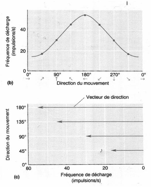
7 An example of population coding of movement direction. The blue lines represent the vectorial contributions of individual cells in the population (N = 475). The movement direction is in yellow and the direction of the population vector in red. A 95% directional variability cone around the population vector (red). The population is the same as in Figure 1, but the movement direction (yellow) is different. 7

8 Evolution of the population vector in time. Front and side views of time series of population (P) and movement (M) vectors are shown. Population and movement vectors are normalized relative to their respective maximum. Movement vectors are averages from one animal. STZM, onset of target light; MOV, onset of movement 8
9 9
10 10
11 11
12 12
.")
.")

13 Fig. 1. (A) Surface view showing the location of entry points with respect to the CS of 79 penetrations used in the present study. For graphical purposes, data from four hemispheres are transformed and plotted on an outline of a right hemisphere (see figure 6 in ref. 17). The transformation is invariant with respect to the distance between a particular entry point and the CS. The continuous line parallel to, and at a distance d from, the CS represents a borderline that demarcates the cortical surface into two regions (see text). The imaginary sections S1 S2 orthogonal to the CS is shown in B. A, anterior; M, medial. (B) Section view of the cortical depth in a plane orthogonal to the CS (S1 S2, in A). Dotted curves schematically represent the cortical laminae. Double lines illustrate the orientations of two imaginary penetrations P1 and P2 made nearby and farther away from the CS, respectively. The bisecting borderline is shown here in a similar fashion as in A. II VI cortical layers. (C) Examples of two penetrations analogous to P1 and P2. The entry points of the penetrations are color-shape coded in A and C. The PDs of directionally tuned cells isolated along the penetrations are shown as arrows, with the corresponding direction cosines in color. It can be seen that PDs were very similar along the penetration resembling P2 (red square), whereas they differed appreciably in the penetration resembling P1 (blue triangle). The average angle between PDs of all cell pairs was 19 for the former and 96 for the latter penetration. (Amirikian and Georgopoulos, 2003 PNAS) 13
 Three-dimensional directional tuning. The axes (white) meet at the origin of the movement.")
 of")
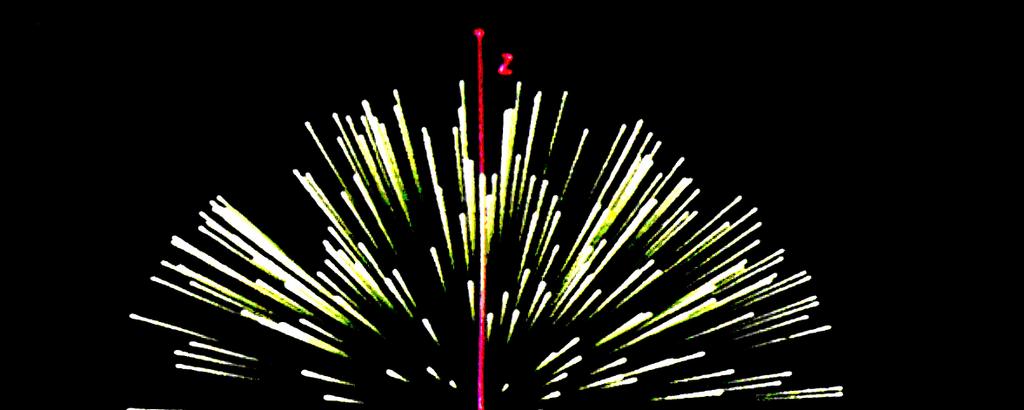
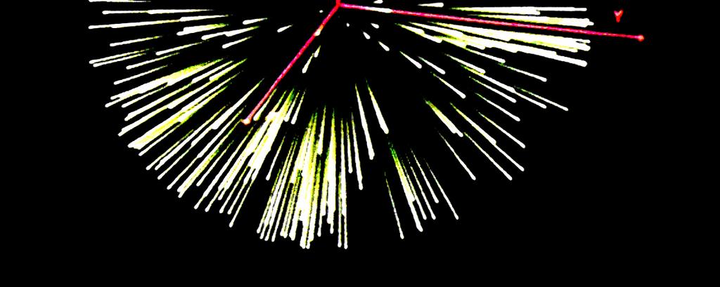
14 Three-dimensional preferred directions (purple) of 634 motor cortical cells studied in three monkeys. The axes are in white. (Georgopoulos et al., 1993 Science) Three-dimensional directional tuning. The axes (white) meet at the origin of the movement. For a particular movement, the discharge rate of the cell predicted by Eq. 1 is proportional to the length of a line pointing in the direction of the movement and drawn from the origin to the surface (purple) of the tuning volume. The cell's preferred direction is indicated by the yellow cone. 14


15 The population vector (green) obtained from the set of cells with preferred directions shown in Fig. 2. The direction of movement is shown in yellow. Images representing functional neuron density fields computed for two mutually orthogonal dimensions (Amirikian and Georgopoulos, 2003 PNAS) 15
16 Objectives Neurophysiology of brain area related to movement and motor control 1. Ascending pathways (sensory input) 2. Sensory input treatment, and thalamo-cortical & cortico-thalamic filter 3. Sensory cortical areas 4. Descending pathways (motor output) &motor unit definition 5. Synthesis of motor output organization 6. Toward parcellation, specialization and complexity 7. Pre-motor area definition, identification 8. Basal-ganglia organization direct & indirect pathways 9. Sensory motor transformation 10. Preferential direction of M1 neurons extrinsic-like code 11. Neuronal population coding 12. Emergence of muscle-like neurons in M1 13. Global movement induced by electrical stimulation of the cortex 14. Cortical plastcity Kakei, Hoffman, Strick Science,
 the monkey right hand gripping the handle of the manipulandum in 3 forearm postures. Pro, pronated; Sup, supinated, Mid, midway between pronation and supination.")
17 Fig. 1. Experimental design: dissociation of extrinsic, joint and muscle coordinate frames with changes in forearm posture. (A) the monkey right hand gripping the handle of the manipulandum in 3 forearm postures. Pro, pronated; Sup, supinated, Mid, midway between pronation and supination. Ext, extension; Flx, flexion; Rad, radial deviation; Uln, ulnar deviation. Up and Down indicate direction of movements in space. (B, C) PDs of the seven task-related muscles ((B) 4 wrist muscles and (C) 3 finger muscles, respectively) when the limb was in the 3 forearm postures. APL, abductor pollicis longus; ECRB, extensor carpi radialis brevis; ECRL, extensor carpi radialis longus; ECU, extensor carpi ulnaris; ED23, extensor digitorum 2,3; EDC, extensor digitorum communis; FCR, flexor carpi radialis. (D) Normalized shifts of PDs of the task-related muscles in the three forearm postures. Left circle, the single vector represents the PDs of the task-related muscles in the Pro position, which were normalized to the Up direction. Middle circle, the unlabeled vectors represent the relative shifts of the PDs of the task-related muscles with forearm rotation from Pro to Mid. Right circle, the unlabeled vectors represent the relative shifts of the PDs of the task-related muscles with forearm rotation from Pro to Sup. The vectors labeled Extrinsic represent the PDs of ideal vectors fixed to an extrinsic coordinate frame. The vectors labeled Joint represent the PDs of ideal vectors fixed to the wrist joint. Note that the unlabeled vectors are clearly separated from the Extrinsic and Joint vectors. Muscle-like neuron in M1 Shift de 79 Pro-Sup 28 out 88 Extrinsic-like neuron in M1 44 out 88 Extrinsic-like neuron In M1 influenced by joint Posture 16 out 88 Kakei, Hoffman, Strick Science,
, (B), and (C) in this figure correspond to neurons A, B, and C in Fig. 2.")

.")
18 Fig. 3. Spatiotemporal maps of activity of the same M1 neurons illustrated in Fig. 2. Neurons (A), (B), and (C) in this figure correspond to neurons A, B, and C in Fig. 2. To construct these maps, we calculated averaged spike numbers in a 50-ms time window, sliding the time window by 25 ms, from2500 to 1500 ms relative to the movement onset. The calculation was performed for each movement direction in each wrist posture. Then, contour plots of the spatiotemporal distribution of the neuron activity were generated with Surfer (Golden Software, Golden, Colorado). The maximum activity for any of the three wrist postures in the 50-ms analysis window was normalized to 100% (A: 155; B: 98; C: 124 spikes persecond). PDs in the Pro position were set to 0 in order to demonstrate the amount of the shift. PDs of neuron activity (18) for each posture are indicated by arrows. Kakei, Hoffman, Strick1 Science, 1999 Pre-motor cortex function 18
19 Kakei et al 2003 Neuroscience Research 19

20 Sensori-motor transformation with gain modulation 20
 that innervate the muscle.")
21 Fig. 1. Retrograde transneuronal transport of rabies virus from single muscles. When rabies virus is injected into a single digit muscle, it is transported in the retrograde direction to infect the motoneurons (i.e., first-order neurons) that innervate the muscle. Then virus is transported transneuronally in the retrograde direction to label all those second-order neurons that synapse on the infected motoneurons. These include dorsal root ganglion cells that supply group Ia muscle spindle afferents, spinal cord interneurons, and cortical neurons in layer V (CM cells). At longer survival times, virus can undergo another stage of retrograde transneuronal transport and label all those third-order neurons that synapse on the infected second-order neurons. For example, virus can move from second-order neurons in layer V to third-order neurons in layer III. Similarly, virus can move from second-order inter-neurons in the spinal cord to third-order cortical neurons in layer V. DRG, dorsal root ganglion cell; Int, interneuron; Mn, motoneuron; 1, first-order neuron; 2, second-order neuron; 3, third-order neuron. 21
22 How are the neurons that directly influence the motoneurons of a muscle distributed in the primary motor cortex (M1)? To answer this classical question we used retrograde transneuronal transport of rabies virus from single muscles of macaques. This enabled us to define cortico-motoneuronal (CM) cells that make monosynaptic connections with the motoneurons of the injected muscle. We examined the distribution of CM cells that project to motoneurons of three thumb and finger muscles. We found that the CM cells for these digit muscles are restricted to the caudal portion of M1, which is buried in the central sulcus. Within this region of M1, CM cells for one muscle display a remarkably widespread distribution and fill the entire mediolateral extent of the arm area. In fact, CM cells for digit muscles are found in regions of M1 that are known to contain the shoulder representation. The cortical territories occupied by CM cells for different muscles overlap extensively. Thus, we found no evidence for a focal representation of single muscles in M1. Instead, the overlap and intermingling among the different populations of CM cells may be the neural substrate to create a wide variety of muscle synergies.wefound two additional surprising results. First, 15 16% of the CM cells originate from area 3a, a region of primary somatosensory cortex. Second, the size range of CM cells includes both fast and slow pyramidal tract neurons. These observations are likely to lead to dramatic changes in views about the function of the CM system (Rathelot & Strick, PNAS, 2006) Objectives Neurophysiology of brain area related to movement and motor control 1. Ascending pathways (sensory input) 2. Sensory input treatment, and thalamo-cortical & cortico-thalamic filter 3. Sensory cortical areas 4. Descending pathways (motor output) &motor unit definition 5. Synthesis of motor output organization 6. Toward parcellation, specialization and complexity 7. Pre-motor area definition, identification 8. Basal-ganglia organization direct & indirect pathways 9. Sensory motor transformation 10. Preferential direction of M1 neurons extrinsic-like code 11. Neuronal population coding 12. Emergence of muscle-like neurons in M1 13. Global movement induced by electrical stimulation of the cortex 14. Cortical plastcity 22
23 Figure 1. An Example of a Complex Posture Evoked from Monkey 1 by Microstimulation of Precentral Cortex When this site was stimulated the left hand closed into a grip posture, turned to the face, moved toward the mouth, and the mouth opened. Stimulation was for 500 ms at 100 A and 200 Hz. Drawings were traced from video footage acquired at 30 frames per second. The 11 dotted lines show the frameby-frame position of the hand for 11 different stimulation trials. Each dot shows the part of the video image of the hand that was farthest from the elbow. The start point of each trajectory was distant from the mouth; the end point was at or near the mouth. Graziano et al EBR, 2004 Figure 2. Examples of Postures Evoked by Microstimulation of Precentral Cortex(A F) Six examples of postures of the left arm evoked from monkey 2. Stimulation on the right side of the brain caused movements mainly of the left side of the body. Postures of the right limbs shown in these traced video frames are incidental and not dependant on the stimulation. Final postures that involved the left hand near the edge of the workspace, such as in (F), could not be tested from all directions, but still showed convergence from the range of initial positions tested.(g) A posture of the mouth and tongue evoked from monkey 1. When this site was stimulated, the mouth opened partly and the tongue pointed toward the left canine (final posture). Three initial postures of the mouth and tongue are shown. For the evoked movements shown in (A), (D), and (E), stimulation was at 50 A; (B), (C), and (G), 100 A; (F), 75 A. For all sites, stimulation trains were presented for 500 ms at 200 Hz. 23
24 Figure 3. Electromyographic (EMG) Activity from Muscles of the Upper Arm during Stimulation of One Site in Primary Motor Cortex Stimulation at this site for 500 ms at 100 A evoked a final posture of the arm and hand including a partial flexion of the elbow. When the elbow was fully extended, stimulation caused it to flex to its final posture. When the elbow was fully flexed, stimulation caused it to extend to the final posture. Graziano et al, 2004, JNP Figure 5. Eight Example Postures Illustrating the Topographic Map Found in Precentral Cortex of Monkey 1A similar map (not shown) was obtained in monkey 2. The circle on the brain shows the area that could be reached with the electrode. The magnified view at the bottom shows the locations of the stimulation sites. The area to the left of the lip of the central sulcus represents the anterior bank of the sulcus. Stimulation on the right side of the brain caused movements mainly of the left side of the body. Postures of the right arm shown in these traced video frames are incidental and not dependant on the stimulation. For the evoked movements shown in (A) and (G), stimulation was at 50 A. In (B) (F) and (H), stimulation was at 100 A. For all sites, stimulation trains were presented for 500 ms at 200 Hz. 24
25 Objectives Neurophysiology of brain area related to movement and motor control 1. Ascending pathways (sensory input) 2. Sensory input treatment, and thalamo-cortical & cortico-thalamic filter 3. Sensory cortical areas 4. Descending pathways (motor output) &motor unit definition 5. Synthesis of motor output organization 6. Toward parcellation, specialization and complexity 7. Pre-motor area definition, identification 8. Basal-ganglia organization direct & indirect pathways 9. Sensory motor transformation 10. Preferential direction of M1 neurons extrinsic-like code 11. Neuronal population coding 12. Emergence of muscle-like neurons in M1 13. Global movement induced by electrical stimulation of the cortex 14. Cortical plastcity Classen et al,1998, JNP 25
26 conclusions From these results, it appears likely that the motor cortex undergoes continuous plastic modifications. Frequently repeated movements reinforce particular network connectional patterns, but those patterns weaken if the movements have not been recently executed. This principle may underlie the beneficial effect of preperformance practice ( e.g., in athletics or musical performance). It also may be a requirement for purposeful skill acquisition in intact humans and in the rehabilitation of persons with brain damage The main mechanisms that have been suggested for mediating reorganization in the cerebral cortex involve the unmasking of existing, but latent, horizontal connections (for a review, see Sanes and Donoghue 2000) Modulation of synaptic efficacy such as long-term potentiation (LTP) (Hess and Donoghue 1994; Hess and others 1996) or long-term depression (LTD) (Hess and Donoghue 1996). Such modification of synaptic efficacy was recently demonstrated in the horizontal connections in the motor cortex of rats that underwent training of a skilled motor task (Rioult-Pedotti and others 1998). Concept that the motor cortex contains multiple overlapping motor representations (Donoghue and others 1992; Schieber and Hibbard 1993; Sanes and others 1995) functionally connected through an extensive horizontal network (Huntley and Jones 1991). Although connections are abundant within somatic representations, they are sparse between them (Huntley and Jones 1991). By changing the strength of horizontal connections between motor neurons, functionally different neuronal assemblies can form, thereby providing a substrate to construct dynamic motor output zones. 26
27 Pharmacological modulations Lorazepan (LZ): GABAA enhancement, blocks induction of LTP Dextromethorphan (DM): blocks NMDA receptors involved in LTP induction Lamotrigine (LTG): gating of NA+ and Ca++ without affecting LTP induction 27
Cortical Control of Movement
 Strick Lecture 2 March 24, 2006 Page 1 Cortical Control of Movement Four parts of this lecture: I) Anatomical Framework, II) Physiological Framework, III) Primary Motor Cortex Function and IV) Premotor
Cortical Involvement in the Recruitment of Wrist Muscles
 Journal of Neurophysiology 9: 2445 2456; June 2004 Cortical Involvement in the Recruitment of Wrist Muscles Ashvin Shah, Andrew H. Fagg 2, and Andrew G. Barto 2 Neuroscience and Behavior Program 2 Department
Neurophysiology of systems
 Neurophysiology of systems Motor cortex (voluntary movements) Dana Cohen, Room 410, tel: 7138 danacoh@gmail.com Voluntary movements vs. reflexes Same stimulus yields a different movement depending on context
Cortical Involvement in the Recruitment of Wrist Muscles
 Cortical Involvement in the Recruitment of Wrist Muscles Ashvin Shah Andrew H. Fagg Andrew G. Barto January 23, 24 Abstract In executing a voluntary movement, one is faced with the problem of translating
 Peripheral facial paralysis (right side). The patient is asked to close her eyes and to retract their mouth (From Heimer) Hemiplegia of the left side. Note the characteristic position of the arm with
Cortical Involvement in the Recruitment of Wrist Muscles
 J Neurophysiol 91: 2445 2456, 2004. First published January 28, 2004; 10.1152/jn.00879.2003. Cortical Involvement in the Recruitment of Wrist Muscles Ashvin Shah, 1 Andrew H. Fagg, 2 and Andrew G. Barto
Circuits & Behavior. Daniel Huber
 Circuits & Behavior Daniel Huber How to study circuits? Anatomy (boundaries, tracers, viral tools) Inactivations (lesions, optogenetic, pharma, accidents) Activations (electrodes, magnets, optogenetic)
Variety of muscle responses to tactile stimuli
 Variety of muscle responses to tactile stimuli Julita Czarkowska-Bauch Department of Neurophysiology, Nencki Institute of Experimental Biology, 3 Pasteur St., 02-093 Warsaw, Poland Abstract. Influences
At the highest levels of motor control, the brain represents actions as desired trajectories of end-effector
 At the highest levels of motor control, the brain represents actions as desired trajectories of end-effector Normal condition, using fingers and wrist Using elbow as folcrum Using shoulder as folcrum (outstretched
W. S. Smith and E. E. Fetz J Neurophysiol 102: , First published Jun 10, 2009; doi: /jn
 W. S. Smith and E. E. Fetz J Neurophysiol 12:14-148, 29. First published Jun 1, 29; doi:1.1152/jn.9152.28 You might find this additional information useful... This article cites 21 articles, 15 of which
Lecture 9: Forearm bones and muscles
 Lecture 9: Forearm bones and muscles Remember, the region between the shoulder and the elbow = brachium/arm, between elbow and wrist = antebrachium/forearm. Forearm bones : Humerus (distal ends) Radius
Motor Functions of Cerebral Cortex
 Motor Functions of Cerebral Cortex I: To list the functions of different cortical laminae II: To describe the four motor areas of the cerebral cortex. III: To discuss the functions and dysfunctions of
Brain-Computer Interfaces to Replace or Repair the Injured Central Nervous System
 Three approaches to restore movement Brain-Computer Interfaces to Replace or Repair the Injured Central Nervous System 1. Replace: Brain control of 2. Replace & Repair: Intra-Spinal Stimulation 3. Repair:
A Computational Model of Muscle Recruitment for Wrist Movements
 J Neurophysiol 88: 3348 3358, 2002. First published October 10, 2002; 10.1152/jn.00621.2002. A Computational Model of Muscle Recruitment for Wrist Movements ANDREW H. FAGG, 1 ASHVIN SHAH, 2 AND ANDREW
Somatosensation. Recording somatosensory responses. Receptive field response to pressure
 Somatosensation Mechanoreceptors that respond to touch/pressure on the surface of the body. Sensory nerve responds propotional to pressure 4 types of mechanoreceptors: Meissner corpuscles & Merkel discs
STRUCTURAL BASIS OF MEDICAL PRACTICE EXAMINATION 5 October 6, 2006
 STRUCTURAL BASIS OF MEDICAL PRACTICE EXAMINATION 5 October 6, 2006 PART l. Answer in the space provided. (8 pts) 1. Identify the structures. (2 pts) B C A. _pisiform B. _ulnar artery A C. _flexor carpi
Nerves of the upper limb Prof. Abdulameer Al-Nuaimi. E. mail:
 Nerves of the upper limb Prof. Abdulameer Al-Nuaimi E-mail: a.al-nuaimi@sheffield.ac.uk E. mail: abdulameerh@yahoo.com Brachial plexus Median nerve After originating from the brachial plexus in the axilla,
OpenSim Tutorial #2 Simulation and Analysis of a Tendon Transfer Surgery
 OpenSim Tutorial #2 Simulation and Analysis of a Tendon Transfer Surgery Laboratory Developers: Scott Delp, Wendy Murray, Samuel Hamner Neuromuscular Biomechanics Laboratory Stanford University I. OBJECTIVES
HUMAN MOTOR CONTROL. Emmanuel Guigon
 HUMAN MOTOR CONTROL Emmanuel Guigon Institut des Systèmes Intelligents et de Robotique Université Pierre et Marie Curie CNRS / UMR 7222 Paris, France emmanuel.guigon@upmc.fr e.guigon.free.fr/teaching.html
Human Anatomy Biology 351
 1 Human Anatomy Biology 351 Upper Limb Exam Please place your name on the back of the last page of this exam. You must answer all questions on this exam. Because statistics demonstrate that, on average,
MCQWeek2. All arise from the common flexor origin. The posterior aspect of the medial epicondyle is the common flexor origin.
 MCQWeek2. 1. Regarding superficial muscles of anterior compartment of the forearm: All arise from the common flexor origin. The posterior aspect of the medial epicondyle is the common flexor origin. Flexor
The Physiology of the Senses Chapter 8 - Muscle Sense
 The Physiology of the Senses Chapter 8 - Muscle Sense www.tutis.ca/senses/ Contents Objectives... 1 Introduction... 2 Muscle Spindles and Golgi Tendon Organs... 3 Gamma Drive... 5 Three Spinal Reflexes...
Clinical examination of the wrist, thumb and hand
 Clinical examination of the wrist, thumb and hand 20 CHAPTER CONTENTS Referred pain 319 History 319 Inspection 320 Functional examination 320 The distal radioulnar joint.............. 320 The wrist.......................
Nature Neuroscience doi: /nn Supplementary Figure 1. Characterization of viral injections.
 Supplementary Figure 1 Characterization of viral injections. (a) Dorsal view of a mouse brain (dashed white outline) after receiving a large, unilateral thalamic injection (~100 nl); demonstrating that
Direction of action is represented in the ventral premotor cortex
 articles 21 Nature Publishing Group http://neurosci.nature.com Direction of action is represented in the ventral premotor cortex 21 Nature Publishing Group http://neurosci.nature.com Shinji Kakei 1,2,4,
in Motion analysis TRAMA Project September th 2007
 First Course Basics in Motion analysis TRAMA Project September 10-12 th 2007 Prof. Guy CHERON Laboratory of Neurophysiology and Movement Biomechanics Université Libre de Bruxelles, Belgium Objectives Neurophysiology
Nerves of Upper limb. Dr. Brijendra Singh Professor & Head Department of Anatomy AIIMS Rishikesh
 Nerves of Upper limb Dr. Brijendra Singh Professor & Head Department of Anatomy AIIMS Rishikesh 1 Objectives Origin, course & relation of median & ulnar nerves. Motor & sensory distribution Carpal tunnel
10/15/2014. Wrist. Clarification of Terms. Clarification of Terms cont
 Wrist Clarification of Terms Palmar is synonymous with anterior aspect of the wrist and hand Ventral is also synonymous with anterior aspect of the wrist and hand Dorsal refers to the posterior aspect
Biological Bases of Behavior. 8: Control of Movement
 Biological Bases of Behavior 8: Control of Movement m d Skeletal Muscle Movements of our body are accomplished by contraction of the skeletal muscles Flexion: contraction of a flexor muscle draws in a
DELSYS. Purpose. Hardware Concepts. Software Concepts. Technical Note 101: EMG Sensor Placement
 Technical Note 101: EMG Sensor Placement Purpose This technical note addresses proper placement technique for Delsys Surface EMG Sensors. The technique is demonstrated through an experiment in which EMG
Practical 2 Worksheet
 Practical 2 Worksheet Upper Extremity BONES 1. Which end of the clavicle is on the lateral side (acromial or sternal)? 2. Describe the difference in the appearance of the acromial and sternal ends of the
The Motor Systems. What s the motor system? Plan
 The Motor Systems What s the motor system? Parts of CNS and PNS specialized for control of limb, trunk, and eye movements Also holds us together From simple reflexes (knee jerk) to voluntary movements
Key Points for Success:
 SELF WRIST & HAND 1 2 All of the stretches described in this chapter are detailed to stretch the right side. Key Points for Success: Sit comfortably in a position where you can straighten or fully extend
Multi-joint Mechanics Dr. Ted Milner (KIN 416)
") Multi-joint Mechanics Dr. Ted Milner (KIN 416) Muscle Function and Activation It is not a straightforward matter to predict the activation pattern of a set of muscles when these muscles act on multiple
Multi-joint Mechanics Dr. Ted Milner (KIN 416) Muscle Function and Activation It is not a straightforward matter to predict the activation pattern of a set of muscles when these muscles act on multiple
CNS Control of Movement
 CNS Control of Movement Cognitive Neuroscience, Fall, 2011 Joel Kaplan, Ph.D. Dept of Clinical Neuroscience Karolinska Institute joel.kaplan@ki.se Charles Sherrington (1857-1952) Basic Concepts Localization
The Nervous System S P I N A L R E F L E X E S
 The Nervous System S P I N A L R E F L E X E S Reflexes Rapid, involuntary, predictable motor response to a stimulus Spinal Reflexes Spinal somatic reflexes Integration center is in the spinal cord Effectors
The Muscular System. Chapter 10 Part C. PowerPoint Lecture Slides prepared by Karen Dunbar Kareiva Ivy Tech Community College
 Chapter 10 Part C The Muscular System Annie Leibovitz/Contact Press Images PowerPoint Lecture Slides prepared by Karen Dunbar Kareiva Ivy Tech Community College Table 10.9: Muscles Crossing the Shoulder
Constraints on Somatotopic Organization in the Primary Motor Cortex
 INVITED REVIEW Constraints on Somatotopic Organization in the Primary Motor Cortex MARC H. SCHIEBER Departments of Neurology, Neurobiology and Anatomy, Brain and Cognitive Science, and Physical Medicine
Corticospinal excitation of presumed cervical propriospinal neurones and its reversal to inhibition in humans
 11911 Journal of Physiology (2001), 533.3, pp.903 919 903 Corticospinal excitation of presumed cervical propriospinal neurones and its reversal to inhibition in humans Guillaume Nicolas, Véronique Marchand-Pauvert,
Chapter 12b. Overview
 Chapter 12b Spinal Cord Overview Spinal cord gross anatomy Spinal meninges Sectional anatomy Sensory pathways Motor pathways Spinal cord pathologies 1 The Adult Spinal Cord About 18 inches (45 cm) long
Nature Methods: doi: /nmeth Supplementary Figure 1. Activity in turtle dorsal cortex is sparse.
 Supplementary Figure 1 Activity in turtle dorsal cortex is sparse. a. Probability distribution of firing rates across the population (notice log scale) in our data. The range of firing rates is wide but
Introduction to Ultrasound Examination of the Hand and upper
 Introduction to Ultrasound Examination of the Hand and upper Emil Dionysian, M.D. Ultrasound of upper ext. Upside Convenient Opens another exam dimension Can be like a stethoscope Helps 3-D D visualization
The Nervous System: Sensory and Motor Tracts of the Spinal Cord
 15 The Nervous System: Sensory and Motor Tracts of the Spinal Cord PowerPoint Lecture Presentations prepared by Steven Bassett Southeast Community College Lincoln, Nebraska Introduction Millions of sensory
Restoration of Reaching and Grasping Functions in Hemiplegic Patients with Severe Arm Paralysis
 Restoration of Reaching and Grasping Functions in Hemiplegic Patients with Severe Arm Paralysis Milos R. Popovic* 1,2, Vlasta Hajek 2, Jenifer Takaki 2, AbdulKadir Bulsen 2 and Vera Zivanovic 1,2 1 Institute
Lecture VIII. The Spinal Cord, Reflexes and Brain Pathways!
 Reflexes and Brain Bio 3411! Monday!! 1! Readings! NEUROSCIENCE 5 th ed: Review Chapter 1 pp. 11-21;!!Read Chapter 9 pp. 189-194, 198! THE BRAIN ATLAS 3 rd ed:! Read pp. 4-17 on class web site! Look at
Plasticity of Cerebral Cortex in Development
 Plasticity of Cerebral Cortex in Development Jessica R. Newton and Mriganka Sur Department of Brain & Cognitive Sciences Picower Center for Learning & Memory Massachusetts Institute of Technology Cambridge,
Levels of the anatomical cuts of the upper extremity RADIUS AND ULNA right
 11 CHAPTER 2 Levels of the anatomical cuts of the upper extremity AND right CUT 1 CUT 4 1 2 3 4 5 6 Isolated fixation of the radius is difficult at this level because of the anterolateral vessels and the
Peripheral Nerve Injuries of the Upper Limb.
 Peripheral Nerve Injuries of the Upper Limb www.fisiokinesiterapia.biz Definitions Radiculopathy Process affecting the nerve root, most commonly by a herniated disc Weakness in muscles supplied by the
MLT Muscle(s) Patient Position Therapist position Stabilization Limb Position Picture Put biceps on slack by bending elbow.
 Patient Position Therapist position Stabilization Limb Position Picture Put biceps on slack by bending elbow.") MLT Muscle(s) Patient Position Therapist position Stabilization Limb Position Picture Put biceps on slack by bending elbow. Pectoralis Minor Supine, arm at side, elbows extended, supinated Head of Table
MLT Muscle(s) Patient Position Therapist position Stabilization Limb Position Picture Put biceps on slack by bending elbow. Pectoralis Minor Supine, arm at side, elbows extended, supinated Head of Table
Electrical stimulation of the upper limb. Paul Taylor
 Electrical stimulation of the upper limb Paul Taylor Three upper limb Pathways Shoulder subluxation Upper Limb Active Function Upper limb Passive All differ in aims but follow a similar 6 month protocol
SOMATOSENSORY SYSTEMS
 SOMATOSENSORY SYSTEMS Schematic diagram illustrating the neural pathways that convey somatosensory information to the cortex and, subsequently, to the motor system. Double arrows show reciprocal connections.
Module 7 - The Muscular System Muscles of the Arm and Trunk
 Module 7 - The Muscular System Muscles of the Arm and Trunk This Module will cover the muscle anatomy of the arms and trunk. We have already seen the muscles that move the humerus, so this module will
STRUCTURAL ORGANIZATION OF THE NERVOUS SYSTEM
 STRUCTURAL ORGANIZATION OF THE NERVOUS SYSTEM STRUCTURAL ORGANIZATION OF THE BRAIN The central nervous system (CNS), consisting of the brain and spinal cord, receives input from sensory neurons and directs
Lab Activity 11: Group II
 Lab Activity 11: Group II Muscles Martini Chapter 11 Portland Community College BI 231 Origin and Insertion Origin: The place where the fixed end attaches to a bone, cartilage, or connective tissue. Insertion:
Functional Anatomy of the Elbow
 Functional Anatomy of the Elbow Orthopedic Institute Daryl C. Osbahr, M.D. Chief of Sports Medicine, Orlando Health Chief Medical Officer, Orlando City Soccer Club Orthopedic Consultant, Washington Nationals
The hand is full with sweat glands, activated at times of stress. In Slide #2 there was a mistake where the doctor mentioned lateral septum twice.
 We should only know: Name, action & nerve supply Layers - Skin - Superficial fascia - Deep fascia The hand is full with sweat glands, activated at times of stress. Deep fascia In Slide #2 there was a mistake
Motor Systems I Cortex. Reading: BCP Chapter 14
 Motor Systems I Cortex Reading: BCP Chapter 14 Principles of Sensorimotor Function Hierarchical Organization association cortex at the highest level, muscles at the lowest signals flow between levels over
A computational model for optimal muscle activity considering muscle viscoelasticity in wrist movements
 J Neurophysiol 19: 2145 216, 213. First published January 16, 213; doi:1.1152/jn.542.211. A computational model for optimal muscle activity considering muscle viscoelasticity in wrist movements Hiroyuki
Medical Terminology. Anatomical Position, Directional Terms and Movements
 Medical Terminology Anatomical Position, Directional Terms and Movements What we will cover... Content Objectives Students will be able to gain a better understanding and application of medical terminology
Medical Neuroscience Tutorial Notes
 Medical Neuroscience Tutorial Notes Finding the Central Sulcus MAP TO NEUROSCIENCE CORE CONCEPTS 1 NCC1. The brain is the body's most complex organ. LEARNING OBJECTIVES After study of the assigned learning
Microcircuitry coordination of cortical motor information in self-initiation of voluntary movements
 Y. Isomura et al. 1 Microcircuitry coordination of cortical motor information in self-initiation of voluntary movements Yoshikazu Isomura, Rie Harukuni, Takashi Takekawa, Hidenori Aizawa & Tomoki Fukai
Biceps Brachii. Muscles of the Arm and Hand 4/4/2017 MR. S. KELLY
 Muscles of the Arm and Hand PSK 4U MR. S. KELLY NORTH GRENVILLE DHS Biceps Brachii Origin: scapula Insertion: radius, fascia of forearm (bicipital aponeurosis) Action: supination and elbow flexion Innervation:
Motor systems.... the only thing mankind can do is to move things... whether whispering or felling a forest. C. Sherrington
 Motor systems... the only thing mankind can do is to move things... whether whispering or felling a forest. C. Sherrington 1 Descending pathways: CS corticospinal; TS tectospinal; RS reticulospinal; VS
Kinesiology of The Wrist and Hand. Cuneyt Mirzanli Istanbul Gelisim University
 Kinesiology of The Wrist and Hand Cuneyt Mirzanli Istanbul Gelisim University Bones The wrist and hand contain 29 bones including the radius and ulna. There are eight carpal bones in two rows of four to
divided by the bones ( redius and ulna ) and interosseous membrane into :
 and interosseous membrane into :") fossa Cubital Has: * floor. * roof : - Skin - superficial fasica - deep fascia ( include bicipital aponeurosis ) Structures within the roof : -cephalic and basilic veins -and between them median cubital
fossa Cubital Has: * floor. * roof : - Skin - superficial fasica - deep fascia ( include bicipital aponeurosis ) Structures within the roof : -cephalic and basilic veins -and between them median cubital
Motor systems III: Cerebellum April 16, 2007 Mu-ming Poo
 Motor systems III: Cerebellum April 16, 2007 Mu-ming Poo Population coding in the motor cortex Overview and structure of cerebellum Microcircuitry of cerebellum Function of cerebellum -- vestibulo-ocular
POSTSYNAPTIC INHIBITION OF CRAYFISH TONIC FLEXOR MOTOR NEURONES BY ESCAPE COMMANDS
 J. exp. Biol. (1980), 85, 343-347 343 With a figures Printed in Great Britain POSTSYNAPTIC INHIBITION OF CRAYFISH TONIC FLEXOR MOTOR NEURONES BY ESCAPE COMMANDS BY J. Y. KUWADA, G. HAGIWARA AND J. J. WINE
Anatomical Substrates of Somatic Sensation
 Anatomical Substrates of Somatic Sensation John H. Martin, Ph.D. Center for Neurobiology & Behavior Columbia University CPS The 2 principal somatic sensory systems: 1) Dorsal column-medial lemniscal system
Lateral view of human brain! Cortical processing of touch!
 Lateral view of human brain! Cortical processing of touch! How do we perceive objects held in the hand?! Touch receptors deconstruct objects to detect local features! Information is transmitted in parallel
Note: Waxman is very sketchy on today s pathways and nonexistent on the Trigeminal.
 Dental Neuroanatomy Thursday, February 3, 2011 Suzanne Stensaas, PhD Note: Waxman is very sketchy on today s pathways and nonexistent on the Trigeminal. Resources: Pathway Quiz for HyperBrain Ch. 5 and
Medical Terminology. Unit 2
 Medical Terminology Unit 2 Students will apply medical terminology. Objective 1: Identify and utilize anatomical positions, planes, and directional terms. Demonstrate what anatomical position is and how
Nerve Injury. 1) Upper Lesions of the Brachial Plexus called Erb- Duchene Palsy or syndrome.
 Upper Lesions of the Brachial Plexus called Erb- Duchene Palsy or syndrome.") Nerve Injury - Every nerve goes to muscle or skin so if the nerve is injured this will cause paralysis in the muscle supplied from that nerve (paralysis means loss of function) then other muscles and other
Nerve Injury - Every nerve goes to muscle or skin so if the nerve is injured this will cause paralysis in the muscle supplied from that nerve (paralysis means loss of function) then other muscles and other
Human Anatomy and Physiology I Laboratory Spinal and Peripheral Nerves and Reflexes
 Human Anatomy and Physiology I Laboratory Spinal and Peripheral Nerves and Reflexes 1 This lab involves the second section of the exercise Spinal Cord, Spinal Nerves, and the Autonomic Nervous System,
Main Menu. Wrist and Hand Joints click here. The Power is in Your Hands
 1 The Wrist and Hand Joints click here Main Menu K.5 http://www.handsonlineeducation.com/classes/k5/k5entry.htm[3/23/18, 1:40:40 PM] Bones 29 bones, including radius and ulna 8 carpal bones in 2 rows of
Reflexes. Dr. Baizer
 Reflexes Dr. Baizer 1 Learning objectives: reflexes Students will be able to describe: 1. The clinical importance of testing reflexes. 2. The essential components of spinal reflexes. 3.The stretch reflex.
Peripheral Nervous Sytem: Upper Body
 Peripheral Nervous Sytem: Upper Body MSTN121 - Neurophysiology Session 10 Department of Myotherapy Cervical Plexus Accessory nerve (CN11 + C1-5) Motor: trapezius and sternocleidomastoid Greater auricular
Degree of freedom problem
 KINE 4500 Neural Control of Movement Lecture #1:Introduction to the Neural Control of Movement Neural control of movement Kinesiology: study of movement Here we re looking at the control system, and what
Year 2004 Paper one: Questions supplied by Megan
 QUESTION 47 A 58yo man is noted to have a right foot drop three days following a right total hip replacement. On examination there is weakness of right ankle dorsiflexion and toe extension (grade 4/5).
Ultrasonography of the wrist - a step-by-step approach to study protocols and normal findings
 Ultrasonography of the wrist - a step-by-step approach to study protocols and normal findings Poster No.: C-1779 Congress: ECR 2016 Type: Educational Exhibit Authors: R. R. Domingues Madaleno, A. P. Pissarra,
Skin types: hairy and glabrous (e.g. back vs. palm of hand)
") Lecture 19 revised 03/10 The Somatic Sensory System Skin- the largest sensory organ we have Also protects from evaporation, infection. Skin types: hairy and glabrous (e.g. back vs. palm of hand) 2 major
Lecture 19 revised 03/10 The Somatic Sensory System Skin- the largest sensory organ we have Also protects from evaporation, infection. Skin types: hairy and glabrous (e.g. back vs. palm of hand) 2 major
Announcement. Danny to schedule a time if you are interested.
 Announcement If you need more experiments to participate in, contact Danny Sanchez (dsanchez@ucsd.edu) make sure to tell him that you are from LIGN171, so he will let me know about your credit (1 point).
KINE 4500 Neural Control of Movement. Lecture #1:Introduction to the Neural Control of Movement. Neural control of movement
 KINE 4500 Neural Control of Movement Lecture #1:Introduction to the Neural Control of Movement Neural control of movement Kinesiology: study of movement Here we re looking at the control system, and what
Robot control using electromyography (EMG) signals of the wrist
 signals of the wrist") Robot control using electromyography (EMG) signals of the wrist C. DaSalla,J.Kim and Y. Koike,2 Tokyo Institute of Technology, R2-5, 4259 Nagatsuta-cho, Midori-ku, Yokohama 226-853, Japan 2 CREST, Japan
Robot control using electromyography (EMG) signals of the wrist C. DaSalla,J.Kim and Y. Koike,2 Tokyo Institute of Technology, R2-5, 4259 Nagatsuta-cho, Midori-ku, Yokohama 226-853, Japan 2 CREST, Japan
Thalamo-Cortical Relationships Ultrastructure of Thalamic Synaptic Glomerulus
 Central Visual Pathways V1/2 NEUR 3001 dvanced Visual Neuroscience The Lateral Geniculate Nucleus () is more than a relay station LP SC Professor Tom Salt UCL Institute of Ophthalmology Retina t.salt@ucl.ac.uk
Role of brainstem in somatomotor (postural) functions
 functions") Role of brainstem in somatomotor (postural) functions (vestibular apparatus) The muscle tone and its regulation VESTIBULAR SYSTEM (Equilibrium) Receptors: Otolith organs Semicircular canals Sensation (information):
Role of brainstem in somatomotor (postural) functions (vestibular apparatus) The muscle tone and its regulation VESTIBULAR SYSTEM (Equilibrium) Receptors: Otolith organs Semicircular canals Sensation (information):
medial half of clavicle; Sternum; upper six costal cartilages External surfaces of ribs 3-5
 MUSCLE ORIGIN INSERTION ACTION NERVE Pectoralis Major medial half of clavicle; Sternum; upper six costal cartilages Lateral lip of intertubercular groove of horizontal adduction Medial and lateral pectoral
Nature Neuroscience: doi: /nn Supplementary Figure 1. Distribution of starter cells for RV-mediated retrograde tracing.
 Supplementary Figure 1 Distribution of starter cells for RV-mediated retrograde tracing. Parcellation of cortical areas is based on Allen Mouse Brain Atlas and drawn to scale. Thick white curves, outlines
Lesson 33. Objectives: References: Chapter 16: Reading for Next Lesson: Chapter 16:
 Lesson 33 Lesson Outline: Nervous System Structure and Function Neuronal Tissue Supporting Cells Neurons Nerves Functional Classification of Neuronal Tissue Organization of the Nervous System Peripheral
Voluntary Movements. Lu Chen, Ph.D. MCB, UC Berkeley. Outline. Organization of the motor cortex (somatotopic) Corticospinal projection
 Corticospinal projection") Voluntary Movements Lu Chen, Ph.D. MCB, UC Berkeley 1 Outline Organization of the motor cortex (somatotopic) Corticospinal projection Physiology of motor neurons Direction representation, population coding
Voluntary Movements Lu Chen, Ph.D. MCB, UC Berkeley 1 Outline Organization of the motor cortex (somatotopic) Corticospinal projection Physiology of motor neurons Direction representation, population coding
Medical Terminology. Anatomical Position, Directional Terms and Movements
 Medical Terminology Anatomical Position, Directional Terms and Movements What we will cover... Content Objectives Students will be able to gain a better understanding and application of medical terminology
Body Planes & Positions
 Learning Objectives Objective 1: Identify and utilize anatomical positions, planes, and directional terms. Demonstrate what anatomical position is and how it is used to reference the body. Distinguish
Forearm and Wrist Regions Neumann Chapter 7
 Forearm and Wrist Regions Neumann Chapter 7 REVIEW AND HIGHLIGHTS OF OSTEOLOGY & ARTHROLOGY Radius dorsal radial tubercle radial styloid process Ulna ulnar styloid process ulnar head Carpals Proximal Row
The Cerebellum. Outline. Lu Chen, Ph.D. MCB, UC Berkeley. Overview Structure Micro-circuitry of the cerebellum The cerebellum and motor learning
 The Cerebellum Lu Chen, Ph.D. MCB, UC Berkeley 1 Outline Overview Structure Micro-circuitry of the cerebellum The cerebellum and motor learning 2 Overview Little brain 10% of the total volume of the brain,
STRUCTURAL BASIS OF MEDICAL PRACTICE EXAMINATION 5. September 30, 2011
 STRUCTURAL BASIS OF MEDICAL PRACTICE EXAMINATION 5 September 30, 2011 PART l. Answer in the space provided. (12 pts) 1. Identify the structures. (2 pts) EXAM NUMBER A. Suprascapular nerve B. Axillary nerve
2. Name and give the neurotransmitter for two of the three shown (Fig. 26.8) brainstem nuclei that control sleep and wakefulness.
 brainstem nuclei that control sleep and wakefulness.") Put your name here-> BL A-415 Nerve cell mechanisms in behavior - Prof. Stark BL A-615 Neural bases of behavior Final examination - Tuesday, Dec. 12, 2000 12 noon - 1:50 p.m. Keep "essays" brief. Pay close
Put your name here-> BL A-415 Nerve cell mechanisms in behavior - Prof. Stark BL A-615 Neural bases of behavior Final examination - Tuesday, Dec. 12, 2000 12 noon - 1:50 p.m. Keep "essays" brief. Pay close
Nervous System: Spinal Cord and Spinal Nerves (Chapter 13)
") Nervous System: Spinal Cord and Spinal Nerves (Chapter 13) Lecture Materials for Amy Warenda Czura, Ph.D. Suffolk County Community College Eastern Campus Primary Sources for figures and content: Marieb,
Nervous System: Spinal Cord and Spinal Nerves (Chapter 13) Lecture Materials for Amy Warenda Czura, Ph.D. Suffolk County Community College Eastern Campus Primary Sources for figures and content: Marieb,
Three-dimensional tuning profile of motor cortical activity during arm. [Running Head] motor cortical activity during arm movements
![Three-dimensional tuning profile of motor cortical activity during arm. [Running Head] motor cortical activity during arm movements](/thumbs/92/109936344.jpg "Three-dimensional tuning profile of motor cortical activity during arm. [Running Head] motor cortical activity during arm movements") [Title] Three-dimensional tuning profile of motor cortical activity during arm movements [Running Head] motor cortical activity during arm movements [Authors] Takashi Mitsuda (1) Corresponding Author Paolo
[Title] Three-dimensional tuning profile of motor cortical activity during arm movements [Running Head] motor cortical activity during arm movements [Authors] Takashi Mitsuda (1) Corresponding Author Paolo
Overview of the Human Arm Anatomy
 Senior MESA Day Overview of the Human Arm Anatomy Bones, Joints, Muscles Review Arm Motion Kinematics: types of motion, location of motion, direction of motion, magnitude of motion, and degrees of freedom
Estimation of the Upper Limb Lifting Movement Under Varying Weight and Movement Speed
 1 Sungyoon Lee, 1 Jaesung Oh, 1 Youngwon Kim, 1 Minsuk Kwon * Jaehyo Kim 1 Department of mechanical & control engineering, Handong University, qlfhlxhl@nate.com * Department of mechanical & control engineering,