Virological Synapse-Mediated Spread of Human Immunodeficiency Virus. Type-1 between T cells is Sensitive to Entry Inhibition
|
|
|
- Clemence Kelly
- 5 years ago
- Views:
Transcription
1 JVI Accepts, published online ahead of print on 20 January 2010 J. Virol. doi: /jvi Copyright 2010, American Society for Microbiology and/or the Listed Authors/Institutions. All Rights Reserved Virological Synapse-Mediated Spread of Human Immunodeficiency Virus Type-1 between T cells is Sensitive to Entry Inhibition Nicola Martin 1, Sonja Welsch 2,4, Clare Jolly 3, John A. G. Briggs 4, David Vaux 1 and Quentin J. Sattentau 1* 1 The Sir William Dunn School of Pathology, University of Oxford, OX1 3RE, UK; 2 Structural Biology Unit, Wellcome Trust Centre for Human Genetics, University of Oxford, OX3 7BN, UK; 3 Wohl Virion Centre and MRC/UCL Centre for Medical Molecular Virology, University College London, London W1T 4JF, UK; 4 Structural and Computational Biology Unit, European Molecular Biology Laboratory, D Heidelberg, Germany *Corresponding author: Quentin J. Sattentau The Department of Pathology, The University of Oxford, South Parks Road, Oxford OX1 3R, UK Phone: Fax: quentin.sattentau@path.ox.ac.uk Running title: Inhibition of HIV-1 cell-cell spread
2 Abstract HIV-1 can disseminate between CD4 + T cells via diffusion-limited cellfree viral spread or by directed cell-cell transfer using virally-induced structures termed virological synapses. Although T cell virological synapses have been well characterized, it is unclear whether this mode of viral spread is susceptible to inhibition by neutralizing antibodies and entry inhibitors. Here we show that both cell-cell and cell-free viral spread are equivalently sensitive to entry inhibition. Fluorescence imaging analysis measuring virological synapse lifetimes and inhibitor time-of-addition studies implied that inhibitors can access pre-formed virological synapses and interfere with HIV-1 cell-cell infection. This concept was supported by electron tomography that revealed the T cell virological synapse to be a relatively permeable structure. VS-mediated HIV-1 spread is thus efficient, but is not an immune or entry inhibitor evasion mechanism, a result that is encouraging for vaccine and drug design. [138 words]
3 INTRODUCTION As with enveloped viruses from several viral families, the Human Immunodeficiency Virus type-1 (HIV-1) can disseminate both by fluid-phase diffusion of viral particles and by directed cell-cell transfer (39). The primary target cell for HIV-1 replication in vivo is the CD4 + T cell (13), that is infectable by CCR5-tropic (R5) and CXCR4-tropic (X4) viral variants (29). R5 HIV-1 is the major transmitted viral phenotype and dominates the global pandemic, whereas X4 virus is found later in infection in approximately 50% of infected individuals, and its presence indicates a poor disease progression prognosis (23). Cell-cell HIV-1 transfer between T cells is more efficient than diffusionlimited spread (8, 16, 32, 38), although recent estimates for the differential range from approximately one (42) to four (6) orders of magnitude. Two structures have been proposed to support contact-mediated intercellular movement of HIV-1 between T cells: membrane nanotubes (33, 43) and macromolecular adhesive contacts termed virological synapses (VS) (15, 17, 33). VS appear to be the dominant structure involved in T cell-t cell spread (33), and both X4 (17) and R5 HIV-1 (6, 15, 42) can spread between T cells via this mechanism. VS assembly and function are dependent on HIV-1 envelope glycoprotein (Env) engaging its primary cellular receptor CD4 (2, 6, 17). This interaction recruits more CD4 and coreceptor to the site of cell-cell contact in an actindependent manner (17). Adhesion molecules cluster at the intercellular junction and are thought to stabilize the VS (18). In parallel, viral Env and Gag are recruited to the interface by a microtubule-dependent mechanism (19),
4 where polarized viral budding may release virions into the synaptic space across which the target cell is infected (17). The precise mechanism by which HIV-1 subsequently enters the target T cell cytoplasm remains unclear: by fusion directly at the plasma membrane, fusion from within an endosomal compartment, or both (4, 6, 15, 25, 34). Viruses from diverse families including herpesviruses (9), poxviruses (22) and hepatitis C virus (44) evade neutralizing antibody attack by direct cell-cell spread, as the tight junctions across which the these viruses move are antibody-impermeable. It has been speculated that transfer of HIV-1 across VS may promote evasion from immune or therapeutic intervention with the inference that the junctions formed in retroviral VS may be non-permissive to antibody entry (39). However, available evidence regarding whether neutralizing antibodies (NAb) and other entry inhibitors can inhibit HIV-1 cellcell spread is inconsistent (25). An early analysis suggested that HIV-1 T cell- T cell spread is relatively resistant to neutralizing monoclonal antibodies (NMAb)(12). A later study agreed with this conclusion by demonstrating a lack of permissivity of HIV-1 T cell-t cell spread, measured by transfer of viral Gag, to interference with viral fusion using a gp41-specific NMAb and a peptidic fusion inhibitor (6). By contrast, another analysis reported that antigp41-specific NMAb interfered effectively with HIV-1 spread between T cells (26). Inhibitors of the HIV-1 surface glycoprotein (gp120)-cd4 or gp120- CXCR4 interaction reduced X4 HIV-1 VS assembly and viral transfer if applied prior to mixing of infected and receptor-expressing target cells (17, 19), but the effect of these inhibitors has not been tested on pre-formed VS. Thus the
5 field is currently unclear on whether direct T cell-t cell infectious HIV-1 spread is susceptible or not to antibody and entry inhibitor-mediated disruption of VS assembly, and the related question, whether the VS is permeable to viral entry inhibitors including NAb. Addressing these questions is of central importance to understanding HIV-1 pathogenesis and informing future drug and vaccine design. Since estimates reported in the literature of the relative efficiency of direct HIV-1 T cell-t cell spread compared to cell-free spread vary by approximately 3 orders of magnitude (6, 38, 42), and the evidence for the activity of viral entry inhibitors on cell-cell spread is conflicting, we set out to quantify the efficiency of infection across the T cell VS and analyze the susceptibility of this structure to NAb and viral entry inhibitors. Assays reporting on events proximal to productive infection show that the R5 HIV-1 T cell VS is approximately 1 order of magnitude more efficient than cell-free virus infection, and imaging analyses reveal that the VS assembled by HIV-1 is most likely permeable to inhibitors both during, and subsequent to, VS assembly. Thus we conclude that the T cell VS does not provide a privileged environment allowing HIV-1 escape from entry inhibition. MATERIALS AND METHODS Cell culture and virus infection. The cell lines Jurkat CE6.1, Jurkat.Tat.CCR5, A201, A301.R5 and ACH-2 were obtained from the National Institute for Biological Standards and Controls (NIBSC) Centre for AIDS Reagents (CFAR), Potters Bar, UK. A301.R5 cells were derived by retroviral
6 transduction and cultured as previously described (7). Cells were cultured in Complete Medium (CM): RPMI (GIBCO, Invitrogen) 10% FCS 100U/ml Penicillin 100ug/ml Streptomycin (PAA, Yeovil, UK). Unless otherwise stated, cells were cultured at 37 0 C in 5% CO 2. Jurkat.Tat.CCR5 and A301.R5 were maintained on CM supplemented with 1mg/ml G-418 (Sigma, Poole UK) to maintain expression of CCR5. Peripheral blood mononuclear cells (PBMC) were isolated from whole blood or buffy coats (from The National Blood Transfusion Service, Bristol, UK) by Ficoll-Hypaque (Sigma-Aldrich) gradient centrifugation. A MACS CD4 T cell Kit-II (Miltenyi UK Inc) was used to negatively isolate CD4 + T cells that were routinely >99% pure. For experiments with the R5 HIV-1 BaL isolate, primary CD4 + T cells were activated with CM supplemented with 5 µg/ml PHA-L (Sigma) and 10 U/mL IL-2 (NIBSC) for 3 days, then CM with IL-2 alone until use on day 7 of culture. For experiments with the X4 HIV-1 IIIB isolate, primary CD4 + T cells were purified from PBMC and used immediately. Cells (10 7 ) were infected in a volume of 1 ml with IIIB or BaL (NISBC) virus-containing supernatant overnight at a multiplicity of infection of approximately 0.01, then cultured in 50 ml CM and typically used on day 7-9 of infection. At this timepoint surface expression of Env was highest as detected by anti-gp120 antibody 2G12 (NIBSC) and antihuman IgG-phycoerythrin conjugate (Jackson Laboratories) and analyzed by flow cytometry on a FACScalibur using Flow-Jo software (Becton-Dickinson UK). Inhibitors used. Inhibitors used were as follows. NMAbs with relatively broad neutralizing activity: 2G12 (CFAR, NIBSC), glycan-specific, binds to mannose
7 groups on gp120 (37, 40); IgG1b12 (B12, D. Burton, Scripps Institute), binds the CD4 binding surface on gp120 (5); 2F5 (D. Katinger, Polymun Inc) that binds the membrane-proximal extracellular region (MPER) of gp41 and inhibits virus-cell fusion (30). Q4120 is a CD4 domain 1-specific MAb that efficiently blocks the CD4-gp120 interaction and viral entry (14), T20 (Enfuvirtide) is a broadly active synthetic peptide inhibitor of gp41-mediated fusion licensed for human use (28), TAK779 (NIH AIDS Reagent Repository, USA) a small molecule inhibitor of the gp120-ccr5 interaction (1), and PRO 2000 (Indevus Inc) a polyanionic candidate topical microbicide that interferes with viral entry, at least in part by binding to elements of the coreceptor binding surface (36). Cell-cell and cell-free infection assays. Three different methods were used to separate infected and target cells to measure cell-free infection, which was then compared to samples where the cells were directly cultured together to allow cell-cell viral transfer. In each case, 5x10 5 infected cells (Jkt BaL ) and 5x10 5 target cells (A301.R5) were used, and A201 (CD4 - /CCR5 - ) were used as negative control target cells. For infection of target cells by cell-free supernatants, the Jkt BaL were cultured alone in 150 µl GM for the 6 or 12 h to produce virus. Cells were centrifuged at 2000 x g for 5 mins and the supernatant was carefully aspirated and immediately added to a culture of 5x10 5 target cells in 150 µl CM. Target cells were further cultured to achieve a total incubation time of 12 h, then centrifuged and harvested for DNA extraction. For infection of target cells with and without transwells, cells were either cultured directly together, or separated by a 3 µm 24-well transwell,
8 allowing full diffusion of virus but not migration of cells: data not shown). 5x10 5 infected cells were suspended in 50 µl CM and placed in the upper well and 5x10 5 target cells were suspended in 250 µl in the lower well. Target cells were harvested by removing transwells and were lyzed and processed for DNA extraction. For the agitatied culture method we used an adaptation of that described in (42). Briefly, 5x10 5 of each cell type were resuspended in 1 ml CM in a 6-well plate. Paired samples were either left static, or rocked at 70 rpm in 5% CO 2. At the relevant time point the culture was harvested and processed for DNA extraction for qpcr. Cell-free and cell-cell infection inhibition assays. To measure inhibition of cell-free or cell-cell infection, 5x10 5 A301.R5 target cells were either cultured with an equal number of Jkt BaL, or with 100 ng PBMC-grown cell-free BaL virus. At 0 h inhibitors were titrated onto the cells, starting at 100 µg/ml in a 5- fold dilution series for all inhibitors except TAK, which was started at 50 nm. Cells were incubated for 12 h (cell-cell) or 24 h (cell-free), corresponding to a single cycle of viral infection. For B12, inhibition was also measured when the NMAb was added at 10 µg/ml at either 1 h or 3 h post-mixing. The antibody was diluted in 7.5 µl CM and carefully added to existing cocultures of 5x10 5 Jkt BaL and 5x10 5 A301.R5 target cells to avoid disturbing pre-formed conjugates. Quantitative Real-Time PCR for HIV-1 pol DNA detection. Samples were prepared by mixing 5x10 5 infected and 5x10 5 target A301.R5 or control target A201.R5 cells in CM, and were incubated at for the required time with or
9 without the addition of inhibitors. Samples were pelleted, supernatant aspirated and pellets frozen at -80ºC for later extraction with Qiagen DNEasy Kit. Primers and probe to detect HIV-1 DNA were designed using PrimerExpress3 against a pol sequence common to BaL and HXB2. The following primers were used: forward primer, TGGGTTATGAACTCCATCCTGAT, reverse primer, TGTCATTGACAGTCCAGCTGTCT. The probe had sequence TTCTGGCAGCACTATAGGCTGTACTGTCCATT and was labelled with the fluorophore FAM and the quencher TAMRA. For detection of the β-globin cellular reference gene, the following primers and probes were used: forward primer AACTGGGCATGTGGAGACAGA, reverse primer CTAAGGGTGGGAAAATAGACCAATAG, probe TCTTGGGTTTCTGATAGGCACTGACTCTCTCTG. The probe was labelled with VIC and the quencher TAMRA. PCR was performed in 25 µl total volume, using 1 x Eurogentec Mastermix (Eurogentec Ltd., Southampton, UK) 0.3 µm primers, 0.1 µm probe, and 2.5 µl sample DNA. A standard curve was created by titrating DNA from ACH-2 cells. Reactions were performed in single-plex using the ABI7500 (Applied Biosystems, Warrington, UK) standard run. For all qpcr measurements, the results of triplicate single-plex reactions detecting pol and β-globin were first averaged to give a mean for each, then pol values were divided by β-globin values to give a relative abundance. For experiments reported in Fig. 1 A and B, this was further normalised by dividing the value at each time point by the relevant 0 h result, to adjust for the starting level of infection and to enable comparison between cells with differing efficiency of HIV-1 infection. For experiments reported in Fig 1C and 1D,
10 where the target cell cultures for cell-free infection did not contain any infected cells, the 0 h value is shown in the graph. For all other PCR data presented where target and infected cells were harvested together, the result from a matched sample using the non-permissive A201 cells as targets was subtracted from the test value at each time point to account for the signal arising from the infected cells. To calculate a percentage inhibition of HIV-1 infection, the relative abundance of pol was normalised against A201 signal as described above. This value was subtracted from the no-treatment control value at the relevant timepoint, and the result divided by the no-treatment control value to give percentage inhibition. Laser Scanning Confocal Microscopy (LSCM). Target A301.R5 cells were prelabelled with the non-blocking, CD4 domain 4-specific MAb L120 (14, 17), mixed 1:1 with infected cells (Jkt BaL or Jkt IIIB ), incubated on poly-l-lysine coated coverslips for the required time, then fixed at room temperature with 4% paraformaldehyde (PFA). Samples were quenched with 50mM NH 4 Cl and permeabilized with PBS containing 0.1% Triton-X and 5% FCS. Env was labeled with 10 µg/ml 2G12 (NIBSC) and Gag with 1:1000 polyclonal rabbit anti-p24/anti-p17 (NIBSC). Samples were counterstained with anti-mouse- Alexa 488 (Invitrogen UK), anti-human-tritc, and anti-rabbit-cy5 (Jackson- Immunotech, UK) and mounted on glass slides using ProLong Gold (Invitrogen). Images were acquired on a Zeiss Pascal Axiovert 200M using sequential capture and processed using Adobe Photoshop CS2. Gag polarization and transfer between cells were quantified in 10 randomlyacquired low-power fields, each containing an average of approximately 20
11 conjugates. Conjugate interfaces were defined as contacts between an infected (Env + and/or Gag + ) and a target (CD4 + ) cell. Polarization of Gag, Env or CD4 was defined as signal enrichment in the membrane contact zone compared to the non-contact regions quantified using Metamorph software. VS were defined by colocalization of all three markers at the intercellular interface, and percentages calculated by dividing the number of colocalization events by the number of interfaces. Gag transfer was defined as Gag signal within the target cell in the absence of colocalized Env signal. The total number of these events/field was divided either by the number of VS, or by the total number of conjugate interfaces. Time-lapse LSCM analysis of conjugate lifetimes. To measure the duration of interaction times between HIV-1-infected and uninfected CD4 + target cells, Jkt BaL were labelled with CellTracker Orange CMRA (Invitrogen) and uninfected primary CD4 + T cells labelled with CFSE (Invitrogen). A total of 10 4 Jkt BaL and 10 4 primary CD4 + T cells were mixed in 50 µl CM and placed in one well of an Ibidi IbiTreat 24-well angiogenesis slide (Ibidi GmBH, Munich, Germany), and the plastic cover replaced. The slide was placed within a heated stage and supplemented with humidified 5% CO 2. Using a Zeiss Pascal LSM4 with sequential channel capture, images were recorded every 1 min up to 5 h. To quantify infected cell-target cell interaction times, each frame of the time-lapse series was processed in Metamorph to binarize the cell color labels into red (infected) and green (target), the cell profile was dilated to create defined interaction zones (yellow) at cell-cell contact regions, and interaction sites were isolated for quantification. Interaction sites for each x-y
12 frame of the movie were stacked into a 3-D box with time along the z-axis allowing each interaction to be tracked over time as an individual object Electron Microscopy and Tomography. Freshly purified primary CD4 + T cells were mixed 1:1 with Jkt IIIB, or 3 day PHA/IL-2-activated primary CD4 + T cells were mixed with Jkt BaL at 10 6 cells/ml in RPMI 10% FCS for 3 h at 37ºC in 5% CO 2, followed by gentle pelleting, aspiration of the supernatant and fixation in 4% PFA for 10 min at 37 ºC and 8% PFA for 50 min at RT. Samples were left in 2.5% glutaraldehyde in cacodylate buffer (100 mm sodium cacodylate, 50 mm KCl, 2.5 mm CaCl 2, ph 7.2) overnight, post-fixed for 1 h on ice with 1% osmium tetroxide, washed, stained overnight at 4 C with 0.5% uranyl acetate and dehydrated in a graded ethanol series at RT. Samples were embedded in epoxy resin (Roth, Karlsruhe, Germany) according to the manufacturer s instructions. 300 nm serial thick sections were cut, and dualaxis digital image series were recorded on an FEI Technai TF30 microscope at 300kV with FEI Eagle 4k CCD camera (pixel size after binning to 2k 1.99 nm or 2.53 nm at the specimen level) over a -60 to +60 tilt range (increment 1 ) and defocus -0.2µm, using the SerialEM software package (27). Tomogram reconstruction, joining of serial tomograms, manual 3D image segmentation and calculation of membrane model point coordinates used IMOD software v (20). Nearest approach points between the two membrane models in each tomogram were calculated and displayed as coloured patches using MATLAB (Mathworks, Natick, MA, USA) and UCSF Chimera (31), respectively.
13 Statistical analyses. Unless otherwise stated, quantitative data represent the mean values of replicate independent experiments each carried out in triplicate, and error bars represent SEM. Significance testing throughout was by 2-tailed Student's t-test, with Bonferroni correction for multiple tests where appropriate. RESULTS Efficiency of direct T cell-t cell spread of R5 HIV-1. Since R5 HIV-1 is the virus type that is principally transmitted between individuals, its study is central to understanding viral infection and spread and inhibition of these processes. We therefore adapted a previously-established qpcr-based assay for detection of de-novo DNA viral transcripts in X4 virus-infected cells (19) for use with R5 viral DNA. The pol region was chosen as a template for primers and probe since this is a late reverse transcript and therefore relevant to infection events proximal to proviral integration and productive replication. Values normalized to β-globin were expressed as copies of HIV-1 DNA/cell, and assay specificity was confirmed by co-culturing CD4 + /CCR5 + Jurkat.Tat.R5 cells infected with HIV-1 BaL (Jkt BaL ) with either the permissive CD4 + /CCR5 + T cell line A301.R5, or the non-permissive CD4 - /CCR5 - line A201. As previously observed with X4 HIV-1 (18, 19), de-novo pol DNA production was not observed in A2.01 target cells (data not shown), whereas A3.01.R5 target cells demonstrated a significant (p<0.05) increase of approximately 5-fold over background in pol DNA over 12 h (Fig. 1A). Because model systems using immortalized T cell lines may not accurately
14 represent interactions taking place between primary CD4 + T cells, we measured relative R5 HIV-1 pol production after transfer between infected and uninfected primary CD4 + T cells. Activated purified primary CD4 + T cells infected to levels >50% as determined by Gag and Env staining and flow cytometric analysis (data not shown) were mixed with uninfected autologous CD4 + T cells for the times shown, and samples processed for qpcr. We observed significant levels of increased pol in mixtures of infected and uninfected primary CD4 + T cells (Fig. 1B) that were similar in magnitude to virus spread between the cell lines, but with slower kinetics of pol synthesis. This confirmed that the use of T cell lines appears qualitatively justified for the study of cell-cell spread of R5 HIV-1, with the caveat that it is more rapid than the primary cell system. The HIV-1 BaL -infected T cell line Jkt BaL was subsequently used in all experiments hereafter for reasons of reproducibility, and A301.R5 cells were used as targets unless otherwise stated. To quantify differences between cell-free and cell-cell R5 HIV-1 transmission we used three different assay formats: i) use of a virus-permeable transwell to separate Jkt BaL from target cells; ii) culture of A3.01/R5 target cells with supernatants from Jkt BaL or direct A3.01/R5 mixing with Jkt BaL ; iii) an adaptation of the approach adopted by Sourisseau et al (42), in which cultures were maintained static for cell-cell interactions to take place, or were agitated to prevent stable cell-cell interactions. We confirmed that in each assay infected and target cell viability was maintained at an equivalent level over the time course of the experiment (data not shown). Fig. 1C shows that by 12 h post Jkt Bal -target cell mixing there was a robust pol signal detectable that was
15 approximately 13-fold greater than cell-free signal (p<0.01). Free diffusion of virus across the transwell membrane was determined by sampling supernatant for p24 Gag ELISA: there was no significant difference in p24 levels above or below the membrane (Fig. 1D). The signal obtained when Jkt Bal -target were mixed was 6.5-fold greater than the signal obtained with viral supernatant (Fig. 1E, p<0.01), and 5.4-fold greater when cells were maintained in a static, as opposed to an agitated culture (Fig 6F, p<0.01). Taken together, the mean increase in cell-cell compared to cell-free infection at 12 h corresponds to 8.3 ± 3.3-fold. Although we do not know which assay most precisely mimics the in vivo situation relating to cell-cell compared to cell-free viral spread, we assume that the mean value of approximately 10-fold is broadly representative of relative in vitro efficiency of HIV-1 spread between T cells. Cell-free and cell-cell spread of R5 HIV-1 are equally susceptible to entry inhibition. Having established that three different cell-cell assays for R5 virus spread gave broadly similar results, and having compared efficiency of cell-free and cell-cell spread quantitatively, we next analyzed whether these modes of infection are similarly susceptible to interference with viral entry. We chose the supernatant-based cell-free assay alongside the cell-cell infectivity assay (as shown in Fig 1E) for these experiments as this was the most technically feasible for this type of inhibition experiment and gave an intermediate result (6.5-fold) in terms of relative viral transfer efficiency. We selected a panel of HIV-1 entry inhibitors based upon their modes of action (receptor blocking or fusion inhibition) their chemical type (neutralizing
16 antibody, peptide, small molecule inhibitor, polyanion) and their relative molecular mass (0.5 kda to 150 kda). Inhibitors were titrated into each system at time of cell free infectious supernatant or infected cell mixing with target cells. De novo synthesized viral DNA was measured by qpcr within a time-frame allowing robust pol detection and corresponding to single round of viral replication in both systems, approximating to 12 h in the cell-cell system and 24 h in the cell-free system. Titration curves from combined experiments are shown for each inhibitor tested in Fig. 2A, and the mean IC 50 data are summarized in Fig 2B. All agents showed increasing inhibition of both cell-cell and cell-free infection with increasing concentration, and all achieved approximately 100% inhibition at the higher doses in the range. Mean ID 50 values varied across 4-orders of magnitude, representing their relative potency against R5 HIV-1. Considerable biological variation was observed between experiments, particularly with the CCR5 antagonist TAK779, for reasons that are currently unclear. Two of the inhibitors that interfere with gp120-cd4 binding (B12 and Q4120) showed a clear trend at low concentrations towards less effective inhibition of cell-free viral spread compared to cell-cell spread (Fig. 2A). This might reflect a modest selective reduction in inhibition of cell-cell spread as a result of reduced inhibitor access to the VS, increased velocity of gp120 engagement of CD4 on the target cell, or both. Nevertheless, no overall significant difference in inhibitor efficacy as measured at the ID 50 was observed between the two systems, whether inhibitors of low (TAK779, ~0.5 kda (1)), or high (NAb, ~150 kda) molecular weight were tested. This implies that the inhibitors were either acting rapidly to
17 prevent VS assembly and cell-cell virus transmission, or that these molecules enter assembled VS to block viral infection, or both Lifespan of HIV-1 R5 T cell VS. To investigate the possibility that inhibitors might interfere with HIV-1 spread across existing VS, we first established the longevity of R5 VS in T cells. For this we assumed that longlived conjugates formed between HIV-1 infected and uninfected, receptorexpressing CD4 + T cells represented VS, a proportion of which would be functional in cell-cell transfer of HIV-1 infection. Differentially labelled Jkt BaL, or control uninfected Jurkats, were mixed with uninfected primary CD4 + T cells and their interactions imaged using time-lapse laser-scanning confocal microscopy (LSCM). A frame from a representative movie (Supplementary Movie 1) is shown in Fig. 3A, in which multiple conjugates between infected (red) and uninfected target cells (green) are identified by arrows. To quantify infected cell-target cell interaction times, each frame of the time-lapse series was processed to create binary versions of the two colors and to dilate the cell profile to create defined interaction zones (yellow) at cell-cell contact regions (Fig. 3B). Interaction sites were then isolated for quantification (Fig 3C). Interaction sites for each x-y frame of the movie were stacked into a 3- dimensional box with time along the z-axis allowing each interaction to be tracked over time as an individual object. Lifetimes of control interactions 419 obtained from uninfected Jurkat and primary CD4 + T cells labeled and processed in the same manner as the test samples were compared to the test, and the data are quantified in Fig 3D and E. Control and test sample conjugate lifespans were similarly represented at times of up to 10 min, and
18 are consistent with previously-described intravitally-measured lifespans of normal homotypic T cell interactions (35). Since ~100% of the Jkt BaL cells were infected, this implies that many infected-target cell interactions are similarly transient to uninfected cell-cell interactions: whether these rapid interactions result in viral transfer is unknown. However, none of the control interactions exceeded 10 min, demonstrating that the formation of long-lived T cell-t cell contacts is HIV-1-driven. The mean contact time in the test samples was 62 min, and a small number were stable to the end of the assay at 300 min. Since these sustained (>10 min) interactions representing ~21% of all events approximates the percentage of VS found within T cell conjugates in other studies with X4 virus (17), we hypothesized that these were likely to represent VS. However, since X4 and R5 HIV-1 VS kinetics of assembly and frequency may vary, we investigated R5 virus VS dynamics in the current system using LSCM of Jkt BaL -primary CD4 + T cell conjugates. The percentage of CD4, Gag and Env capping at different time points was quantified from randomly-selected low-power fields as previously described (17). All three markers independently showed increased capping from 1 to 3 h post-cell mixing, plateauing between 3 and 6 h (Fig 4A). Similarly, co-capping of all 3 markers at a conjugate interface (our definition of a VS) increased significantly between 1 and 3 h but not between 3 and 6 h, reaching a maximum value of ~30% at 6 h. Gag transfer across the VS measured by distinct localization of Gag within the target cell revealed increasing transfer over the 6 h period. When this was expressed as percentage of total VS, approximately 20% VS showed Gag transfer at 1 h, rising to ~60 and ~80% at 3 and 6 h respectively (Fig. 4B). Expression of these values as a percentage of all conjugate
19 interfaces showed accumulation of target cell-associated Gag to a plateau of about 20% between 3 and 6 h (Fig 4B), consistent with the percentage of long-lived conjugates seen in Fig 3E. We therefore hypothesize that the majority of the long-lived conjugates forming between HIV-1-infected and CD4 + target cells are likely to contain a VS functional for Gag transfer, and that the average functional VS lifetime is probably >1 h. Inhibition of HIV-1 cell-cell spread in preformed VS. With this knowledge we next addressed the question of whether inhibitors added after conjugate formation interfered with R5 HIV-1-driven VS assembly. Since this concept has not been interrogated for X4 HIV-1 spread across VS, we investigated this in parallel, basing time of addition on the data reported in (17) that demonstrate X4 HIV-1 VS assembly reaching a plateau in about 30% of conjugates by 3 h post infected-target cell mixing. Jkt BaL - or Jkt IIIB - A3.01.R5 conjugates were prepared and inhibitors at a single inhibitory concentration (10 µg/ml for antibodies, 50 µm for PRO 2000, 1µM for TAK 779 and AMD3100, which had previously been shown to be close to 100% inhibitory when added at 0 h: Fig 2A), were added either at time of cell mixing or at 1 h post-cell mixing when approximately 30% of VS had formed (Fig 4A). Conjugates were then fixed at 3 h when VS frequency was at a plateau (Fig 4). Multiple fields were scored for percentage of conjugates forming VS, and pooled data are shown in Fig. 5. Approximately 30% of Jkt BaL -A3.01.R5 conjugates formed VS over the 3 h time-course in these experiments (Fig. 5A), consistent with the data reported in Fig 4A, and a very similar number was also observed for Jkt IIIB -A3.01.R5 conjugates (Fig 5B). When added at T
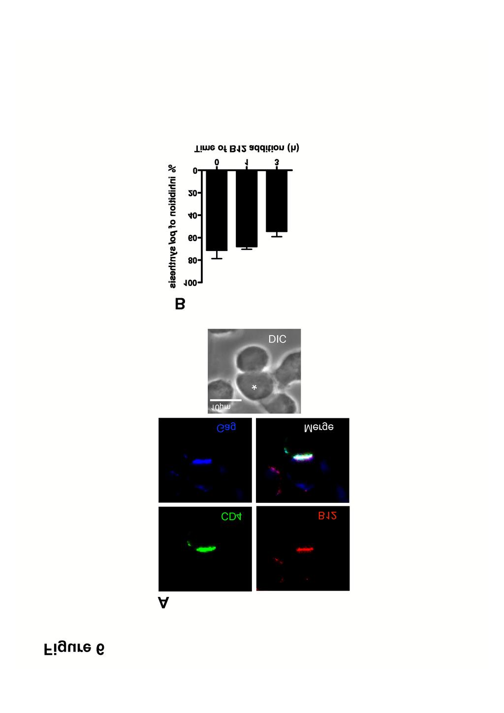
20 = 0, R5 HIV-1-mediated VS assembly was most strongly inhibited by blockers of the CD4-gp120 interaction such as B12 (~60% inhibition) and Q4120 (~80% inhibition), as previously observed for the X4 VS (17). By contrast, antagonists of the gp120-ccr5 interaction (TAK779 and PRO 2000) were more weakly inhibitory. When added after 1 h of coculture, VS inhibition was significantly reduced for B12 and Q4120, whereas no significant increase in VS number was observed with the other inhibitors. Thus once formed, the R5 VS appears moderately resistant to disassembly by HIV-1 receptor blockers. By contrast, the effect of CD4-gp120 or CXCR4-gp120 antagonists on the X4 HIV-1 VS was more pronounced. Whether added at 0 or 1 h, all inhibitors were ~75-100% effective at inhibiting VS assembly, suggesting that the preformed X4 VS may be susceptible to disassembly by antagonists of gp120-receptor interactions. Taken together these data imply that R5 VS assembly is less easily inhibited than the X4 VS by blockers of gp120- receptor interactions, whether applied before or during VS formation. The relative resistance of the R5 VS structure to inhibition may represent an inability of inhibitors to access a pre-formed VS, or alternatively may demonstrate that pre-formed VS structure may be maintained independent of function in terms of HIV-1 cell-cell spread. To investigate these possibilities we chose a single high-molecular weight (~150 kda) inhibitor, the NMAb B12, for further analysis. Since B12 prevents most VS assembly when introduced at the time of infected-uninfected cell mixing, we hypothesized that the presence of B12 within a VS would most likely signify incorporation of NMAb into a pre-existing VS. Jkt BaL and A3.01 were cocultured for 1 h, which results in at least 30% of total VS formation (Fig. 5A), prior to addition of B12 to the
21 culture, and cultures were maintained for a further 2 h prior to fixing and labelling for LSCM. Fig. 6A shows a representative conjugate in which B12 has robustly labelled the conjugate interface, and colocalizes with CD4 on the target cell and viral Gag in the infected cell. These data support the notion that inhibitors of relatively high molecular mass can enter a VS without disassembling the supramolecular structure. To investigate the functional effects of inhibitors on pre-formed R5 HIV-1 VS, we set up a similar experiment to that described for Fig. 6A with the same concentrations of inhibitors added either at the time of cell mixing or 1 or 3 h later, but lyzed the cells after a total of 12 h culture and analyzed for relative pol signal. Addition of inhibitors at time of mixing resulted in % inhibition of relative pol signal (Fig. 6), consistent with the results in Fig. 2. Taken together, the results presented in Figs 5 and 6 suggest that inhibitors can either prevent VS assembly if present during initial infected cell-target cell contact, or if introduced subsequent to VS formation, can enter intact VS and inhibit viral transfer into the target cell and subsequent reverse trasncription. The ultrastructure of the VS. Although the data shown in Figs 5 and 6 are suggestive of a VS structure permeable to inhibitors, this is not unequivocal since some inhibitor may conceivably be incorporated into forming VS without necessarily blocking further assembly. We therefore used electron tomography (ET) to probe the structure of mature R5 and X4 HIV-1 VS. Conventional thin-section electron microscopy yields ultrastructural details of cellular interactions in 2 dimensions, but is unable to resolve the 3- dimensional details of a complex intercellular interface. We therefore carried
22 out ET of serial thick sections spanning approximately 1.2 µm total thickness, of epoxy-embedded conjugates between infected Jurkats and primary CD4 + T cells fixed 3 h after cell mixing. The associated single-section movies are Supplemental movies 2 and 3. Fig 7 shows single slices from representative conjugates formed between Jkt IIIB (A-C) or Jkt BaL (D and E) and primary CD4 + T target cells. The infected cells were identified based on the presence of budding structures in HIV-1 IIIB (Fig. 7B, C) and HIV-1 BaL (Fig 7E) infected cells. The plasma membrane interfaces for the two VS are clearly defined, with multiple virus-like vesicles present at the interface between the cells, but few points of obvious intracellular contact. Three-dimensional models reconstructed from the tomographic analysis of the serial sections are shown in Fig 7F and G. It is evident that the VS interface for both virus types is complex with multiple membrane invaginations and projections, and yet the interface between the cells appears loosely-structured. A large number of X4 HIV-1 virions are present in gaps between the two cells, many of which are adherent to the target cell plasma membrane, presumably via Env-receptor and/or adhesion interactions. Fewer virions can be seen in the R5 HIV-1 infected VS, and most of these are either free or are associated with the infected cell membrane, possibly reflecting a lower avidity for target cell receptors than the X4 virions. The infected-target cell plasma membranes are relatively distant (>100nm) over most of the synapse, but are punctuated by limited areas of close membrane apposition. Analysis of distance between target cell and the nearest infected cell plasma membrane with virions removed to simplify analysis revealed regions that were 30 nm or closer (Figures 7H and I), consistent with integrin interactions such as those which
23 stabilize the immunological synapse (41) and implicated in X4 HIV-1 VS structure and function (18). These discrete adhesive regions were typically concentrated between interlocking villi surrounded by large areas of noninteracting plasma membrane. We have observed that the plasma membranes are often dramatically ruffled or invaginated (e.g. target cell membrane in Figure 7D), that might lead to the interpretation, from a single section, of a large internal virus-containing compartment. However, the rendering in 7G and I, imply that this is not a closed compartment but open to the external milieu, and many virions can be seen at the edges of, and outside the zone of cell membrane contact. Despite analysis of multiple tomographic reconstructions, we have yet to observe virus within a closed compartment within the target T cell. Nevertheless, since the sections that we have analyzed generally span only a proportion of the entire VS area, we cannot exclude the possibility that some virus is internalized into internal compartments within the target cell. In conclusion, these models reveal a porous intercellular interface containing mature virus particles that would allow access of even high molecular weight compounds to virus without imposition of obvious steric constraints. DISCUSSION Enveloped viruses from several different families use direct passage between cells to propagate themselves (39), and HIV-1 is no exception. We find here using three different assays that cell-cell spread is approximately an order of magnitude more efficient than cell-free spread, a figure that agrees with one
24 recent study (42), but disagrees with another (6). We believe that the discrepancy comes from the viral endpoint of cell-cell spread measured: the Sourisseau study assayed for productive infection by measuring Gag release into the supernatant, whereas the Chen study quantified Gag transfer into target cells. Gag transfer does not necessarily correspond to infection, particularly since the authors report that much of the Gag appears to be endocytosed by the target cell, an entry pathway that may result in a high proportion of non-infectious events (2, 4, 6, 15). Like the Sourisseau study, we have assayed for a proximal surrogate of productive viral infection, synthesis of late viral DNA product, which we believe is most likely to represent an infectious outcome in the target cell. Divergent experimental conditions may also influence the rate and extent of viral transfer between T cells, including the ratio of infected to uninfected T cells, the chronicity of the infection in the donor cells, and the type of cell, or cell line used. The mechanism of enhanced efficiency of cell-cell spread compared to cellfree spread of HIV-1 remains to be formally elucidated. Studies by others (8, 16, 32, 38) and our data herein and previously published (17), however, suggest that there may be several factors determining efficiency of viral dissemination in these systems. i) The proximity of the target and effector cells. Since the infected and uninfected cells are in relatively close and stable apposition during cell-cell spread, the rate-limiting factor of fluid-phase diffusion is essentially eliminated. ii) Receptor clustering. The VS is defined as the polarization of CD4 and coreceptor to the site of infected-target cell contact. This would allow de-novo budded HIV-1 to engage an optimal
25 number of receptors rapidly without the delay implicit in receptor recruitment required for fusion that is imposed upon individual cell surface-associated virions (21). iii) The multiplicity of infection (MOI) of target cells. The rate of spread of HIV-1 in culture is directly related to the MOI (8). Although we have not been able to quantify the number of infectious virions traversing a VS, the MOI via VS will be much higher than that occurring via cell-free spread both because of the increased total number of particles engaged by the target cell, and the reduced rate of time-dependent inactivation of viruses undergoing cell-cell spread compared to cell-free spread. We intend that the assays used here reflect as far as possible the in vivo situation regarding viral spread between lymphocytes. One obvious difference is that our cultures are approximately 100-fold less dense than the estimated lymphocyte density in vivo (45). Such a high in vivo density of lymphocytes would increase the chances of both cell-cell and cell-free infection occurring, however, making it difficult to model how this might affect the differential efficiency of viral spread by the two modes. Our demonstration that a panel of HIV-1 entry inhibitors interferes equivalently with cell-cell and cell-free HIV-1 infection when inhibitors are added at the time of mixing target cells with infected cells or free virus suggests that VSmediated spread of HIV-1 between T cells is unlikely to be an antibody or drug evasion strategy for the virus. In most scenarios in vivo, the inhibitor (antibody or therapeutic entry inhibitor) would be present prior to addition of virus to cells and so would not have to enter a pre-formed VS in order to interfere with cell-cell viral spread. However, even if inhibitor was added
26 subsequent to encounter between a virus infected and an uninfected T cell, our data suggest that such an inhibitor could access the pre-formed VS and prevent viral cell-cell spread. These results are encouraging for use of prophylactic vaccines designed to elicit neutralizing antibodies and for entry inhibitors applied in a prophylactic or therapeutic setting. Our results showing that all entry inhibitors tested target both X4 and R5 HIV-1 cell-cell spread similarly to diffusion-limited spread, are inconsistent with certain of the data obtained in T cell systems by others (2-4, 6). We consider that the most likely explanation again relates to systems measuring viral antigen uptake by endocytic pathways that are resistant to viral coreceptor antagonists and fusion inhibitors, but their contribution to productive infection is unclear. Although when using electron microscopy or tomography we have not observed evidence of endocytic uptake of HIV-1 by primary CD4 + T cells across a VS in the current study or previously (17), our analyses do not exclude this as an outcome of cell-cell transfer of virus. However, we assume that such events may be rare in primary CD4 + T cells compared to the immortalized cell lines used predominantly in other analyses (2-4, 6). Regardless of the route of viral uptake across VS by target cells, the outcome of importance must clearly be target cell infection. In this respect, the poldetection assay used here is more likely to represent events proximal to infection of the target T cell than uptake of Gag. Our study shows that that inhibitors of all stages of viral entry (CD4 engagement, coreceptor engagement and fusion) interfere to a similar degree with viral infection via VS. Our results do not agree with other recent analyses that failed to observe
27 inhibition of Gag transfer across VS by coreceptor antagonists and fusion inhibitors (2, 6). We assume that this is due to these studies measuring events mostly unrelated to HIV-1 infection of target cells. In this respect the same group that reported an absence of inhibition of VS-mediated Gag transfer by coreceptor antagonists in one study (6), reported inhibition by the same agent in a later study when a better surrogate of viral infection (transcription of HIV-1 LTR-driven GFP) was measured (15). Our measurements of uninfected CD4 + T cell-t cell interactions are consistent with previously-described intravitally-measured lifespans of homotypic T cell interactions (35), and confirm that T cells undergo transient adhesive interactions under normal conditions. A proportion of HIV-1-infected T cells undergo longer interactions, however, and we assume that a percentage of these relates to VS assembly and viral cell-cell transfer. A small minority of events lasted >300 min: these may be very long-lived interactions perhaps resulting from high-level infection of the donor T cell leading to enhanced cellcell interactions, and are consistent with the observed conjugate lifespans (18-32 h) reported in (15). Alternatively these long-lived interactions may represent a small proportion of conjugates undergoing membrane fusion to form syncytia. Although the electron-tomographic analyses carried out here and thin-section electron microscopy carried out previously (17) reveal the T cell VS to be loosely-structured, other cell types may assemble VS-like structures with distinct functional and structural features. We recently described a VS formed
28 between HIV-1-infected macrophages and CD4 + T cells: thin-section electron microscopic analysis revealed a broad interaction surface between the cells that had the appearance of a tight junction, but that formed only transiently (11). A study evaluating the ultrastructure of the VS formed between Human T Cell Leukemia Virus type-1 (HTLV-1) and permissive target T cells also found large surfaces of tighly-apposed membrane with occasional pockets containing virions (24). Thus the type of VS assembled is likely to be both celland virus-type specific, and this may influence sensitivity of this mode of viral spread to inhibition. Indeed, in this respect, dendritic cell transfer of HIV-1 to CD4 + T cells in trans across infectious synapses have been demonstrated to be resistant to a variety of NMAbs (10, 46). Further comparative analyses of VS formed between different cell types, and their sensitivity to Nabs and entry inhibitors are therefore warranted. ACKNOWLEDGEMENTS We thank the EM core facility at EMBL Heidelberg for equipment and assistance with electron tomography, Dr. Sylvain Lacomble for assistance with image processing and Leo Kong, Becky Russell, Will Hillson and Kate Gartlan for proofreading the manuscript. This work was supported by The Medical Research Council UK, The International AIDS Vaccine Initiative (IAVI) Neutralizing Antibody Consortum (NAC) and Fondation Dormeur. SW and the electron tomography part of this study were funded by Wellcome Trust grant H5RCYV0 to Stephen Fuller, and Deutsche Forschungsgemeinschaft grant SPP1175 to JAGB. QJS is a Jenner Institute Fellow.
29 FIGURE LEGENDS Figure 1. R5 HIV-1 spreads more efficiently cell-cell than by cell-free virion diffusion. (A) HIV-1 BaL transfer between infected and uninfected T cell lines Jkt BaL were mixed with A301.R5 target cells and at the time points indicated samples were lyzed and analyzed for HIV-1 pol by qpcr, and normalized against cellular β-globin. The background signal obtained from non-permissive A201 cells was subtracted from all values before calculation of percentage inhibition compared to no-treatment or no-virus controls. (B) HIV-1 transfer from primary CD4 BaL to autologous activated primary CD4 + T cells, assayed by qpcr and presented as in (A) over 24 h. Data represent means of 8 independent experiments each carried out in triplicate. (C) Jkt BaL were mixed directly with A301.R5 target cells in a transwell chamber without membrane (white bars), or were separated by the transwell membrane (black bars). At the times shown cultures were lyzed and processed for qpcr. Data represent the mean pol copy number relative to β-globin from 3 independent experiments each carried out in triplicate. (D) Supernatant was harvested from the lower (containing A301.R5 target cells) chamber of a transwell without (white bars) or with (black bars) a transwell membrane to separate the top well (containing Jkt Bal ), and assayed for p24 Gag by ELISA. Triplicate wells were sampled from one representative experiment and the bars represent the mean + 1 SD. (E) Jkt BaL (white bars) or cell-free supernatants harvested from Jkt BaL over a 12 h culture (black bars) were mixed with A301.R5 target cells at T = 0. Samples were processed as in (A) and data represent the mean pol copy number relative to β-globin from 3 independent experiments each carried
30 out in triplicate. (F) Jkt BaL were mixed with A301.R5 target cells at T = 0, after which the cultures were either left static (white bars) or were agitated to prevent stable cell-cell contacts forming (black bars). Samples were processed as in (A) and data represent the mean pol copy number relative to β-globin from 3 independent experiments each carried out in triplicate. Error bars represent SEM, *p<0.05; **p<0.01; ***p<0.001 Figure 2. Cell-free and Cell-cell infection are equally susceptible to entry inhibition. (A) A301.R5 target cells were either mixed 1:1 with Jkt BaL or with 12 h cell-free supernatants derived from Jkt BaL in the presence of titrations of the inhibitors shown. Cells were harvested after 12 h and processed for qpcr of pol and β-globin. Reduction in the pol PCR signal relative to the β-globin signal was expressed for each dilution of each inhibitor. The background signal obtained from non-permissive A201 cells was subtracted from all values before calculation of percentage inhibition compared to no-treatment or no virus controls. Solid lines represent cell-free virus inhibition and dashed lines represent cell-cell virus inhibition. (B) Percentage inhibition data from 3 independent experiments each carried out in triplicate were used to calculate IC 50 values. ( ) represent cell-free infection inhibition and ( ) represents cellcell infection inhibition. Error bars represent SEM. Figure 3. The lifespan of the R5 HIV-1 VS. (A) Jkt BaL pre-stained with CTO (red) and activated primary CD4 + T cells pre-stained with CFSE (green) were mixed in a 1:1 ratio and seeded onto MatTek poly-d-lysine coated glassbottomed tissue culture dishes. Cells were imaged by time-lapse LSCM, one
31 frame acquired every 30 s for 250 min. A representative frame is shown with red, green and DIC merged, with cell-cell interactions indicated by yellow arrows. (B) The same 2-color image in (A) after setting the threshold, binarizing the green and red images, and dilating the individual cell profiles as described in materials and methods. Cell-cell interaction sites are visible as yellow crescents. (C) The interactions are isolated in x-y 2-dimensional frames as discrete white regions. Individual x-y frames were stacked together into a 3-dimensional box, allowing tracking of the individual events over time. The test and control series were processed as described in methods to give the duration of each cell contact, which were grouped into the temporal categories shown in (D). The events falling into the >10 min category for the test movie are depicted as individual points, with the red line representing the mean duration in min (E). Figure 4. Assembly dynamics of the R5 HIV-1 VS. (A) A301.R5 target cells prelabeled with the non-blocking CD4 mab L120 were mixed with Jkt BaL for the times shown before fixing, permeabilizing and labelling with the appropriate secondary reagent for CD4 (grey bars), or with the appropriate primary and secondary reagents for Gag (hatched bars) or Env (black bars). Labeling and colocalization of all 3 markers in the same samples was also carried out (white bars). Data represent the combined percentage values from multiple conjugates counted in randomly-selected low-power fields, and error bars represent + 1 SD, *p<0.05. (B) As in (A) but the percentage of VS in which Gag was observed independently of Env in the target cell was divided
32 by the number of VS (white bars) or by the total number of conjugate interfaces (hatched bars) Figure 5. Time of inhibitor addition effects on VS formation. (A) Target A301.R5 cells pre-labeled with the non-blocking CD4 mab L120 were mixed 1:1 with Jkt BaL and inhibitor was either added simultaneously (white bars) or after 1 h of cell coculture (black bars). The hatched bars represent VS assembly in the absence of inhibitor (Non-Treated Control, NTC). Cells were cultured for a total of 3 h before fixing, permeabilizing and labelling for Gag and Env. Randomly-selected conjugate interfaces were analyzed for colocalization of the three markers defined as VS, as described for Fig. 4. Data are means of values from 3 independent experiments each carried out in triplicate, and error bars represent SEM. (B) As for (A) except that the infected cells were Jkt IIIB. Figure 6. Effects of NMAb B12 on HIV-1 infection across VS. (A) Target A301.R5 cells pre-labelled with the non-blocking CD4-specific mab L120 were mixed with Jkt BaL and cocultured on poly-l-lysine-coated coverslips. After 1 h of coculture, B12 was gently added to yield a final concentration of 10 µg/ml. Cells were cocultured for a further 2 h then fixed, permeabilized and labeled for B12 with anti-human IgG-TRITC and for Gag with anti-p24 antiserum, and Gag and CD4 labeled with the appropriate secondary detection reagents. Coverslips were mounted and analyzed by LSCM. (B) Target A301.R5 cells were mixed 1:1 with Jkt Bal in the presence of 10 µg/ml B12 (T = 0) or the same concentration of B12 was added 1 h or 3 h after cell mixing. Cells were
33 then cultured for a total of 12 h prior to lysis and qpcr for pol and β-globin products. Results are expressed as % inhibition of relative pol synthesis compared to no inhibitor or mixtures of Jkt BaL with the non-permissive A201 target cell control. Each bar represents the mean of data from 3 independent experiments carried out in triplicate, and error bars represent SEM. Figure 7. Electron tomographic reconstruction of the HIV-1 T cell VS. (A) A single 2 nm thick digital slice from a tomographic reconstruction of a conjugate between a primary CD4 + T cell (left) and a Jkt IIIB cell (right) cocultured at a 1:1 ratio for 3 h. (B) Single higher magnification 5 nm thick digital slice from a tomographic reconstruction of a conjugate between a primary CD4 + T cell (left) and a Jkt BaL cell (right). Arrows indicate viral budding structures on the infected cell, white asterisks indicate nuclei of infected cells. (C) Another single digital slice at a different z-height within the same tomogram as in (A). Arrows indicate budding structures. (D) As in (A) but image from a conjugate between a Jkt BaL and a primary CD4 + T cell. (E) The higher magnification image represents a diagonal slice through the 3D volume for better visualization of the budding structure (arrow) on the surface of the Jkt BaL cell. (F) 3-dimensional model of the Jkt IIIB cell surface (red), the primary CD4 + T cell surface (green) and virions (red spheres), generated from the same tomogram as in (A). (G) 3-dimensional model of the Jkt BaL cell surface (red), the primary CD4 + T cell surface (green) and virions (red spheres), generated from the same tomogram as in (D). (H) Higher-power representation of cell membranes shown in the boxed area of (F). Yellow membrane patches indicate areas of close apposition (<30 nm) of the two membranes. (I) Higher-
34 power representation of cell membranes shown in the boxed area of (G), yellow label as in (H). 823
35 REFERENCES Baba, M., O. Nishimura, N. Kanzaki, M. Okamoto, H. Sawada, Y. Iizawa, M. Shiraishi, Y. Aramaki, K. Okonogi, Y. Ogawa, K. Meguro, and M. Fujino A small-molecule, nonpeptide CCR5 antagonist with highly potent and selective anti-hiv-1 activity. Proc Natl Acad Sci U S A 96: Blanco, J., B. Bosch, M. T. Fernandez-Figueras, J. Barretina, B. Clotet, and J. A. Este High level of coreceptor-independent HIV transfer induced by contacts between primary CD4 T cells. J Biol Chem 279: Bosch, B., J. Blanco, E. Pauls, I. Clotet-Codina, M. Armand-Ugon, B. Grigorov, D. Muriaux, B. Clotet, J. L. Darlix, and J. A. Este Inhibition of coreceptor-independent cell-to-cell human immunodeficiency virus type 1 transmission by a CD4-immunoglobulin G2 fusion protein. Antimicrob Agents Chemother 49: Bosch, B., B. Grigorov, J. Senserrich, B. Clotet, J. L. Darlix, D. Muriaux, and J. A. Este A clathrin-dynamin-dependent endocytic pathway for the uptake of HIV-1 by direct T cell-t cell transmission. Antiviral Res 80: Burton, D. R., J. Pyati, R. Koduri, S. J. Sharp, G. B. Thornton, P. W. Parren, L. S. Sawyer, R. M. Hendry, N. Dunlop, P. L. Nara, and et al Efficient neutralization of primary isolates of HIV-1 by a recombinant human monoclonal antibody. Science. 266: Chen, P., W. Hubner, M. A. Spinelli, and B. K. Chen Predominant mode of HIV transfer between T cells is mediated by sustained Env-dependent neutralization-resistant virological synapses. J Virol. 7. Chenine, A., Q. J. Sattentau, and M. Moulard Selective HIV-1- induced downmodulation of CD4 and coreceptors. Arch. Virol. 145: Dimitrov, D. S., R. L. Willey, H. Sato, L. J. Chang, R. Blumenthal, and M. A. Martin Quantitation of human immunodeficiency virus type 1 infection kinetics. J Virol 67: Favoreel, H. W., G. Van Minnebruggen, G. R. Van de Walle, J. Ficinska, and H. J. Nauwynck Herpesvirus interference with virus-specific antibodies: bridging antibodies, internalizing antibodies, and hiding from antibodies. Vet Microbiol 113: Ganesh, L., K. Leung, K. Lore, R. Levin, A. Panet, O. Schwartz, R. A. Koup, and G. J. Nabel Infection of specific dendritic cells by CCR5- tropic human immunodeficiency virus type 1 promotes cell-mediated transmission of virus resistant to broadly neutralizing antibodies. J Virol 78: Groot, F., S. Welsch, and Q. J. Sattentau Efficient HIV-1 transmission from macrophages to T cells across transient virological synapses. Blood 111: Gupta, P., R. Balachandran, M. Ho, A. Enrico, and C. Rinaldo Cell-to-cell transmission of human immunodeficiency virus type 1 in the presence of azidothymidine and neutralizing antibody. J Virol 63:
36 Haase, A Population biology of HIV-1 infection: viral and CD4+ T cell demographics and dynamics in lymphatic tissue. Annu. Rev. Immunol. 17: Healey, D., L. Dianda, J. P. Moore, J. S. McDougal, M. J. Moore, P. Estess, D. Buck, P. D. Kwong, P. C. L. Beverley, and Q. J. Sattentau Novel anti-cd4 monoclonal antibodies separate human immunodeficiency virus infection and fusion of CD4+ cells from virus binding. J. Exp Med. 172: Hubner, W., G. P. McNerney, P. Chen, B. M. Dale, R. E. Gordon, F. Y. Chuang, X. D. Li, D. M. Asmuth, T. Huser, and B. K. Chen Quantitative 3D video microscopy of HIV transfer across T cell virological synapses. Science. 323: Johnson, D., and M. Huber Directed egress of animal viruses promotes cell-to-cell spread. J. Virol. 76: Jolly, C., K. Kashefi, M. Hollinshead, and Q. J. Sattentau HIV-1 cell to cell transfer across an Env-induced, actin-dependent synapse. J Exp Med 199: Jolly, C., I. Mitar, and Q. J. Sattentau Adhesion molecule interactions facilitate human immunodeficiency virus type-1-induced virological synapse formation between T cells. J Virol 81: Jolly, C., I. Mitar, and Q. J. Sattentau Requirement for an intact T- cell actin and tubulin cytoskeleton for efficient assembly and spread of human immunodeficiency virus type 1. J Virol 81: kremer, J., D. Mastronarde, and M. JR Computer visualization of three-dimensional image data using IMOD. J. Structural Biology 116: Kuhmann, S. E., E. Platt, S. Kozak, and D. Kabat Cooperationof multiple CCR5 coreceptors is required for infections by human immunodeficiency virus type 1. J. Virol. 74: Law, M., R. Hollinshead, and G. L. Smith Antibody-sensitive and antibody-resistant cell-to-cell spread by vaccinia virus: role of the A33R protein in antibody-resistant spread. J Gen Virol 83: Levy, J. A HIV pathogenesis: 25 years of progress and persistent challenges. AIDS (London, England) 23: Majorovits, E., M. Nejmeddine, Y. Tanaka, G. P. Taylor, S. D. Fuller, and C. R. Bangham Human T-lymphotropic virus-1 visualized at the virological synapse by electron tomography. PLoS ONE 3:e Martin, N., and Q. Sattentau Cell-to-cell HIV-1 spread and its implications for immune evasion. Curr Opin HIV AIDS 4: Massanella, M., I. Puigdomenech, C. Cabrera, M. T. Fernandez-Figueras, A. Aucher, G. Gaibelet, D. Hudrisier, E. Garcia, M. Bofill, B. Clotet, and J. Blanco Anti-gp41 antibodies fail to block early events of virological synapses but inhibit HIV spread between T cells. AIDS (London, England) 23: Mastronarde, D Automated electron microscope tomography using robust prediction of specimen movements. J. Structural Biology 152: Matthews, T., M. Salgo, M. Greenberg, J. Chung, R. DeMasi, and D. Bolognesi Enfuvirtide: the first therapy to inhibit the entry of HIV-1 into host CD4 lymphocytes. Nat Rev Drug Discov 3: Moore, J. P., S. G. Kitchen, P. Pugach, and J. A. Zack The CCR5 and CXCR4 coreceptors--central to understanding the transmission and
37 pathogenesis of human immunodeficiency virus type 1 infection. AIDS Res Hum Retroviruses 20: Muster, T., F. Steindl, M. Purtscher, A. Trkola, A. Klima, G. Himmler, F. Ruker, and H. Katinger A conserved neutralizing epitope on gp41 of human immunodeficiency virus type 1. J Virol 67: Pettersen, E., T. Goddard, C. Huang, G. Couch, D. Greenblatt, E. Meng, and T. Ferrin UCSF Cimera - a visualization system for exploratory research and analysis. J Computational Biol 25: Phillips, D The role of cell-to-cell transmission in HIV infection. AIDS 8: Rudnicka, D., J. Feldmann, F. Porrot, S. Wietgrefe, S. Guadagnini, M. C. Prevost, J. Estaquier, A. T. Haase, N. Sol-Foulon, and O. Schwartz Simultaneous cell-to-cell transmission of human immunodeficiency virus to multiple targets through polysynapses. J Virol 83: Ruggiero, E., R. Bona, C. Muratori, and M. Federico Virological consequences of early events following cell-cell contact between human immunodeficiency virus type 1-infected and uninfected CD4+ cells. J Virol 82: Sabatos, C. A., J. Doh, S. Chakravarti, R. S. Friedman, P. G. Pandurangi, A. J. Tooley, and M. F. Krummel A synaptic basis for paracrine interleukin-2 signaling during homotypic T cell interaction. Immunity. 29: Sachdev, D. D., B. Zerhouni-Layachi, M. Ortigoza, A. T. Profy, M. Tuen, C. E. Hioe, and M. E. Klotman The differential binding and activity of PRO 2000 against diverse HIV-1 envelopes. J Acquir Immune Defic Syndr 51: Sanders, R. W., M. Venturi, L. Schiffner, R. Kalyanaraman, H. Katinger, K. O. Lloyd, P. D. Kwong, and J. P. Moore The mannose-dependent epitope for neutralizing antibody 2G12 on human immunodeficiency virus type 1 glycoprotein gp120. J Virol 76: Sato, H., J. Orenstein, D. Dimitrov, and M. Martin Cell-cell spread of HIV-1 occurs within minutes and may not involve the participation of virus particles. Virology 186: Sattentau, Q Avoiding the void: cell-to-cell spread of human viruses. Nat Rev Microbiol 6: Scanlan, C. N., R. Pantophlet, M. R. Wormald, E. Ollmann Saphire, R. Stanfield, I. A. Wilson, H. Katinger, R. A. Dwek, P. M. Rudd, and D. R. Burton The broadly neutralizing anti-human immunodeficiency virus type 1 antibody 2G12 recognizes a cluster of alpha1-->2 mannose residues on the outer face of gp120. J Virol 76: Sims, T. N., and M. L. Dustin The immunological synapse: integrins take the stage. Immunol Rev 186: Sourisseau, M., N. Sol-Foulon, F. Porrot, F. Blanchet, and O. Schwartz Inefficient human immunodeficiency virus replication in mobile lymphocytes. J Virol 81: Sowinski, S., C. Jolly, O. Berninghausen, M. A. Purbhoo, A. Chauveau, K. Kohler, S. Oddos, P. Eissmann, F. M. Brodsky, C. Hopkins, B. Onfelt, Q. Sattentau, and D. M. Davis Membrane nanotubes physically connect T cells over long distances presenting a novel route for HIV-1 transmission. Nat Cell Biol 10:
38 Timpe, J. M., Z. Stamataki, A. Jennings, K. Hu, M. J. Farquhar, H. J. Harris, A. Schwarz, I. Desombere, G. L. Roels, P. Balfe, and J. A. McKeating Hepatitis C virus cell-cell transmission in hepatoma cells in the presence of neutralizing antibodies. Hepatology 47: Trepel, F Number and distribution of lymphocytes in man. A critical analysis. Klin. Wschr 52: van Montfort, T., A. A. Nabatov, T. B. Geijtenbeek, G. Pollakis, and W. A. Paxton Efficient capture of antibody neutralized HIV-1 by cells expressing DC-SIGN and transfer to CD4+ T lymphocytes. J Immunol 178: Downloaded from on September 30, 2018 by guest
39
40
41

42
43
44
45


46 Figure 7 * C D G 11m E Figure 4 11m F B *
Virological Synapse-Mediated Spread of Human Immunodeficiency Virus Type 1 between T Cells Is Sensitive to Entry Inhibition
 JOURNAL OF VIROLOGY, Apr. 2010, p. 3516 3527 Vol. 84, No. 7 0022-538X/10/$12.00 doi:10.1128/jvi.02651-09 Copyright 2010, American Society for Microbiology. All Rights Reserved. Virological Synapse-Mediated
The Regulated Secretory Pathway in CD4 + T cells Contributes to Human Immunodeficiency Virus Type-1 Cell-to-Cell Spread at the Virological Synapse
 The Regulated Secretory Pathway in CD4 + T cells Contributes to Human Immunodeficiency Virus Type-1 Cell-to-Cell Spread at the Virological Synapse Clare Jolly 1 *, Sonja Welsch 2,3, Stefanie Michor 4,
Title: Neutralization resistance of HIV-1 virological synapse-mediated infection is. Running Title: Virological-synapse neutralization resistance
 JVI Accepts, published online ahead of print on 2 May 2012 J. Virol. doi:10.1128/jvi.00230-12 Copyright 2012, American Society for Microbiology. All Rights Reserved. 1 2 Title: Neutralization resistance
Influenza virus exploits tunneling nanotubes for cell-to-cell spread
 Supplementary Information Influenza virus exploits tunneling nanotubes for cell-to-cell spread Amrita Kumar 1, Jin Hyang Kim 1, Priya Ranjan 1, Maureen G. Metcalfe 2, Weiping Cao 1, Margarita Mishina 1,
HIV-1 p24 ELISA Pair Set Cat#: orb54951 (ELISA Manual)
") HIV-1 p24 ELISA Pair Set Cat#: orb54951 (ELISA Manual) BACKGROUND Human Immunodeficiency Virus ( HIV ) can be divided into two major types, HIV type 1 (HIV-1) and HIV type 2 (HIV-2). HIV-1 is related to
HIV-1 p24 ELISA Pair Set Cat#: orb54951 (ELISA Manual) BACKGROUND Human Immunodeficiency Virus ( HIV ) can be divided into two major types, HIV type 1 (HIV-1) and HIV type 2 (HIV-2). HIV-1 is related to
Human Immunodeficiency Virus type 1 (HIV-1) p24 / Capsid Protein p24 ELISA Pair Set
 p24 / Capsid Protein p24 ELISA Pair Set") Human Immunodeficiency Virus type 1 (HIV-1) p24 / Capsid Protein p24 ELISA Pair Set Catalog Number : SEK11695 To achieve the best assay results, this manual must be read carefully before using this product
Human Immunodeficiency Virus type 1 (HIV-1) p24 / Capsid Protein p24 ELISA Pair Set Catalog Number : SEK11695 To achieve the best assay results, this manual must be read carefully before using this product
Commercially available HLA Class II tetramers (Beckman Coulter) conjugated to
 conjugated to") Class II tetramer staining Commercially available HLA Class II tetramers (Beckman Coulter) conjugated to PE were combined with dominant HIV epitopes (DRB1*0101-DRFYKTLRAEQASQEV, DRB1*0301- PEKEVLVWKFDSRLAFHH,
Class II tetramer staining Commercially available HLA Class II tetramers (Beckman Coulter) conjugated to PE were combined with dominant HIV epitopes (DRB1*0101-DRFYKTLRAEQASQEV, DRB1*0301- PEKEVLVWKFDSRLAFHH,
Supplementary information. MARCH8 inhibits HIV-1 infection by reducing virion incorporation of envelope glycoproteins
 Supplementary information inhibits HIV-1 infection by reducing virion incorporation of envelope glycoproteins Takuya Tada, Yanzhao Zhang, Takayoshi Koyama, Minoru Tobiume, Yasuko Tsunetsugu-Yokota, Shoji
Human Immunodeficiency Virus type 1 (HIV-1) gp120 / Glycoprotein 120 ELISA Pair Set
 gp120 / Glycoprotein 120 ELISA Pair Set") Human Immunodeficiency Virus type 1 (HIV-1) gp120 / Glycoprotein 120 ELISA Pair Set Catalog Number : SEK11233 To achieve the best assay results, this manual must be read carefully before using this product
Human Immunodeficiency Virus type 1 (HIV-1) gp120 / Glycoprotein 120 ELISA Pair Set Catalog Number : SEK11233 To achieve the best assay results, this manual must be read carefully before using this product
Supplemental Materials and Methods Plasmids and viruses Quantitative Reverse Transcription PCR Generation of molecular standard for quantitative PCR
 Supplemental Materials and Methods Plasmids and viruses To generate pseudotyped viruses, the previously described recombinant plasmids pnl4-3-δnef-gfp or pnl4-3-δ6-drgfp and a vector expressing HIV-1 X4
Gladstone Institutes, University of California (UCSF), San Francisco, USA
, San Francisco, USA") Fluorescence-linked Antigen Quantification (FLAQ) Assay for Fast Quantification of HIV-1 p24 Gag Marianne Gesner, Mekhala Maiti, Robert Grant and Marielle Cavrois * Gladstone Institutes, University of
Fluorescence-linked Antigen Quantification (FLAQ) Assay for Fast Quantification of HIV-1 p24 Gag Marianne Gesner, Mekhala Maiti, Robert Grant and Marielle Cavrois * Gladstone Institutes, University of
Supplementary Materials for
 advances.sciencemag.org/cgi/content/full/3/6/e1700338/dc1 Supplementary Materials for HIV virions sense plasma membrane heterogeneity for cell entry Sung-Tae Yang, Alex J. B. Kreutzberger, Volker Kiessling,
Direct ex vivo characterization of human antigen-specific CD154 + CD4 + T cells Rapid antigen-reactive T cell enrichment (Rapid ARTE)
") Direct ex vivo characterization of human antigen-specific CD154 + CD4 + T cells Rapid antigen-reactive T cell enrichment (Rapid ARTE) Introduction Workflow Antigen (ag)-specific T cells play a central
Direct ex vivo characterization of human antigen-specific CD154 + CD4 + T cells Rapid antigen-reactive T cell enrichment (Rapid ARTE) Introduction Workflow Antigen (ag)-specific T cells play a central
Identification of Mutation(s) in. Associated with Neutralization Resistance. Miah Blomquist
 in. Associated with Neutralization Resistance. Miah Blomquist") Identification of Mutation(s) in the HIV 1 gp41 Subunit Associated with Neutralization Resistance Miah Blomquist What is HIV 1? HIV-1 is an epidemic that affects over 34 million people worldwide. HIV-1
Identification of Mutation(s) in the HIV 1 gp41 Subunit Associated with Neutralization Resistance Miah Blomquist What is HIV 1? HIV-1 is an epidemic that affects over 34 million people worldwide. HIV-1
Optimization of the Fuse-It-mRNA Protocol for L929 Cells in the µ-plate 24 Well
 Optimization of the Fuse-It-mRNA Protocol for L929 Cells in the µ-plate 24 Well 1. General Information... 1 2. Background... 1 3. Material and Equipment Required... 2 4. Experimental Procedure and Results...
EMERGING ISSUES IN THE HUMORAL IMMUNE RESPONSE TO HIV. (Summary of the recommendations from an Enterprise Working Group)
") AIDS Vaccine 07, Seattle, August 20-23, 2007 EMERGING ISSUES IN THE HUMORAL IMMUNE RESPONSE TO HIV (Summary of the recommendations from an Enterprise Working Group) The Working Group Reston, Virginia,
AIDS Vaccine 07, Seattle, August 20-23, 2007 EMERGING ISSUES IN THE HUMORAL IMMUNE RESPONSE TO HIV (Summary of the recommendations from an Enterprise Working Group) The Working Group Reston, Virginia,
Supplemental Data Figure S1 Effect of TS2/4 and R6.5 antibodies on the kinetics of CD16.NK-92-mediated specific lysis of SKBR-3 target cells.
 Supplemental Data Figure S1. Effect of TS2/4 and R6.5 antibodies on the kinetics of CD16.NK-92-mediated specific lysis of SKBR-3 target cells. (A) Specific lysis of IFN-γ-treated SKBR-3 cells in the absence
J. Cell Sci. 129: doi: /jcs : Supplementary information
 Movie 1. AgLDL is contained in small sub-regions of the lysosomal synapse that are acidic. J774 cells were incubated with agldl dual labeled with a ph sensitive and a ph insensitive fluorophore for 1 hr.
Figure S1. PMVs from THP-1 cells expose phosphatidylserine and carry actin. A) Flow
 Flow") SUPPLEMENTARY DATA Supplementary Figure Legends Figure S1. PMVs from THP-1 cells expose phosphatidylserine and carry actin. A) Flow cytometry analysis of PMVs labelled with annexin-v-pe (Guava technologies)
SUPPLEMENTARY DATA Supplementary Figure Legends Figure S1. PMVs from THP-1 cells expose phosphatidylserine and carry actin. A) Flow cytometry analysis of PMVs labelled with annexin-v-pe (Guava technologies)
Supplementary Materials for
 www.sciencesignaling.org/cgi/content/full/8/366/ra25/dc1 Supplementary Materials for Viral entry route determines how human plasmacytoid dendritic cells produce type I interferons Daniela Bruni, Maxime
Ali Alabbadi. Bann. Bann. Dr. Belal
 31 Ali Alabbadi Bann Bann Dr. Belal Topics to be discussed in this sheet: Particles-to-PFU Single-step and multi-step growth cycles Multiplicity of infection (MOI) Physical measurements of virus particles
Supplementary Figure 1
 Supplementary Figure 1 Supplementary Figure 1: Cryopreservation alters CD62L expression by CD4 T cells. Freshly isolated (left) or cryopreserved PBMCs (right) were stained with the mix of antibodies described
Instructions. Fuse-It-mRNA easy. Shipping and Storage. Overview. Kit Contents. Specifications. Note: Important Guidelines
 Membrane fusion is a highly efficient method for transfecting various molecules and particles into mammalian cells, even into sensitive and primary cells. The Fuse-It reagents are cargo-specific liposomal
Application of μmacs Streptavidin MicroBeads for the analysis of HIV-1 directly from patient plasma
 Excerpt from MACS&more Vol 8 1/2004 Application of μmacs Streptavidin MicroBeads for the analysis of HIV-1 directly from patient plasma L. Davis Lupo and Salvatore T. Butera HIV and Retrovirology Branch,
Influenza A H1N1 HA ELISA Pair Set
 Influenza A H1N1 HA ELISA Pair Set for H1N1 ( A/Puerto Rico/8/1934 ) HA Catalog Number : SEK11684 To achieve the best assay results, this manual must be read carefully before using this product and the
genome edited transient transfection, CMV promoter
 Supplementary Figure 1. In the absence of new protein translation, overexpressed caveolin-1-gfp is degraded faster than caveolin-1-gfp expressed from the endogenous caveolin 1 locus % loss of total caveolin-1-gfp
In vitro human regulatory T cell expansion
 - 1 - Human CD4 + CD25 + regulatory T cell isolation, Workflow in vitro expansion and analysis In vitro human regulatory T cell expansion Introduction Regulatory T (Treg) cells are a subpopulation of T
Rapid antigen-specific T cell enrichment (Rapid ARTE)
") Direct ex vivo characterization of human antigen-specific CD154+CD4+ T cell Rapid antigen-specific T cell enrichment (Rapid ARTE) Introduction Workflow Antigen (ag)-specific T cells play a central role
Direct ex vivo characterization of human antigen-specific CD154+CD4+ T cell Rapid antigen-specific T cell enrichment (Rapid ARTE) Introduction Workflow Antigen (ag)-specific T cells play a central role
Instructions. Fuse-It-Color. Overview. Specifications
 Membrane fusion is a novel and highly superior method for incorporating various molecules and particles into mammalian cells. Cargo-specific liposomal carriers are able to attach and rapidly fuse with
In vitro human regulatory T cell suppression assay
 Human CD4 + CD25 + regulatory T cell isolation, in vitro suppression assay and analysis In vitro human regulatory T cell suppression assay Introduction Regulatory T (Treg) cells are a subpopulation of
ADCC Assay Protocol Vikram Srivastava 1, Zheng Yang 1, Ivan Fan Ngai Hung 2, Jianqing Xu 3, Bojian Zheng 3 and Mei- Yun Zhang 3*
 ADCC Assay Protocol Vikram Srivastava 1, Zheng Yang 1, Ivan Fan Ngai Hung 2, Jianqing Xu 3, Bojian Zheng 3 and Mei- Yun Zhang 3* 1 Department of Microbiology, Li Ka Shing Faculty of Medicine, University
VIRAL TITER COUNTS. The best methods of measuring infectious lentiviral titer
 VIRAL TITER COUNTS The best methods of measuring infectious lentiviral titer FLUORESCENCE CYCLES qpcr of Viral RNA SUMMARY Viral vectors are now routinely used for gene transduction in a wide variety of
Supplementary Data 1. Alanine substitutions and position variants of APNCYGNIPL. Applied in
 Supplementary Data 1. Alanine substitutions and position variants of APNCYGNIPL. Applied in Supplementary Fig. 2 Substitution Sequence Position variant Sequence original APNCYGNIPL original APNCYGNIPL
were isolated from the freshly drawn blood of healthy donors and ACS patients using the
 Supplemental Figure 1. Quality control of CD4 + T-cell purification. CD4 + T cells were isolated from the freshly drawn blood of healthy donors and ACS patients using the RosetteSep CD4 + T Cell Enrichment
Adhesion molecule interactions facilitate human immunodeficiency. virus type-1-induced virological synapse formation between T cells ACCEPTED
 JVI Accepts, published online ahead of print on October 00 J. Virol. doi:.1/jvi.0-0 Copyright 00, American Society for Microbiology and/or the Listed Authors/Institutions. All Rights Reserved. 1 1 1 1
In vitro human regulatory T cell expansion
 - 1 - Human CD4 + CD25 + CD127 dim/- regulatory T cell Workflow isolation, in vitro expansion and analysis In vitro human regulatory T cell expansion Introduction Regulatory T (Treg) cells are a subpopulation
In Vivo Imaging of Virological Synapses
 In Vivo Imaging of Virological Synapses Xaver Sewald 1, David G. Gonzalez 2, Ann M. Haberman 2, and Walther Mothes 1 * 1 Department of Microbial Pathogenesis, Yale University School of Medicine, New Haven,
Influenza B Hemagglutinin / HA ELISA Pair Set
 Influenza B Hemagglutinin / HA ELISA Pair Set Catalog Number : SEK11053 To achieve the best assay results, this manual must be read carefully before using this product and the assay is run as summarized
Nature Medicine: doi: /nm.2109
 HIV 1 Infects Multipotent Progenitor Cells Causing Cell Death and Establishing Latent Cellular Reservoirs Christoph C. Carter, Adewunmi Onafuwa Nuga, Lucy A. M c Namara, James Riddell IV, Dale Bixby, Michael
Department of Medicine, Division of Infectious Diseases, Immunology Institute, Mount Sinai School of Medicine, New York, New York 10029
 JOURNAL OF VIROLOGY, Nov. 2007, p. 12582 12595 Vol. 81, No. 22 0022-538X/07/$08.00 0 doi:10.1128/jvi.00381-07 Copyright 2007, American Society for Microbiology. All Rights Reserved. Predominant Mode of
Serafino et al. Thymosin α1 activates complement receptor-mediated phagocytosis in human monocyte-derived macrophages. SUPPLEMENTARY FIGURES
 Supplementary Fig. S1. Evaluation of the purity and maturation of macrophage cultures tested by flow cytometry. The lymphocytic/monocytic cellular fraction was isolated from buffy coats of healthy donors
Section 6. Junaid Malek, M.D.
 Section 6 Junaid Malek, M.D. The Golgi and gp160 gp160 transported from ER to the Golgi in coated vesicles These coated vesicles fuse to the cis portion of the Golgi and deposit their cargo in the cisternae
Supplementary Material
 Supplementary Material Nuclear import of purified HIV-1 Integrase. Integrase remains associated to the RTC throughout the infection process until provirus integration occurs and is therefore one likely
Phosphate buffered saline (PBS) for washing the cells TE buffer (nuclease-free) ph 7.5 for use with the PrimePCR Reverse Transcription Control Assay
 for washing the cells TE buffer (nuclease-free) ph 7.5 for use with the PrimePCR Reverse Transcription Control Assay") Catalog # Description 172-5080 SingleShot Cell Lysis Kit, 100 x 50 µl reactions 172-5081 SingleShot Cell Lysis Kit, 500 x 50 µl reactions For research purposes only. Introduction The SingleShot Cell Lysis
Catalog # Description 172-5080 SingleShot Cell Lysis Kit, 100 x 50 µl reactions 172-5081 SingleShot Cell Lysis Kit, 500 x 50 µl reactions For research purposes only. Introduction The SingleShot Cell Lysis
Evidence for a biphasic mode of respiratory syncytial virus transmission in permissive HEp2 cell monolayers
 Huong et al. Virology Journal (2016) 13:12 DOI 10.1186/s12985-016-0467-9 RESEARCH Open Access Evidence for a biphasic mode of respiratory syncytial virus transmission in permissive HEp2 cell monolayers
Hepatitis B Antiviral Drug Development Multi-Marker Screening Assay
 Hepatitis B Antiviral Drug Development Multi-Marker Screening Assay Background ImQuest BioSciences has developed and qualified a single-plate method to expedite the screening of antiviral agents against
CD4 and CD8 T cells show a similar accumulation in the tumor stroma.
 Fig S1 CD4 Fibronectin EpCM CD8 CD4 and CD8 T cells show a similar accumulation in the tumor stroma. Fluorescently-labeled CD4 (CMFD, green) and CD8 (Hoechst, yellow) T cells were added to a human lung
Instructions for Use. APO-AB Annexin V-Biotin Apoptosis Detection Kit 100 tests
 3URGXFW,QIRUPDWLRQ Sigma TACS Annexin V Apoptosis Detection Kits Instructions for Use APO-AB Annexin V-Biotin Apoptosis Detection Kit 100 tests For Research Use Only. Not for use in diagnostic procedures.
Evaluation of directed and random motility in microslides Assessment of leukocyte adhesion in flow chambers
 Evaluation of directed and random motility in microslides Motility experiments in IBIDI microslides, image acquisition and processing were performed as described. PMN, which ended up in an angle < 180
MedChem 401~ Retroviridae. Retroviridae
 MedChem 401~ Retroviridae Retroviruses plus-sense RNA genome (!8-10 kb) protein capsid lipid envelop envelope glycoproteins reverse transcriptase enzyme integrase enzyme protease enzyme Retroviridae The
PBMC from each patient were suspended in AIM V medium (Invitrogen) with 5% human
 with 5% human") Anti-CD19-CAR transduced T-cell preparation PBMC from each patient were suspended in AIM V medium (Invitrogen) with 5% human AB serum (Gemini) and 300 international units/ml IL-2 (Novartis). T cell proliferation
Anti-CD19-CAR transduced T-cell preparation PBMC from each patient were suspended in AIM V medium (Invitrogen) with 5% human AB serum (Gemini) and 300 international units/ml IL-2 (Novartis). T cell proliferation
Supplemental information contains 7 movies and 4 supplemental Figures
 1 2 3 4 5 6 7 8 9 10 11 12 13 14 15 16 17 18 19 20 21 22 23 24 25 26 27 Supplemental information contains 7 movies and 4 supplemental Figures Movies: Movie 1. Single virus tracking of A4-mCherry-WR MV
Supplementary table and figures
 3D single molecule tracking with multifocal plane microscopy reveals rapid intercellular transferrin transport at epithelial cell barriers Sripad Ram, Dongyoung Kim, Raimund J. Ober and E. Sally Ward Supplementary
Figure S1. Schematic presentation of genomic replication of idsiv after transfection and infection. After transfection of idsiv plasmid DNA into 293T
 Figure S1. Schematic presentation of genomic replication of idsiv after transfection and infection. After transfection of idsiv plasmid DNA into 293T cells, the RNA genomes with all modifications are generated
Supplementary Information
 Supplementary Information Supplementary Figure 1. CD4 + T cell activation and lack of apoptosis after crosslinking with anti-cd3 + anti-cd28 + anti-cd160. (a) Flow cytometry of anti-cd160 (5D.10A11) binding
Influenza A H1N1 (Swine Flu 2009) Hemagglutinin / HA ELISA Pair Set
 Hemagglutinin / HA ELISA Pair Set") Influenza A H1N1 (Swine Flu 2009) Hemagglutinin / HA ELISA Pair Set Catalog Number : SEK001 To achieve the best assay results, this manual must be read carefully before using this product and the assay
Influenza A H1N1 (Swine Flu 2009) Hemagglutinin / HA ELISA Pair Set Catalog Number : SEK001 To achieve the best assay results, this manual must be read carefully before using this product and the assay
VEGFR2-Mediated Vascular Dilation as a Mechanism of VEGF-Induced Anemia and Bone Marrow Cell Mobilization
 Cell Reports, Volume 9 Supplemental Information VEGFR2-Mediated Vascular Dilation as a Mechanism of VEGF-Induced Anemia and Bone Marrow Cell Mobilization Sharon Lim, Yin Zhang, Danfang Zhang, Fang Chen,
NF-κB p65 (Phospho-Thr254)
") Assay Biotechnology Company www.assaybiotech.com Tel: 1-877-883-7988 Fax: 1-877-610-9758 NF-κB p65 (Phospho-Thr254) Colorimetric Cell-Based ELISA Kit Catalog #: OKAG02015 Please read the provided manual
Assay Biotechnology Company www.assaybiotech.com Tel: 1-877-883-7988 Fax: 1-877-610-9758 NF-κB p65 (Phospho-Thr254) Colorimetric Cell-Based ELISA Kit Catalog #: OKAG02015 Please read the provided manual
Ultrastructural Study of Human Natural Killer CNK) Cell*)
 Cell*)") Hiroshima Journal of Medical Sciences Vol. 31, No. 1, March, 1982 HJIM 31-6 31 Ultrastructural Study of Human Natural Killer CNK) Cell*) Yoshinori KAWAGUCHI, Eishi KITTAKA, Yoshito TANAKA, Takeo TANAKA
Hiroshima Journal of Medical Sciences Vol. 31, No. 1, March, 1982 HJIM 31-6 31 Ultrastructural Study of Human Natural Killer CNK) Cell*) Yoshinori KAWAGUCHI, Eishi KITTAKA, Yoshito TANAKA, Takeo TANAKA
Proteomic profiling of small-molecule inhibitors reveals dispensability of MTH1 for cancer cell survival
 Supplementary Information for Proteomic profiling of small-molecule inhibitors reveals dispensability of MTH1 for cancer cell survival Tatsuro Kawamura 1, Makoto Kawatani 1, Makoto Muroi, Yasumitsu Kondoh,
Lecture Readings. Vesicular Trafficking, Secretory Pathway, HIV Assembly and Exit from Cell
 October 26, 2006 1 Vesicular Trafficking, Secretory Pathway, HIV Assembly and Exit from Cell 1. Secretory pathway a. Formation of coated vesicles b. SNAREs and vesicle targeting 2. Membrane fusion a. SNAREs
RNA extraction, RT-PCR and real-time PCR. Total RNA were extracted using
 Supplementary Information Materials and Methods RNA extraction, RT-PCR and real-time PCR. Total RNA were extracted using Trizol reagent (Invitrogen,Carlsbad, CA) according to the manufacturer's instructions.
Mechanistic Studies of Pentamidine Analogs on Leishmania donovani Promastigotes
 Mechanistic Studies of Pentamidine Analogs on Leishmania donovani Promastigotes Undergraduate Honors Thesis The Ohio State University, College of Pharmacy Division of Medicinal Chemistry and Pharmacognosy
Supplementary Data Table of Contents:
 Supplementary Data Table of Contents: - Supplementary Methods - Supplementary Figures S1(A-B) - Supplementary Figures S2 (A-B) - Supplementary Figures S3 - Supplementary Figures S4(A-B) - Supplementary
Total Histone H3 Acetylation Detection Fast Kit (Colorimetric)
") Total Histone H3 Acetylation Detection Fast Kit (Colorimetric) Catalog Number KA1538 48 assays Version: 02 Intended for research use only www.abnova.com Table of Contents Introduction... 3 Intended Use...
Total Histone H3 Acetylation Detection Fast Kit (Colorimetric) Catalog Number KA1538 48 assays Version: 02 Intended for research use only www.abnova.com Table of Contents Introduction... 3 Intended Use...
Annexin V-PE Apoptosis Detection Kit
 Annexin V-PE Apoptosis Detection Kit Catalog Number KA0716 100 assays Version: 02 Intended for research use only www.abnova.com Table of Contents Introduction... 3 Background... 3 General Information...
Annexin V APC Assay Kit
 Annexin V APC Assay Kit Item No. 601410 www.caymanchem.com Customer Service 800.364.9897 Technical Support 888.526.5351 1180 E. Ellsworth Rd Ann Arbor, MI USA TABLE OF CONTENTS GENERAL INFORMATION 3 Materials
SIV p27 Antigen ELISA Catalog Number:
 INTENDED USE The RETRO-TEK SIV p27 Antigen ELISA is for research use only and is not intended for in vitro diagnostic use. The RETRO-TEK SIV p27 Antigen ELISA is an enzyme linked immunoassay used to detect
ACTG Laboratory Technologist Committee Revised Version 2.0 ACTG Lab Man HIV Syncytium-Inducing (MT-2) assay 29 April 2004
 assay 29 April 2004") HIV SYNCYTIUM-INDUCING (MT-2) ASSAY 1. BACKGROUND and CLINICAL SIGNIFICANCE Host and viral factors may play a role in determining the way in which an individual responds to anti-retroviral therapy. Presence
HIV SYNCYTIUM-INDUCING (MT-2) ASSAY 1. BACKGROUND and CLINICAL SIGNIFICANCE Host and viral factors may play a role in determining the way in which an individual responds to anti-retroviral therapy. Presence
Constitutive Reporter Lentiviral Vectors Expressing Fluorescent Proteins
 Constitutive Reporter Lentiviral Vectors Expressing Fluorescent Proteins www.vectalys.com/products/ Constitutive Reporter Lentiviral Vectors Catalog Number referring to this User Manual: 0008VCT; 0009VCT;
Influenza A H7N9 (A/Anhui/1/2013) Hemagglutinin / HA ELISA Pair Set
 Hemagglutinin / HA ELISA Pair Set") Influenza A H7N9 (A/Anhui/1/2013) Hemagglutinin / HA ELISA Pair Set Catalog Number : SEK40103 To achieve the best assay results, this manual must be read carefully before using this product and the assay
Influenza A H7N9 (A/Anhui/1/2013) Hemagglutinin / HA ELISA Pair Set Catalog Number : SEK40103 To achieve the best assay results, this manual must be read carefully before using this product and the assay
Supplementary Materials for
 immunology.sciencemag.org/cgi/content/full/2/16/eaan6049/dc1 Supplementary Materials for Enzymatic synthesis of core 2 O-glycans governs the tissue-trafficking potential of memory CD8 + T cells Jossef
ab CytoPainter Golgi/ER Staining Kit
 ab139485 CytoPainter Golgi/ER Staining Kit Instructions for Use Designed to detect Golgi bodies and endoplasmic reticulum by microscopy This product is for research use only and is not intended for diagnostic
7-AAD/CFSE Cell-Mediated Cytotoxicity Assay Kit
 7-AAD/CFSE Cell-Mediated Cytotoxicity Assay Kit Item No. 600120 www.caymanchem.com Customer Service 800.364.9897 Technical Support 888.526.5351 1180 E. Ellsworth Rd Ann Arbor, MI USA TABLE OF CONTENTS
CRISPRaTest Functional dcas9-activator Assay Kit v1 Last update: 2018/07/04 Cellecta, Inc.
 CRISPRaTest Functional dcas9-activator Assay Kit v1 Last update: 2018/07/04 Cellecta, Inc. Copyright (c) 2018 Cellecta, Inc. All Rights Reserved. Table of Contents 1. CRISPRaTest Functional dcas9-activator
Mammalian Membrane Protein Extraction Kit
 Mammalian Membrane Protein Extraction Kit Catalog number: AR0155 Boster s Mammalian Membrane Protein Extraction Kit is a simple, rapid and reproducible method to prepare cellular protein fractions highly
ab Membrane fluidity kit Instructions for Use For the detection of membrane fluidity in cells
 ab189819 Membrane fluidity kit Instructions for Use For the detection of membrane fluidity in cells This product is for research use only and is not intended for diagnostic use. Version 1 Last Updated
Isolation, Propagation, and Titration of Human Immunodeficiency Virus Type 1 From Peripheral Blood of Infected Individuals
 Isolation of HIV-1 From PBMC of Infected Individuals 17 2 Isolation, Propagation, and Titration of Human Immunodeficiency Virus Type 1 From Peripheral Blood of Infected Individuals Hanneke Schuitemaker
Tricks in HIV s bag to counter vaginal or rectal microbicides
 Tricks in HIV s bag to counter vaginal or rectal microbicides Florian Hladik, MD, PhD University of Washington Fred Hutchinson Cancer Research Center Seattle Contribution of the various HIV invasion routes
Bead Based Assays for Cytokine Detection
 Bead Based Assays for Cytokine Detection September 27, 2014 6 th EFIS-EJI South East European Immunology School SEEIS 2014 Timisoara, Romania The Cells of the Immune System The Immune Reaction (Th2) (Th1)
Recombinant Protein Expression Retroviral system
 Recombinant Protein Expression Retroviral system Viruses Contains genome DNA or RNA Genome encased in a protein coat or capsid. Some viruses have membrane covering protein coat enveloped virus Ø Essential
Islet viability assay and Glucose Stimulated Insulin Secretion assay RT-PCR and Western Blot
 Islet viability assay and Glucose Stimulated Insulin Secretion assay Islet cell viability was determined by colorimetric (3-(4,5-dimethylthiazol-2-yl)-2,5- diphenyltetrazolium bromide assay using CellTiter
04_polarity. The formation of synaptic vesicles
 Brefeldin prevents assembly of the coats required for budding Nocodazole disrupts microtubules Constitutive: coatomer-coated Selected: clathrin-coated The formation of synaptic vesicles Nerve cells (and
Endogenous TNFα orchestrates the trafficking of neutrophils into and within lymphatic vessels during acute inflammation
 SUPPLEMENTARY INFORMATION Endogenous TNFα orchestrates the trafficking of neutrophils into and within lymphatic vessels during acute inflammation Samantha Arokiasamy 1,2, Christian Zakian 1, Jessica Dilliway
Supplementary Figure 1. ALVAC-protein vaccines and macaque immunization. (A) Maximum likelihood
 Maximum likelihood") Supplementary Figure 1. ALVAC-protein vaccines and macaque immunization. (A) Maximum likelihood tree illustrating CRF01_AE gp120 protein sequence relationships between 107 Envs sampled in the RV144 trial
Supplementary Figure 1. ALVAC-protein vaccines and macaque immunization. (A) Maximum likelihood tree illustrating CRF01_AE gp120 protein sequence relationships between 107 Envs sampled in the RV144 trial
T H E J O U R N A L O F C E L L B I O L O G Y
 Supplemental material Beck et al., http://www.jcb.org/cgi/content/full/jcb.201011027/dc1 T H E J O U R N A L O F C E L L B I O L O G Y Figure S1. Membrane binding of His-tagged proteins to Ni-liposomes.
Cell isolation. Spleen and lymph nodes (axillary, inguinal) were removed from mice
 were removed from mice") Supplementary Methods: Cell isolation. Spleen and lymph nodes (axillary, inguinal) were removed from mice and gently meshed in DMEM containing 10% FBS to prepare for single cell suspensions. CD4 + CD25
Supplementary Methods: Cell isolation. Spleen and lymph nodes (axillary, inguinal) were removed from mice and gently meshed in DMEM containing 10% FBS to prepare for single cell suspensions. CD4 + CD25
Human Urokinase / PLAU / UPA ELISA Pair Set
 Human Urokinase / PLAU / UPA ELISA Pair Set Catalog Number : SEK10815 To achieve the best assay results, this manual must be read carefully before using this product and the assay is run as summarized
BMDCs were generated in vitro from bone marrow cells cultured in 10 % RPMI supplemented
 Supplemental Materials Figure S1. Cultured BMDCs express CD11c BMDCs were generated in vitro from bone marrow cells cultured in 10 % RPMI supplemented with 15 ng/ml GM-CSF. Media was changed and fresh
Human LDL ELISA Kit. Innovative Research, Inc.
 Human LDL ELISA Kit Catalog No: IRKTAH2582 Lot No: SAMPLE INTRODUCTION Human low-density lipoprotein (LDL) transports cholesterol from the liver to tissues where it is incorporated into cell membranes.
Cover Page. The handle holds various files of this Leiden University dissertation.
 Cover Page The handle http://hdl.handle.net/1887/23854 holds various files of this Leiden University dissertation. Author: Marel, Sander van der Title: Gene and cell therapy based treatment strategies
Detailed step-by-step operating procedures for NK cell and CTL degranulation assays
 Supplemental methods Detailed step-by-step operating procedures for NK cell and CTL degranulation assays Materials PBMC isolated from patients, relatives and healthy donors as control K562 cells (ATCC,
SUPPLEMENTARY INFORMATION
 Supplementary Figures Supplementary Figure S1. Binding of full-length OGT and deletion mutants to PIP strips (Echelon Biosciences). Supplementary Figure S2. Binding of the OGT (919-1036) fragments with
Solid-in-oil peptide nanocarriers for transcutaneous cancer vaccine delivery. against melanoma
 Supplementary Information for Solid-in-oil peptide nanocarriers for transcutaneous cancer vaccine delivery against melanoma Rie Wakabayashi,,a,b Masato Sakuragi,,a Shuto Kozaka, a Yoshiro Tahara, a Noriho
For in vitro Veterinary Diagnostics only. Kylt Rotavirus A. Real-Time RT-PCR Detection.
 For in vitro Veterinary Diagnostics only. Kylt Rotavirus A Real-Time RT-PCR Detection www.kylt.eu DIRECTION FOR USE Kylt Rotavirus A Real-Time RT-PCR Detection A. General Kylt Rotavirus A products are
Essential Medium, containing 10% fetal bovine serum, 100 U/ml penicillin and 100 µg/ml streptomycin. Huvec were cultured in
 Supplemental data Methods Cell culture media formulations A-431 and U-87 MG cells were maintained in Dulbecco s Modified Eagle s Medium. FaDu cells were cultured in Eagle's Minimum Essential Medium, containing
NOTES. Adhesion Molecule Interactions Facilitate Human Immunodeficiency Virus Type 1-Induced Virological Synapse Formation between T Cells
 JOURNAL OF VIROLOGY, Dec. 2007, p. 13916 13921 Vol. 81, No. 24 0022-538X/07/$08.00 0 doi:10.1128/jvi.01585-07 Copyright 2007, American Society for Microbiology. All Rights Reserved. NOTES Adhesion Molecule
SUPPORTING MATREALS. Methods and Materials
 SUPPORTING MATREALS Methods and Materials Cell Culture MC3T3-E1 (subclone 4) cells were maintained in -MEM with 10% FBS, 1% Pen/Strep at 37ºC in a humidified incubator with 5% CO2. MC3T3 cell differentiation
SUPPLEMENT. Materials and methods
 SUPPLEMENT Materials and methods Cell culture and reagents Cell media and reagents were from Invitrogen unless otherwise indicated. Antibiotics and Tet-certified serum were from Clontech. In experiments
WHO Prequalification of In Vitro Diagnostics PUBLIC REPORT. Product: Alere q HIV-1/2 Detect WHO reference number: PQDx
 WHO Prequalification of In Vitro Diagnostics PUBLIC REPORT Product: Alere q HIV-1/2 Detect WHO reference number: PQDx 0226-032-00 Alere q HIV-1/2 Detect with product codes 270110050, 270110010 and 270300001,