Skeletal System. Bones & Joints
|
|
|
- Imogen Neal
- 5 years ago
- Views:
Transcription


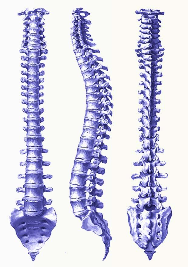
1 Skeletal System Bones & Joints


2 Vertebral Column Upper Limb Lower Limb OUTLINE Clinical Related Features Arrangements Features of the Joints


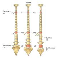
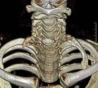
3 Vertebral Column (Overview) Costal Element Regional Features

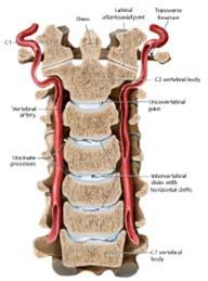
4 Uncovertebral Joint Cervical Spine Sections of Uncovertebral Joint Connections


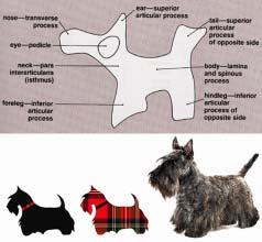
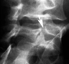


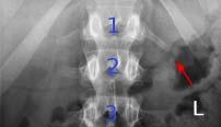
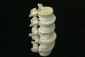
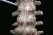
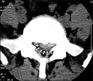
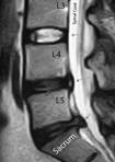
5 Lumbar Spine Scotty Dog Scotty Dog Fracture/Sign Lumbar Spine


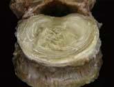


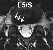
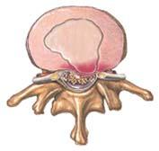
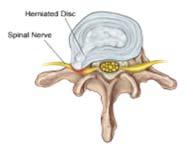
6 Connections of Vertebral Column Herniation of Nucleus Pulposus Anulus Fibrosus & Nucleus Pulposus Ligaments & Intervertebral Disc


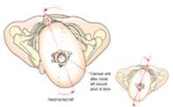
7 Atlanto-occipital Joints (Condyloid Joint) Overview Craniovertebral Joints Median Atlanto-axial Joint (Pivot Joint) Lateral Atlanto-axial Joints atlanto-occipital joints median atlanto-axial joint lateral atlanto-axial joints Movements


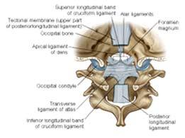
8 anterior longitudinal ligament anterior atlanto-occipital membrane & anterior atlanto-axial membrane Craniocervical Ligaments posterior longitudinal ligament tectorial membrane ligamentum flavum posterior atlanto-occipital membrane & posterior atlanto-axial membrane Tectorial Membrane Apical Ligament Alar Ligament

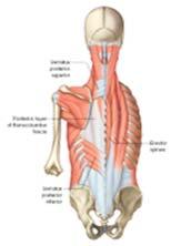
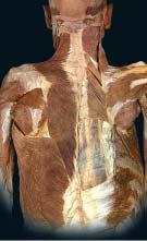
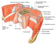
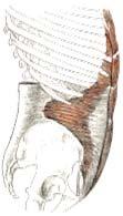
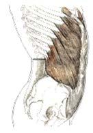
9 Superficial Muscles Back Muscles Thoracolumbar Fascia Abdominal Flat Muscles

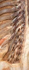
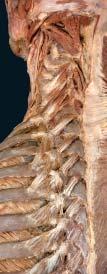
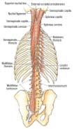
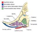
10 Deep Back Muscles intermediate intrinsic mm. deep intrinsic mm.

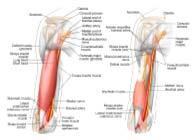
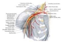
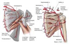
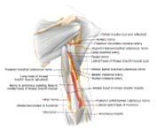
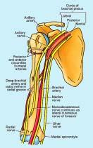
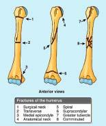

11 Subclaviain Artery Neurovasculatures Brachial ofplexus Anterior & Posterior Arm Dorsal Scapular Arterial Network Fractures of Humerus & Related Neurovasculatures Upper Limb Overview (Shoulder & Arm)
12 Intermuscular Septum


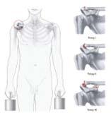
13 Coracoacromial Ligament & Coracoacromial Arch Sup. transverse Scapular Lig. & Scapular Notch Acromioclavicular Joint Acromioclavicular Lig. Coracoclavicular Acromioclavicular Lig. Coracoclavicular Lig. Injuries of Acromioclavicular Joint


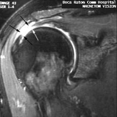
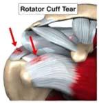
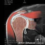
14 Musculotendinous /Rotator Cuff Rotator Cuff Tendon Tear Extensive Rupture of Left Cuff Normal Structures Test of Partial Tear of Cuff Patrick Omoumi, et al. Radiology, 2012 (264-3)


15 SGHL: superior glenohumeral ligament; BT: biceps tendon; CHL: coracohumeral ligament; MCHL: medial coracohumeral ligament; LCHL: lateral coracohumeral ligament; RIC: rotator interval capsule. David Y. Ding, et al. J Bone Joint Surg Am. 2014;96:e176(1-11) Popeye Sign Biceps Brachii
 Capsular Ligament")


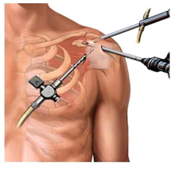
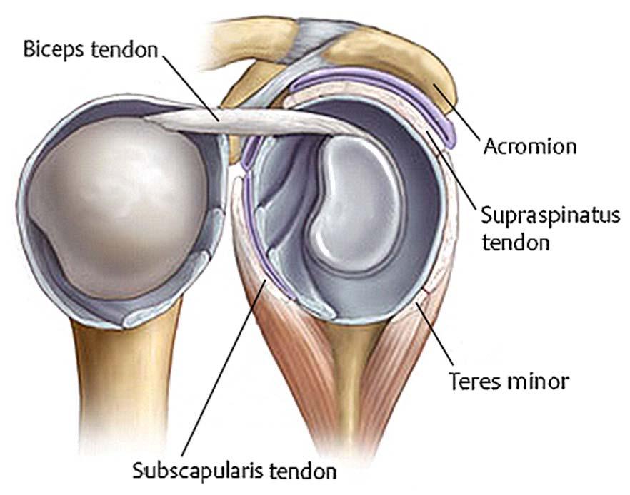
16 Biceps Brachii Tendon (Long Head) Capsular Ligament coracohumeral ligament glenohumeral ligament - superior, middle & inferior parts Shoulder Injection Shoulder/ Glenohumeral Joint
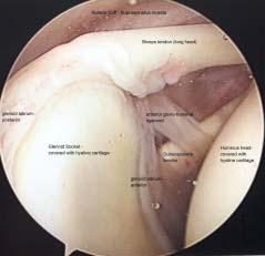
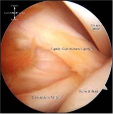

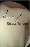


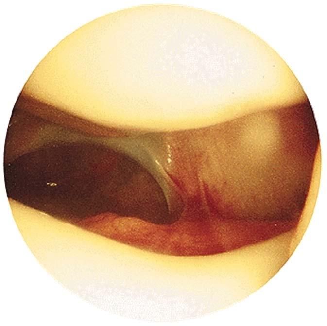
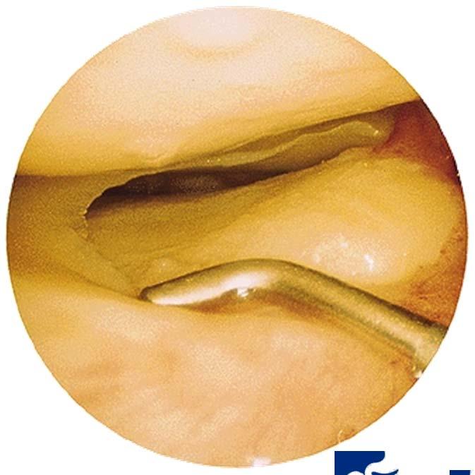
17 Shoulder Arthroscopy Images Reference Webpage:


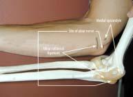
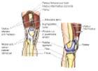
18 Hueter Line & Triangle Elbow Joint 1 capsule for 3 joints Capsular Ligaments radial collateral lig. ulnar collateral lig. anular lig. of radius

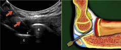


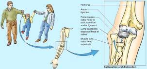
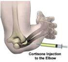
19 Elbow Injection Dislocation of Radal Head


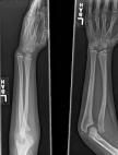
20 Fracture of Unla Radialis M. Group Bones & Muscles Forearm & Hand


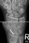
21 Wrist Joint TFCC Injury Note: nutrient arteries only enters the distal half of scaphoid Fracture of Scaphoid TFCC


22 lateral intermuscular septum - attaches to the iliotibial tract posteromedial intermuscular septum - sciatic nerve inside Arteries of the Thigh Arrangement of Lower Limb Thigh Leg
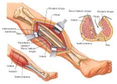
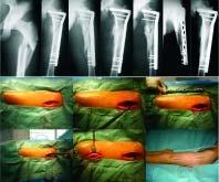
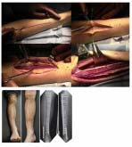
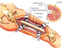
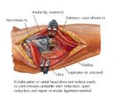
23 Surgical Approaches

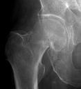
24 Hip Bone Bony Structures Femur Leg Bones


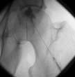
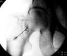
25 Arteriral Supply Network to Femoral Head Synovial Cavity Hip Joint Hip Injection


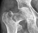
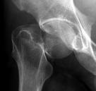
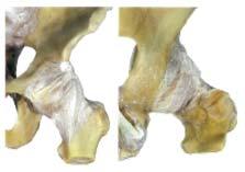
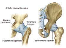
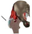
26 Fibrous Capsule & Capsular Ligament Fibrous Capsule Intracapsular & Extracapsular Fracture

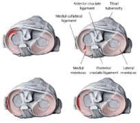
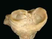
27 Capsule & Ligaments Synovial Capsule & Suprapatellar Bursa Knee Joint Menisci Alar Fold & Infrapatellar Fold


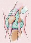
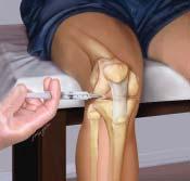
28 Meniscal Tears Suprapatellar Bursa Centesis & Knee Injection


29 Pes Anserinus & Anserine Bursa Synovial Bursae around the Knee
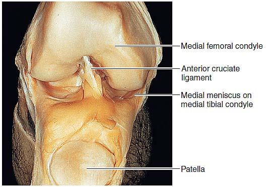
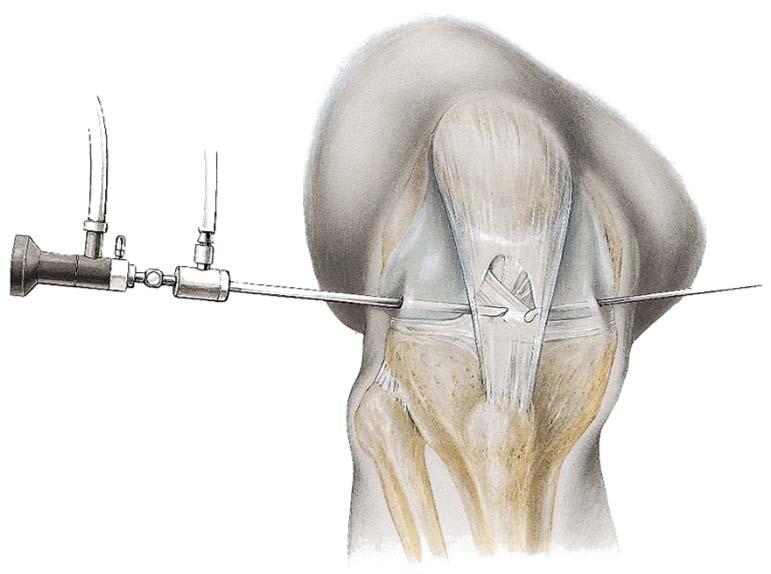
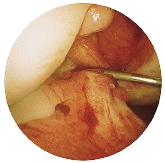
30 Arthroscope Structures inside Fibrous Capsule


31 articular branches of popliteal a. genicular branches of femoral a. recurrent branches of ant. tibial a. recurrent branches of circumflex fibular a. descending branch of lateral circumflex femoral a. Periarticular Genicular Anastomosis - medial sup. genicular a. - lateral sup. genicular a. - middle genicular a. - medial inf. genicular a. - lateral inf. genicular a. Zoran Blagojević, et al. International Orthopaedics (SICOT) (2015) 39:
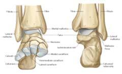
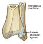
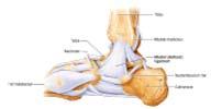
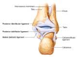
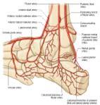
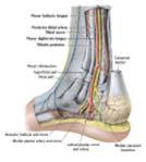
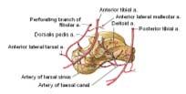
32 Articulation Ankle Joint Ligaments Arterial Inferior Anaostomoses Tibiofibular Joint & & Malleolar Ankle Canal Joint Arterial Network


33 Accessory Soleus Muscle Rupture of Achilles Tendon usually rupture appears of the as a distal calcaneal belly tendon medial is to the calcaneal often sustained tendon. by clinically, poorly conditioned an ccessory soleus people may with be a associated history of calcaneal with pain & edema tendinitis. during prolonged exercise.


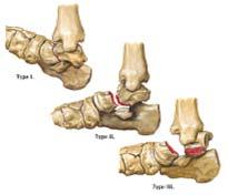
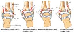


34 Fractures of Talar Neck Ankle Fracture (Lauge-Hansen) Hallucis Nerve Block Anterolater Apporach to Ankle
Anatomy of the Musculoskeletal System
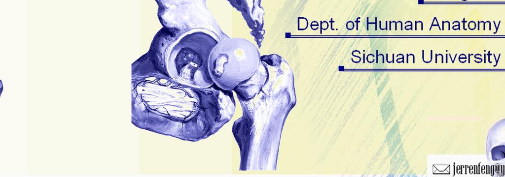
 Anatomy of the Musculoskeletal System Kyle E. Rarey, Ph.D. Department of Anatomy & Cell Biology and Otolaryngology University of Florida College of Medicine Outline of Presentation Vertebral Column Upper
Chapter 9 Articulations Articulations joints where two bones interconnect. Two classification methods are used to categorize joints:
 Chapter 9 Articulations Articulations joints where two bones interconnect Two classification methods are used to categorize joints: Functional classification Structural classification Functional classification
Joints of the Lower Limb II
 Joints of the Lower Limb II Lecture Objectives Describe the components of the knee and ankle joint. List the ligaments associated with these joints and their attachments. List the muscles acting on these
The Back OUTLINE. Vertebral Column (review) Craniovertebral Joints Dorsal Scapular Region(review) Muscles of the Back Suboccipital Region
 Craniovertebral Joints Dorsal Scapular Region(review) Muscles of the Back Suboccipital Region") The Back OUTLINE Vertebral Column (review) Craniovertebral Joints Dorsal Scapular Region(review) Muscles of the Back Suboccipital Region Dept. of Human Anatomy, Si Chuan University Zhou hongying eaglezhyxzy@163.com
The Back OUTLINE Vertebral Column (review) Craniovertebral Joints Dorsal Scapular Region(review) Muscles of the Back Suboccipital Region Dept. of Human Anatomy, Si Chuan University Zhou hongying eaglezhyxzy@163.com
Anatomy Workshop Upper Extremity David Ebaugh, PT, PhD Workshop Leader. Lab Leaders: STATION I BRACHIAL PLEXUS
 Anatomy Workshop Upper Extremity David Ebaugh, PT, PhD Workshop Leader Lab Leaders: STATION I BRACHIAL PLEXUS A. Posterior cervical triangle and axilla B. Formation of plexus 1. Ventral rami C5-T1 2. Trunks
Chiropractic ICD-10 Common Codes List
 Chiropractic ICD-10 Common Codes List This is a preliminary list of Common ICD-10 Codes for chiropractic diagnoses. This is a common code list to be used as a guide for coding and is not intended to represent
Functional anatomy and variability of the blood vessels of the upper and lower limbs. Anastasia Bendelic Human Anatomy Departament
 Functional anatomy and variability of the blood vessels of the upper and lower limbs Anastasia Bendelic Human Anatomy Departament Plan: 1. Variations of the branching pattern of the aortic arch 2. Arterial
Netter's Anatomy Flash Cards Section 6 List 4 th Edition
 Netter's Anatomy Flash Cards Section 6 List 4 th Edition https://www.memrise.com/course/1577581/ Section 6 Upper Limb (66 cards) Plate 6-1 Humerus and Scapula: Anterior View 1.1 Acromion 1.2 Greater tubercle
DISSECTION SCHEDULE. Session I - Hip (Front) & Thigh (Superficial)
 & Thigh (Superficial)") DISSECTION SCHEDULE Session I - Hip (Front) & Thigh (Superficial) Surface anatomy Inguinal region Gluteal region Thigh Leg Foot bones Hip bone Femur Superficial fascia Great saphenous vein Superficial
DISSECTION SCHEDULE Session I - Hip (Front) & Thigh (Superficial) Surface anatomy Inguinal region Gluteal region Thigh Leg Foot bones Hip bone Femur Superficial fascia Great saphenous vein Superficial
The Knee. Prof. Oluwadiya Kehinde
 The Knee Prof. Oluwadiya Kehinde www.oluwadiya.sitesled.com The Knee: Introduction 3 bones: femur, tibia and patella 2 separate joints: tibiofemoral and patellofemoral. Function: i. Primarily a hinge joint,
FUNCTIONAL ANATOMY OF SHOULDER JOINT
 FUNCTIONAL ANATOMY OF SHOULDER JOINT ARTICULATION Articulation is between: The rounded head of the Glenoid cavity humerus and The shallow, pear-shaped glenoid cavity of the scapula. 2 The articular surfaces
Joints of the upper limb II
 Joints of the upper limb II Prof. Abdulameer Al-Nuaimi E-mail: a.al-nuaimi@sheffield.ac.uk E. mail: abdulameerh@yahoo.com Elbow joint The elbow joint is connecting the upper arm to the forearm. It is classed
Exercise Science Section 4: Joint Mechanics and Joint Injuries
 Exercise Science Section 4: Joint Mechanics and Joint Injuries An Introduction to Health and Physical Education Ted Temertzoglou Paul Challen ISBN 1-55077-132-9 Types of Joints Fibrous joint Cartilaginous
P-DTR Intermediate Series Curriculum
 P-DTR Intermediate Series Curriculum MODULE 1 PRIORITY DYSFUNCTIONS Structure of fractals Rules of priority dysfunctions o Super basic mode Rules of Double UTL SEQUENCE DYSFUNCTIONS Reflexes vs. sequences
Scapular and Deltoid Regions
 M1 Gross and Developmental Anatomy Scapular and Deltoid Regions Dr. Peters 1 Outline I. Skeleton of the Shoulder and Attachment of the Upper Extremity to Trunk II. Positions and Movements of the Scapula
The Upper Limb II. Anatomy RHS 241 Lecture 11 Dr. Einas Al-Eisa
 The Upper Limb II Anatomy RHS 241 Lecture 11 Dr. Einas Al-Eisa Sternoclavicular joint Double joint.? Each side separated by intercalating articular disc Grasp the mid-portion of your clavicle on one side
Index. Note: Page numbers of article titles are in boldface type.
 Note: Page numbers of article titles are in boldface type. A ACJ. See Acromioclavicular joint (ACJ) Acromioclavicular joint (ACJ) procedures of, 557 559 Ankle and foot procedures of, 649 671 (See also
3/15/15. Chapter 8: Joints. Classification of Joints. Classification of Joints. } Objectives. } Functional Classifications
 Chapter 8: Joints Classification of Joints } Objectives } Define Joint or Articulation } Classify Joints by Structure and by Function } Describe the general structure, know the properties of, and provide
The Leg. Prof. Oluwadiya KS
 The Leg Prof. Oluwadiya KS www.oluwadiya.sitesled.com Compartments of the leg 4 Four Compartments: 1. Anterior compartment Deep fibular nerve Dorsiflexes the foot and toes 2. Lateral Compartment Superficial
STRUCTURAL BASIS OF MEDICAL PRACTICE EXAMINATION 5. September 30, 2011
 STRUCTURAL BASIS OF MEDICAL PRACTICE EXAMINATION 5 September 30, 2011 PART l. Answer in the space provided. (12 pts) 1. Identify the structures. (2 pts) EXAM NUMBER A. Suprascapular nerve B. Axillary nerve
Joints: Part B 10/30/14. Classification of Synovial Joints. Six types, based on shape of articular surfaces: Plane Joints
 PowerPoint Lecture Slides prepared by Janice Meeking, Mount Royal College C H A P T E R 8 Joints: Part B Classification of Synovial Joints Six types, based on shape of articular surfaces: Plane Hinge Pivot
CLASSIFICATION OF JOINTS STRUCTURAL VS FUNCTIONAL
 CHAPTER 8 JOINTS CLASSIFICATION OF JOINTS STRUCTURAL VS FUNCTIONAL The most moveable type of joint is a 1) Synarthrosis 2) Amphiarthrosis 3) Diarthrosis FIBROUS JOINTS Figure 8.1 Fibrous joints. (a) Suture
The thigh. Prof. Oluwadiya KS
 The thigh Prof. Oluwadiya KS www.oluwadiya.com The Thigh: Boundaries The thigh is the region of the lower limb that is approximately between the hip and knee joints Anteriorly, it is separated from the
Bones of Thorax (Rib Cage)
") Musculoskeletal System (Part A-2) Module 7 -Chapter 10 Overview Muscles Attachments Bones Bone types Surface features of bones Divisions of the skeletal system Joints or Articulations Susie Turner, M.D.
Musculoskeletal System (Part A-2) Module 7 -Chapter 10 Overview Muscles Attachments Bones Bone types Surface features of bones Divisions of the skeletal system Joints or Articulations Susie Turner, M.D.
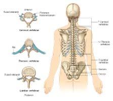
THE VERTEBRAL COLUMN. Average adult length: In male: about 70 cms. In female: about 65 cms.
 THE VERTEBRAL COLUMN Average adult length: In male: about 70 cms. In female: about 65 cms. 1 Vertebral Column (Regions and Curvatures) Curvatures of the vertebral column: A. Primary curvature: C-shaped;
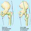
rotation of the hip Flexion of the knee Iliac fossa of iliac Lesser trochanter Femoral nerve Flexion of the thigh at the hip shaft of tibia
 Anatomy of the lower limb Anterior & medial compartments of the thigh Dr. Hayder The fascia lata encloses the entire thigh like a sleeve/stocking. Three intramuscular fascial septa (lateral, medial, and
Upper limb Arm & Cubital region 黃敏銓
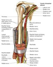
 Upper limb Arm & Cubital region 黃敏銓 1 Arm Lateral intermuscular septum Anterior (flexor) compartment: stronger Medial intermuscular septum Posterior (extensor) compartment 2 Coracobrachialis Origin: coracoid
The Knee Joint By Prof. Dr. Muhammad Imran Qureshi
 The Knee Joint By Prof. Dr. Muhammad Imran Qureshi Structurally, it is the Largest and the most complex joint in the body because of the functions that it performs: Allows mobility (flexion/extension)
Gluteal region DR. GITANJALI KHORWAL
 Gluteal region DR. GITANJALI KHORWAL Gluteal region The transitional area between the trunk and the lower extremity. The gluteal region includes the rounded, posterior buttocks and the laterally placed
STRUCTURAL BASIS OF MEDICAL PRACTICE EXAMINATION 5 October 6, 2006
 STRUCTURAL BASIS OF MEDICAL PRACTICE EXAMINATION 5 October 6, 2006 PART l. Answer in the space provided. (8 pts) 1. Identify the structures. (2 pts) B C A. _pisiform B. _ulnar artery A C. _flexor carpi
Arthrology joint, articulation or union between two or more bones Classification by degree of movement or tissue that bind the bones together
 ARTICULATIONS OF THE SPINE AND THORAX Pages 8-12, 42 and 57 Arthrology joint, articulation or union between two or more bones Classification by degree of movement or tissue that bind the bones together
MUSCULOSKELETAL LOWER LIMB
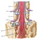
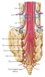
 MUSCULOSKELETAL LOWER LIMB Spinal Cord Lumbar and Sacral Regions Spinal cord Dorsal root ganglion Conus medullaris Cauda equina Dorsal root ganglion of the fifth lumbar nerve End of subarachnoid space
8.2: Fibrous Joints. There are three (3) types of fibrous joints (synarthroses): Syndesmosis Suture Gomphosis. Interosseus membrane of leg.
 types of fibrous joints (synarthroses): Syndesmosis Suture Gomphosis. Interosseus membrane of leg.") 8.1: Introduction Are known as articulations Functional junctions between bones Bind parts of skeletal system together Make bone growth possible Permit parts of the skeleton to change shape during childbirth
8.1: Introduction Are known as articulations Functional junctions between bones Bind parts of skeletal system together Make bone growth possible Permit parts of the skeleton to change shape during childbirth
Slides of Anatomy. Spring Dr. Maher Hadidi, University of Jordan
 Slides of Anatomy Please note : These slides are Dr. Maher Hadidi s slides of spring 2016 and were edited by the Premed Academic Team to fit the slides of spring 2019. Spring 2019 Dr. Maher Hadidi, University
Ligaments of Elbow hinge: sagittal plane so need lateral and medial ligaments
 Ligaments of Elbow hinge: sagittal plane so need lateral and medial ligaments Ulnar Collateral ligament on medial side; arising from medial epicondyle and stops excess valgus movement (lateral movement)
Misc Anatomy. Upper Limb! 2. Lower Limb! 5. Venous Drainage! Head & neck! 8
 Misc Anatomy Upper Limb! 2 Arteries!... 2 Veins!... 2 Spaces!... 4 Lower Limb! 5 Arteries!... 5 Venous Drainage!... 6 Spaces!... 7 Head & neck! 8 Artery!... 8 Ultrasound View for IJ CVL!... 8 Arteries
ARTICULATIONS and MUSCULAR SYSTEM
 ARTICULATIONS and MUSCULAR SYSTEM PART #1 ARTICULATIONS 1. Introduction A. Articulation C. Kinesiology B. Arthrology D. Rheumatology 2. Structural Classifications for Joints A. Fibrous Joints i. Suture
Joints Dr. Ali Ebneshahidi
 Joints Dr. Ali Ebneshahidi Function of Joints 1. Serve as functional junctions between bones. 2. Bind bones, strokes, and other related tissues together. 3. Allow bone growth to occur. 4. Permit certain
The Knee. Tibio-Femoral
 The Knee Tibio-Femoral Osteology Distal Femur with Proximal Tibia Largest Joint Cavity in the Body A modified hinge joint with significant passive rotation Technically, one degree of freedom (Flexion/Extension)
To describe he knee joint, ligaments, structure & To list the main features of other lower limb joints
 To describe he knee joint, ligaments, structure & neurovascular supply To demonstrate the ankle joint anatomy To list the main features of other lower limb joints To list main groups of lymph nodes in
region of the upper limb between the shoulder and the elbow Superiorly communicates with the axilla.
 1 region of the upper limb between the shoulder and the elbow Superiorly communicates with the axilla. Inferiorly, a number of important structures pass between arm & forearm through cubital fossa. 2 medial
Connects arm to thorax 3 joints. Glenohumeral joint Acromioclavicular joint Sternoclavicular joint
 Connects arm to thorax 3 joints Glenohumeral joint Acromioclavicular joint Sternoclavicular joint Scapula Elevation Depression Protraction (abduction) Retraction (adduction) Downward Rotation Upward Rotation
Gross Anatomy Coloring Book Series. Lower Extremity Arteries
 Gross Anatomy Coloring Book Series Lower Extremity Arteries 1 Femoral Artery and Associated Branches For the life of the flesh is in the blood. Leviticus 17:11 Femoral Artery and Associated Branches After
Leg. Dr. Heba Kalbouneh Associate Professor of Anatomy and Histology
 Leg Dr. Heba Kalbouneh Associate Professor of Anatomy and Histology Skin of the Leg Cutaneous Nerves Medially: The saphenous nerve, a branch of the femoral nerve supplies the skin on the medial surface
The Dance Hall by Vincent van Gogh,1888
 The Dance Hall by Vincent van Gogh,1888 Articulations of the pelvic girdle Lumbosacral joints, sacroiliac joints & pubic symphysis The remaining joints of the lower limb Hip joint Knee joint Tibiofibular
To classify the joints relative to structure & shape
 To classify the joints relative to structure & shape To describe the anatomy of the hip joint To describe the ankle joint To memorize their blood & nerve supply JOINTS: Joints are sites where skeletal
Anatomy. Anatomy deals with the structure of the human body, and includes a precise language on body positions and relationships between body parts.
 Anatomy deals with the structure of the human body, and includes a precise language on body positions and relationships between body parts. Proper instruction on safe and efficient exercise technique requires
UNIT 2 - CHAPTER 8: JOINTS OF THE SKELETAL SYSTEM LEARNING OUTCOMES:
 LEARNING OUTCOMES: 8.1 Introduction 1. List the functions of joints. 2. Explain how joints can be classified according to the type of tissue that binds the bones together and the degree of movement possible
Ligaments of the vertebral column:
 In the last lecture we started talking about the joints in the vertebral column, and we said that there are two types of joints between adjacent vertebrae: 1. Between the bodies of the vertebrae; which
locomotice system Plastinated specimensⅠ: Silicone specimens Regional specimens and organs
 locomotice system Plastinated specimensⅠ: Silicone specimens Regional specimens and organs Art-No. Name Description The locomotor system SL001 Two hundred pieces of plastinated bones (without six The bones
The Appendicular Skeleton
 8 The Appendicular Skeleton PowerPoint Lecture Presentations prepared by Jason LaPres Lone Star College North Harris 8-1 The Pectoral Girdle The Pectoral Girdle Also called shoulder girdle Connects the
UNIT 2 - CHAPTER 8: JOINTS OF THE SKELETAL SYSTEM LEARNING OUTCOMES:
 LEARNING OUTCOMES: 8.1 Types of Joints 1. Explain how joints can be classified according to the type of tissue that binds the bones together and the degree of movement possible at the joint. (p. 268) 2.
Femoral Artery. Its entrance to the thigh Position Midway between ASIS and pubic symphysis
 Lower Limb Vessels Lecture Objectives Describe the major arteries of the lower limb. Describe the deep and superficial veins of the lower limb. Describe the topographical relationships of the arteries
Lecture 9: Arthrology
 Lecture 9: Arthrology M/O Chapter 9 45. Classify joints based on the degree of movement allowed and give examples of each classification. 46. Classify joints based on anatomical structure and give examples
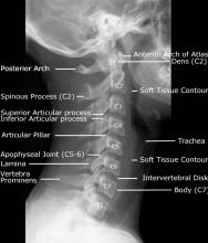
Demonstrate the bony features of Cl and C2 vertebrae evident on this Xray
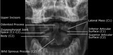
 SUBJECT: ANATOMY 7 September 2007 am. TOPIC: X-ray: Lateral C spine NUMBER: JL Demonstrate the bony features of Cl and C2 vertebrae evident on this Xray 1 Odontoid peg (dens) 2 Bodies of Cl andc2 3 anterior
VERTEBRAL COLUMN VERTEBRAL COLUMN
 VERTEBRAL COLUMN FUNCTIONS: 1) Support weight - transmits weight to pelvis and lower limbs 2) Houses and protects spinal cord - spinal nerves leave cord between vertebrae 3) Permits movements - *clinical
Pectoral (Shoulder) Girdle
 Girdle") Chapter 8 Skeletal System: Appendicular Skeleton Pectoral girdle Pelvic girdle Upper limbs Lower limbs 8-1 Pectoral (Shoulder) Girdle Consists of scapula and clavicle Clavicle articulates with sternum
Chapter 8 Skeletal System: Appendicular Skeleton Pectoral girdle Pelvic girdle Upper limbs Lower limbs 8-1 Pectoral (Shoulder) Girdle Consists of scapula and clavicle Clavicle articulates with sternum
Clarification of Terms
 Clarification of Terms The Spine, Spinal Column, and Vertebral Column are synonymous terms referring to the bony components housing the spinal cord Spinal Cord = made of nervous tissue Facet = a small,
Chapter 8. The Pectoral Girdle & Upper Limb
 Chapter 8 The Pectoral Girdle & Upper Limb Pectoral Girdle pectoral girdle (shoulder girdle) supports the arm consists of two on each side of the body // clavicle (collarbone) and scapula (shoulder blade)
Artery 1 Head and Thoracic Arteries. Arrange the parts in the order blood flows through them.
 Artery 1 Head and Thoracic Arteries 1. Given the following parts of the aorta: 1. abdominal aorta 2. aortic arch 3. ascending aorta 4. thoracic aorta Arrange the parts in the order blood flows through
The arm: *For images refer back to the slides
 The arm: *For images refer back to the slides Muscles of the arm: deltoid, triceps (which is located at the back of the arm), biceps and brachialis (it lies under the biceps), brachioradialis (it lies
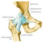
The Hip (Iliofemoral) Joint. Presented by: Rob, Rachel, Alina and Lisa
 Joint. Presented by: Rob, Rachel, Alina and Lisa") The Hip (Iliofemoral) Joint Presented by: Rob, Rachel, Alina and Lisa Surface Anatomy: Posterior Surface Anatomy: Anterior Bones: Os Coxae Consists of 3 Portions: Ilium Ischium Pubis Bones: Pubis Portion
The Hip (Iliofemoral) Joint Presented by: Rob, Rachel, Alina and Lisa Surface Anatomy: Posterior Surface Anatomy: Anterior Bones: Os Coxae Consists of 3 Portions: Ilium Ischium Pubis Bones: Pubis Portion
and K n e e J o i n t Is the most complicated joint in the body!!!!
 K n e e J o i n t K n e e J o i n t Is the most complicated joint in the body!!!! 1-Consists of two condylar joints between: A-The medial and lateral condyles of the femur and The condyles of the tibia
The Lower Limb II. Anatomy RHS 241 Lecture 3 Dr. Einas Al-Eisa
 The Lower Limb II Anatomy RHS 241 Lecture 3 Dr. Einas Al-Eisa Tibia The larger & medial bone of the leg Functions: Attachment of muscles Transfer of weight from femur to skeleton of the foot Articulations
Region of upper limb attachment to the trunk Proximal segment of limb overlaps parts of the trunk (thorax and back) and lower lateral neck.
 and lower lateral neck.") Region of upper limb attachment to the trunk Proximal segment of limb overlaps parts of the trunk (thorax and back) and lower lateral neck. includes Pectoral Scapular Deltoid regions of the upper limb
Region of upper limb attachment to the trunk Proximal segment of limb overlaps parts of the trunk (thorax and back) and lower lateral neck. includes Pectoral Scapular Deltoid regions of the upper limb
MUSCLES OF SHOULDER REGION
 Dr Jamila EL Medany OBJECTIVES At the end of the lecture, students should: List the name of muscles of the shoulder region. Describe the anatomy of muscles of shoulder region regarding: attachments of
Index. Note: Page numbers of article titles are in boldface type.
 Note: Page numbers of article titles are in boldface type. A Abscess, epidural, 822 824 Achilles tendon rupture, 894 895, 981 982 Acromioclavicular separations, shoulder pain in, 751 753 Adhesive capsulitis,
CHAPTER 9 LECTURE OUTLINE INTRODUCTION
 CHAPTER 9 LECTURE OUTLINE INTRODUCTION I. A joint (articulation or arthrosis) is a point of contact between two or more bones, between cartilage and bones, or between teeth and bones. A. The scientific
Clarification of Terms
 Clarification of Terms The Spine, Spinal Column, and Vertebral Column are synonymous terms referring to the bony components housing the spinal cord Spinal Cord = made of nervous tissue Facet = a small,
Clarification of Terms
 Clarification of Terms The Spine, Spinal Column, and Vertebral Column are synonymous terms referring to the bony components housing the spinal cord Spinal Cord = made of nervous tissue Facet = a small,
10/12/2010. Upper Extremity. Pectoral (Shoulder) Girdle. Clavicle (collarbone) Skeletal System: Appendicular Skeleton
 Girdle. Clavicle (collarbone) Skeletal System: Appendicular Skeleton") Skeletal System: Appendicular Skeleton Pectoral girdle Pelvic girdle Upper limbs Lower limbs 8-1 Pectoral (Shoulder) Girdle Consists of scapula and clavicle Clavicle articulates with sternum (Sternoclavicular
Skeletal System: Appendicular Skeleton Pectoral girdle Pelvic girdle Upper limbs Lower limbs 8-1 Pectoral (Shoulder) Girdle Consists of scapula and clavicle Clavicle articulates with sternum (Sternoclavicular
Knee Joint Anatomy 101
 Knee Joint Anatomy 101 Bone Basics There are three bones at the knee joint femur, tibia and patella commonly referred to as the thighbone, shinbone and kneecap. The fibula is not typically associated with
Exercise 13. Articulations and Body Movements
 Exercise 13 Articulations and Body Movements Articulations Articulations, or joints, are points where a bone is connected to one or more other bones. Articulations hold the skeleton together. Articulations
Lecture 09. Popliteal Fossa. BY Dr Farooq Khan Aurakzai
 Lecture 09 Popliteal Fossa BY Dr Farooq Khan Aurakzai Dated: 14.02.2018 What is popliteus? Introduction Anything relating to, or near the part of the leg behind the knee. From New Latin popliteus the muscle
Human Anatomy, First Edition McKinley & O'Loughlin
 Human Anatomy, First Edition McKinley & O'Loughlin Chapter 8 : Appendicular Skeleton 8-1 Appendicular Skeleton Includes the bones of the upper and lower limbs. The girdles of bones that attach the upper
The Arm and Cubital Fossa
 The Arm and Cubital Fossa Dr. Andrew Gallagher School of Anatomical Sciences University of the Witwatersrand Introduction The ARM (BRACHIUM) is the most proximal segment of the upper limb musculoskeletal
3 Mohammad Al-Mohtasib Areej Mosleh
 3 Mohammad Al-Mohtasib Areej Mosleh ***Muscles Connecting the Upper Limb to the Vertebral Column 1.Trapezius Muscle ***The first muscle on the back is trapezius muscle, it s called so according
Copyright 2010 Pearson Education, Inc. Copyright 2010 Pearson Education, Inc. Figure Sectioned spinous process. Interspinous.
 PowerPoint Lecture Slides prepared by Janice Meeking, Mount Royal College C H A P T E R 7 The Skeleton: Part B Vertebral Column Transmits weight of trunk to lower limbs Surrounds and protects spinal cord
2. The vertebral arch is composed of pedicles (projecting from the body) and laminae (uniting arch posteriorly).
 and laminae (uniting arch posteriorly).") VERTEBRAL COLUMN 2018zillmusom I. VERTEBRAL COLUMN - functions to support weight of body and protect spinal cord while permitting movements of trunk and providing for muscle attachments. A. Typical vertebra
VERTEBRAL COLUMN 2018zillmusom I. VERTEBRAL COLUMN - functions to support weight of body and protect spinal cord while permitting movements of trunk and providing for muscle attachments. A. Typical vertebra
The Elbow and the cubital fossa. Prof Oluwadiya Kehinde
 The Elbow and the cubital fossa Prof Oluwadiya Kehinde www.oluwadiya.com Elbow and Forearm Anatomy The elbow joint is formed by the humerus, radius, and the ulna Bony anatomy of the elbow Distal Humerus
Copyright 2003 Pearson Education, Inc. publishing as Benjamin Cummings. Dr. Nabil khouri
 Dr. Nabil khouri Appendicular Skeleton The appendicular skeleton is made up of the bones of the upper and lower limbs and their girdles Two girdles: Pectoral girdles attach the upper limbs to the body
Topic list for the Anatomy end-semester exam of the First semester Academic year 2018/2019, Autumn term
 University of Szeged, Faculty of Medicine Department of Anatomy, Histology and Embryology Chairman: Prof. Antal Nógrádi MD, PhD, DSc Kossuth L. sgt. 40., H-6724 Szeged, Hungary Tel.: +36-62-545-665 P.
Schedule. Quiz 3: Lower Extremity Bones (10pts) Assignment 3 was due New Material: Joints and Ligaments Assignment 4 and EC 2 Posted
 Assignment 3 was due New Material: Joints and Ligaments Assignment 4 and EC 2 Posted") Schedule Quiz 3: Lower Extremity Bones (10pts) Assignment 3 was due New Material: Joints and Ligaments Assignment 4 and EC 2 Posted JOINTS & LIGAMENTS What is a joint? AKA Articulations Definition: Union
Schedule Quiz 3: Lower Extremity Bones (10pts) Assignment 3 was due New Material: Joints and Ligaments Assignment 4 and EC 2 Posted JOINTS & LIGAMENTS What is a joint? AKA Articulations Definition: Union
Functional Anatomy of the Elbow
 Functional Anatomy of the Elbow Orthopedic Institute Daryl C. Osbahr, M.D. Chief of Sports Medicine, Orlando Health Chief Medical Officer, Orlando City Soccer Club Orthopedic Consultant, Washington Nationals
Definition of anatomy 1 Questions 5
 Contents Chapter 1: Introduction 1 5 Definition of anatomy 1 Questions 5 Chapter 2: Skeletal System 6 74 Skeleton 6 Skeletal system 8 Bones of superior extremity 12 Articulated skeleton of hand 17 Clinical
DART Diagnosis and Related Treatments
 DART Diagnosis and Related Treatments The DART Tool allows a user to obtain Treatment recommendations based on the disorder/condition. These recommendations are provided by ACOEM (American College of Occupational
The Skeletal System THE APPENDICULAR SKELETON
 The Skeletal System THE APPENDICULAR SKELETON The appendicular skeleton consists of the girdles and the skeleton of the limbs. The upper (anterior) limbs are attached to the pectoral (shoulder) girdle
Chapter 9 Joints. Classification of Joints. Fibrous Joints. Structural classification based upon: Functional classification based upon movement:
 Chapter 9 Joints Joints hold bones together but permit movement Point of contact between 2 bones between cartilage and bone between teeth and bones Arthrology = study of joints Kinesiology = study of motion
Lecture 08 THIGH MUSCLES ANTERIOR COMPARTMENT. Dr Farooq Khan Aurakzai. Dated:
 Lecture 08 THIGH MUSCLES ANTERIOR COMPARTMENT BY Dr Farooq Khan Aurakzai Dated: 11.02.2017 INTRODUCTION to the thigh Muscles. The musculature of the thigh can be split into three sections by intermuscular
Introduction. Fibrous Joints. 8.1: Types of Joints. Cartilaginous Joints. Fibrous Joints 12/14/2016. Chapter 08 Lecture Outline
 Introduction Chapter 08 Lecture Outline See separate PowerPoint slides for all figures and tables preinserted into PowerPoint without notes. Joints (Articulations): Functional junctions between bones Bind
Anterior and Medial compartments of the thigh. Dr. Heba Kalbouneh Associate Professor of Anatomy and Histology
 Anterior and Medial compartments of the thigh Dr. Heba Kalbouneh Associate Professor of Anatomy and Histology Terms Related to Movements Movement Flexion Extension Abduction Adduction Medial (internal)
Fascial Compartments of the Upper Arm
 Fascial Compartments of the Upper Arm The upper arm is enclosed in a sheath of deep fascia and has two fascial septa: 1- Medial fascial septum (medial intermuscular septum): attached to the medial supracondylar
Joints. Judi Laprade. Illustrations from: Essential Clinical Anatomy 3 rd ed. (ECA3) Moore, K. and Agur, A. Lippincott Williams and Wilkins, 2007
 Moore, K. and Agur, A. Lippincott Williams and Wilkins, 2007") Slide 1 Joints Judi Laprade Illustrations from: Essential Clinical Anatomy 3 rd ed. (ECA3) Moore, K. and Agur, A. Lippincott Williams and Wilkins, 2007 Grant s Atlas of Anatomy 12 th ed. (GA12) Agur, A.
Slide 1 Joints Judi Laprade Illustrations from: Essential Clinical Anatomy 3 rd ed. (ECA3) Moore, K. and Agur, A. Lippincott Williams and Wilkins, 2007 Grant s Atlas of Anatomy 12 th ed. (GA12) Agur, A.
The Muscular System. Chapter 10 Part C. PowerPoint Lecture Slides prepared by Karen Dunbar Kareiva Ivy Tech Community College
 Chapter 10 Part C The Muscular System Annie Leibovitz/Contact Press Images PowerPoint Lecture Slides prepared by Karen Dunbar Kareiva Ivy Tech Community College Table 10.9: Muscles Crossing the Shoulder
Elbow Elbow Anatomy. Flexion extension. Pronation Supination. Anatomy. Anatomy. Romina Astifidis, MS., PT., CHT
 Elbow Elbow Anatomy Romina Astifidis, MS., PT., CHT Curtis National Hand Center Baltimore, MD October 6-8, 2017 Link between the arm and forearm to position the hand in space Not just a hinge Elbow = 70%
Anatomy and Physiology 1 Chapter 9 self quiz Pro, Dima Darwish,MD.
 Anatomy and Physiology 1 Chapter 9 self quiz Pro, Dima Darwish,MD. 1) Joints can be classified structurally as A) bony. B) fibrous. C) cartilaginous. D) synovial. E) All of the answers are correct. 2)
Anatomage Table Instructors Guide- Lower Limb
 The Lower Limb Anatomage Table Instructors Guide- Lower Limb Table of Contents Lower Limb 1- The Skeletal System...3 1: Hip Bone...3 2: Hip Joint and Femur...4 3: Patella and Knee Joint...7 4: Tibia, Fibula,
Human Anatomy Laboratory Manual with Cat Dissections Marieb Mitchell Smith Seventh Edition
 Human Anatomy Laboratory Manual with Cat Dissections Marieb Mitchell Smith Seventh Edition Pearson Education Limited Edinburgh Gate Harlow Essex CM20 2JE England and Associated Companies throughout the
A. Incorrect! The appendicular skeleton includes bones of the shoulder, arm, hand, pelvis, leg and foot.
 Anatomy and Physiology - Problem Drill 08: The Skeletal System III No. 1 of 10 1. Which of the following statements about the appendicular skeleton is correct? A. The appendicular skeleton includes bones
Structure and Function of the Vertebral Column
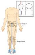
 Structure and Function of the Vertebral Column Posture Vertebral Alignment Does it really matter? Yes it does! Postural Curves The vertebral column has a series of counterbalancing curves posterior anterior