RSV infection and lung ultrasound
|
|
|
- Domenic Caldwell
- 5 years ago
- Views:
Transcription
1 RSV infection and lung ultrasound Neonatology Clinic, University Hospital, Krakow Joanna Hurkała Joanna Pietras Agnieszka Ochoda-Mazur Poznań,
2 Disclosure In relations to this presentation, I declare NO conflicts of interest 2
3 RSV infection RSV- enveloped negative sense, single-strand RNA virus, Pneumoviridae Respiratory syncytial virus (RSV) is the single most important cause of severe respiratory infection in very young infants Most common viral pathogen of pneumonia in children <2 years (28-31% identified) 3

, 2,1% (without BPD) * *Resh B et al.")
4 RSV infection RISK FACTORS prematurity BPD CHD immunodeficiency chromosomal abnormalities eg. Down syndrome Incidents of HOSPITALIZATION among preterm infants <32 weeks: Pre- palivizumab era - 7,5-35% (infants with BPD), 2-12% (without BPD) Palivizumab profilaxis - 5,8% (BPD), 2,1% (without BPD) * *Resh B et al. International Jour Infec Disease 57 (2017) 4

5 RSV infection EPIDEMIOLOGY Temperate climate- annual epidemicswinter RSV circulation weeks in a community, overlaps with influenza epidemics In the tropics circulation more variable throughout the year TRANSMITION Older children introduce into family, spread to infants Nasal secretion, aerosol Virus stable for several hours on hands and hard surface 5
6 RSV infection Acute illness, pneumonia 1-2% of infants in their first year of life are hospitalized (US) *Walsh E, Clin Chest Med % emergency patients with acute respiratory symptoms RSV (+) Mortality in developed countries low ( anually in US), Developing world deaths, and >3 milion hospitalization Subsequent wheezing in childhood, may be related to asthma in later life 6
7 Radiologic findings NOT typical, variable 31% asymptomatic 16% overinflation without infiltrates in X-ray 48% consolidation (diffuse areas, lobar consolidation, unilateral single-lower zone involvement) Interstitial lung disease Ground- glass opacity *Guo W, Brit Jour of Radiol, 2012 *Wang S, AJR
8 CT scans Viral pneumonia- variable, described in adults Poorly defined centrilobular nodules Patchy areas of peribronchial ground- glass opacity Segmental consolidation, parenchymal airspace consolidation Diffuse ground- glass attenuation with bronchial wall thickening (tree in bud) *Syha R, Eur J Radiol 2012, *Perez Padilla R, N. Engl J Med
9 LUS signs according to references Lack of studies, but: LUS found to be valuable tool in the diagnosis of bronchiolitis * Caiulo VA et al., Eur j Pediatr, 2011 Drastically reduce the need for chest X-ray * Volpicelli G et al. Intensive Care Med., Longitudinal, transversal sections, ANT, LAT, POST * Copetti R, Catarosi L, Neonatology 2007, 2008 No particular findings characteristic only for RSV *Tsung JW, Kessler DO, ShahVP, Crit Ultrasound J, BPD could be similar to bronchiolitis in RSV *Basile V et al., Pediatrics,
10 Ultrasound signs according to references Pleural line thickening B- lines, pulmonary intersitial syndrome- concentration of vertical artifacts - confluent or coalescing Areas of lung dysventilation- consolidation adjacent to the pleural line associated or not with local pleural effusion Comet tail sign Signs of bronchiolitis 10
11 Ultrasound signs according to reffernces Ultrasound anomalies on posterior and paravertebral scans gravity of supine newborns and infants Quantitive classification of interstitial syndrome (eg.involved intercostal spaces post bilaterally) corresponds with the need for oxygen supplementation and severity of bronchiolitis Subpleural consolidation of 1 cm or more in the posterior area- need of oxygen supplementation *Basile V et al., Pediatrics,
12 Mild pneumonia Patient W. Male, Hbd 32, BW 1780g Surfactant INSURE after birth, ncpap 7 days 26th day of hospitalisation need of oxygen, cracles, feeding problems On 28th RSV diagnosis, need of ncpap that was continued for consecutive 6 days Oxygen for next several days On 57th day of life patient discharge Patients W. Twin 1 and Twin 2 Male, Hbd 27, BW 1160g & 1140g Surfactant INSURE after birth, ncpap RSV diagnosis on 60th day T2, on 67th T1 Twin 2 CPAP,Twin1 oxygen Discharge on 78th day of life 12
13 Severe complicated pneumonia Patient S. Male, Hbd 28, BW 780g Trisomy 21 ncpap continued for 4 weeks ASD II, VSD treated with enalapryl On 54-56th day of life episodes of desaturation 3 days of ncpap, then invasive ventilation (21 days) Coinfection with Acinetobacter Extubated on 76th day of life Discharged on 118th day of life 13
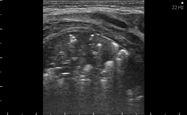
14 Our observations Pleural line thickening and numerous B lines Consolidations prodromal and advenced phases Paravertebral atelectasis Pleural effusion Accidental finding by ino therapy Role of positioning 14

15 Prodromal phase Pleural line and B lines Patient S small consolidations and B lines in front areas, thickening and interruption of pleural line a.ochoda@gmail.com Neonatus

16 Proromal phase Pleural line and B lines Patient W small consolidations in front areas, thickening and interruption of pleural line 16

17 Pleural line and B lines Patient S more intense consolidations and B lines in back areas, thickening and interruption of pleural line a.ochoda@gmail.com Neonatus
18 Prodromal phase Consolidations Patient S. triangle (qouin) like consolidation in the prodromal phase 18
19 Prodromal phase Consolidation Patient W. triangle (qouin) like consolidation in the prodromal phase Neonatus

20 AO3 Consolidations prodromal phase Patient S Patient W twin1 Patient Patient WW Patient W twin2 20
21 Slajd 20 AO3 scapular, midvertebral line Agnieszka Ochoda;


22 Progression of consolidations Intensification of B lines, white lung Progresion of consolidations 21

23 Progression of consolidations Intensification of B lines, white lung Progresion of consolidations 22


24 Paravertebral consolidations S. W. W. Twin 1 W. Twin 2 23
25 D4 Patient W D4 Patient S respiratory failure, ncpap CRP- <4,8mg/l PCT- 0,12ng/ml RSV a.ochoda@gmail.com respiratory failure, intubation CRP- 15,3 mg/l PCT- 0,25 ng/ml RSV Neonatus
26 D4 Patient W D4 Patient S 25


27 D4 Patient S Consolidation with air static bronchogram 26
28 Patient S D5 Mild atelectasis 27

29 D8 - Patient S Pleural effusion 28

30 D8 - Patient S ino therapy 29

31 D8 - Patient S Rich pulmonary vasculature 30


32 Atelectasis - right inferior lobe partly aerated and fully colapsed 31


33 Patient S Day 5 Day 10 a.ochoda@gmail.com Neonatus
34 D10 - Patient S dynamic air bronchogram 33
35 D13 - Patient S Atelectasis of the inferior left lobe 34
")
36 D17 - Patient S better aeration of front parts (baby mainly in supine position) 35


37 D17 - Patient S Partly inflated atelectasis of the back parts 36

38 D31 - Patient S on ncpap 37

39 D31 - Patient S - ncpap thymus and small consolidation 38

40 D31 -Patient S - ncpap small pericardial consolidation 39
41 Summarise LUS is more sensitive in prodromal phase than X-ray Sudden respiratory deterioration (even mild) in preterms should always be checked by LUS Triangle-like consolidations are probably the typical sign in early phase of RSV pneumonia Other signs present in all patients: pleural line thickening, numerous B lines, paravertebral consolidations LUS is a non-invasive method that may be used every day to monitor the state and helps to chose the right intervention LUS is a 4D method (real time, localises precisely change in all dimensions) 40
42 THANK YOU 41
Definitions and diagnostic implications of terms used in the chest radiograph and lung ultrasound diagnoses of pneumonia.
 Supplementary 1 Definitions and diagnostic implications of terms used in the chest radiograph and lung ultrasound diagnoses of pneumonia. Imaging finding Definition Implication CR Consolidation Interstitial
Pediatric Lung Ultrasound (PLUS) In Diagnosis of Community Acquired Pneumonia (CAP)
 In Diagnosis of Community Acquired Pneumonia (CAP)") Pediatric Lung Ultrasound (PLUS) In Diagnosis of Community Acquired Pneumonia (CAP) Dr Neetu Talwar Senior Consultant, Pediatric Pulmonology Fortis Memorial Research Institute, Gurugram Study To compare
Pediatric Lung Ultrasound (PLUS) In Diagnosis of Community Acquired Pneumonia (CAP) Dr Neetu Talwar Senior Consultant, Pediatric Pulmonology Fortis Memorial Research Institute, Gurugram Study To compare
Chest X rays and Case Studies. No disclosures. Outline 5/31/2018. Carlo Manalo, M.D. Department of Radiology Loma Linda University Children s Hospital
 Chest X rays and Case Studies Carlo Manalo, M.D. Department of Radiology Loma Linda University Children s Hospital No disclosures. Outline Importance of history Densities delineated on radiography An approach
Immunocompromised patients. Immunocompromised patients. Immunocompromised patients
 Value of CT in Early Pneumonia in Immunocompromised Patients Nantaka Kiranantawat, PSU Preventative Factors Phagocyts Cellular immunity Humoral immunity Predisposing Factors Infection, Stress, Poor nutrition,
Cryptogenic Organizing Pneumonia Diagnosis Approach Based on a Clinical-Radiologic-Pathologic Consensus
 Cryptogenic Organizing Pneumonia Diagnosis Approach Based on a Clinical-Radiologic-Pathologic Consensus Poster No.: C-1622 Congress: ECR 2012 Type: Scientific Exhibit Authors: C. Cordero Lares, E. Zorita
and localized ground glass opacities, or bronchiolar focal or multifocal micronodules;
 E1 Chest CT scan and Pneumoniae_YE Claessens et al- Supplementary methods Level of CAP probability according to CT scan - definite CAP: systematic alveolar condensation, or alveolar condensation with peripheral
PULMONARY TUBERCULOSIS RADIOLOGY
 PULMONARY TUBERCULOSIS RADIOLOGY RADIOLOGICAL MODALITIES Medical radiophotography Radiography Fluoroscopy Linear (conventional) tomography Computed tomography Pulmonary angiography, bronchography Ultrasonography,
Differential diagnosis
 Differential diagnosis Idiopathic pulmonary fibrosis (IPF) is part of a large family of idiopathic interstitial pneumonias (IIP), one of four subgroups of interstitial lung disease (ILD). Differential
The Intensive Care Unit
 Imaging of the ADRS patient: Risk of transportation and alternative to repetitive radiation exposure Jean-Jacques Rouby Pitié-Salpêtrière Hospital The Intensive Care Unit The Intensive Care Unit Multidisciplinary
Eun-Young Kang, M.D., Jae Wook Lee, M.D., Ji Yung Choo, M.D., Hwan Seok Yong, M.D., Ki Yeol Lee, M.D., Yu-Whan Oh, M.D.
 Eun-Young Kang, M.D., Jae Wook Lee, M.D., Ji Yung Choo, M.D., Hwan Seok Yong, M.D., Ki Yeol Lee, M.D., Yu-Whan Oh, M.D. Department of Radiology, Korea University Guro Hospital, College of Medicine, Korea
Key messages. CXR interpretation in TB/HIV setting. Training course
 Key messages CXR interpretation in TB/HIV setting Training course Normal CXR Front view and lateral view Good notions of technical conditions to obtain a good CXR Good knowledge of criteria for quality
Web Chapter 3. Image Gallery: Lesion detection on low dose chest CT
 Web Chapter 3 Image Gallery: Lesion detection on low dose chest CT Sarabjeet Singh, MD Mannudeep K. Kalra, MD *Eugene J. Mark, MD *James Stone, MD James H. Thrall, MD Department of Radiology and *Department
An Image Repository for Chest CT
 An Image Repository for Chest CT Francesco Frajoli for the Chest CT in Antibody Deficiency Group An Image Repository for Chest CT he Chest CT in Antibody Deficiency Group is an international and interdisciplinary
Diagnosis of TB: Radiology David Finlay, MD
 TB Intensive Tyler, Texas June 2-4, 2010 Diagnosis of TB: Radiology David Finlay, MD June 3, 2010 2stages stages- Tuberculosis 1. primary infection 2. reactivation, or post primary disease 2 1 Primary
Imaging Small Airways Diseases: Not Just Air trapping. Eric J. Stern MD University of Washington
 Imaging Small Airways Diseases: Not Just Air trapping Eric J. Stern MD University of Washington What we are discussing SAD classification SAD imaging with MDCT emphasis What is a small airway? Airway with
Radiographic Features of SARS in Paediatric Patients: A Review of Cases in Singapore
 340 Original Article Radiographic Features of SARS in Paediatric Patients: A Review of Cases in Singapore Jaiman V Emmanuel, 1 MBBS, FRCR, Uei Pua, 1 MBBS, FRCR, Gervais KL Wansaicheong, 1 FRCR, M Med,
Systemic lupus erythematosus (SLE): Pleuropulmonary Manifestations
: Pleuropulmonary Manifestations") 08/30/10 09/26/10 Systemic lupus erythematosus (SLE): Pleuropulmonary Manifestations Camila Downey S. Universidad de Chile, School of Medicine, Year VII Harvard University, School of Medicine Sept 17,
08/30/10 09/26/10 Systemic lupus erythematosus (SLE): Pleuropulmonary Manifestations Camila Downey S. Universidad de Chile, School of Medicine, Year VII Harvard University, School of Medicine Sept 17,
TB Intensive Houston, Texas
 TB Intensive Houston, Texas October 15-17, 17 2013 Diagnosis of TB: Radiology Rosa M Estrada-Y-Martin, MD MSc FCCP October 16, 2013 Rosa M Estrada-Y-Martin, MD MSc FCCP, has the following disclosures to
Acute and Chronic Lung Disease
 KATHOLIEKE UNIVERSITEIT LEUVEN Faculty of Medicine Acute and Chronic Lung Disease W De Wever, JA Verschakelen Department of Radiology, University Hospitals Leuven, Belgium Clinical utility of HRCT To detect
ARDS - a must know. Page 1 of 14
 ARDS - a must know Poster No.: C-1683 Congress: ECR 2016 Type: Authors: Keywords: DOI: Educational Exhibit M. Cristian; Turda/RO Education and training, Edema, Acute, Localisation, Education, Digital radiography,
Tuberculosis: The Essentials
 Tuberculosis: The Essentials Kendra L. Fisher, MD, PhD THORACIC TUBERCULOSIS: THE BARE ESSENTIALS Kendra Fisher MD, FRCP (C) Department of Radiology Loma Linda University Medical Center TUBERCULOSIS ()
10/17/2016. Nuts and Bolts of Thoracic Radiology. Objectives. Techniques
 Nuts and Bolts of Thoracic Radiology October 20, 2016 Carleen Risaliti Objectives Understand the basics of chest radiograph Develop a system for interpreting chest radiographs Correctly identify thoracic
ACUTE RESPIRATORY DISTRESS SYNDROME
 ACUTE RESPIRATORY DISTRESS SYNDROME Angel Coz MD, FCCP, DCE Assistant Professor of Medicine UCSF Fresno November 4, 2017 No disclosures OBJECTIVES Identify current trends and risk factors of ARDS Describe
Acute pneumonia Simple complement
 Acute pneumonia Simple complement 1. Clinical variants of acute pneumonia in children are, except: A. Bronchopneumonia B. Lobar confluent pneumonia C. Viral pneumonia D. Interstitial pneumonia E. Chronic
Pediatric High-Resolution Chest CT
 Pediatric High-Resolution Chest CT Alan S. Brody, MD Professor of Radiology and Pediatrics Chief, Thoracic Imaging Cincinnati Children s s Hospital Cincinnati, Ohio, USA Pediatric High-Resolution CT Short
Radiological and clinical characteristics of plastic bronchitis complicated with H1N1 influenza viral pneumonia in children
 Radiological and clinical characteristics of plastic bronchitis complicated with H1N1 influenza viral pneumonia in children Poster No.: C-1727 Congress: ECR 2012 Type: Scientific Exhibit Authors: H. Fujisawa,
Thoracic lung involvement in rheumatoid arthritis: Findings on HRCT
 Thoracic lung involvement in rheumatoid arthritis: Findings on HRCT Poster No.: C-2488 Congress: ECR 2015 Type: Educational Exhibit Authors: R. E. Correa Soto, M. J. Martín Sánchez, J. M. Fernandez 1 1
Shedding Light on Neonatal X-rays. Objectives. Indications for X-Rays 5/14/2018
 Shedding Light on Neonatal X-rays Barbara C. Mordue, MSN, NNP-BC Neonatal Nurse Practitioner LLUH Children s Hospital, NICU Objectives Utilize a systematic approach to neonatal x-ray interpretation Identify
Chest radiography findings in adults with pandemic H1N influenza
 The British Journal of Radiology, 83 (2010), 499 504 Chest radiography findings in adults with pandemic H1N1 2009 influenza 1 REMCEWEN, MB, ChB, MRCP, MTID, 1 J E SCRIVEN, MB, ChB, MRCP, 1 C A GREEN, MB,
ACUTE PULMNARY INFECTIONS: UNDERSTANDING THE CHEST RADIOGRAPH. Leonard E. Swischuk, M.D. University of Texas Medical Branch
 ACUTE PULMNARY INFECTIONS: UNDERSTANDING THE CHEST RADIOGRAPH Leonard E. Swischuk, M.D. University of Texas Medical Branch AUTHOR HAS NOTHING TO DECLARE LEARNING OBJETIVES Understand the pathophysiology
Bronchial syndrome. Atelectasis Draining bronchus Bronchiectasis
 Bronchial syndrome Atelectasis Draining bronchus Bronchiectasis Etienne Leroy Terquem Pierre L Her SPI / ISP Soutien Pneumologique International / International Support for Pulmonology Atelectasis Consequence
An Introduction to Radiology for TB Nurses
 An Introduction to Radiology for TB Nurses Garold O. Minns, MD September 14, 2017 TB Nurse Case Management September 12 14, 2017 EXCELLENCE EXPERTISE INNOVATION Garold O. Minns, MD has the following disclosures
A Practical Approach to Ultrasound Assessment of Respiratory Distress
 A Practical Approach to Ultrasound Assessment of Respiratory Distress Yanick Beaulieu, MD, FRCPC Director, Bedside Ultrasound Curriculum Division of Cardiology and Critical Care Hôpital du Sacré-Coeur
NATIONAL INSTITUTE FOR HEALTH AND CARE EXCELLENCE SCOPE
 NATIONAL INSTITUTE FOR HEALTH AND CARE EXCELLENCE 1 Guideline title SCOPE Bronchiolitis: diagnosis and management of bronchiolitis in children. 1.1 Short title Bronchiolitis in children 2 The remit The
Pulmonary Computed Tomography Findings in 39 Cases of Streptococcus pneumoniae Pneumonia
 ORIGINAL ARTICLE Pulmonary Computed Tomography Findings in 39 Cases of Streptococcus pneumoniae Pneumonia Attiya Haroon 1, Futoshi Higa 1, Jiro Fujita 1, Akira Watanabe 2, Nobuki Aoki 3, Yoshihito Niki
TB Radiology for Nurses Garold O. Minns, MD
 TB Nurse Case Management Salina, Kansas March 31-April 1, 2010 TB Radiology for Nurses Garold O. Minns, MD April 1, 2010 TB Radiology for Nurses Highway Patrol Training Center Salina, KS April 1, 2010
Chest Ultrasound: Pneumothorax
 WINFOCUS BASIC ECHO (WBE) Chest Ultrasound: Pneumothorax Mark Hamlin, MD, MS Associate Professor of Anesthesiology and Surgery University of Vermont College of Medicine Co-Director of Surgical Critical
Thin-Section CT Findings in 32 Immunocompromised Patients with Cytomegalovirus Pneumonia Who Do Not Have AIDS
 Tomás Franquet 1,2 Kyung S. Lee 3 Nestor L. Müller 1 Received January 27, 2003; accepted after revision April 21, 2003. 1 Department of Radiology, Vancouver Hospital and Health Sciences Center and University
2009 H1N1 Influenza Infection: Spectrum Of Chest CT Findings, With Radiologic- Pathologic Correlation
 ISPUB.COM The Internet Journal of Radiology Volume 12 Number 2 2009 H1N1 Influenza Infection: Spectrum Of Chest CT Findings, With Radiologic- Pathologic Correlation A Nachiappan, E Weihe, B Akkanti, V
Congenital Lung Malformations: Radiologic-Pathologic Correlation
 Acta Radiológica Portuguesa, Vol.XVIII, nº 70, pág. 51-60, Abr.-Jun., 2006 Congenital Lung Malformations: Radiologic-Pathologic Correlation Marilyn J. Siegel Mallinckrodt Institute of Radiology, Washington
Potential public health impact of RSV vaccines. R. Karron December 2016
 Potential public health impact of RSV vaccines R. Karron December 2016 1. RSV is The leading cause of hospitalization in infants and in many high-income countries; >2 million medical visits annually in
Surgical indications: Non-malignant pulmonary diseases. Punnarerk Thongcharoen
 Surgical indications: Non-malignant pulmonary diseases Punnarerk Thongcharoen Non-malignant Malignant as a pathological term: Cancer Non-malignant = not cancer Malignant as an adjective: Disposed to cause
Chronic lung diseases in children Simple choice 1. Finger clubbing is not characteristic for: a) Diffuse bronchiectasis b) Cystic fibrosis c)
 Diffuse bronchiectasis b) Cystic fibrosis c)") Chronic lung diseases in children Simple choice 1. Finger clubbing is not characteristic for: a) Diffuse bronchiectasis b) Cystic fibrosis c) Bronchiolitis obliterans d) Complicated acute pneumonia e)
Chronic lung diseases in children Simple choice 1. Finger clubbing is not characteristic for: a) Diffuse bronchiectasis b) Cystic fibrosis c) Bronchiolitis obliterans d) Complicated acute pneumonia e)
POCUS for the Internist: Lungs & Pericardial Effusions
 POCUS for the Internist: Lungs & Pericardial Effusions Jeremy S. Boyd, MD, FACEP Asst. Professor of Emergency Medicine Vanderbilt University Medical Illustrations courtesy of Robinson Ferre, MD, FACEP
Chest Radiology Interpretation: Findings of Tuberculosis
 Chest Radiology Interpretation: Findings of Tuberculosis Get out your laptops, smart phones or other devices pollev.com/chestradiology Case #1 1 Plombage Pneumonia Cancer 2 Reading the TB CXR Be systematic!
4/16/2017. Learning Objectives. Interpretation of the Chest Radiograph. Components. Production of the Radiograph. Density & Appearance
 Interpretation of the Arthur Jones, EdD, RRT Learning Objectives Identify technical defects in chest radiographs Identify common radiographic abnormalities This Presentation is Approved for 1 CRCE Credit
HRCT in Diffuse Interstitial Lung Disease Steps in High Resolution CT Diagnosis. Where are the lymphatics? Anatomic distribution
 Steps in High Resolution CT Diagnosis Pattern of abnormality Distribution of disease Associated findings Clinical history Tomás Franquet MD What is the diagnosis? Hospital de Sant Pau. Barcelona Secondary
Severe adenovirus community-acquired pneumonia in immunocompetent adults: chest radiographic and CT findings
 Original Article Severe adenovirus community-acquired pneumonia in immunocompetent adults: chest radiographic and CT findings Dingyu Tan 1 *, Yangyang Fu 1 *, Jun Xu 1 *, Zhiwei Wang 2, Jian Cao 2, Joseph
Neonatal Chest X-Ray Interpretation
 CHAPTER 7 Neonatal Chest X-Ray Interpretation Prof. Praveen Kumar Neonatal unit, Department of Pediatrics, PGIMER, Chandigarh Learning Objectives At the end of this session, you should be able to: 1. Schematically
Liebow and Carrington's original classification of IIP
 Liebow and Carrington's original classification of IIP-- 1969 Eric J. Stern MD University of Washington UIP Usual interstitial pneumonia DIP Desquamative interstitial pneumonia BIP Bronchiolitis obliterans
Lung sonography in the diagnosis of pneumothorax.
 Lung sonography in the diagnosis of pneumothorax. Poster No.: C-0526 Congress: ECR 2011 Type: Educational Exhibit Authors: K. Stefanidis, K. Vintzilaios, D. D. Cokkinos, E. Antypa, S. Dimopoulos, S. Nanas,
Financial disclosure COMMON DIAGNOSES IN HRCT. High Res Chest HRCT. HRCT Pre test. I have no financial relationships to disclose. Anatomy Nomenclature
 Financial disclosure I have no financial relationships to disclose. Douglas Johnson D.O. Cardiothoracic Imaging Gaston Radiology COMMON DIAGNOSES IN HRCT High Res Chest Anatomy Nomenclature HRCT Sampling
The McMaster at night Pediatric Curriculum
 The McMaster at night Pediatric Curriculum Community Acquired Pneumonia Based on CPS Practice Point Pneumonia in healthy Canadian children and youth and the British Thoracic Society Guidelines on CAP Objectives
BRONCHIOLITIS. Introduction
 BRONCHIOLITIS Introduction Bronchiolitis is the most common lower respiratory infection in infants and a leading cause of hospital admission in this age group. It is a viral infection and is most commonly
Community-Acquired Acinetobacter baumannii Pneumonia: Initial Chest Radiographic Findings and Follow-up CT Findings in Helping Predict Patient Outcome
 Community-Acquired Acinetobacter baumannii Pneumonia: Initial Chest Radiographic Findings and Follow-up CT Findings in Helping Predict Patient Outcome Jeong Joo Woo, Dong Hyun Lee, Jin Kyung An Department
ASSESSMENT OF LUNG PARENCHYMAL ABNORMALITIES
 2016 by the author Thank you for viewing this presentation. We would like to remind you that this material is the property of the author. It is provided to you by the ERS for your personal use only, as
SUMPh N. Testemitanu Radiology and Medical imaging department PEDIATRIC IMAGING. M. Crivceanschii, assistant professor
 SUMPh N. Testemitanu Radiology and Medical imaging department PEDIATRIC IMAGING M. Crivceanschii, assistant professor GOALS AND OBJECTIVES to be aware of the role of modern diagnostic imaging modalities
Radiological features of Legionella Pneumophila Pneumonia
 Radiological features of Legionella Pneumophila Pneumonia Poster No.: E-0048 Congress: ESTI 2012 Type: Scientific Exhibit Authors: M. Vinciguerra, L. Stefanetti, E. Teti, G. Argentieri, L. G. 1 1 1 1 1
X-Rays. Prepared by Prof.Dr. Magda Hassab Allah Assist.lecturer Marwa Al Hady
 X-Rays Prepared by Prof.Dr. Magda Hassab Allah Assist.lecturer Marwa Al Hady CHEST X-RAYS Normal Chest X-ray Comments on chest X ray includes examination of 1- Bony cage (ribs,clavicles &vertebral column
CT Findings in Pediatric Novel Influenza A (H1N1)-Associated Pneumonia
-Associated Pneumonia") Original Article Iran J Pediatr Jun 2012; Vol 22 (No 2), Pp: 213-217 CT Findings in Pediatric Novel Influenza A (H1N1)-Associated Pneumonia Takashi Yoshinobu 1, MD; Katsumi Abe* 1, MD, PhD; Hisashi Shimizu
Original Article Iran J Pediatr Jun 2012; Vol 22 (No 2), Pp: 213-217 CT Findings in Pediatric Novel Influenza A (H1N1)-Associated Pneumonia Takashi Yoshinobu 1, MD; Katsumi Abe* 1, MD, PhD; Hisashi Shimizu
Interpretation of Chest Radiographs Paul Christensen, MD 10/21/09. Diagnostic Evaluation. Medical Evaluation & CXR Interpretation.
 Diagnostic Evaluation Medical Evaluation & CXR Interpretation University of Michigan TB Consultant Washtenaw County Medical history Physical examination Testing for TB exposure (previously covered) Radiologic
Volume Guarantee Initiation and ongoing clinical management of an infant supported by Volume Guarantee A Case Study
 D-32084-2011 Volume Guarantee Initiation and ongoing clinical management of an infant supported by Volume Guarantee A Case Study Robert DiBlasi RRT-NPS, FAARC Respiratory Care Manager of Research & Quality
Viral Infection. Pulmonary Infections with Respiratory Viruses. Wallace T. Miller, Jr., MD. Objectives: Viral Structure: Significance:
 Viral Infection Wallace T. Miller, Jr., MD Pulmonary Infections with Respiratory Viruses Wallace T. Miller, Jr. MD Associate Professor of Radiology and Pulmonary and Critical Care Medicine University of
Name and title of the investigators responsible for conducting the research: Dr Anna Lavizzari, Dr Mariarosa Colnaghi
 Protocol title: Heated, Humidified High-Flow Nasal Cannula vs Nasal CPAP for Respiratory Distress Syndrome of Prematurity. Protocol identifying number: Clinical Trials.gov NCT02570217 Name and title of
Eosinophilic lung diseases - what the radiologist needs to know
 Eosinophilic lung diseases - what the radiologist needs to know Poster No.: C-0803 Congress: ECR 2014 Type: Authors: Keywords: DOI: Educational Exhibit E.-M. Heursen, R. Reina Cubero, F. Japon Sola; Cádiz/ES
Disclosures. Learning Objectives. Mechanical Ventilation of Infants with Severe BPD: An Interdisciplinary Approach 3/10/2017
 Mechanical Ventilation of Infants with Severe BPD: An Interdisciplinary Approach Steven H. Abman, MD Professor, Department of Pediatrics Director, Pediatric Heart Lung Center University of Colorado School
Lung ultrasound in follow-up of low birth weight with respiratory distress syndrome: clinical application and reduction of x-rays examinations
 Lung ultrasound in follow-up of low birth weight with respiratory distress syndrome: clinical application and reduction of x-rays examinations Poster No.: C-1724 Congress: ECR 2011 Type: Scientific Paper
CT Signs of Solitary Pulmonary Lesions: Revisited
 CT Signs of Solitary Pulmonary Lesions: Revisited Poster No.: C-1764 Congress: ECR 2015 Type: Educational Exhibit Authors: H. Hayashi, K. Ashizawa, Y. Ogihara, A. Nishida, T. Tanaka, 1 1 2 1 1 1 1 1 2
Lung Allograft Dysfunction
 Lung Allograft Dysfunction Carlos S. Restrepo M.D. Ameya Baxi M.D. Department of Radiology University of Texas Health San Antonio Disclaimer: We do not have any conflict of interest or financial gain to
TBLB is not recommended as the initial biopsy option in cases of suspected IPF and is unreliable in the diagnosis of rare lung disease (other than
 TBLB is not recommended as the initial biopsy option in cases of suspected IPF and is unreliable in the diagnosis of rare lung disease (other than PAP) BAL is not required as a diagnostic tool in patients
OVERVIEW. Need for USG. Weaning assessment. Mechanics of USG. Pneumonia / VAP. Principles of lung USG. Prone position ventilation assessment
 OVERVIEW Need for USG Mechanics of USG Principles of lung USG BLUE protocol Alveolar syndrome Interstitial syndrome Weaning assessment Pneumonia / VAP Prone position ventilation assessment ETT positioning
11/10/2014. Multi-disciplinary Approach to Diffuse Lung Disease: The Imager s Perspective. Radiology
 Multi-disciplinary Approach to Diffuse Lung Disease: The Imager s Perspective Radiology Pathology Clinical 1 Role of HRCT Diagnosis Fibrosis vs. inflammation Next step in management Response to treatment
5/9/2015. Multi-disciplinary Approach to Diffuse Lung Disease: The Imager s Perspective. No, I am not a pulmonologist! Radiology
 Multi-disciplinary Approach to Diffuse Lung Disease: The Imager s Perspective No, I am not a pulmonologist! Radiology Pathology Clinical 1 Everyone needs a CT Confidence in diagnosis Definitive HRCT +
Lecture Notes. Chapter 16: Bacterial Pneumonia
 Lecture Notes Chapter 16: Bacterial Pneumonia Objectives Explain the epidemiology Identify the common causes Explain the pathological changes in the lung Identify clinical features Explain the treatment
Initially for cardiac echo Subsequent studies non-cardiac applications
 No disclosures But Heavy accent Initially for cardiac echo Subsequent studies non-cardiac applications 1973: Goldberg et al in JCUS 30 mediastinal masses in pts. age 1-84 yrs. 1977: Kangarloo et al in
David E. Griffith, MD has the following disclosures to make:
 Diagnosis of TB: Radiology David E. Griffith, MD March 13, 2015 TB for Pulmonologist March 13, 2015 Phoenix, AZ EXCELLENCE EXPERTISE INNOVATION David E. Griffith, MD has the following disclosures to make:
Novel Influenza A (H1N1) Virus Infection in Children: Chest Radiographic and CT Evaluation
 Virus Infection in Children: Chest Radiographic and CT Evaluation") Novel Influenza A (H1N1) Virus Infection in Children: Chest Radiographic and CT Evaluation Min Jeong Choi, MD 1 Young Seok Lee, MD 1 Jee Young Lee, MD 1 Kun Song Lee, MD 2 Index terms: H1N1 Influenza virus
Novel Influenza A (H1N1) Virus Infection in Children: Chest Radiographic and CT Evaluation Min Jeong Choi, MD 1 Young Seok Lee, MD 1 Jee Young Lee, MD 1 Kun Song Lee, MD 2 Index terms: H1N1 Influenza virus
Pediatric TB Intensive Houston, Texas October 14, 2013
 Pediatric TB Intensive Houston, Texas October 14, 2013 Radiologic Presentation of Childhood TB Susan D. John, MD, FACR October 14, 2013 Disclosures I have no disclosures or conflicts of interest to report
Introduction to Radiology for TB Nurses
 Introduction to Radiology for TB Nurses Juzar Ali, MD; FRCP(C); FCCP May 4, 2018 Essential Skills for the TB Nurse Case Manager Little Rock, AR May 3 4, 2017 Juzar Ali, MD; FRCP(C); FCCP has the following
Case 1 : Question. 1.1 What is the intralobular distribution? 1. Centrilobular 2. Perilymphatic 3. Random
 Interesting case Case 1 Case 1 : Question 1.1 What is the intralobular distribution? 1. Centrilobular 2. Perilymphatic 3. Random Case 1: Answer 1.1 What is the intralobular distribution? 1. Centrilobular
Collapse, Crowding, Consolidation, and Contrast: Imaging Findings of Atelectasis on Computed Tomography
 Collapse, Crowding, Consolidation, and Contrast: Imaging Findings of Atelectasis on Computed Tomography Garrana SH 1,2, Desouches SL 1,2, Rosado-de-Christenson ML 1,2, Henry TS 3, Kunin JR 1,2, Walker
September 2014 Imaging Case of the Month. Michael B. Gotway, MD. Department of Radiology Mayo Clinic Arizona Scottsdale, AZ
 September 2014 Imaging Case of the Month Michael B. Gotway, MD Department of Radiology Mayo Clinic Arizona Scottsdale, AZ Clinical History: A 57-year-old non-smoking woman presented to her physician as
24. An infant with recurrent pneumonia underwent a frontal chest radiograph (Fig 24-A) followed by
 followed by") 24. An infant with recurrent pneumonia underwent a frontal chest radiograph (Fig 24-A) followed by diagnosis? ndings, what is the most likely A. Pulmonary sequestration B. Congenital pulmonary airway malformation
24. An infant with recurrent pneumonia underwent a frontal chest radiograph (Fig 24-A) followed by diagnosis? ndings, what is the most likely A. Pulmonary sequestration B. Congenital pulmonary airway malformation
The University of Arizona Pediatric Residency Program. Primary Goals for Rotation. Pulmonary
 The University of Arizona Pediatric Residency Program Primary Goals for Rotation Pulmonary 1. GOAL: Diagnose and manage patients with asthma. 2. GOAL: Understand the role of the pediatrician in preventing
Chest X-ray Interpretation
 Chest X-ray Interpretation Introduction Routinely obtained Pulmonary specialist consultation Inherent physical exam limitations Chest x-ray limitations Physical exam and chest x-ray provide compliment
Lung- and airway emergencies
 Lung- and airway emergencies Charlotte de Lange,MD,PhD Pediatric Radiology unit, Oslo University Hospital, Norway 5th Nordic course - Emergency Radiology Oslo 18-21.5.2015 clange@ous-hf.no How come pediatric
Chronic Lung Disease Of Prematurity. Dr Jo Harrison
 Chronic Lung Disease Of Prematurity Dr Jo Harrison 9.9.14 Chronic Neonatal Lung Disease Bronchopulmonary dysplasia (BPD) first described in 1967 by Northway Defined as O 2 dependence at 28 days post birth
Surfactant Administration
 Approved by: Surfactant Administration Gail Cameron Senior Director Operations, Maternal, Neonatal & Child Health Programs Dr. Paul Byrne Medical Director, Neonatology Neonatal Policy & Procedures Manual
Middle East Respiratory Syndrome-Coronavirus Infection: A Case Report of Serial Computed Tomographic Findings in a Young Male Patient
 Case Report Thoracic Imaging http://dx.doi.org/10.3348/kjr.2016.17.1.166 pissn 1229-6929 eissn 2005-8330 Korean J Radiol 2016;17(1):166-170 Middle East Respiratory Syndrome-Coronavirus Infection: A Case
Postmortem Computed Tomography Finding of Lungs in Sudden Infant Death.
 Postmortem Computed Tomography Finding of Lungs in Sudden Infant Death. Poster No.: C-1147 Congress: ECR 2013 Type: Educational Exhibit Authors: Y. Kawasumi, A. Usui, Y. Hosokai, M. Sato, A. Nakajima,
How to Analyse Difficult Chest CT
 How to Analyse Difficult Chest CT Complex diseases are:- - Large lesion - Unusual or atypical pattern - Multiple discordant findings Diffuse diseases are:- - Numerous findings in both sides 3 basic steps
NONE OVERVIEW FINANCIAL DISCLOSURES UPDATE ON IDIOPATHIC PULMONARY FIBROSIS/IPF (UIP) FOR PATHOLOGISTS. IPF = Idiopathic UIP Radiologic UIP Path UIP
 FOR PATHOLOGISTS. IPF = Idiopathic UIP Radiologic UIP Path UIP") UPDATE ON IDIOPATHIC PULMONARY FIBROSIS/IPF () FOR PATHOLOGISTS Thomas V. Colby, M.D. Professor of Pathology (Emeritus) Mayo Clinic Arizona FINANCIAL DISCLOSURES NONE OVERVIEW IPF Radiologic Dx Pathologic
UPDATE ON IDIOPATHIC PULMONARY FIBROSIS/IPF () FOR PATHOLOGISTS Thomas V. Colby, M.D. Professor of Pathology (Emeritus) Mayo Clinic Arizona FINANCIAL DISCLOSURES NONE OVERVIEW IPF Radiologic Dx Pathologic
Respiratory Viruses. Respiratory Syncytial Virus
 Adam Ratner, MD Respiratory Viruses Respiratory viruses are among the most common causes of disease throughout life. Often mild and self-limited, they are still associated with tremendous economic and
Bronchioloalveolar Carcinoma Mimicking DILD:
 Bronchioloalveolar Carcinoma Mimicking DILD: A Case Report 1 Ju Young Lee, M.D., In Jae Lee, M.D., Dong Gyu Kim, M.D. 2, Soo Kee Min, M.D. 3, Min-Jeong Kim, M.D., Sung Il Hwang, M.D., Yul Lee, M.D., Sang
Thoracic CT pattern in lung cancer: correlation of CT and pathologic diagnosis
 19 th Congress of APSR PG of Lung Cancer (ESAP): Update of Lung Cancer Thoracic CT pattern in lung cancer: correlation of CT and pathologic diagnosis Kazuma Kishi, M.D. Department of Respiratory Medicine,
Sorting the sheep from the goats
 Sorting the sheep from the goats How do we improve the diagnosis of pediatric respiratory diseases under low-resource conditions? Pediatric Grand Rounds February 27, 2015 It doesn t matter. refugee camp