Research Article The Capabilities and Limitations of Clinical Magnetic Resonance Imaging for Detecting Kidney Stones: A Retrospective Study
|
|
|
- Rachel Walton
- 5 years ago
- Views:
Transcription
1 Biomedical Imaging Volume 2016, Article ID , 6 pages Research Article The Capabilities and Limitations of Clinical Magnetic Resonance Imaging for Detecting Kidney Stones: A Retrospective Study El-Sayed H. Ibrahim, 1,2 Joseph G. Cernigliaro, 1 Mellena D. Bridges, 1 Robert A. Pooley, 1 and William E. Haley 1 1 Mayo Clinic, 4500 San Pablo Rd, Jacksonville, FL 32224, USA 2 UniversityofMichigan,1500E.MedicalCenterDr,AnnArbor,MI48109,USA Correspondence should be addressed to El-Sayed H. Ibrahim; elsayei@umich.edu Received 3 May 2016; Revised 27 September 2016; Accepted 26 October 2016 Academic Editor: Jyh-Cheng Chen Copyright 2016 El-Sayed H. Ibrahim et al. This is an open access article distributed under the Creative Commons Attribution License, which permits unrestricted use, distribution, and reproduction in any medium, provided the original work is properly cited. The purpose of this work was to investigate the performance of currently available magnetic resonance imaging (MRI) for detecting kidney stones, compared to computed tomography (CT) results, and to determine the characteristics of successfully detected stones. Patients who had undergone both abdominal/pelvic CT and MRI exams within 30 days were studied. The images were ed by two expert radiologists blinded to the patients respective radiological diagnoses. The study consisted of four steps: (1) ing the MRI images and determining whether any kidney stone(s) are identified; (2) ing the corresponding CT images and confirming whether kidney stones are identified; (3) ing the MRI images a second time, armed with the information from the corresponding CT, noting whether any kidney stones are positively identified that were previously missed; (4) for all stones MRI-confirmed on previous steps, the radiologist experts being asked to answer whether in retrospect, with knowledge of size and location on corresponding CT, these stones would be affirmed as confidently identified on MRI or not. In this best-case scenario involving knowledge of stones and their locations on concurrent CT, radiologist experts detected 19% of kidney stones on MRI, with stone size being a major factor for stone identification. 1. Introduction 1.1. Nephrolithiasis. Approximately 11% of men and 7% of women are stone formers, and this is an increasingly prevalent problem [1]. The lifetime risk for US adults is estimated to be 1 : 5. Further, recurrent kidney stone formation is very common. Nephrolithiasis is associated with high treatment costs, estimated at over $5 billion per year in the United States [2]. In addition, recent studies showed that nephrolithiasis is associated with a number of diseases, including cardiovascular and cerebrovascular disease, diabetes, obesity, hypertension, and chronic kidney disease [3] CT Imaging of Kidney Stones. With % sensitivity and specificity, computed tomography (CT) has been established as the method of choice for imaging kidney stones [4]. Further, the recent introduction of dual-energy CT (DECT) adds the capability of differentiating uric-acid (UA) from non-ua stones, which is important insofar as different treatment strategies may be required for best outcomes [5]. Nevertheless, CT is associated with exposure to ionizing radiation, which is a concern in young patients, pregnant women, and recurrent stone formers. One study found that, on average, kidney stone patients receive about 2.5 CT scans, with 10% of the patients receiving 5 or more scans [6]. Although low-dose CT scanning is gaining traction, it has been recently reported that the median total effective radiation dose per kidney stone patient was 29.7 msv, while 20% of the patients received total radiation doses greater than 50 msv [7]. Taken together, we deduce that finding alternative imaging techniques for kidney stones is warranted, especially for the most vulnerable patients.
2 2 Biomedical Imaging 1.3. MR Imaging of the Kidneys. Among various imaging modalities, magnetic resonance imaging (MRI) is characterized by high tissue contrast and spatial resolution, lack of ionizing radiation or radioactive materials, and a large number of imaging parameters that can be adjusted to accentuate the visualization of certain tissue or physiological function. Specifically, for kidney imaging, MRI has the potential of obtaining both functional and anatomical information in the same exam. Currently, noninvasive evaluation of multiple renal function parameters is possible, such as glomerular filtration, tubular concentration, regional perfusion, water movement, and oxygenation [8] MR Imaging of Kidney Stones. Despite the successes of MRI for anatomical and functional imaging of the kidneys, its role in renal stone imaging has traditionally been limited [9]. Using conventional MR imaging, stones appear as nonspecific signal voids, easily overlooked or confused with other structures or artifacts. As a result of this limitation, guidelines have excluded MRI as a kidney stone imaging modality and radiologists do not attempt to identify stones on MRI images [10]. In this work, we conducted a retrospective study to document the performance of currently available clinical MRI images for detecting kidney stones, compared to gold standard CT, and to determine the characteristics of successfully detected stones. 2. Methods 2.1. Study Design. In this IRB-approved, retrospective study, patients treated at our institution between 2009 and 2012 who underwent both abdominal/pelvic CT and MRI exams within 30 days were studied. The CT reports of the patients were ed to identify those diagnosed with kidney stones. Patients who passed stones or had them extracted during the period between the CT and MR scans were excluded. A total of 160 patients were identified with an interval between the CT and MR examinations of 11 ± 9 days. These cases were ed by two radiologists (80 cases each): M. B. (Reviewer 1) and J. C. (Reviewer 2), both highly qualified and experienced fellowship trained experts in abdominal imaging with CT and MRI. The ers were blinded to the patients radiological diagnoses, except that they were aware that all patients had kidney stones noted on CT. The study protocol provided to our radiologist ers consisted of four steps: (1) ing the MRI images and determining whether any kidney stone(s) are identified, noting size and location; (2) ing the corresponding CT images and confirming whether kidney stones are identified, noting size and location; (3) ing the MRI images a second time, armed with the information from the corresponding CT, noting whether any kidney stones are positively identified that were previously missed on the first read; (4) for all stones MRI-confirmed on previous steps, on a third of the MRI images, the radiologist experts being asked to answer yes/no whether in retrospect, with knowledge of size and location on corresponding CT, these stones would be affirmed as confidently identified on MRI or not. At each step, identified kidney stones were characterized by size and location. Interobserver differences were assessed Imaging Protocols. The CT images were acquired on Siemens CT scanners (either single-source or dual-source scanners, Siemens Healthcare, Forchheim, Germany) using renal stone imaging protocols. Continuous images were acquired from just above the diaphragm through the pubic symphysis. For patients with a cross-sectional diameter of 35 cm and below, the tube voltages/reference effective tube current-time products were set to 80 kvp/419 mas and 140 kvp/162 mas with quality reference CTDIvol = mgy. In patients with a cross-sectional diameter greater than 35cm, the tube voltages/reference effective tube current-time products were set to 100 kvp/210 mas and 140 kvp/162 mas with quality reference CTDIvol = mgy. For dual-source DECT, other scan parameters were constant regardless of cross-sectional diameter: collimation = mm and pitch = 0.7. Image reconstruction was performed using a mixed (low and high kvp) dataset with 3-mm slice thickness and 2.5-mm slice interval with a standard soft tissue (B30f) convolution kernel. Syngo software (Siemens Healthcare) was utilized to create material-specific chromatic images using 1-mm slice thickness and 0.8-mm slice interval with D30f kernel. In the single-source scans, the tube voltage (kvp) was set to 120 with reference effective tube current-time product of 240 mas and CTDIvol = mgy. Collimation = mmandpitch=1.Reconstructionwas performed using slice thickness and slice interval of 0.5 mm with B30f convolution kernel. The MRI images were acquired on Siemens MRI scanners (Siemens Healthcare, Erlangen, Germany), with the following imaging parameters: matrix = ; resolution = mm 2 ; slice thickness = 6 mm; flip angle = 150 ;echo train length = 256; echo time (TE) = 84 ms; repetition time (TR) = 1200 ms; # of averages = 1; and readout bandwidth = 362 Hz/pixel. All but 8 MRI studies were performed with IV gadolinium, and standard sequences were available for including HASTE, T1, T2, diffusion, and pre- and postgadolinium fat-saturated T1. 3. Results Figures 1 3 show representative CT and MRI images of kidney stones both detected and missed on the MRI images, as well as stone mimics on MRI. Figure 4 illustrates the organization of the study and provides a flow sheet of the findings. Of the 160 patients (Figure 4(a)), 14 were excluded duetoinadequateanatomicalcoverageorimagequalityofthe MRI images. Eight additional cases were excluded because of the existence of stone mimics in the CT images. The characteristics of the remaining 138 cases were as follows: 84 males and 54 females; age 62 ± 12 years (30 89 years); stone size (based on CT): 6±3mm (1 19 mm); and stone locations: lower pole (56), mid pole (14), upper pole (33), interpolar (18), renal pelvis (7), bladder (1), and bilateral (9) locations (Table 1, all cases).
 3 (b)")

 Enhanced")


 Coronal HASTE")

 against")

 (b) Figure 2:")


.")


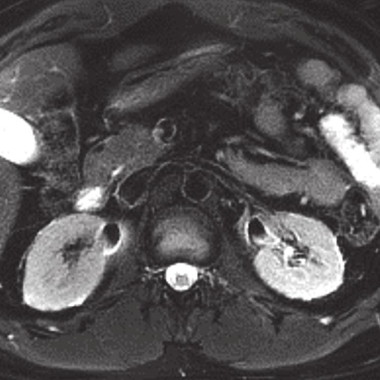
3 Biomedical Imaging (a) 3 (b) Figure 1: Stone visible on both CT and MR. (a) Enhanced axial CT demonstrates a large stone in the left renal pelvis (arrow). (b) Coronal HASTE MRI image clearly depicts the hypointense stone (arrow) against the backdrop of hyperintense urine. (a) (b) Figure 2: Stone visible on CT, but not on MR. (a) Contrast-enhanced CT performed for tumor staging reveals a stone in a right renal interpolar calyx (arrow). (b) Axial HASTE MRI image fails to detect the stone in the same calyx (arrow). Figure 3: MRI artifacts mimicking kidney stones. An MRI image showing signal void due to T2 artifact from concentrated gadolinium in both collecting systems (arrows). These signal voids could readily be mistaken for stones.
4 4 Biomedical Imaging 160 cases 138 CT-confirmed stone cases Step 1: initial MR (14 cases excluded, not adequate MRI anatomical coverage or image quality) 146 cases with adequate MRI 32 (23%) Step 3: 2nd MR 9 (28%) 23 (72%) 106 (77%) Step 3: 2nd MR 12 (11%) 94 (89%) Step 2: CT (8 cases excluded: CT stone mimics) Step 4: 3rd MR 138 CT-confirmed stone cases (a) Initial of 160 MRI and CT scans 9 (26%) 26 (74%) 26/138 (19%) (b) 138 CT-confirmed kidney stones: results of Steps 1 4 Figure 4: Study design and results. Following Steps 1 and 2, 138 CT-confirmed stones represented the included cases. Green boxes represent number and % detected by MRI at each step; blue boxes represent those not detected by MRI at each step. = overall final MRI detected rate. Based on the initial of the MRI images, the stones in 32 cases (23%) were identified (Figure 4(b)), with the following characteristics: stone size = 8±4mm and locations: lower pole (12), upper pole (8), interpolar (7), renal pelvis (4), and bladder (1) locations (Table 1). The characteristics of the unidentified 106 stones (77%) were as follows: stone size = 5±3mm and locations: lower pole (44), mid pole (14), upper pole (25), interpolar (11), renal pelvis (3), and bilateral (9) locations. There was a significant (P < ) difference between the sizes of the identified and unidentified stones; larger stones were more likely identified on MRI images. However, for these 32 identified stones, there was no significant (P = 0.451) difference between the stone size determined by CT (8 ±5mm) and that determined by MRI (8±4mm). On the second reading of the MRI images (Step 3, Figure 4(b)), of the 106 stones originally not identified on the first look at the MRI images, 12 stones (11%) were successfully identified with the following characteristics: stone size: 7± 2 mm and locations: lower pole (7), upper pole (3), and interpolar (2) locations. The characteristics of the 94 stones (89%) that remained unidentified on the MRI images on this second read were as follows: stone size = 5±2mm and locations: lower pole (37), mid pole (14), upper pole (22), interpolar (10), renal pelvis (3), and bilateral (8) locations. The difference between the size of the stones that were successfully identified on the second MRI reading and the size of those that remained unidentifiable was significant (P = 0.001). Regarding the 32 stones that were originally identified on the first reading of the MRI images, ing the CT images along with a second look at the MRI resulted in excluding 9 (28%) (Figure 4(b)) which were determined to be stone mimics (artifacts) with the following characteristics: size: 6± 3 mm and locations: lower pole (4), upper pole (3), interpolar (1), and bladder (1) locations. The characteristics of the 23 (72%) stones that were positively identified on MRI at this step were as follows: stone size = 9±4mm and locations: lower pole (8), upper pole (5), interpolar (5), renal pelvis (4), and bilateral (1) locations. Ultimately, 35 total stones were positively identified on the MRI images, including 12 that were initially not identified as of the first read of the MRI images, plus the 23 that were confirmed on second read (vide supra and Table 1). In Step 4, the ers appraised the MRI images a third timeandreportedthattheywould,inretrospect,havecalled 26 (74%) of the 35 MRI-confirmed stones. This yielded a final rate of stone detection by MRI in this study: 26/138 = 19%; those stones had the following characteristics: stone size = 9±4mm and locations: lower pole (10), upper pole (6), interpolar (6), renal pelvis (3), and bilateral (1) locations. In the cases of the remaining 9 stones (26%), although these had been confirmed on MRI by the ers when armed with
5 Biomedical Imaging 5 Table 1: Stone location by steps. Location in urinary tract All CT-confirmed stones Initial MRI 2nd MRI 3rd MRI N=138 N=32 N=35 N=26 Lower pole 56 (5 ± 3 mm) 12 (5±3mm) 15 (7±3mm) 10 (8±2mm) Mid pole 14 (5±3mm) Upper pole 33 (6±4mm) 8 (10 ± 6 mm) 8 (10 ± 6 mm) 6 (11 ± 6 mm) Interpolar 18 (6±3mm) 7 (8±3mm) 7 (8±3mm) 6 (8±3mm) Renal pelvis 7 (9±3mm) 4 (9±3mm) 4 (9±3mm) 3 (9±4mm) Ureter 1 (3 mm) Bladder 1 (8 mm) 1 (8 mm) 0 0 Bilateral 9 (4±3mm) 0 1 (10 mm) 1 (10 mm) Number of stones (size of stones, mean and SD). corresponding CT images at Step 3, the ers determined that, in retrospect, these would not be called stones based on this third of the MRI images. Characteristics of theselatterstoneswereasfollows:stonesize=7±3mm and locations: lower pole (5), upper pole (2), interpolar (1), and renal pelvis (1) locations (Figure 4(b); Table 1). There was no significant difference between stone sizes at Step 4, comparing those called on this final step to those that in retrospect would not be called stones (P = ). The characteristics of the 138 included cases ed by the two ers were similar. Reviewer 1 ed 64 cases (38 males) with ages of 62 ± 12 years (32 87 years) and Reviewer 2 ed 74 cases (46 males) with ages of 62 ± 12 years (30 89 years). The performances of the two ers were as follows: Reviewers 1 and 2 identified 27% and 20% of the stones based on the first MRI reading. The sizes of the identified/not identified stones were 8±4/5 ±3mm and 8±4/5 ±2mm for Reviewers 1 and 2, respectively. The percentages (sizes) of the stones identified by Reviewers 1 and 2 on the second MRI images were 27% (9 ± 3 mm) and 24% (8 ±4mm), respectively. The sizes of the unidentified stones were 5±3mm and 5±2mm by the firstandseconders,respectively.onthefinalstep, third of MRI images, Reviewers 1 and 2 reported that theywouldhavecalled82%and67%ofthe35totalmriidentified stones, respectively, in retrospect. The sizes of the stones that Reviewers 1 and 2 would have called/not called were 9±3/8±4and 9±4/6±2mm, respectively. At all steps, there were no significant differences in performance between thetwoersintermsofratesorstonesize. 4. Discussion The current study reported the capabilities of clinical MR imaging for detecting kidney stones. The results showed thataboutone-fifth(overall:19%)ofthestonescouldbe confidently detected on clinical MRI images, using modern technology. The stone size and background contrast are known factors for determining kidney stone visibility on MRI [9]. In the present study, the stones detected on MRI were on average 60% larger than those not detected, with significant differences in the stone sizes between the two groups. Knowing the stone size and location from corresponding CT images at the second MRI did not result in identification of many of the 106 previously not identified stones; only 12/106 = 11% of the missed stones were subsequently identified (Figure 4(b), Step 3). Important as well, ofthe32identifiedstonesontheinitialofmri images 9 (9/32 = 28%) were subsequently excluded on the second as MRI stone artifacts. Such false positive MRI findings may be due to the nonspecific nature of signal void foci on T2-weighted images. After the third of the MRI images, our expert radiologists decision to confidently call 26 of the total 35 MRI-identified stones (Figure 4(b), Step 4), even armed with the knowledge of size and location on corresponding CT, reflects the different nature of stoneto-tissue contrast in CT and MRI. The MRI performance herein reported in identifying large stones, especially in regions where they are surrounded by bright signal (Figure 1(b)), is in agreement with a previous report that noted a size threshold of 1 cm [9]. In the current study, 9 mm was the average size of the detected stones, with the smallest detected size being 4-5 mm. Although just one-fifth of the stones confirmed on gold standard CT were detected confidently from clinical MRI images, recent advances in MRI hardware capabilities and pulse sequence design hold promise for improving MRI detection of kidney stones. Together with the anatomical and functional information about the kidneys that can be obtained with MRI, a complete MRI exam could be available in the future for comprehensive kidney imaging, including scanning for kidney stones. Such exams could be particularly valuable for imaging vulnerable patients, for example, young patients, pregnant women or those with childbearing age, or recurrent stone formers. Further, the capability of imaging kidney stones with MRI could allow for obtaining this information at no extra cost in patients who are undergoing abdominal MRI scans for other diagnoses. Although the limited capability of MRI for detecting kidney stones has been previously reported, to the best of our knowledgethisisthefirststudythatconductedasystematic retrospective analysis of abdominal/pelvis MRI images on a relatively large number of patients with kidney stones against ground truth CT results. The study design along with the
6 6 Biomedical Imaging interobserver analysis conducted here allowed for quantitatively assessing the capabilities of MRI for detecting kidney stones along with associated characteristics. Although, due to the retrospective nature of the study, we evaluated only standard clinically implemented MRI sequences, future work by our group includes evaluating newly developed sequences, for example, the ultrashort echo time (UTE) technique, which allows for imaging tissues with very short T2 time constants and could be of potential for imaging kidney stones in the future [11]. Although CT is expected to remain the gold standard for kidney stone imaging, the possibility of reliably imaging the stones with MRI would be a valuable alternative for patients with concerns for repeated exposure to ionizing radiation, for example, young patients, pregnant women (or those of childbearing age), and recurrent stone formers. In conclusion, MRI has the potential for imaging kidney stones, especially medium-to-large stones (mean ± SD = 9± 4 mm) with sufficient background contrast. Further study, using newly developed techniques, is under way and promises to improve the ability of MRI to detect kidney stones, which maybeusefulinvulnerablegroupsforwhomctscanning with its attendant radiation exposure may not be recommended. MRI in the setting of the emergency department in a patient with renal calculi? Magnetic Resonance Imaging,vol. 32,no.5,pp ,2010. [10] Imaging in the Management of Ureteral Calculi, AUA Update Series, pp , [11] E.-S. H. Ibrahim, J. G. Cernigliaro, R. A. Pooley et al., Detection of different kidney stone types: an ex vivo comparison of ultrashort echo time MRI to reference standard CT, Clinical Imaging,vol.40,no.1,pp.90 95,2016. Competing Interests The authors declare that they have no competing interests. References [1] F. L. Coe, A. Evan, and E. Worcester, Kidney stone disease, The JournalofClinicalInvestigation,vol.115,no.10,pp , [2] E. M. Worcester and F. L. Coe, Nephrolithiasis, Primary Care, vol. 35, no. 2, pp , [3]V.Romero,H.Akpinar,andD.G.Assimos, Kidneystones: aglobalpictureofprevalence,incidence,andassociatedrisk factors, Reviews in Urology,vol.12,pp.e86 e96,2010. [4]I.Boulay,P.Holtz,W.D.Foley,B.White,andF.P.Begun, Ureteral calculi: diagnostic efficacy of helical CT and implications for treatment of patients, American Roentgenology,vol.172,no.6,pp ,1999. [5] D. T. Boll, N. A. Patil, E. K. Paulson et al., Renal stone assessment with dual-energy multidetector CT and advanced postprocessing techniques: improved characterization of renal stone composition pilot study, Radiology, vol. 250, no. 3, pp , [6] J. Broder, J. Bowen, J. Lohr, A. Babcock, and J. Yoon, Cumulative CT exposures in emergency department patients evaluated for suspected renal colic, Emergency Medicine, vol. 33,no.2,pp ,2007. [7] M. N. Ferrandino, A. Bagrodia, S. A. Pierre et al., Radiation exposure in the acute and short-term management of urolithiasis at 2 academic centers, The Urology, vol.181,no. 2, pp , [8] N. Grenier, F. Basseau, M. Ries, B. Tyndal, R. Jones, and C. Moonen, Functional MRI of the kidney, Abdominal Imaging, vol.28,no.2,pp ,2003. [9] B.Kalb,P.Sharma,K.Salman,K.Ogan,J.G.Pattaras,andD. R. Martin, Acute abdominal pain: is there a potential role for


7 Rotating Machinery Engineering The Scientific World Journal Distributed Sensor Networks Sensors Control Science and Engineering Advances in Civil Engineering Submit your manuscripts at Electrical and Computer Engineering Robotics VLSI Design Advances in OptoElectronics Navigation and Observation Chemical Engineering Active and Passive Electronic Components Antennas and Propagation Aerospace Engineering Modelling & Simulation in Engineering Shock and Vibration Advances in Acoustics and Vibration
Case Report Three-Dimensional Dual-Energy Computed Tomography for Enhancing Stone/Stent Contrasting and Stone Visualization in Urolithiasis
 Case Reports in Urology Volume 2013, Article ID 646087, 4 pages http://dx.doi.org/10.1155/2013/646087 Case Report Three-Dimensional Dual-Energy Computed Tomography for Enhancing Stone/Stent Contrasting
HHS Public Access Author manuscript Abdom Imaging. Author manuscript; available in PMC 2017 June 27.
 Motion Artifacts in Kidney Stone Imaging Using Single-Source and Dual-Source Dual-Energy CT Scanners. A Phantom Study El-Sayed H. Ibrahim 1,2,*, Joseph G. Cernigliaro 1, Robert A. Pooley 1, James C. Williams
AUA Guidelines for Imaging Known or Suspected Ureteral Calculi. Michael Ferrandino, MD Assoc Professor of Urology Duke University Medical Center
 AUA Guidelines for Imaging Known or Suspected Ureteral Calculi Michael Ferrandino, MD Assoc Professor of Urology Duke University Medical Center Imaging for Urolithiasis Justification for the Guidelines
Dual energy computed tomography for non-invasive differentiation of renal stone composition
 Dual energy computed tomography for non-invasive differentiation of renal stone composition Poster No.: C-0079 Congress: ECR 2012 Type: Scientific Exhibit Authors: R. D. Langer, K. F. W. Neidl van Gorkom,
Preparing for Medical Physics Components of the ABR Core Examination
 Preparing for Medical Physics Components of the ABR Core Examination The ABR core examination for radiologists contains material on medical physics. This content is based on the medical physics that is
Dual Energy CT: a new tool in evaluation of the urinary tract stones composition in clinical practice - initial study
 Dual Energy CT: a new tool in evaluation of the urinary tract stones composition in clinical practice - initial study Poster No.: C-2279 Congress: ECR 2013 Type: Scientific Exhibit Authors: M. Guzi#ski,
Research Article Kidney Modelling for FDG Excretion with PET
 Biomedical Imaging Volume 2007, Article ID 63234, 4 pages doi:10.1155/2007/63234 Research Article Kidney Modelling for FDG Excretion with PET Huiting Qiao, 1 Jing Bai, 1 Yingmao Chen, 2 and Jiahe Tian
Managing Radiation Risk in Pediatric CT Imaging
 Managing Radiation Risk in Pediatric CT Imaging Mahadevappa Mahesh, MS, PhD, FAAPM, FACR, FACMP, FSCCT. Professor of Radiology and Cardiology Johns Hopkins University School of Medicine Chief Physicist
Non Contrast MRA. Mayil Krishnam. Director, Cardiovascular and Thoracic Imaging University of California, Irvine
 Non Contrast MRA Mayil Krishnam Director, Cardiovascular and Thoracic Imaging University of California, Irvine No disclosures Non contrast MRA-Why? Limitations of CTA Radiation exposure Iodinated contrast
True Dual Energy. Dr. Stefan Ulzheimer, Siemens Healthcare GmbH. DEfinitely Siemens
 DEfinitely Siemens True Dual Energy Dr. Stefan Ulzheimer, Siemens Healthcare GmbH International version. Not for distribution in the US. Unrestricted Siemens AG 2015 All rights reserved. The products/features
Toshiba Aquillion 64 CT Scanner. Phantom Center Periphery Center Periphery Center Periphery
 Comparison of radiation dose and imaging performance for the standard Varian x-ray tube and the Richardson Healthcare ALTA750 replacement tube for the Toshiba Aquillion CT scanners. by Robert L. Dixon,
Assessment of Adipose Tissue from Whole Body 3T MRI Scans
 Assessment of Adipose Tissue from Whole Body 3T MRI Scans Ting Song 1, Jing An 2, Qun Chen 2, Vivian Lee 2, Andrew Laine 1 1 Department of Biomedical Engineering, Columbia University, New York, NY, USA
Diffusion weighted MRI in evaluation of transplanted kidney: Preliminary clinical experience
 African Journal of Nephrology (2009) 13: 26-30 Original Article AJN Diffusion weighted MRI in evaluation of transplanted kidney: Preliminary clinical experience Mohamed Abou El-Ghar; M.D, Huda Refaie;
MRI Assessment of the Right Ventricle and Pulmonary Blood Flow, Perfusion and Ventilation
 MRI Assessment of the Right Ventricle and Pulmonary Blood Flow, Perfusion and Ventilation Dr. Richard Thompson Department of Biomedical Engineering University of Alberta Heart and Lung Imaging Many Constantly
Ultralow Dose Chest CT with MBIR
 Ultralow Dose Chest CT with MBIR Ella A. Kazerooni, M.D. Professor & Director Cardiothoracic Radiology Associate Chair for Clinical Affairs University of Michigan Disclosures Consultant: GE Healthcare
CT Head Dose Reduction Using Spiral Scanning Protocol
 CT Head Dose Reduction Using Spiral Scanning Protocol Reed, William J MD; Broderick, Daniel F, MD; Weindling, Steven M, MD; Czervionke, Leo F MD; and Morin, Richard L; PhD; Mayo Clinic, Department of Radiology;
Justification of the use of CT for individual health assessment of asymptomatic people: the Chinese experience
 Justification of the use of CT for individual health assessment of asymptomatic people: the Chinese experience Liang Wang, MD, PhD Professor, attending radiologist wang6@tjh.tjmu.edu.cn Dept. of Radiology
How do the Parameters affect Image Quality and Dose for Abdominal CT? Image Review
 How do the Parameters affect Image Quality and Dose for Abdominal CT? Image Review Mannudeep K. Kalra, MD, DNB Massachusetts General Hospital Harvard Medical School Financial Disclosure This presentation
Vascular CT Protocols
 Vascular CT Protocols V 1D: Chest and abdominal CT angiogram (aortic dissection protocol) V 1T: Chest CT angiogram (aortic trauma protocol) V 2: Abdominal and pelvis CT angiogram (aortic aneurysm protocol)
Low-Dose CT: Clinical Studies & the Radiologist Perspective
 Low-Dose CT: Clinical Studies & the Radiologist Perspective RD-ASiR RD-MBIR SD-FBP RD=0.35 msv (80% dose reduction) Perry J. Pickhardt, MD UW School of Medicine & Public Health Low-Dose CT: Clinical Overview
Doses from pediatric CT examinations in Norway Are pediatric scan protocols developed and in daily use?
 Doses from pediatric CT examinations in Norway Are pediatric scan protocols developed and in daily use? Eva Godske Friberg * Norwegian Radiation Protection Authority, P.O. Box, Østerås, Norway Abstract.
Customizing Contrast Injection for Body MDCT: Algorithmic Approach
 Customizing Contrast Injection for Body MDCT: Algorithmic Approach Lincoln L. Berland, M.D., F.A.C.R. University of Alabama at Birmingham Before Contrast Prep and Hydration Hydration single most important
Imaging Features of Acute Pyelonephritis in Contrast Computed Tomography as Predictors of Need for Intervention
 Imaging Features of Acute Pyelonephritis in Contrast Computed Tomography as Predictors of Need for Intervention Poster No.: C-0088 Congress: ECR 2014 Type: Scientific Exhibit Authors: C. Y. Lee, C. W.
Dual-Energy 101: Principles, Methods and Dose
 Dual-Energy 101: Principles, Methods and Dose Juan Carlos Ramirez-Giraldo, Ph.D Staff Scien2st, Collabora2ons Manager SE Region ISCT San Francisco, 2017 Siemens Medical Solu2ons USA, Inc., 2017 Page 1
Research Article Usefulness of Nonenhanced Computed Tomography for Diagnosing Urolithiasis without Pyuria in the Emergency Department
 BioMed Research International Volume 2015, Article ID 810971, 6 pages http://dx.doi.org/10.1155/2015/810971 Research Article Usefulness of Nonenhanced Computed Tomography for Diagnosing Urolithiasis without
Modifi ed CT perfusion contrast injection protocols for improved CBF quantifi cation with lower temporal sampling
 Investigations and research Modifi ed CT perfusion contrast injection protocols for improved CBF quantifi cation with lower temporal sampling J. Wang Z. Ying V. Yao L. Ciancibello S. Premraj S. Pohlman
X-Ray & CT Physics / Clinical CT
 Computed Tomography-Basic Principles and Good Practice X-Ray & CT Physics / Clinical CT INSTRUCTORS: Dane Franklin, MBA, RT (R) (CT) Office hours will be Tuesdays from 5pm to 6pm CLASSROOM: TIME: REQUIRED
Low-dose CT Lung Cancer Screening Guidelines for Pulmonary Nodules Management Version 2
 Low-dose CT Lung Cancer Screening Guidelines for Pulmonary Nodules Management Version 2 The Committee for Management of CT-screening-detected Pulmonary Nodules 2009-2011 The Japanese Society of CT Screening
Case Report Crossed Renal Ectopia without Fusion An Unusual Cause of Acute Abdominal Pain: A Case Report
 Case Reports in Urology Volume 2012, Article ID 728531, 4 pages doi:10.1155/2012/728531 Case Report Crossed Renal Ectopia without Fusion An Unusual Cause of Acute Abdominal Pain: A Case Report D. P. Ramaema,
BrightSpeed Elite CT with ASiR: Comparing Dose & Image Quality Rule Out Pulmonary Embolism on Initial & Follow-Up Exam
 GE Healthcare BrightSpeed Elite CT with ASiR: Comparing Dose & Image Quality Rule Out Pulmonary Embolism on Initial & Follow-Up Exam Michael Swack, MD Diagnostic Radiologist Irvington Radiologists, PC
Journal of Radiology Case Reports
 Critical Pitfall: Varices in Cancer Patients mimicking Lymphadenopathy; Differentiation of varicose veins and enlarged lymph nodes in routine staging Tilman Schubert 1*, Michele Pansini 1, Georg Bongartz
Digital tomosynthesis (DT) has been well described as a
 has been well described as a") Case Report The Usefulness of Digital Tomosynthesis (DT) in Assisting in Cases of Doubtful Routine Radiography and/or Computed Tomography (CT) Image. Abstract Digital tomosynthesis is useful in assisting
Case Report The Usefulness of Digital Tomosynthesis (DT) in Assisting in Cases of Doubtful Routine Radiography and/or Computed Tomography (CT) Image. Abstract Digital tomosynthesis is useful in assisting
ACR MRI Accreditation: Medical Physicist Role in the Application Process
 ACR MRI Accreditation: Medical Physicist Role in the Application Process Donna M. Reeve, MS, DABR, DABMP Department of Imaging Physics University of Texas M.D. Anderson Cancer Center Educational Objectives
Quantifying Dual-energy computed tomography (DECT) in patients with renal calculi using a Toshiba Aquilion One Scanner.
 in patients with renal calculi using a Toshiba Aquilion One Scanner.") Quantifying Dual-energy computed tomography (DECT) in patients with renal calculi using a Toshiba Aquilion One Scanner. Poster No.: C-2613 Congress: ECR 2015 Type: Authors: Keywords: DOI: Scientific Exhibit
Quantifying Dual-energy computed tomography (DECT) in patients with renal calculi using a Toshiba Aquilion One Scanner. Poster No.: C-2613 Congress: ECR 2015 Type: Authors: Keywords: DOI: Scientific Exhibit
CT SCAN PROTOCOL. Shoulder
 CT SCAN PROTOCOL Shoulder Purpose and Summary CT images made with this protocol are used to provide the orthopedic surgeon with a detailed 3D anatomical reconstruction of the patient s scapula and proximal
PROFESSIONAL SKILLS 1 3RD YEAR SEMESTER 6 RADIOGRAPHY. THE URINARY SYSTEM Uz. Fatema shmus aldeen Tel
 PROFESSIONAL SKILLS 1 3RD YEAR SEMESTER 6 RADIOGRAPHY THE URINARY SYSTEM Uz. Fatema shmus aldeen Tel. 0925111552 Professional skills-2 THE URINARY SYSTEM The urinary system (review anatomy and physiology)
Case Report Sinus Venosus Atrial Septal Defect as a Cause of Palpitations and Dyspnea in an Adult: A Diagnostic Imaging Challenge
 Case Reports in Medicine Volume 2015, Article ID 128462, 4 pages http://dx.doi.org/10.1155/2015/128462 Case Report Sinus Venosus Atrial Septal Defect as a Cause of Palpitations and Dyspnea in an Adult:
Combined Anatomical and Functional Imaging with Revolution * CT
 GE Healthcare Case studies Combined Anatomical and Functional Imaging with Revolution * CT Jean-Louis Sablayrolles, M.D. Centre Cardiologique du Nord, Saint-Denis, France Case 1 Whole Brain Perfusion and
Computed tomography. Department of Radiology, University Medical School, Szeged
 Computed tomography Department of Radiology, University Medical School, Szeged voxel +1-4 +2 +5 +3 +1 0-2 pixel -2 0 +1-4 -6 +5 +2 +1 Department of Radiology, University Medical School, Szeged
An Overview of Ultrasound Testing For Lesion Detection in Human Kidney
 Journal of Tomography System & Sensors Application Vol.1, Issue 1, June 2018 An Overview of Ultrasound Testing For Lesion Detection in Human Kidney Aina Fadhilah Abd Rahim 1, Zawin Najah Abd Halim 1, Jaysuman
Austin Radiological Association Nuclear Medicine Procedure PET SODIUM FLUORIDE BONE SCAN (F-18 NaF)
") Austin Radiological Association Nuclear Medicine Procedure PET SODIUM FLUORIDE BONE SCAN (F-18 NaF) Overview Indication Sodium Fluoride F18 injection is a radioactive diagnostic agent for positron emission
Austin Radiological Association Nuclear Medicine Procedure PET SODIUM FLUORIDE BONE SCAN (F-18 NaF) Overview Indication Sodium Fluoride F18 injection is a radioactive diagnostic agent for positron emission
Why is CT Dose of Interest?
 Why is CT Dose of Interest? CT usage has increased rapidly in the past decade Compared to other medical imaging CT produces a larger radiation dose. There is direct epidemiological evidence for a an increase
Intravenous Pyelogram (IVP)
") Scan for mobile link. Intravenous Pyelogram (IVP) Intravenous pyelogram (IVP) is an x-ray exam that uses an injection of contrast material to evaluate your kidneys, ureters and bladder and help diagnose
Scan for mobile link. Intravenous Pyelogram (IVP) Intravenous pyelogram (IVP) is an x-ray exam that uses an injection of contrast material to evaluate your kidneys, ureters and bladder and help diagnose
SPECIFIC PRINCIPLES FOR DOSE REDUCTION IN HEAD CT IMAGING. Rajiv Gupta, MD, PhD Neuroradiology, Massachusetts General Hospital Harvard Medical School
 SPECIFIC PRINCIPLES FOR DOSE REDUCTION IN HEAD CT IMAGING Rajiv Gupta, MD, PhD Neuroradiology, Massachusetts General Hospital Harvard Medical School OUTLINE 1 st Presentation: Dose optimization strategies
Research Article Comparison of Colour Duplex Ultrasound with Computed Tomography to Measure the Maximum Abdominal Aortic Aneurysmal Diameter
 International Vascular Medicine, Article ID 574762, 4 pages http://dx.doi.org/10.1155/2014/574762 Research Article Comparison of Colour Duplex Ultrasound with Computed Tomography to Measure the Maximum
CT dose survey data acquisition form
 CT dose survey data acquisition form CT Protocol page CT Head (acute stroke) C-spine (fracture) Chest (lung cancer) Chest High-Res. (interstitial lung disease) CTA (blood vessels) CTPA (PE) Abdomen (liver
Translating Protocols Across Patient Size: Babies to Bariatric
 Translating Protocols Across Patient Size: Babies to Bariatric Cynthia H. McCollough, PhD, FACR, FAAPM Professor of Radiologic Physics Director, CT Clinical Innovation Center Department of Radiology Mayo
Excretory urography (EU) or IVP US CT & radionuclide imaging
 or IVP US CT & radionuclide imaging") Excretory urography (EU) or IVP US CT & radionuclide imaging MRI arteriography studies requiring catherization or direct puncture of collecting system EU & to a lesser extent CT provide both functional
Excretory urography (EU) or IVP US CT & radionuclide imaging MRI arteriography studies requiring catherization or direct puncture of collecting system EU & to a lesser extent CT provide both functional
Radiology Rounds A Newsletter for Referring Physicians Massachusetts General Hospital Department of Radiology
 Radiology Rounds A Newsletter for Referring Physicians Massachusetts General Hospital Department of Radiology Minimizing CT Radiation Dose CT examinations improve health care and are an essential part
Sensitivity and Specificity in Detection of Labral Tears with 3.0-T MRI of the Shoulder
 Magee and Williams MRI for Detection of Labral Tears Musculoskeletal Imaging Clinical Observations C M E D E N T U R I C L I M G I N G JR 2006; 187:1448 1452 0361 803X/06/1876 1448 merican Roentgen Ray
Measurement of organ dose in abdomen-pelvis CT exam as a function of ma, KV and scanner type by Monte Carlo method
 Iran. J. Radiat. Res., 2004; 1(4): 187-194 Measurement of organ dose in abdomen-pelvis CT exam as a function of ma, KV and scanner type by Monte Carlo method M.R. Ay 1, M. Shahriari 2, S. Sarkar 3, P.
Fast and easy diagnostic imaging from head to toe
 Publication for the Philips MRI Community ISSUE 50 2014 / 1 Fast and easy diagnostic imaging from head to toe Ingenia 1.5T with dstream provides speed and convenience, IntelliSpace Portal provides flexibility
RADIOLOGIC AND IMAGING SCIENCE (RIS)
") Kent State University Catalog 2017-2018 1 RADIOLOGIC AND IMAGING SCIENCE (RIS) RIS 34001 INTRODUCTION TO DIAGNOSTIC MEDICAL SONOGRAPHY 1 Credit Provides an introduction to diagnostic medical sonography.
Kent State University Catalog 2017-2018 1 RADIOLOGIC AND IMAGING SCIENCE (RIS) RIS 34001 INTRODUCTION TO DIAGNOSTIC MEDICAL SONOGRAPHY 1 Credit Provides an introduction to diagnostic medical sonography.
Acute flank pain in children: Imaging considerations
 Acute flank pain in children: Imaging considerations Carlos J. Sivit MD Rainbow Babies and Children s Hospital Case Western Reserve School of Medicine Flank pain Results from distention of ureter or renal
The radiation dose in retrospective
 The radiation dose in retrospective gated tdcoronary computed td tomography (CCT) Saeed AL Ahmari, Ghormallah AL Zahrani, Sumiah AL Helali, Samir AL Dulikan, Abdullah Bafagih, HibaKhashojji Prince Sultan
Imaging Ejaculatory Disorders and Hematospermia
 ATHENS 4-6 October 2018 European Society of Urogenital Radiology Imaging Ejaculatory Disorders and Hematospermia Parvati Ramchandani, MD Professor, Radiology and Surgery University of Pennsylvania Medical
Biomarkers and the Future of. John R. Votaw CBIS 5 th Year Anniversary Celebration/Look to the future February 8, 2013
 Biomarkers and the Future of Radiology John R. Votaw CBIS 5 th Year Anniversary Celebration/Look to the future February 8, 2013 Statistics/Radiology Collaboration The utility of Radiologic procedures
Ultra-low dose CT of the acute abdomen: Spectrum of imaging findings
 Ultra-low dose CT of the acute abdomen: Spectrum of imaging findings Poster No.: C-1452 Congress: ECR 2010 Type: Educational Exhibit Topic: GI Tract Authors: P. A. Vlachou, C. Kloeters, S. Kandel, P. Hein,
CT Low Dose Lung Cancer Screening. Part I. Journey to LDCT LCS Program
 CT Low Dose Lung Cancer Screening Part I Journey to LDCT LCS Program Paul Johnson, M.S., DABHP, DABR Cleveland Clinic September 26, 2015 Lung Caner is No. 1 In Cancer Related Death In The United States
Prof. Dr. NAGUI M. ABDELWAHAB,M.D.; MARYSE Y. AWADALLAH, M.D. AYA M. BASSAM, Ms.C.
 Role of Whole-body Diffusion MR in Detection of Metastatic lesions Prof. Dr. NAGUI M. ABDELWAHAB,M.D.; MARYSE Y. AWADALLAH, M.D. AYA M. BASSAM, Ms.C. Cancer is a potentially life-threatening disease,
B-Flow, Power Doppler and Color Doppler Ultrasound in the Assessment of Carotid Stenosis: Comparison with 64-MD-CT Angiography
 Med. J. Cairo Univ., Vol. 85, No. 2, March: 805-809, 2017 www.medicaljournalofcairouniversity.net B-Flow, Power Doppler and Color Doppler Ultrasound in the Assessment of Carotid Stenosis: Comparison with
Pediatric chest HRCT using the idose 4 Hybrid Iterative Reconstruction Algorithm: Which idose level to choose?
 Journal of Physics: Conference Series PAPER OPEN ACCESS Pediatric chest HRCT using the idose 4 Hybrid Iterative Reconstruction Algorithm: Which idose level to choose? To cite this article: M Smarda et
MR Tumor Staging for Treatment Decision in Case of Wilms Tumor
 MR Tumor Staging for Treatment Decision in Case of Wilms Tumor G. Schneider, M.D., Ph.D.; P. Fries, M.D. Dept. of Diagnostic and Interventional Radiology, Saarland University Hospital, Homburg/Saar, Germany
Fetal Dose Calculations and Impact on Patient Care
 Fetal Dose Calculations and Impact on Patient Care Matt Hough, MS, DABR, DABMP Florida Hospital Diagnostic Medical Physics and Radiation Safety Resource ACR-SPR Practice Parameter for Imaging Pregnant
Ureteral Stenting and Nephrostomy
 Scan for mobile link. Ureteral Stenting and Nephrostomy Ureteral stenting and nephrostomy help restore urine flow through blocked ureters and return the kidney to normal function. Ureters are long, narrow
How I do it: Non Contrast-Enhanced MR Angiography (syngo NATIVE)
") Clinical How-I-do-it Cardiovascular How I do it: Non Contrast-Enhanced MR Angiography (syngo NATIVE) Manuela Rick, Nina Kaarmann, Peter Weale, Peter Schmitt Siemens Healthcare, Erlangen, Germany Introduction
Clinical How-I-do-it Cardiovascular How I do it: Non Contrast-Enhanced MR Angiography (syngo NATIVE) Manuela Rick, Nina Kaarmann, Peter Weale, Peter Schmitt Siemens Healthcare, Erlangen, Germany Introduction
HHS Public Access Author manuscript AJR Am J Roentgenol. Author manuscript; available in PMC 2016 September 02.
 Quantification of Urinary Stone Composition in Mixed Stones Using Dual-Energy CT: A Phantom Study Shuai Leng, PhD 1,*, Alice Huang, BS 1, Juan Montoya, BS 1, Xinhui Duan, PhD 1, James C. Williams, PhD
CT Imaging of Atherosclerotic Plaque. William Stanford MD Professor-Emeritus Radiology University of Iowa College of Medicine Iowa City, IA
 CT Imaging of Atherosclerotic Plaque William Stanford MD Professor-Emeritus Radiology University of Iowa College of Medicine Iowa City, IA PREVALENCE OF CARDIOVASCULAR DISEASE In 2006 there were 80 million
Genitourinary Imaging Original Research
 Genitourinary Imaging Original Research Masch et al. Genitourinary Imaging Original Research William R. Masch 1 Richard H. Cohan 1,2 James H. Ellis 1,2 Jonathan R. Dillman 1,3 Jonathan M. Rubin 1,2 Matthew
Radiation Dose Reduction: Should You Use a Bismuth Breast Shield?
 Radiation Dose Reduction: Should You Use a Bismuth Breast Shield? Lincoln L. Berland, M.D., F.A.C.R. Michael V. Yester, Ph.D. University of Alabama at Birmingham Breast Radiation on CT Use of chest CT
A multicentric study on patient dose in multislice CT
 A multicentric study on patient dose in multislice CT A.Stratis 1, M.Molfetas 1, S.Kottou 2, A.Louizi 2 1. Medical Physics department, Evangelismos General hospital of Athens, Athens, Greece 2. Medical
ASSESSING THE PLAIN ABDOMINAL RADIOGRAPH M A A M E F O S U A A M P O F O
 ASSESSING THE PLAIN ABDOMINAL RADIOGRAPH M A A M E F O S U A A M P O F O Introduction The abdomen (less formally called the belly, stomach, is that part of the body between the thorax (chest) and pelvis,
Doses from Cervical Spine Computed Tomography (CT) examinations in the UK. John Holroyd and Sue Edyvean
 examinations in the UK. John Holroyd and Sue Edyvean") Doses from Cervical Spine Computed Tomography (CT) examinations in the UK John Holroyd and Sue Edyvean Why a new dose survey? Number of enquires received concerning the current NDRL Concern that could
Doses from Cervical Spine Computed Tomography (CT) examinations in the UK John Holroyd and Sue Edyvean Why a new dose survey? Number of enquires received concerning the current NDRL Concern that could
Dual-Energy CT: The Technological Approaches
 Dual-Energy CT: The Technological Approaches Dushyant Sahani, M.D Director of CT Associate Professor of Radiology Massachusetts General Hospital Harvard Medical School Email-dsahani@partners.org Disclosure
ESTABLISHING DRLs in PEDIATRIC CT. Keith Strauss, MSc, FAAPM, FACR Cincinnati Children s Hospital University of Cincinnati College of Medicine
 ESTABLISHING DRLs in PEDIATRIC CT Keith Strauss, MSc, FAAPM, FACR Cincinnati Children s Hospital University of Cincinnati College of Medicine CT Dose Indices CTDI INTRODUCTION CTDI 100, CTDI w, CTDI vol
Bedside Ultrasound in the Emergency Department to Detect Hydronephrosis for the Evaluation of Suspected Ureteric Colic
 Bedside Ultrasound in the Emergency Department to Detect Hydronephrosis for the Evaluation of Suspected Ureteric Colic Shrestha R, Shakya RM, Khan A ABSTRACT Background Department of Emergency Medicine
Accuracy of ultrasonography for renal stone detection and size determination: is it good enough for management decisions?
 Upper Urinary Tract Accuracy of ultrasonography for renal stone detection and size determination: is it good enough for management decisions? Vishnu Ganesan*,, Shubha De*, Daniel Greene*, Fabio Cesar Miranda
OASIS 1.2T: MULTIPARAMETRIC MRI OF PROSTATE CANCER
 OASIS 1.2T: MULTIPARAMETRIC MRI OF PROSTATE CANCER By Dr. John Feller, MD, Radiologist Desert Medical Imaging, Palm Springs, CA MRI is clinically accepted as the best imaging modality for displaying anatomical
Detection of Renal Stones on Portal Venous Phase CT: Comparison of Thin Axial and Coronal Maximum- Intensity-Projection Images
 Genitourinary Imaging Original Research Corwin et al. Detection of Renal Stones on Portal Venous Phase CT Genitourinary Imaging Original Research Michael T. Corwin 1 Justin S. Lee 1 Ghaneh Fananapazir
Use of IV-contrast versus IV-and oral-contrast in the evaluation of abdominal pain on CT in the emergency department
 Use of IV-contrast versus IV-and oral-contrast in the evaluation of abdominal pain on CT in the emergency department Poster No.: B-0693 Congress: ECR 2016 Type: Authors: Scientific Paper M. Wasserman 1,
L. Alexandre Frigini MD; Aaron Thomas, MD; Veronica Lenge de Rosen, MD
 Computed Tomography Urography (CTU) for Evaluation of Asymptomatic microscopic hematuria. Is intravenous contrast administration warranted for all patients? A retrospective evaluation utilizing ACR s Appropriateness
Acknowledgments. A Specific Diagnostic Task: Lung Nodule Detection. A Specific Diagnostic Task: Chest CT Protocols. Chest CT Protocols
 Personalization of Pediatric Imaging in Terms of Needed Indication-Based Quality Per Dose Acknowledgments Duke University Medical Center Ehsan Samei, PhD Donald Frush, MD Xiang Li PhD DABR Cleveland Clinic
RADIOLOGIC TECHNOLOGY (526)
") RADIOLOGIC TECHNOLOGY (526) 526-133 DMS General Procedures 2 Radiologic Technology (526) 1 526-130 Introduction to Diagnostic Medical Sonography This course introduces the student to the history of ultrasound
RADIOLOGIC TECHNOLOGY (526) 526-133 DMS General Procedures 2 Radiologic Technology (526) 1 526-130 Introduction to Diagnostic Medical Sonography This course introduces the student to the history of ultrasound
Case Report Spontaneous Pelvic Rupture as a Result of Renal Colic in a Patient with Klinefelter Syndrome
 Volume 2013, Article ID 374973, 4 pages http://dx.doi.org/10.1155/2013/374973 Case Report Spontaneous Pelvic Rupture as a Result of Renal Colic in a Patient with Klinefelter Syndrome Sergey Reva and Yuri
Utility of Dual-Energy CT to Evaluate Patients with Hip and Pelvis Pain in the ER Setting
 Utility of Dual-Energy CT to Evaluate Patients with Hip and Pelvis Pain in the ER Setting Johnson, T., Moran, E., Glazebrook, K., Leng, S., Fletcher, J., and McCollough, C. An educational review ER011
Bone PET/MRI : Diagnostic yield in bone metastases and malignant primitive bone tumors
 Bone PET/MRI : Diagnostic yield in bone metastases and malignant primitive bone tumors Lars Stegger, Benjamin Noto Department of Nuclear Medicine University Hospital Münster, Germany Content From PET to
ACR MRI Accreditation Program. ACR MRI Accreditation Program Update. Educational Objectives. ACR accreditation. History. New Modular Program
 ACR MRI Accreditation Program Update Donna M. Reeve, MS, DABR, DABMP Department of Imaging Physics University of Texas M.D. Anderson Cancer Center Educational Objectives Present requirements of the new
JMSCR Vol 05 Issue 06 Page June 2017
 www.jmscr.igmpublication.org Impact Factor 5.84 Index Copernicus Value: 83.27 ISSN (e)-2347-176x ISSN (p) 2455-0450 DOI: https://dx.doi.org/10.18535/jmscr/v5i6.29 MRI in Clinically Suspected Uterine and
High Field MR of the Spine
 Department of Radiology University of California San Diego 3T for MR Applications Advantages High Field MR of the Spine Increased signal-to-noise Better fat suppression Increased enhancement with gadolinium
Computed tomography evaluation of urinary stones densities compared to in vitro analysis of its chemical composition
 International Journal of Medical Imaging 2014; 2(6): 141-145 Published online December 22, 2014 (http://www.sciencepublishinggroup.com/j/ijmi) doi: 10.11648/j.ijmi.20140206.14 ISSN: 2330-8303 (Print);
Half-Fourier Acquisition Single-Shot Turbo Spin-Echo (HASTE) MR: Comparison with Fast Spin-Echo MR in Diseases of the Brain
 MR: Comparison with Fast Spin-Echo MR in Diseases of the Brain") Half-Fourier Acquisition Single-Shot Turbo Spin-Echo (HASTE) MR: Comparison with Fast Spin-Echo MR in Diseases of the Brain Mahesh R. Patel, Roman A. Klufas, Ronald A. Alberico, and Robert R. Edelman PURPOSE:
Half-Fourier Acquisition Single-Shot Turbo Spin-Echo (HASTE) MR: Comparison with Fast Spin-Echo MR in Diseases of the Brain Mahesh R. Patel, Roman A. Klufas, Ronald A. Alberico, and Robert R. Edelman PURPOSE:
TORNIER BLUEPRINT. 3D Planning + PSI SCAN PROTOCOL
 TORNIER BLUEPRINT 3D Planning + PSI SCAN PROTOCOL Contents 3 Introduction 3 Patient preparation 3 Scanning instructions 4 Image instructions 5 Scanning parameters 6 Technical instructions 2 BLUEPRINT 3D
Imaging Of The Pelvis
 Imaging Of The Pelvis 1 / 6 2 / 6 3 / 6 Imaging Of The Pelvis MRI of the pelvis may be more focused on the organs, soft tissues, and vessels, rather than on the bones themselves. In many instances, MRI
Conducting Research Involving Medical Imaging
 Conducting Research Involving Medical Imaging J. Keith Smith, MD, PhD Terry Hartman, MPH, MS, CCRC Angela Creighton, MBA April 19, 2018 Objectives Summarize the risks of medical imaging involving ionizing
Radiation Safety. Disclosures
 Radiation Safety Timothy D. Averch, M.D., F.A.C.S Professor and Vice Chair for Quality Department of Urology, UPMC Chair, AUA QIPS Committee Disclosures None Objectives Review patient radiation exposure
MR QA/QC for MRgRT. Rick Layman, PhD, DABR Department of Radiology July 13, 2015
 MR QA/QC for MRgRT Rick Layman, PhD, DABR Department of Radiology July 13, 2015 The Ohio State University Comprehensive Cancer Center Arthur G. James Cancer Hospital and Richard J. Solove Research Institute
Radiation Dose Reduction Strategies in Coronary CT Angiography
 Radiation Dose Reduction Strategies in Coronary CT Angiography Noor Diyana Osman, PhD noordiyana@usm.my Contents: Introduction Radiation dosimetry in CT Radiation risk associated with coronary CT angiography
MR Advance Techniques. Vascular Imaging. Class II
 MR Advance Techniques Vascular Imaging Class II 1 Vascular Imaging There are several methods that can be used to evaluate the cardiovascular systems with the use of MRI. MRI will aloud to evaluate morphology