Acute flank pain in children: Imaging considerations
|
|
|
- Jerome Lang
- 6 years ago
- Views:
Transcription


1 Acute flank pain in children: Imaging considerations Carlos J. Sivit MD Rainbow Babies and Children s Hospital Case Western Reserve School of Medicine Flank pain Results from distention of ureter or renal capsule Pain associated with inflammation not as sudden or severe as that caused by acute obstruction Pain severity usually related to acuteness of onset 1


2 Flank pain Often associated with other symptoms including fever, nausea and vomiting Association with fever requires prompt diagnosis and relief of obstruction Infection associated with obstruction causes rapid renal damage and risk for sepsis Urolithiasis Acute pyelonephritis Obstructive uropathy 2
3 Urolithiasis Urolithiasis Calcium based stones % Calcium oxalate and calcium phosphate Struvite stones % Magnesium ammonium phosphate Uric acid stones % Occur in acidic urine; dissolved with alkanization Cysteine, xanthine & protein matrix <5% 3
4 Urolithiasis Risk factors Conditions that alter urine composition Hypercalciuria, hyperoxaluria, hyperuricosuria, hypercitrauria Medical conditions Neurogenic bladder, obstructive uropathy, diabetes, hypertension Medications that crystallize urine Urolithiasis Flank pain and hematuria in most Renal colic due to ureteral obstruction Similar rates of flank pain and hematuria with and without calculi History of fever negative predictor Personal history strong predictor 4
5 Urolithiasis Role of imaging Establish diagnosis Provide information regarding stone burden and distribution Identify associated hydronephrosis Diagnose predisposing abnormalities Urolithiasis Location of calculi Ureteral 52% Renal 24% Renal & ureteral 21% Bladder 4% Persaud. Pediatrics 2009;124:888 5
 LONG TRAN TRAN")
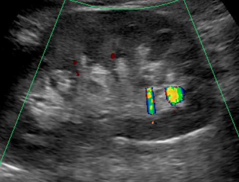
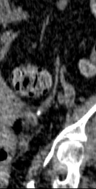
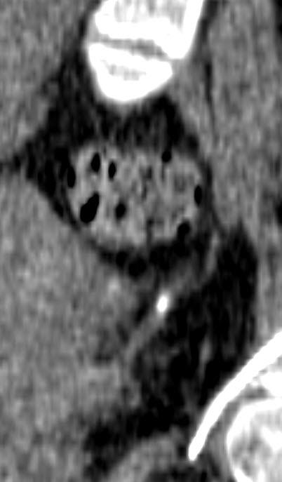
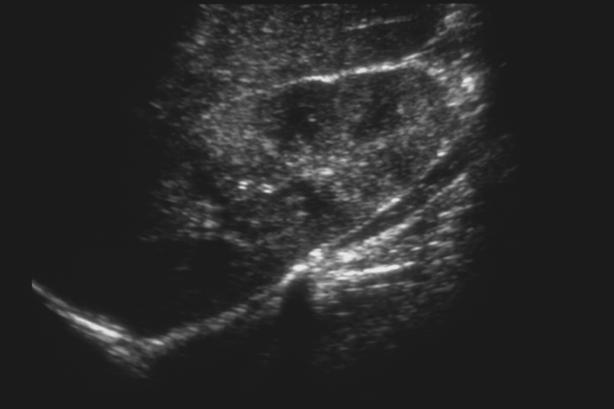
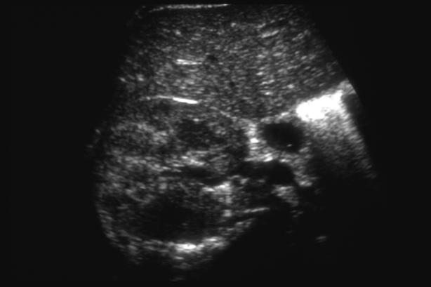
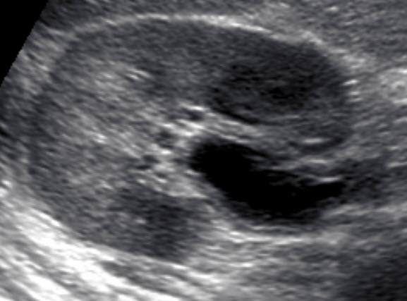
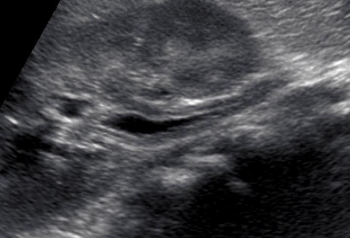
6 US High sensitivity for renal (>90%) Low sensitivity for ureteral (<40%) Sonographic findings Echogenic foci within collecting system Acoustic shadowing >5 mm Color doppler twinkling artifact Hydronephrosis (indirect) LONG TRAN TRAN 6

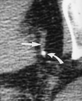
7 Twinkling artifact Observed at color Doppler when insonating tough reflective surfaces Discrete focus of alternating colors with or without a comet tail Increases detection of stones Many false-negatives and positives Dillman. AJR 2011;259:911 7

8 8

9 9
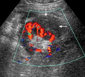
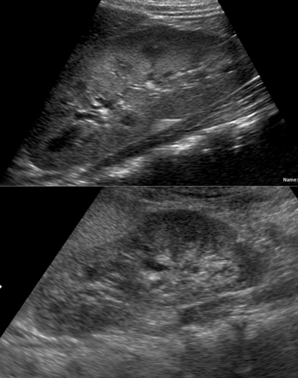
 effective since imaging high contrast lesions 10")
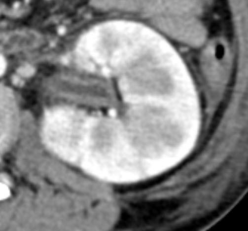
10 CT Unenhanced CT exam of choice High diagnostic sensitivity Low dose protocols (< 3 msv) effective since imaging high contrast lesions 10

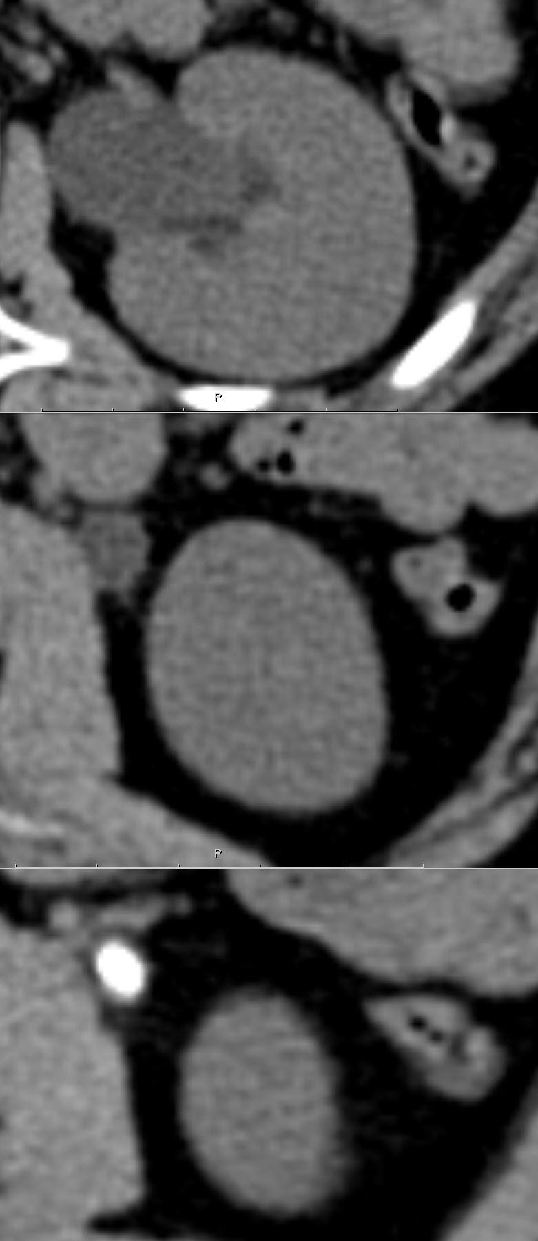
11 CT Primary finding Visualization of stone within collecting system Attenuation value higher than surrounding soft tissue Calcium 1000 H.U.; Uric acid H.U. Exception unmineralized matrix stones & stones related to Indavir 11

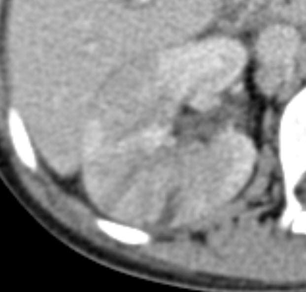
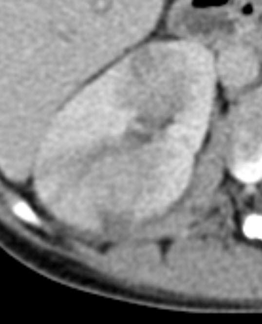
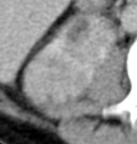
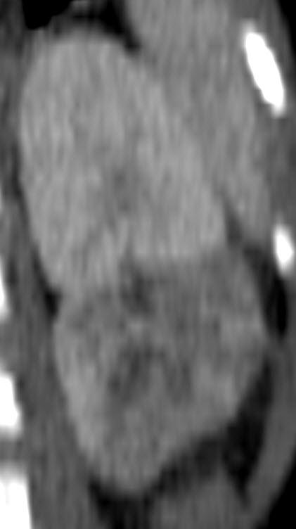
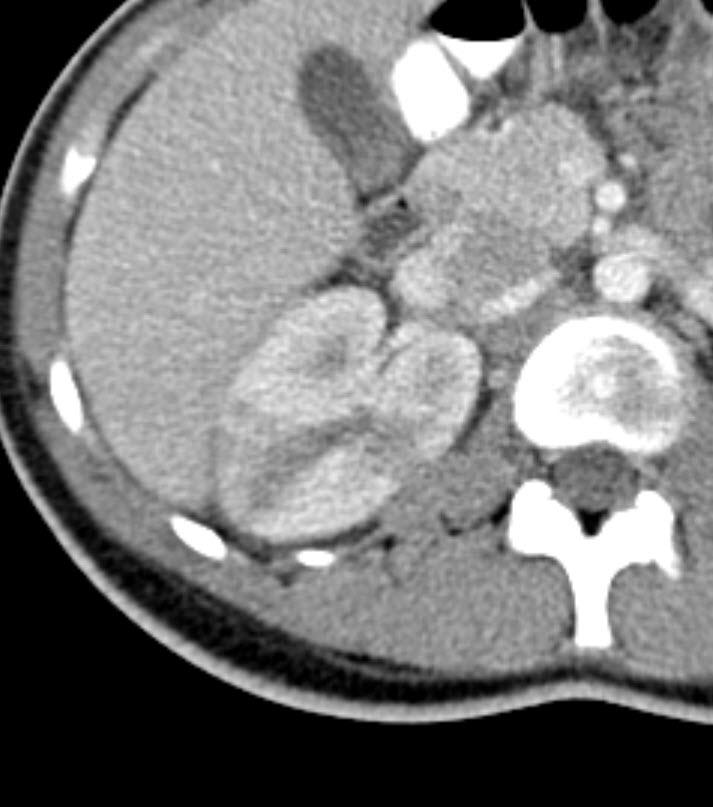
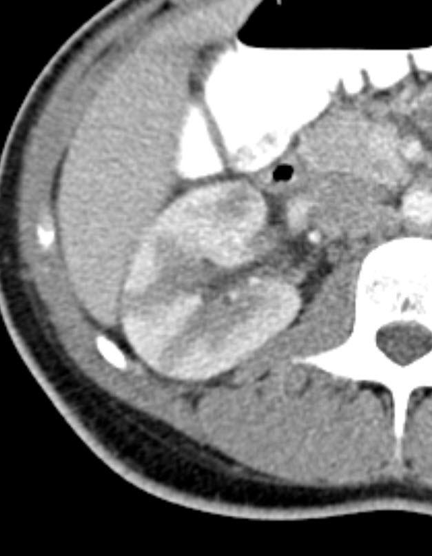
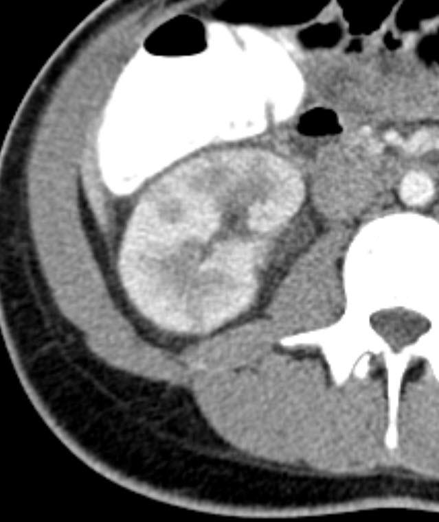
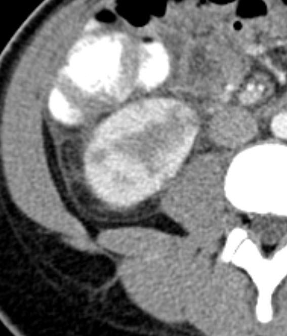
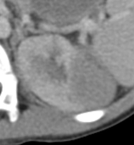
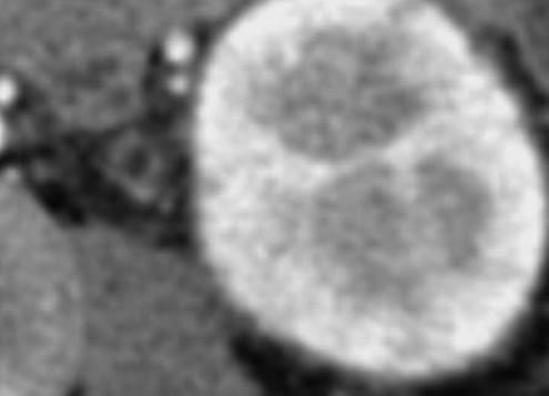
12 CT Secondary findings Hydronephrosis Perinephric fat stranding Hydroureter Soft tissue rim sign Hydronephrosis 12
13 Hydroureter 13


14 Fat stranding Fluid in bridging septa of perinephric fat due to increased lymphatic pressure 14

15 Soft tissue rim sign Halo of soft tissue attenuation around calcification Represents wall of ureter Pitfall - Phleboliths Absence of soft tissue rim Comet tail sign Linear soft tissue area adjacent to calcification 15
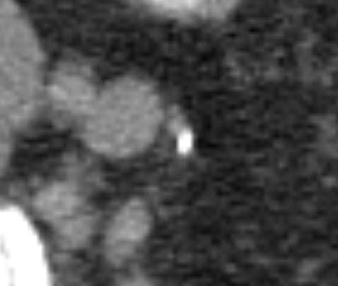
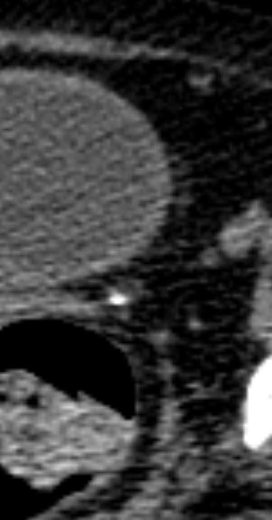
16 16


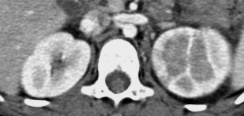
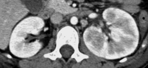
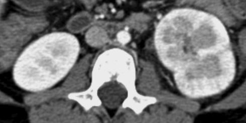
17 Acute Pyelonephritis Acute Pyelonephritis Infection of collecting system uroepithelium & renal interstitium Most commonly ascending infection but may be hematogenous Increased risk with obstruction or vesicoureteral reflux E. Coli responsible for >90% 17

18 Acute Pyelonephritis Role of Imaging Establish diagnosis Identify complications requiring surgical or percutaneous drainage Diagnose predisposing abnormalities US Low diagnostic sensitivity Gray scale: 25-45% Color & Power Doppler: 63-75% Sonographic findings Renal enlargement Increased or decreased echogenicity Loss of corticomedullary differentiation Decreased cortical blood flow 18
19 19

20 LT RT LT 20


 Perirenal inflammatory")
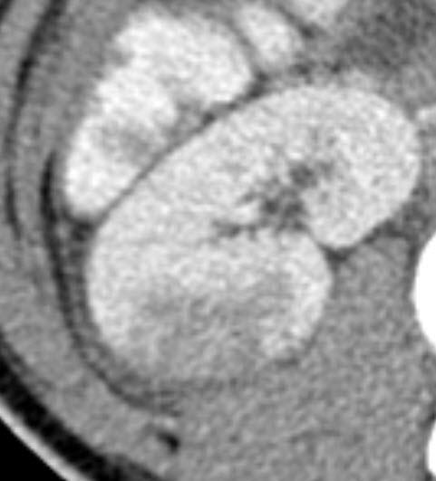
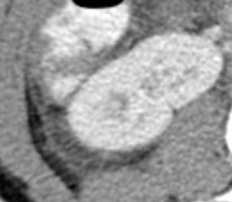
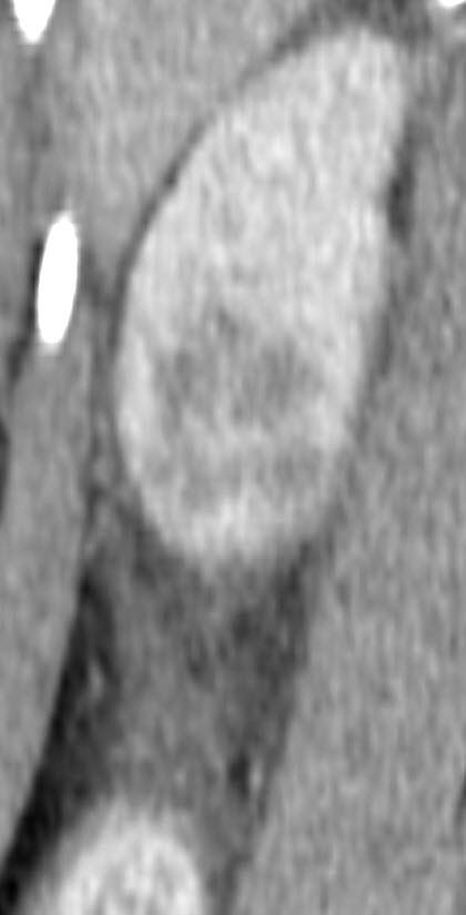
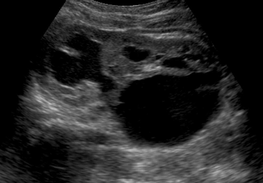
21 CT Highly sensitive for diagnosis Single or multifocal wedged- or oval-shaped peripheral lowattenuation defects Striated nephrogram Renal enlargement (diffuse) Perirenal inflammatory changes Focal 21

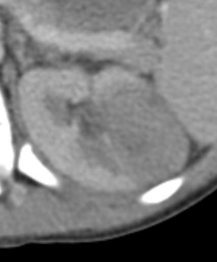
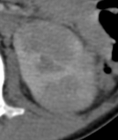
22 Multifocal 22
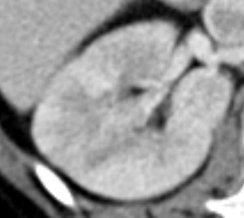
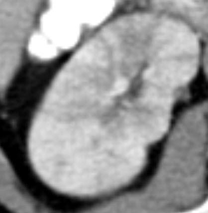
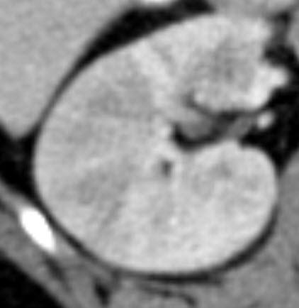

23 Striated nephrogram Enlargement 23
24 Inflammatory changes Perinephric space Perirenal fascia Urothelium 24


25 MR Similar findings as CT Increased signal intensity on contrast enhanced imaging Alternative to CT to avoid radiation exposure Ddx Segmental infarct Leukemia Lymphoma 25


26 Complications Renal abscess Pyonephrosis Renal abscess Necrotic cavity filled with pus Typically result from inadequately treated parenchymal infection Most commonly with DM, SSD or in immunocompromised children Conservative treatment if small Drainage required if large 26


27 Renal abscess Rounded low-attenuation lesion(s) that fail to enhance CT/MR Hypoechoic or anechoic thick walled mass +/- septations & debris US Air may be seen with gas forming organisms Inflammation may extend into or outside of perirenal space 27

 with pus")

28 Pyonephrosis Pyelonephritis + Obstruction Infection associated with obstructive uropathy Collecting system dilated (under pressure) with pus Present with urosepsis Rapid parenchymal destruction Hydronephrosis with debris +/- gas RT LT 28


29 Scarring Almost exclusively < 6 years Only at sites of acute pyelonephritis Acute pyelonephritis results in scarring in approximately 1/2 of cases Scarring begins w/in 24 hrs of infection Risk of hypertension & end stage renal disease directly linked to scarring Initial CT DMSA 8 months later 29

30 Obstructive uropathy Obstructive uropathy UPJ obstruction Ureterocele Urolithiasis 30
 & calices Secondary cortical thinning or dysplasia may be present")
31 UPJ obstruction Most common urinary tract obstruction 10% of neonatal hydronephrosis Abnormal development of short segment of ureteral smooth muscle at UPJ (adynamic segment) versus extrinsic factors including bands or aberrant crossing vessels UPJ obstruction Imaging findings Dilated renal pelvis (AP diameter >10 mm) & calices Secondary cortical thinning or dysplasia may be present 31

32 1 day 9 months 2 years 32


33 Diagnosis UPJ obstruction Requires functional examination to confirm impediment in urine flow Sonography can not address functional significance of hydronephrosis Diuretic radionuclide renography with Tc-99m MAG3 current exam of choice 33


34 MAG 3 Post lasix Ureterocele Cystic dilatation of intravesical segment of distal ureter Ureteral meatal obstruction vs incomplete muscularization of distal ureter vs excessive dilatation of distal ureter Associated with duplex > single ureter 34


35 Duplex ureter with ureterocele 2 separate collecting systems Ectopic insertion of upper pole ureter below trigone, bladder neck, urethra or genital structures 35


36 Simple ureterocele Obstruction of normally positioned ureteral orifice Congenital stenosis Inflammatory stricture Balloning of proximal ureteral segment Secondary hydronephrosis 36


37 Acute flank pain in children: Imaging considerations 37
US in non-traumatic acute abdomen. Lalita, M.D. Radiologist Department of radiology Faculty of Medicine ChiangMai university
 US in non-traumatic acute abdomen Lalita, M.D. Radiologist Department of radiology Faculty of Medicine ChiangMai university Sagittal Orientation Transverse (Axial) Orientation Coronal Orientation Intercostal
Abdominal ultrasound:
 Abdominal ultrasound: Non-traumatic acute abdomen Wittanee Na-ChiangMai, MD Department of Radiology ChiangMai University 26/04/2017 Contents Technique of examination Normal anatomy Emergency conditions
이학종분당서울대학교병원. Ultrasound in Urinary Colic
 이학종분당서울대학교병원 Ultrasound in Urinary Colic U l t r a s o u n d i n U r i n a US: Normal Kidney r y C o l i c Contents 1. 1. Definition and clinical consideration 2. 2. Pathophysiology 3. 3. US in in obstructive
Excretory urography (EU) or IVP US CT & radionuclide imaging
 or IVP US CT & radionuclide imaging") Excretory urography (EU) or IVP US CT & radionuclide imaging MRI arteriography studies requiring catherization or direct puncture of collecting system EU & to a lesser extent CT provide both functional
Excretory urography (EU) or IVP US CT & radionuclide imaging MRI arteriography studies requiring catherization or direct puncture of collecting system EU & to a lesser extent CT provide both functional
Proceedings of the 34th World Small Animal Veterinary Congress WSAVA 2009
 www.ivis.org Proceedings of the 34th World Small Animal Veterinary Congress WSAVA 2009 São Paulo, Brazil - 2009 Next WSAVA Congress : Reprinted in IVIS with the permission of the Congress Organizers IMAGING
Outline. Introduction to imaging modalities of the urinary system. Case base learning of common diseases in urinary tract
 Outline Introduction to imaging modalities of the urinary system Case base learning of common diseases in urinary tract Diagnostic Investigations in Urinary System PLAIN KUB EXCRETORY UROGRAPHY RETROGRADE
Outline. Introduction to imaging modalities of the urinary system. Case base learning of common diseases in urinary tract
 Outline Introduction to imaging modalities of the urinary system Case base learning of common diseases in urinary tract Outline Introduction to imaging modalities of the urinary system Case base learning
Obstetrics Content Outline Obstetrics - Fetal Abnormalities
 Obstetrics Content Outline Obstetrics - Fetal Abnormalities Effective February 2007 10 16% renal agenesis complete absence of the kidneys occurs when ureteric buds fail to develop Or degenerate before
La calcolosi urinaria :patologia di interesse multidisciplinare
 La calcolosi urinaria :patologia di interesse multidisciplinare Nuovi standard radiologici e di medicina nucleare nello studio della litiasi urinaria CT Dott. PAOLO BRESCIANI U.O.C. RADIOLOGIA Azienda
Kidney & Urinary Tract Ultrasound. Fatina Fadel Hafez Bazaraa
 Kidney & Urinary Tract Ultrasound Fatina Fadel Hafez Bazaraa Ultrasonography Ultrasound Available Rapid Inexpensive Painless & no sedation needed No adverse effects/ complications Can be repeated Useful
CYSTIC DISEASES of THE KIDNEY. Dr. Nisreen Abu Shahin
 CYSTIC DISEASES of THE KIDNEY Dr. Nisreen Abu Shahin 1 Types of cysts 1-Simple Cysts 2-Dialysis-associated acquired cysts 3-Autosomal Dominant (Adult) Polycystic Kidney Disease 4-Autosomal Recessive (Childhood)
Urinary system Ultrasound (Renal & Urinary bladder)
") Urinary system Ultrasound (Renal & Urinary bladder) Edited & Presented by ; Hussien A.B ALI DINAR. Msc.Phd ISRRT Associate Member Lecturer (National university) Reporting Sonographer (PHC) Objective By
Urinary system Ultrasound (Renal & Urinary bladder) Edited & Presented by ; Hussien A.B ALI DINAR. Msc.Phd ISRRT Associate Member Lecturer (National university) Reporting Sonographer (PHC) Objective By
Urinary tract obstruction
 Urinary tract obstruction Common causes : stone, blood clot Radiographic findings depend on I. Level of obstruction II. Severity of obstruction : partial or complete III. Timing of obstruction Pathophysiology
Acute renal colic Radiological investigation in patients with renal colic
 Acute renal colic Radiological investigation in patients with renal colic Mikael Hellström Professor Department of Radiology Sahlgrenska University Hospital Göteborg University 0.9-1.8/1.000 inhabitants
Contents. Review anatomy of the urinary tract Imaging modalities
 Contents Review anatomy of the urinary tract Imaging modalities The Urinary Tract Kidneys ตาแหน งไต (position) อย ใน retroperitoneum ระด บ T12-L3 โดยไต ขวาจะม ระด บตากว าไตซ ายเล กน อย ร ปร าง (shape)
Imaging findings in renal infections
 Imaging findings in renal infections Poster No.: C-0221 Congress: ECR 2013 Type: Educational Exhibit Authors: I. lópez blasco, D. Soriano Mena, R. Pastor Toledo, S. Paz Maya, A. M. Julve Parreño, J. Palmero
My Patient Has Abdominal Pain PoCUS of the Biliary Tract and the Urinary Tract
 My Patient Has Abdominal Pain PoCUS of the Biliary Tract and the Urinary Tract Objectives PoCUS for Biliary Disease PoCUS for Renal Colic PoCUS for Urinary Retention Biliary Disease A patient presents
What s Your Diagnosis??? Renée Fahrenholz, Class of 2012
 Renée Fahrenholz, Class of 2012 What s Your Diagnosis??? Signalment Emma, a 9 year old, Female, Spayed, Domestic Short Haired Feline Presenting Complaint Weight loss, vomited the morning of her visit,
Hydronephrosis. What is hydronephrosis?
 What is hydronephrosis? Hydronephrosis Hydronephrosis describes the situation where the urine collecting system of the kidney is dilated. This may be a normal variant or it may be due to an underlying
DISEASES AFFECTING TUBULES AND INTERSTITIUM
 DISEASES AFFECTING TUBULES AND INTERSTITIUM Acute tubular injury (ATI) Pyelonephritis Drug-induced tubulointerstitial nephritis (TIN) Myeloma cast NP Renal stones Urinary outflow obstruction: hydronephrosis
R adio logical investigations of urinary system
 R adio logical investigations of urinary system There are 4 main radiological Ix: 1 IVU: Intravenous urography. 2- U/S 3-CT scan 4-Radioisotope scan. Others (not frequently used): MRI, arteriography, antegrade
Chapter 20 Diseases of the kidney:
 Chapter 20 Diseases of the kidney: 1. Which of the following is seen in Nephrotic syndrome (2000, 2004) (a) Albumin is lost in the urine, while other globulins are unaffected (b) Early hypertension (c)
Role of imaging in RCC. Ultrasonography. Solid lesion. Cystic RCC. Solid RCC 31/08/60. From Diagnosis to Treatment: the Radiologist Perspective
 Role of imaging in RCC From Diagnosis to Treatment: the Radiologist Perspective Diagnosis Staging Follow up Imaging modalities Limitations and pitfalls Duangkamon Prapruttam, MD Department of Therapeutic
Kidneys and Urinary Tract Content Outline. Anatomy Coverings. Location. (Effective February 2007) (16%-24%)
 (16%-24%)") Kidneys and Urinary Tract Content Outline (Effective February 2007) (16%-24%) Anatomy Coverings true capsule perirenal fat surrounds capsule Gerota s fascia separates perirenal from extraperitoneal fat
Kidneys and Urinary Tract Content Outline (Effective February 2007) (16%-24%) Anatomy Coverings true capsule perirenal fat surrounds capsule Gerota s fascia separates perirenal from extraperitoneal fat
The 82 nd UWI/BAMP CME Conference November 18, Jeetu Nebhnani MBBS D.M. Urology Consultant Urologist
 The 82 nd UWI/BAMP CME Conference November 18, 2017 Jeetu Nebhnani MBBS D.M. Urology Consultant Urologist Disclosures Outline Index case Introduction Etiology Risk factors Acute stone event Conservative
Find Medical Solutions to Your Problems HYDRONEPHROSIS. (Distension of Renal Calyces & Pelvis)
") HYDRONEPHROSIS (Distension of Renal Calyces & Pelvis) Hydronephrosis is the distension of the renal calyces and pelvis due to accumulation of the urine as a result of the obstruction to the outflow of
HYDRONEPHROSIS (Distension of Renal Calyces & Pelvis) Hydronephrosis is the distension of the renal calyces and pelvis due to accumulation of the urine as a result of the obstruction to the outflow of
URINARY SYSTEM. Lecturer Dr.Firdous M.Jaafar Department of anatomy/histology section Lecture 3
 URINARY SYSTEM Lecturer Dr.Firdous M.Jaafar Department of anatomy/histology section Lecture 3 Objectives 1- Describe the structure of the urinary bladder, 2- Describe the structure of the ureters, bladder,
Congenital Pediatric Anomalies: A Collection of Abdominal Scintigraphy Findings: An Imaging Atlas
 ISPUB.COM The Internet Journal of Nuclear Medicine Volume 5 Number 1 Congenital Pediatric Anomalies: A Collection of Abdominal Scintigraphy Findings: An Imaging Atlas V Vijayakumar, T Nishino Citation
Abdomen and Retroperitoneum Ultrasound Protocols
 Abdomen and Retroperitoneum Ultrasound Protocols Reviewed By: Anna Ellermeier, MD Last Reviewed: March 2018 Contact: (866) 761-4200, Option 1 **NOTE for all examinations: 1. If documenting possible flow
PROFESSIONAL SKILLS 1 3RD YEAR SEMESTER 6 RADIOGRAPHY. THE URINARY SYSTEM Uz. Fatema shmus aldeen Tel
 PROFESSIONAL SKILLS 1 3RD YEAR SEMESTER 6 RADIOGRAPHY THE URINARY SYSTEM Uz. Fatema shmus aldeen Tel. 0925111552 Professional skills-2 THE URINARY SYSTEM The urinary system (review anatomy and physiology)
Obstructive Uropathy. PATHOPHYSIOLOGIC CHANGES UUO vs BUO. Arry Rodjani Urology Department Ciptomangunkusumo Hospital Jakarta
 Obstructive Uropathy PATHOPHYSIOLOGIC CHANGES UUO vs BUO Arry Rodjani Urology Department Ciptomangunkusumo Hospital Jakarta INTRODUCTION Obstructive uropathy refers to the functional or anatomic obstruction
RENAL SCINTIGRAPHY IN THE 21 st CENTURY
 RENAL SCINTIGRAPHY IN THE 21 st CENTURY 99m Tc- MAG 3 with zero time injection of Furosemide (MAG 3 -F 0 ) : A Fast and Easy Protocol, One for All Indications Clinical Experience Congenital Disorders PROTOCOL
Alterations of Renal and Urinary Tract Function
 Alterations of Renal and Urinary Tract Function Chapter 29 Urinary Tract Obstruction Urinary tract obstruction is an interference with the flow of urine at any site along the urinary tract The obstruction
kingstonegems.com Precious Stones: Gems of the urogenital system Nordic Forum 2017, Helsinki, Finland Ken F Linnau MD, MS Emergency Radiology
 kingstonegems.com Precious Stones: Gems of the urogenital system Nordic Forum 2017, Helsinki, Finland Ken F Linnau MD, MS Emergency Radiology 59 year old woman Intermittent right flank pain Pain radiates
Role of imaging in evaluation of genitourinary i trauma Spectrum of GU injuries Relevance of imaging findings in determining management Focus on MDCT
 Genitourinary Tract Injuries 6 th Nordic Course Scott D. Steenburg, MD Assistant Professor University of Maryland Department of Radiology Division of Trauma and Emergency Radiology R Adams Cowley Shock
UTI are the most common genitourinary disease of childhood. The prevalence of UTI at all ages is girls and 1% of boys.
 UTI are the most common genitourinary disease of childhood. The prevalence of UTI at all ages is girls and 1% of boys. 1-3% of Below 1 yr. male: female ratio is 4:1 especially among uncircumcised males,
Uroradiology For Medical Students
 Uroradiology For Medical Students Lesson 4: Cystography & Urethrography - Part 2 American Urological Association Review Cystography is useful in evaluating the bladder, the urethra and the competence of
Prenatal Hydronephrosis
 Prenatal Hydronephrosis What is hydronephrosis? Hydronephrosis is dilation of the kidney, specifically the renal pelvis (place where urine is stored after its production). This can be the result of an
Abdominal Ultrasound : Aorta, Kidneys, Bladder
 Abdominal Ultrasound : Aorta, Kidneys, Bladder Nilam J. Soni, MD, MSc Associate Professor of Medicine Divisions of Hospital Medicine and Pulmonary/Critical Care Medicine Department of Medicine University
Urolithiasis. Ali Kasraeian, MD, FACS Kasraeian Urology Advanced Laparoscopic, Robotic & Minimally Invasive Urologic Surgery
 Urolithiasis Ali Kasraeian, MD, FACS Kasraeian Urology Advanced Laparoscopic, Robotic & Minimally Invasive Urologic Surgery Urolithiasis: Why should we care? Affects 5% of US men and women Men twice as
Urinary Tract Abnormalities
 Urinary Tract Abnormalities Dr Hennie Lombaard Senior Specialist Maternal and Fetal Medcine Department of Obstetrics and Gynecology Level 7 Pretoria Academic Hospital Pictures from The 18 to 23 weeks scan
Identification and qualitative Analysis. of Renal Calculi
 Identification and qualitative Analysis of Renal Calculi 1 -Renal Calculi: Kidney stones, renal calculi or renal lithiasis (stone formation) are small, hard deposits that form inside your kidneys. The
Normal Sonographic Anatomy
 hapter 2:The Liver DUNSTAN ABRAHAM Normal Sonographic Anatomy Homogeneous, echogenic texture (Figure 2-1) Measures approximately 15 cm in length and 10 12.5 cm anterior to posterior; measurement taken
CT Imaging of the Kidney
 September 2001 CT Imaging of the Kidney Images: Netter, FH: Atlas of Human Anatomy, 2 nd ed. Novartis, 1997 Anthony Powell, HMS IV Beth Israel Deaconess Medical Center Images: BIDMC, Dept of Radiology,
J of Evolution of Med and Dent Sci/ eissn , pissn / Vol. 3/ Issue 42/Sep 08, 2014 Page 10564
 MANAGING LARGE COMPLICATED BILATERAL STAGHORN, URETERIC AND VESICAL CALCULI: IMAGES AND DILEMMAS Ranjith Chaudhary 1, Kulwant Singh 2, Chirag Shanthi Dausage 3, Nidhi Jain 4 HOW TO CITE THIS ARTICLE: Ranjith
Plain Radiographs in Non-Traumatic Abdominal Pain. Plain Radiographs in Non-Traumatic Abdominal Pain
 Jake Block, MD Associate Professor Associate Vice-Chairman for Clinical Operations Director, Musculoskeletal and Emergency Radiology Department of Radiology and Radiological Sciences Vanderbilt University
Case Presentation - Pediatric Endourology
 Case Presentation - Pediatric Endourology PA N E L : E U G ENE M I N EV I C H, U S A J O NAT H A N G L A S S, UK R OY M O R AG, I S R A E L YO R A M M O R, I S R A E L P I N C H AS L I V N E, I S R A E
Urinary Tract Infections KIDNEY INFECTIONS. Dr. AMMAR FADIL
 Urinary Tract Infections KIDNEY INFECTIONS Dr. AMMAR FADIL General principles Urinary tract infections (UTIs) is inflammatory response of the urothelium to bacterial invasion. are common affect men and
PYELONEPHRITIS. Wendy Glaberson 11/8/13
 PYELONEPHRITIS Wendy Glaberson 11/8/13 A 19mo infant girl was seen in the ED 3 days ago and diagnosed with a UTI. She was afebrile at the time and discharged on broad spectrum antibiotics. The child returns
Treatment of choice for end stage renal disease Imaging to establish baseline and diagnosis of potential complications Review common surgical
 Treatment of choice for end stage renal disease Imaging to establish baseline and diagnosis of potential complications Review common surgical techniques Review normal appearance Discuss US diagnosis of
Urinary System. Analyze the Anatomy and Physiology of the urinary system
 Urinary System Analyze the Anatomy and Physiology of the urinary system Kidney Bean-shaped Located between peritoneum and the back muscles (retroperitoneal) Renal pelvis funnelshaped structure at the beginning
XANTHOGRANULOMATOUS PYELONEPHRITIS: radiologic review.
 XANTHOGRANULOMATOUS PYELONEPHRITIS: radiologic review. Poster No.: C-0557 Congress: ECR 2014 Type: Educational Exhibit Authors: M. Barral, J. M. Sánchez Crespo, J. C. Pérez Herrera, J. L. 1 2 3 1 1 1 Ortega
Imaging Features of Acute Pyelonephritis in Contrast Computed Tomography as Predictors of Need for Intervention
 Imaging Features of Acute Pyelonephritis in Contrast Computed Tomography as Predictors of Need for Intervention Poster No.: C-0088 Congress: ECR 2014 Type: Scientific Exhibit Authors: C. Y. Lee, C. W.
Hydronephrosis. Nephrosis. Refers to the kidney
 What is hydronephrosis? Hydro Nephrosis Refers to water or fluid Refers to the kidney A build-up of fluid (urine) in the kidney is the medical term for a build-up of urine in the kidney. As the urine builds
Clinico-radiological Features and Classification of Emphysematous Pyelonephritis: A prospective study
 ORIGINAL ARTICLE Clinico-radiological Features and Classification of Emphysematous Pyelonephritis: A prospective study Singh A Department of Radiodiagnosis, Government Medical College, Amritsar, Punjab,
Chapter 6: Genitourinary and Gastrointestinal Systems 93
 Chapter 6: Genitourinary and Gastrointestinal Systems 93 Chapter 6 Genitourinary and Gastrointestinal Systems Embryology Three sets of excretory organs or kidneys develop in human embryos: Pronephros:
Pediatric urinary tract infection. Dr. Nariman Fahmi Pediatrics/2013
 Pediatric urinary tract infection Dr. Nariman Fahmi Pediatrics/2013 objectives EPIDEMIOLOGY CAUSATIVE PATHOGENS PATHOGENESIS CATEGORIES OF URINARY TRACT INFECTIONS AND CLINICAL MANIFESTATIONS IN pediatrics
8/14/2017. Kidney location & visualization. Brief Review with tips & Case Based Illustrations. Size = x L2. Size =
 Dr. Russell Tucker, DACVR Brief Review with tips & Case Based Illustrations Kidney location & visualization K9 Kidneys: Rt @ T13-L1 Lt @ L2-L4 Kidney visualization K9 Kidneys: Rt @ T13-L1 Lt @ L2-L4 Size
Urinary System. Dr. Thorson
 Urinary System Dr. Thorson Lesson Objectives Upon completion of this lesson, students should be able to Define and spell the terms to learn for this chapter. Describe the purpose and function of the urinary
Pelvi-Ureteric Junction Obstruction Revisited
 Dr. Bimalendu Mukherjee was trained in Urology in the UK between 1956 to 1961. Upon return to India, he took up a teaching position in Calcutta National Medical College and ultimately retired as Professor
Renal masses - the role of diagnostic imaging
 Renal masses - the role of diagnostic imaging Poster No.: C-2471 Congress: ECR 2015 Type: Educational Exhibit Authors: V. Rai#; Bjelovar/HR Keywords: Cysts, Cancer, Structured reporting, Ultrasound, MR,
Case MDCT 3D reconstructed features of posterior urethral valve
 Case 12688 MDCT 3D reconstructed features of posterior urethral valve Hidayatullah Hamidi Third year Resident of Radiology French medical institute for children Radiology Department; Kabul, Afghanistan;
Acute Pyelonephritis
 Acute Pyelonephritis Variant 1: Acute pyelonephritis. Uncomplicated patient (eg, no history of diabetes or immune compromise or history of stones or obstruction or prior renal surgery or lack of response
Fetal Renal Malformations: The Role of Ultrasound in Diagnosis & Management
 Fetal Renal Malformations: The Role of Ultrasound in Diagnosis & Management 12 weeks Alfred Abuhamad, M.D. Eastern Virginia Medical School 13 weeks 2nd trimester Medullary pyramids Renal Sinus Cortex 2nd
Information for Patients
 Information for Patients Congenital Malformation in the Urinary Tract: Ureteral Duplication, Ureterocele, and Ectopic Ureter English Table of contents Ureteral Duplication... 3 Symptoms and Diagnosis...
Pediatric Ure-Radiology*
 Pediatric Ure-Radiology* HERMAN GROSSMAN, M.D. Professor of Radiology and Pediatrics, Duke University Medical Center, Durham, North Carolina "Routine" radiologic studies do not, often enough, concentrate
Genitourinary Imaging Original Research
 Genitourinary Imaging Original Research Masch et al. Genitourinary Imaging Original Research William R. Masch 1 Richard H. Cohan 1,2 James H. Ellis 1,2 Jonathan R. Dillman 1,3 Jonathan M. Rubin 1,2 Matthew
IT 의료융합 1 차임상세미나 복부질환초음파 이재영
 IT 의료융합 1 차임상세미나 2013-4-3 복부질환초음파 이재영 나는오늘누구를위하여 종을울리나? 전통적의료 의사 공학설계자 의사 최첨단진단장비들 USG, CT, MRI 환자 환자 현대의료 사용자중심의사고 US in the Abdomen Detection DDx Look Behavior Response by external stimuli Guiding Tool
RISK FACTORS AND TREATMENT STRATEGIES FOR URINARY STONES Review of NASA s Evidence Reports on Human Health Risks
 Mayo Clinic O Brien Urology Research Center RISK FACTORS AND TREATMENT STRATEGIES FOR URINARY STONES 2017 Review of NASA s Evidence Reports on Human Health Risks John C Lieske, MD July 27, 2017 What types
Caveat sonologist Mistakes to avoid in Kidney Ultrasound
 Caveat sonologist Mistakes to avoid in Kidney Ultrasound Simon Freeman Derriford Hospital, Plymouth simonfreeman@nhs.net Bear trap 1 Report: There is a 4cm solid mass arising from the left kidney likely
Shlomi Albert, M.D., Inc Warner Avenue, Suite 423 Fountain Valley, Ca Tel (714) Fax (714) Kidney Stone Disease in Adults
 Fax (714) Kidney Stone Disease in Adults") Shlomi Albert, M.D., Inc. 11160 Warner Avenue, Suite 423 Fountain Valley, Ca 92708 Tel (714)549-3333 Fax (714)549-3334 Kidney Stone Disease in Adults Overview Kidney stones are one of the most painful
Shlomi Albert, M.D., Inc. 11160 Warner Avenue, Suite 423 Fountain Valley, Ca 92708 Tel (714)549-3333 Fax (714)549-3334 Kidney Stone Disease in Adults Overview Kidney stones are one of the most painful
What s Your Diagnosis?
 What s Your Diagnosis? Signalment: 5 year old MC Belgian Malinois Presenting Complaint: Perineal hernia as well as not eating or defecating History: The patient presented to the KSU VHC on 7/28/2018 for
Multi-detecter CT urography of various ureteral disease
 Multi-detecter CT urography of various ureteral disease Poster No.: C-0435 Congress: ECR 2014 Type: Educational Exhibit Authors: D. Kim, S. K. Yoon, E.-J. Kang, D. H. Ha, S. Choi, K. J. NAM; Busan/KR Keywords:
UROLITHIASIS ADULT & PEDIATRIC
 DEFINITION Calculi (stone) in the urinary tract (kidneys, bladder, urethra). Often causes renal colic, a pain produced by the presence and movement of a stone within the ureter or renal pelvis. Some clients
Sex: 女 Age: 51 Occupation: 無 Admission date:92/07/22
 Sex: 女 Age: 51 Occupation: 無 Admission date:92/07/22 Chief complaint Unknown fever for one month Hand tremor and left huge renal tumor was noted Present illness Suffered from fever for one month, hand
Primary Renal Candidiasis
 Case Series Primary Renal Candidiasis Importance of Imaging and Clinical History in Diagnosis and Management Barry J. Sadegi, MD, Bhargavi K. Patel, MD, Andrew C. Wilbur, MD, Anil Khosla, MD, Ejaz Shamim,
MODULE 7: PEDIATRIC URINARY TRACT INFECTIONS
 MODULE 7: PEDIATRIC URINARY TRACT INFECTIONS KEY WORDS: Cystitis, vesicoureteral reflux (VUR), dysuria, hematuria, pyelonephritis, hydronephrosis, UTI LEARNING OBJECTIVES At the end of this clerkship,
Abstract Submission Form
 Abstract Submission Form All abstracts must be submitted to the AOCR by September 15 th. All information included must be the original work of the author(s) and be in typed form. Incomplete or handwritten
Renal Disease. Please refer to the assignment page Three online modules TBLs
 Renal Disease Please refer to the assignment page Three online modules TBLs 1 Renal Embryology 2 Lab Tests UA CBC Enzymes Creatinine Creatinine clearance Ammonia Abs C Bx 3 BUN Creatinine Creatinine Clearance
Basic of Ultrasound Physics E FAST & Renal Examination. Dr Muhammad Umer Ihsan MBBS,MD, DCH CCPU,DDU1,FACEM
 Basic of Ultrasound Physics E FAST & Renal Examination Dr Muhammad Umer Ihsan MBBS,MD, DCH CCPU,DDU1,FACEM What is Sound? Sound is Mechanical pressure waves What is Ultrasound? Ultrasounds are sound waves
CLINICAL PRESENTATION AND RADIOLOGY QUIZ QUESTION
 Donald L. Renfrew, MD Radiology Associates of the Fox Valley, 333 N. Commercial Street, Suite 100, Neenah, WI 54956 1/22/2011 Radiology Quiz of the Week # 4 Page 1 CLINICAL PRESENTATION AND RADIOLOGY QUIZ
Urologic Stone Disease. Urologic Stone Disease. Urologic Stone Disease. Urologic Stone Disease. Urologic Stone Disease 5/7/2010
 Diagnosis and Treatment Stephen E. Strup MD William Farish Professor and Chief of Urology Director of Minimally Invasive Urologic Surgery University of Kentucky I will not cut, even for the stone, but
Urinary tract infections, renal malformations and scarring
 Urinary tract infections, renal malformations and scarring Yaacov Frishberg, MD Division of Pediatric Nephrology Shaare Zedek Medical Center Jerusalem, ISRAEL UTI - definitions UTI = growth of bacteria
Two cases of retained ureteral stents presenting with breakage and encrustations
 Available online at www.ijmrhs.com ISSN No: 2319-5886 International Journal of Medical Research & Health Sciences, 2016, 5, 10:208-212 Two cases of retained ureteral stents presenting with breakage and
Nephrographic and Pyelographic Analysis of CT Urography: Principles, Patterns, and Pathophysiology
 Genitourinary Imaging Review Wolin et al. CT Urography Principles, Patterns, and Genitourinary Imaging Review FOCUS ON: Ely A. Wolin 1 David S. Hartman J. Ryan Olson Wolin EA, Hartman DS, Olson JR Keywords:
The Diagnostic Value of Color Doppler Ultrasound in Ureteral Calculi
 International Journal of Medical Imaging 2018; 6(2): 12-17 http://www.sciencepublishinggroup.com/j/ijmi doi: 10.11648/j.ijmi.20180602.11 ISSN: 2330-8303 (Print); ISSN: 2330-832X (Online) The Diagnostic
Developmental Abnormalities of the Kidneys and GU System
 A5 Developmental Abnormalities of the Kidneys and GU System Erin Parilla, MD Neonatologist Pediatrix Medical Group, Tampa, FL The speaker has signed a disclosure form and indicated she has no significant
In Canada, there was a 25% reduction in incidence of genitourinary TB in the period compared with An interesting speculation
 Renal T B EPIDEMIOLOGY Young to middle age usually affected, rare in children Male : female ratio = 2:1 True prevalence and incidence not known as patients are usually asymptomatic With HIV pandemic, there
Kidney is Being Attacked: MDCT Findings, Pathology, Clinical Correlation and Algorithmic Approach of Renal Infection.
 Kidney is Being Attacked: MDCT Findings, Pathology, Clinical Correlation and Algorithmic Approach of Renal Infection. Poster No.: C-0986 Congress: ECR 2014 Type: Educational Exhibit Authors: H. M. Shebel,
Excretory System. Biology 2201
 Excretory System Biology 2201 Excretory System How does the excretory system maintain homeostasis? It regulates: Body heat Water-salt concentrations Acid-base concentrations Metabolite concentrations ORGANS
Excretory System. Excretory System
 Excretory System Biology 2201 Excretory System How does the excretory system maintain homeostasis? It regulates: Body heat Water-salt concentrations Acid-base concentrations Metabolite concentrations 1
Urinary System Part of the Excretory System
 Urinary System Part of the Excretory System Bellwork **only write the term and underlined definition INCONTINENCE involuntary urination, often seem in older persons, or due to illness and disease ENURESIS
Renal colic and its mimickers: Pearls and pitfalls on CT to avoid misdiagnosis
 Renal colic and its mimickers: Pearls and pitfalls on CT to avoid misdiagnosis Poster No.: C-2229 Congress: ECR 2013 Type: Educational Exhibit Authors: C. Esteves, C. Maciel, F. Rego Costa, A. F. S. Simões,
An overview of Extracorporeal shock wave lithotripsy (ESWL) and the role of Radiographers in ESWL. Tse Ka Wai, Sam (Rad II, TMH)
 and the role of Radiographers in ESWL. Tse Ka Wai, Sam (Rad II, TMH)") An overview of Extracorporeal shock wave lithotripsy (ESWL) and the role of Radiographers in ESWL Tse Ka Wai, Sam (Rad II, TMH) What is ESWL? ESWL Machine Body Stone Renal Stone Incidence rate in HK population
An overview of Extracorporeal shock wave lithotripsy (ESWL) and the role of Radiographers in ESWL Tse Ka Wai, Sam (Rad II, TMH) What is ESWL? ESWL Machine Body Stone Renal Stone Incidence rate in HK population
Residents Section Structured Review Article
 Residents Section Structured Review rticle O Connor et al. CT Urography Residents Section Structured Review rticle Residents inradiology Owen J. O Connor 1 Michael M. Maher O Connor OJ, Maher MM Keywords:
CASE REPORT RENAL TUBERCULOSIS CAUSE OF RENAL REPLACEMENT LIPOMATOSIS : A RARE ASSOCIATION
 CASE REPORT RENAL TUBERCULOSIS CAUSE OF RENAL REPLACEMENT LIPOMATOSIS : A RARE ASSOCIATION DR ANAND AARTI 1, DR CHANDAK PRIYA 2,DR SURESH PARVATHY 3 1. PROF AND HOD, DEPARTMENT OF RADIODIAGNOSIS, GOVERNMENT
Uroradiology Tutorial For Medical Students
 Uroradiology Tutorial For Medical Students Lesson 3: Cystography & Urethrography Part 1 American Urological Association Introduction Conventional radiography of the urinary tract includes several diagnostic
Day 1 Bell Work We will be discussing one of FIVE excretory organs in the human body. We have already studied four of them. The kidneys are considered
 URINARY SYSTEM 1 Day 1 Bell Work We will be discussing one of FIVE excretory organs in the human body. We have already studied four of them. The kidneys are considered the main organ in the excretory system.
Challenges in Stone Management of Complex Patients
 Challenges in Stone Management of Complex Patients Eugene Minevich, MD Professor, Division of Pediatric Urology Director, Stone Center Cincinnati Children s Hospital, Cincinnati, USA Financial and Other