IV. Tumours of the urinary bladder
|
|
|
- Madeleine Ward
- 5 years ago
- Views:
Transcription

1 Bull. Org. mond. Sant , Bull. Wld Hlth Org. J IV. Tumours of the urinary bladder A. M. PAMUKCU 1 Tumours of the urinary bladder are uncommon in all domestic animals except cattle in certain regions. Where cattle eat bracken (Pteridium aquilinum) there is a high incidence of these tumours. Epithelial tumours are the most frequently encountered neoplasms in cattle and in dogs-the two species most studied. They are described under the following names: papilloma, adenoma, transitional cell carcinoma (with variants), squamous cell carcinoma, adenocarcinoma, and undifferentiated carcinoma. An important reason for studying the occurrence and nature of tumours of the urinary bladder in animals other than man is that the information obtained may help to clarify the etiology of the human disease. Unfortunately, careful studies of the subject are rarely made and most reviews of the epidemiology of cancer in animals provide little information about these tumours. One explanation for this may be that the only lesions reported are large masses plainly visible at autopsy. Small mucosal lesions in a viscus, such as the urinary bladder, may not be noticed if the organ is not distended, bisected, and carefully examined. Urinary bladder tumours have been studied in cattle more than in other domestic animals and reports indicate that these tumours are common in certain parts of the world, reaching a prevalence as high as 25% in slaughtered cattle over 2 years of age. These neoplasms are associated with a syndrome known as chronic enzootic haematuria. All breeds of cattle between the ages of 4 and 12 years may be affected; the disease is rarely seen in younger animals. The tumours occur also in the domestic water buffalo in Turkey, Formosa, and Indonesia. They are found in the male as frequently as in the female in both species. The occurrence of bovine urinary bladder tumours in different parts of the world has often been linked with the geographical distribution of bracken (Pteridium aquilinum). Studies have demonstrated clearly that such tumours are related to the ingestion of this fern. When the plant was fed in small quantities to cattle for a long period (mean: 550 days), the 1 Department of Pathologic Anatomy, Faculty of Veterinary Medicine, Ankara University, Ankara, Turkey. animals developed urinary bladder tumours (including carcinomas) indistinguishable from those that occur naturally in areas of the world where bracken abounds. The tumours are rare in cattle living outside areas of endemicity and relatively infrequent when bracken is not eaten. Thus a prevalence of 0.1 % has been reported in cattle in Kenya and 0.01 % in the USA. Primary tumours of the urinary bladder are seldom reported in sheep, but this does not necessarily mean that they are rare. These animals are mostly killed for food at an early age, before they have had a chance to develop such tumours. Recently carcinoma of the urinary bladder was described in a flock of aged merino wethers in Australia that had had access to bracken for at least 18 months. The high incidence of tumours in the sheep examined, and the number showing clinical haematuria, suggested that 5-8 % of the flock probably had bladder tumours. No information is available on the incidence of primary vesical tumours in goats. Primary tumours of the urinary bladder occur infrequently in dogs, comprising less than 1 % of all canine neoplasms. The average age of the dogs with these tumours was 9-10 years at the time of diagnosis. The frequency of the tumours does not seem to be related to the sex or breed. Although little is known of the etiology of naturally occurring canine bladder tumours, a variety of chemicals can produce urinary bladder neoplasia experimentally. Cats apparently have a very low incidence of bladder tumours-only 14 cases have been reported in this species: 8 primary carcinomas of the bladder, 2 papillomas, 2 lymphosarcomas, 1 myxoma, and 1 leiomyoma. Vesical tumours are not usually

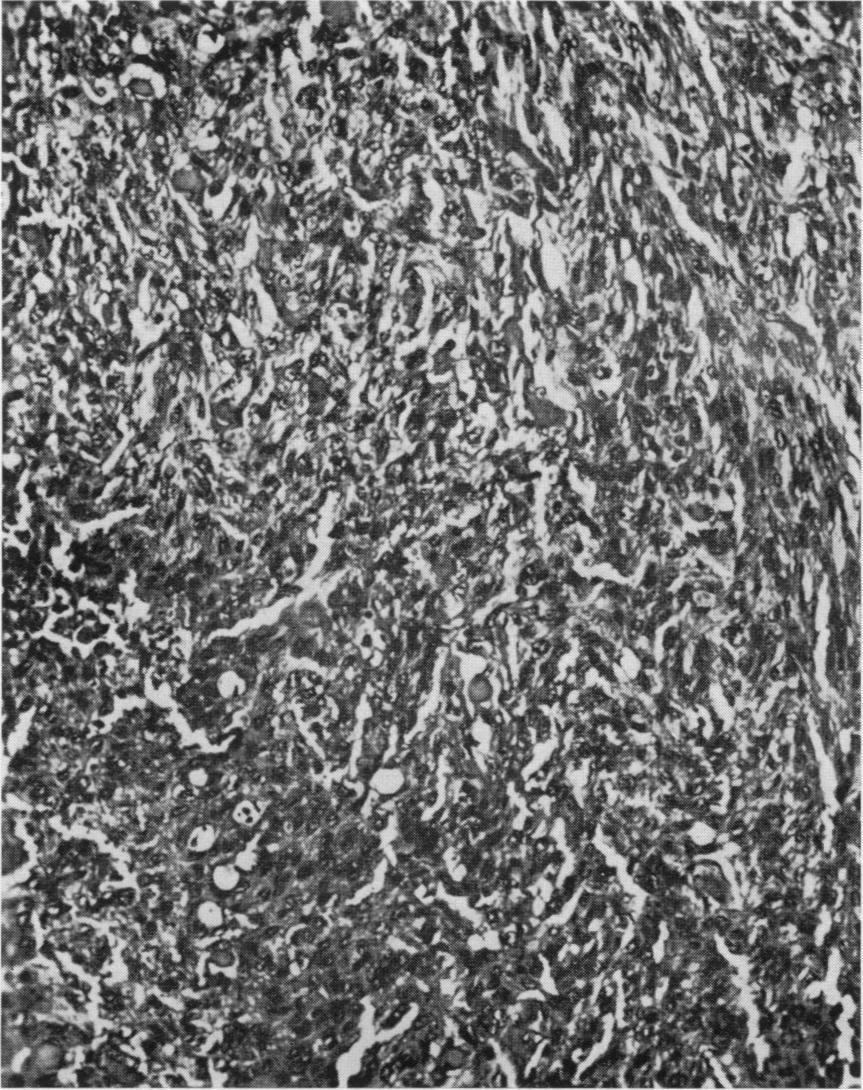
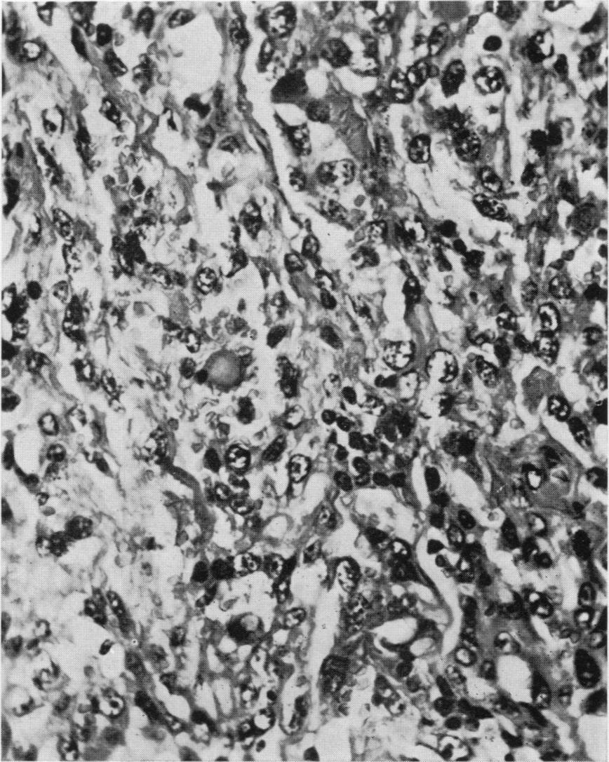
2 44 A. M. PAMUKCU found in cats under 8 years of age. These animals seem to be more resistant to the development of such tumours than are other domestic species that are permitted to live most of their life span. This resistance may be due to differences in metabolic pathways between species rather than to differences of tissue susceptibility. For example, it has been shown that cats metabolize tryptophane-an essential amino acid-by a process that does not involve the production of large quantities of orthoaminophenol metabolites. Consequently, unlike the dog, rat, and man, the cat has an extremely low level of such tryptophane metabolites in the urine. Certain of these metabolites have been implicated as etiologic agents in the genesis of human bladder cancer. Primary neoplasms of the urinary bladder are infrequent in horses. So far, only 37 cases have been reported, nearly all of them in horses over 10 years of age. The tumours were more common in males than in females. Urinary bladder tumours are exceedingly rare in swine. One papilloma was found in the bladder of an animal 6 months old. Because of the small number of such tumours reported in cats, sheep, horses, and swine, this histological classification will be based mainly on the findings in cattle and dogs. However, reference will also be made to other species in the explanatory notes. A wide variety of benign and malignant tumours occur in the urinary bladder. Epithelial tumours are the most common, accounting for approximately 82% (145 of 177 cases) and 77% (123 of 160 cases) of all primary bladder tumours in cattle and dogs, respectively. Malignant epithelial tumours are more frequently found than benign ones. Tumours of the urinary bladder may be of primary or secondary origin. Primary tumours are the most common in cattle and dogs, and most primary malignant tumours are of epithelial origin. HISTOLOGICAL GRADING Grading of carcinomas is of particular importance from the standpoint of prognosis. It is based solely on the degree of anaplasia of the cells and not on the pattern of tumours, the cell type, or the extent of invasion into the bladder wall. All carcinomas may be graded according to the following histological indications of cellular anaplasia: (1) increased cellularity; (2) nuclear crowding; (3) disturbance of cellular polarity; (4) failure of differentiation from base to surface; (5) polymorphism; (6) irregularity in the size of cells; (7) variation in the shape of nuclei and in chromatin pattern; (8) presence of giant cells; and (9) displaced or abnormal mitotic figures. The presence of one or more of these criteria is acceptable as evidence of anaplasia. Care must be taken to exclude reactive or regenerative conditions in which some of these features may be present. Tumours that demonstrate slight anaplasia are designated as Grade I carcinoma (Fig. 3, 4). At the opposite extreme are tumours showing severe anaplasia, which should be classified as Grade III carcinoma (Fig. 6, 7). Any tumour that does not fit readily into Grade I or Grade III is assigned to the intermediate grade, i.e., Grade II (Fig. 5). Sections used for grading should be of adequate size: usually a piece of tissue measuring about 1.5 x 2.0 cm will show the full range of the tumour pattern. Occasionally a definite variation in grade, as distinct from mere variation in structural pattern, may be seen in the same histological section. The grading should then be that of the most malignant part of the growth. Consistency of grading can be acquired only after considerable experience. The author acknowledges the collaboration of Dr F. K. Mostofi in examining slides and reviewing the manuscript. The photographs were reproduced by courtesy of the Armed Forces Institute of Pathology, Washington, D.C., USA. * *

3 URINARY BLADDER 45 HISTOLOGICAL CLASSIFICATION AND NOMENCLATURE OF TUMOURS OF THE URINARY BLADDER I. EPITHELIAL TUMOURS A. PAPILLOMA B. ADENOMA C. TRANSITIONAL CELL CARCINOMA D. VARIANTS OF TRANSITIONAL CELL CARCINOMA 1. With squamous metaplasia 2. With glandular metaplasia 3. With squamous and glandular metaplasia E. SQUAMOUS CELL CARCINOMA F. ADENOCARCINOMA G. UNDIFFERENTIATED CARCINOMA II. NONEPITHELIAL TUMOURS A. MUSCLE TUMOURS B. VAscuLAR TUMOURS C. FIBROBLASTIC TUMOURS D. OTHER NONEPITHELIAL TUMOURS III. TUMOURS WITH COEXISTING EPI- THELIAL AND MESENCHYMAL ELEMENTS IV. SECONDARY TUMOURS V. UNCLASSIFIED TUMOURS VI. PROLIFERATIVE CHANGES I. EPITHELIAL TUMOURS Normally the bladder is lined with 3-6 layers of transitional epithelial cells.a Under certain conditions the bladder mucosa may be in an unstable condition. The mucosa is often seen not to be normal when examined cystoscopically, yet a definite diagnosis of tumour cannot be made histologically. The condition may revert to normal, stabilize, or progress to a tumour. These changes are mainly of a proliferative nature (vide infra). DESCRIPTION OF TUMOURS A. Papilloma (Fig. 1) Bladder papillomas are defined as papillary tumours that have delicate fibrovascular stroma covered with transitional epithelium indistinguishable from the normal bladder epithelium and not more than six cells deep. The individual cells are slender and elongated; they are arranged parallel to one another and at right angles to the basement membrane; they contain basal normal mitotic figures; and there is no evidence of invasion. Histological diagnosis of papilloma is based on a complete absence of cellular anaplasia (vide infra). There is a wide variation in their biological behaviour. In cattle, papillomas account for approximately 17% a The term " urothelium " is used in some departments for this type of epithelium; likewise, transitional cell carcinoma is called " urothelial carcinoma ". (30 of 177 cases) of primary tumours of the bladder and, in dogs, for 14% (23 of 160 cases). It is no known whether these tumours recur after surgica removal. B. Adenoma This is a benign growth of glandular epithelium that occurs rarely in cattle and dogs. These adenomas vary from solitary, cauliflower-shaped masses to multiple, pedunculated growths. Some adenomas are indistinguishable from papillary urothelial tumours. Adenomas are composed of a large number of glandular structures separated from one another by a variable amount of connective tissue. The epithelium forming the glands consists of a single layer of columnar cells. The amount of mucin in the epithelial cells varies. In some there is only a small amount situated in the cell substance nearest the lumen, whereas in others there is an amount large enough to produce goblet-type cells. Frequently the glands are distended into thin-walled cystic spaces filled with mucin and desquamated cells. The arrangement of the cells lining the glands is orderly and mitotic figures are exceedingly rare. Adenomas do not invade the muscular layers of the bladder wall. They develop either from the preexisting glands in the mucosa or from the transitional epithelium through metaplastic changes. The most common
 Carcinomas account for nearly 80% of epithelial tumours of the bladder in cattle (115 of 145) and in dogs (98 of 123).")
4 46 A. M. PAMUKCU adenomas in cattle and dogs are those of metaplastic origin. Endometrial or nephrogenic adenomas have not been reported in animals. C. Transitional cell carcinoma (Fig. 2-7) Carcinomas account for nearly 80% of epithelial tumours of the bladder in cattle (115 of 145) and in dogs (98 of 123). Transitional cell carcinomas are the most common; squamous cell carcinomas and adenocarcinomas occur less frequently. The diagnosis of malignancy is based on cellular anaplasia, invasion, or metastasis. Metastasis is a late phenomenon in carcinoma of the bladder. Transitional cell carcinoma of the bladder may be single or multiple. Multiple tumours are mostly papillary and vary in size and appearance. Some are almost microscopic, whereas others may completely fill the bladder with protuberant growths or may extend over most of the mucosal surface. On the basis of morbid anatomy, both gross and microscopic, transitional cell carcinomas may be subdivided into four kinds as indicated below. Nonpapillary and noninfiltrating carcinoma, in which the tumour is confined to the surface, i.e., carcinoma in situ (Fig. 2). This is defined as a neoplastic entity in which the tumour cells are still within the epithelium of origin without invasion of the basement membrane. Although it is noninvasive and nonpapillary, carcinoma in situ is found most often in the mucosa adjacent to well-formed carcinoma in the bladder. Its occurrence in association with definite carcinomas raises the question of its relationship with invasive as well as with papillary carcinoma. Carcinoma in situ is rarely found in cattle and dogs. It occurs mainly in association with nodular infiltrating transitional cell carcinoma. Papillary carcinoma grows into the lumen of the bladder and resembles the papilloma grossly, but its base is relatively broader and the villi are confluent and clubbed or blunted. The growth may have a cauliflower-like pattern. Microscopically, the fronds of the papillary carcinoma have a thick epithelial coat. The covering epithelium shows an increase in cells, which are not uniform, are crowded together, and are irregularly stratified. Atypical nuclei, anaplasia, giant cells, and mitotic figures are characteristic (Fig. 3). In some tumours, squamous cells are observed among the transitional cells that dominate the pattern of the papillary noninfiltrating carcinoma. The papillary carcinoma does not invade the stroma of its own stalks or the base of the growth. In the veterinary literature, papillary noninfiltrating carcinomas are not reported separately from papillary infiltrating carcinomas. Therefore, it is difficult to give any figure for their frequency in animals. Such differentiation may have prognostic importance. Papillary and infiltrating carcinoma resembles papillary carcinoma in many respects. It also invades the stroma of its own stalk and lamina propria of the bladder wall (Fig. 4, 5). Most of the transitional cell carcinomas are papillary. They account for nearly 55% of carcinomas in cattle and dogs. However, very few adenomas, adenocarcinomas. and squamous cell carcinomas have papillary foci, Infiltrating carcinoma grows into the wall of the bladder and usually appears as a flat, plaque-like lesion or as an ulcerated, infiltrated mucosal patch overlying a nodular growth. Microscopically, the tumour is composed of transitional cells forming cohesive sheets, cords, or nests. The cellular borders are sharp, but prickles cannot be demonstrated. Nuclear and cytoplasmic patterns vary widely. Sessile infiltrating carcinomas carry a poorer prognosis than papillary carcinomas (Fig. 6, 7). Infiltrating transitional cell carcinomas constitute nearly 21 % of all primary carcinomas of the bladder in cattle. Transitional cell carcinomas often metastasize to the regional lymph nodes and the lungs in cattle and dogs, although other organs, such as the liver, spleen, and kidneys, are involved in some instances. D. Variants of transitional cell carcinoma Since the large majority of carcinomas of the bladder start as transitional cell carcinomas and remain as such, they may be referred to simply as " carcinomas ". However, a number of transitional cell carcinomas exhibit areas of squamous cells or glandular structure. It is best not to categorize such tumours as squamous, glandular, or metaplastic carcinomas, but to designate them as follows: 1. With squamous metaplasia 2. With glandular metaplasia 3. With squamous and glandular metaplasia E. Squamous cell carcinoma (Fig. 8, 9) Squamous cell carcinoma is usually not papillary, although it may have papillary foci. The tumour may have firm, raised, and rolled edges and an eroded centre. Microscopically, squamous cell carcinoma of the bladder does not differ in histological appearance from squamous carcinoma elsewhere in the body. The least well differentiated tumours can be distinguished from transitional cell carcinoma


. Fig. 4.")
5 Fig. 1. Papilloma (cow). Fig. 2. Carcinoma in situ (cow). Fig. 3. Transitional cell carcinoma, papillary and noninfiltrating, Grade I (dog). Fig. 4. Transitional cell carcinoma, papillary and infiltrating, Grade I (cow).

. Fig. 6.")


6 Fig. 5. Transitional cell carcinoma, papillary infiltrating, Grade lii(dog). Fig. 6. Transitional cell carcinoma, infiltrating, Grade Ill (dog). Fig. 7. Transitional cell carcinoma, infiltrating, Grade Ill (cow). Fig. 8. Squamous cell carcinoma (cow).

. (cow). Fig.")


7 Fig. 9. Squamous cell carcinoma, tentacular invasion Fig. 10. Adenocarcinoma (cow). (cow). Fig. 11. Undifferentiated carcinoma, spindle cells (cow). Fig. 12. Undifferentiated carcinoma, small round cells (water buffalo).


8 Fig. 13. Leiomyosarcoma (cow). Fig. 14. Haemangioma (cow). Fig. 15. Haemangiosarcoma (cow). Fig. 16. Haemangiosarcoma (cow).

9 URINARY BLADDER 51 because there is frank keratinization (Fig. 8, 9). The most highly differentiated growths show the usual pearls. Squamous cell carcinomas most often result from the epidermidalization of a papillary or transitional cell tumour. They consist entirely of squamous epithelial elements. Transitional cell carcinomas having areas of squamous metaplasia are not regarded as true squamous cell carcinomas. This type of carcinoma is not infrequent in animals, and it accounts for approximately 70% (9 of 115) and 14% (14 of 98) of all primary bladder carcinomas in cattle and dogs, respectively. It occurs in old male dogs more often than in bitches. However, a high incidence of squamous cell carcinoma has been reported in horses (13 of 35 cases). This was explained by the normal presence of squamous cell islands in the urinary bladder epithelium of the horse. Squamous cell carcinomas metastasize readily. In dogs, 9% of such tumours showed metastases. F. Adenocarcinoma (Fig. 10) All adenocarcinomas of the bladder, with the exception of the mucinous varieties, resemble urothelial carcinomas. They present as localized areas of diffusely involved vesical mucosa, as flat ulcerations, or as polypoid growths. When examined grossly and microscopically, adenocarcinomas are seen to fall into three patterns: papillary; papillary and infiltrating; and sessile and infiltrating. The two last-mentioned patterns occur with equal frequency in cattle. Histologically, the tumours are made up of glands of variable sizes and shapes. These glands are lined with mucous-secreting columnar or cuboidal cells. The amount of mucus in the glands varies considerably, and lakes of mucus lined with indistinct epithelial cells may be formed. The histological differentiation of primary and secondary adenocarcinoma may also be difficult. The three most common adenocarcinomas that involve the bladder secondarily are those of the uterus, prostate, and rectum. Features helpful for differentiation include: (a) the presence of foci of transitional epithelium, (b) areas of transition from normal to neoplastic epithelium, (c) an association of proliferative changes in the adjoining epithelium, and (d) areas of carcinoma in situ. Adenocarcinomas of the prostate are generally of the nonmucinous type, whereas those of the bladder commonly produce mucin. Adenocarcinoma metastases may be found in the lungs, prostate, and abdominal walls in dogs (3 of 6). Adenocarcinomas not infrequently arise as a result of glandular metaplasia of the transitional epithelium or normally located glands in the bladder mucosa. Those of dysontogenic origin have not been studied in detail in animals. Most adenocarcinomas reported in cattle fall into the metaplastic category. Primary adenocarcinomas are those with a glandular structure throughout. Transitional carcinomas, having foci of glandular metaplasia, are excluded from the category of true adenocarcinoma. Primary adenocarcinomas comprise 60/ (6 of 98) and 10% (12 of 115) of all primary carcinomas of the bladder in dogs and cattle, respectively. G. Undifferentiated carcinoma (Fig. 11, 12) With some tumours, it is impossible to recognize the cell type as belonging to any of the abovementioned categories. Such tumours are designated as " undifferentiated carcinoma ". The term " undifferentiated" is used here in a histogenic sense to characterize a type of epithelium-not to indicate the degree of anaplasia of the tumour. This category is not synonymous with anaplastic tumours. II. NONEPITHELIAL TUMOURS Nonepithelial bladder tumours comprise fewer than 18% and 230% of all primary tumours in cattle and dogs, respectively. They may arise from practically any tissue component of the bladder. They are classified according to the system described under Tumours of the soft (mesenchymal) tissues (see page 101). Nonepithelial tumours should be recorded as recommended above for epithelial tumours, with observations on the pattern, tissue type, degree of anaplasia, organ or area involved, depth of penetration, and presence of lymphatic or blood vessel invasion. The most common nonepithelial tumours of the bladder are muscle tumours and vascular tumours in dogs and cattle, respectively. A. Muscle tumours Leiomyoma and leiomyosarcoma (Fig. 13) account for 12% (20 of 160) of all primary tumours in dogs. Rhabdomyosarcomas occur infrequently in animals. So far, two cases have been reported in dogs. Muscle tumours are very rare in cattle. Leiomyoma and leiomyosarcoma arise from the muscle of the bladder or are a growth of undifferentiated mesenchyma that differentiates into muscle. Leiomyosarcomas are generally vascular and consist of elongated acidophilic cells arranged in sharply intersecting bundles. The presence of cellular pleo-
, haemangiosarcoma (Fig. 15, 16) and haemangiopericytoma, occur often in the bovine bladder.")
10 52 A. M. PAMUKCU morphism, marked cellularity, tumour giant cells, and atypical mitotic figures helps to distinguish them from their benign counterparts, the leiomyomas. B. Vascular tumours Angioendothelial tumours, such as haemangioma (Fig. 14), haemangiosarcoma (Fig. 15, 16) and haemangiopericytoma, occur often in the bovine bladder. They make up nearly 6% of all primary bladder tumours and are usually found in compound tumours with primary epithelial tumours of the bladder. Vascular tumours are found infrequently in dogs and cats. The microscopic appearance of these tumours is similar to that of vascular tumours found elsewhere in the body. C. Fibroblastic tumours These occur in both cattle and dogs and can be either benign or malignant. So far, fibroma (5 of 160), fibrosarcoma (4 of 160), and sarcoma (unclassified) (4 of 160) have been reported in dogs as primary bladder tumours. Such tumours have a histological structure similar to those seen elsewhere in the body. D. Other nonepithelial tumours Primary malignant lymphomas of the bladder are very rare. Only 8 cases have been reported: 2 in dogs, 2 in cats, and 4 in cows. However, secondary involvement of the bladder in generalized lymphosarcoma is not uncommon, and the histology is similar to that observed in lymphosarcoma of the lymph nodes. III. TUMOURS WITH COEXISTING EPITHELIAL AND MESENCHYMAL ELEMENTS These are defined as tumours composed of neoplastic epithelial components and of mesenchymal tissue, e.g., carcinosarcoma, carcinohaemangioma and carcinoleiomyosarcohaemangioma, adenohaemangioma, adenocarcinohaemangioma, and papillohaemangioma. Frequently, two or more different types of tumour have been recognized in the urinary bladders of cattle that have ingested bracken. These types of tumour are very rare in other animal species. IV. SECONDARY TUMOURS Secondary involvement of the bladder occurs either by direct extension from malignant primary tumours of adjacent organs (chiefly the prostate, rectum, and uterus), by implantation from primary lesions located in the upper urinary tract, or, rarely, by metastasis from distant primary tumours. Secondary tumours comprise 7.8 % of all urinary bladder tumours (13 of 166) in dogs. In 7 of 13 cases, the secondary tumour originated from carcinoma of the prostate. The remainder had metastasized from malignant tumours of the heart, thyroid, bone, and lymphoid tissue. In lymphosarcoma, the wall of the urinary bladder may be infiltrated, but secondary lymphosarcomas occur more frequently in the kidneys. The kidneys were involved in 10 of 52 cases (19.2%) of lymphosarcoma in dogs, whereas the urinary bladder was involved in only 2 (3.8 %). Three cases of secondary lymphosarcoma have been observed with generalized lymphosarcoma in cattle. Secondary tumours of the urinary bladder are seldom found in other animal species. V. UNCLASSIFIED TUMOURS These are primary benign or malignant tumours that cannot be placed in any of the categories described above. VI. PROLIFERATIVE CHANGES Proliferative changes usually result from cystitis. They may be mistaken for bladder tumours macroscopically as well as microscopically, but histological differentiation from bladder tumours does not cause any difficulty. The proliferative changes commonly observed are listed below. Von Brunn's nests. These are compact groups of transitional epithelial cells lying in the lamina propria, with or without connexion to the surface epithelium. Glandular metaplasia (glandular cystitis). This is characterized by mucus-containing columnar epithelial cells either on the surface or forming glands in the lamina propria. Squamous metaplasia. Transitional epithelium shows squamous metaplasia and is replaced by squamous cells with or without keratinization. There is usually hyperplasia of the epithelium. Cystic cystitis. These are groups of transitional epithelial cells with central cavitation, sometimes cystic, lying in the lamina propria, with or without connexion to the surface epithelium. Papillary (polypoid) cystitis. This proliferative change is characterized by hyperplastic epithelium that covers thickened, finger-like projections of the lamina propria. The projections are aedematous, hyperaemic, and infiltrated with inflammatory cells.
XX. Tumours of the nasal cavity *
 XX. Tumours of the nasal cavity * H. STONZI 1 & B. HAUSER2 Tumours of the nasal cavity are rare in domestic animals, most cases occurring in the dog. Epithelial tumours are the most common type in carnivores
Epithelial tumors. Dr. F.F. Khuzin, PhD Dr. M.O. Mavlikeev
 Epithelial tumors Dr. F.F. Khuzin, PhD Dr. M.O. Mavlikeev Epithelial tumors Tumors from the epithelium are the most frequent among tumors. There are 2 group features of these tumors: The presence in most
XIII. Tumours of the liver and biliary system
 XIII. Tumours of the liver and biliary system V. PONOMARKOV 1 & L. J. MACKEY 2 In this histological classification of liver and gall bladder tumours the tumour types largely correspond to those found in
Disorders of Cell Growth & Neoplasia. Histopathology Lab
 Disorders of Cell Growth & Neoplasia Histopathology Lab Paul Hanna April 2010 Case #84 Clinical History: 5 yr-old, West Highland White terrier. skin mass from axillary region. has been present for the
number Done by Corrected by Doctor Maha Shomaf
 number 16 Done by Waseem Abo-Obeida Corrected by Zeina Assaf Doctor Maha Shomaf MALIGNANT NEOPLASMS The four fundamental features by which benign and malignant tumors can be distinguished are: 1- differentiation
Neoplasia 2018 Lecture 2. Dr Heyam Awad MD, FRCPath
 Neoplasia 2018 Lecture 2 Dr Heyam Awad MD, FRCPath ILOS 1. List the differences between benign and malignant tumors. 2. Recognize the histological features of malignancy. 3. Define dysplasia and understand
NEOPLASIA-I CANCER. Nam Deuk Kim, Ph.D.
 NEOPLASIA-I CANCER Nam Deuk Kim, Ph.D. 1 2 Tumor in the hieroglyphics of the Edwin Smith papyrus (1,600 B.C., Breasted s translation 1930) 3 War on Cancer (National Cancer Act, 1971) 4 Cancer Acts in Korea
Neoplasia literally means "new growth.
 NEOPLASIA Neoplasia literally means "new growth. A neoplasm, defined as "an abnormal mass of tissue the growth of which exceeds and is uncoordinated with that of the normal tissues and persists in the
ONCOLOGY. Csaba Bödör. Department of Pathology and Experimental Cancer Research november 19., ÁOK, III.
 ONCOLOGY Csaba Bödör Department of Pathology and Experimental Cancer Research 2018. november 19., ÁOK, III. bodor.csaba1@med.semmelweis-univ.hu ONCOLOGY Characteristics of Benign and Malignant Neoplasms
Synonyms. Nephrogenic metaplasia Mesonephric adenoma
 Nephrogenic Adenoma Synonyms Nephrogenic metaplasia Mesonephric adenoma Definition Benign epithelial lesion of urinary tract with tubular, glandular, papillary growth pattern Most frequently in the urinary
5/21/2018. Prostate Adenocarcinoma vs. Urothelial Carcinoma. Common Differential Diagnoses in Urological Pathology. Jonathan I.
 Common Differential Diagnoses in Urological Pathology Jonathan I. Epstein Prostate Adenocarcinoma vs. Urothelial Carcinoma 1 2 NKX3.1 NKX3.1 3 4 5 6 Proposed ISUP Recommendations Option to use PSA as a
Normal Morphology. Anatomic Considerations. Normal Urothelial Histology and Cytology
 1 Normal Morphology Anatomic Considerations The urinary tract can be divided into three regions: the kidney; the calyces, pelves and ureters (upper collecting system or upper tract); and the bladder and
Pathology of bladder cancer in Egypt; a current study.
 Pathology of bladder cancer in Egypt; a current study. Thesis Submitted for partial fulfillment of Master degree in urology By Mohamed Atef Mohamed Ahmed M.B.B.CH Supervised by Prof.Dr.: Omar Mohamed Abdel-
Diseases of the breast (1 of 2)
") Diseases of the breast (1 of 2) Introduction A histology introduction Normal ducts and lobules of the breast are lined by two layers of cells a layer of luminal cells overlying a second layer of myoepithelial
Diseases of the breast (1 of 2) Introduction A histology introduction Normal ducts and lobules of the breast are lined by two layers of cells a layer of luminal cells overlying a second layer of myoepithelial
A neoplasm is defined as "an abnormal tissue proliferation, which exceeds that of adjacent normal tissue. This proliferation continues even after
 NEOPLASIA Neoplasia is a very important topic in pathology because neoplasms are both common and serious diseases. A neoplasm literally means a new growth, and this term is used interchangeably with a
Mody. AIS vs. Invasive Adenocarcinoma of the Cervix
 Common Problems in Gynecologic Pathology Michael T. Deavers, M.D. Houston Methodist Hospital, Houston, Texas Common Problems in Gynecologic Pathology Adenocarcinoma in-situ (AIS) of the Cervix vs. Invasive
Neoplasia part I. Dr. Mohsen Dashti. Clinical Medicine & Pathology nd Lecture
 Neoplasia part I By Dr. Mohsen Dashti Clinical Medicine & Pathology 316 2 nd Lecture Lecture outline Review of structure & function. Basic definitions. Classification of neoplasms. Morphologic features.
Objectives. Atypical Glandular Cells. Atypical Endocervical Cells. Reactive Endocervical Cells
 2013 California Society of Pathologists 66 th Annual Meeting San Francisco, CA Atypical Glandular Cells to Early Invasive Adenocarcinoma: Cervical Cytology and Histology Christina S. Kong, MD Associate
2 to 3% of All New Visceral Cancers Peak Incidence is 6th Decade M:F = 2:1 Grossly is a Bright Yellow, Necrotic Mass with a Pseudocapsule
 GENITOURINARY PATHOLOGY Kathleen M. O Toole, M.D. Renal Cell Carcinoma 2 to 3% of All New Visceral Cancers Peak Incidence is 6th Decade M:F = 2:1 Grossly is a Bright Yellow Necrotic Mass Grossly is a Bright
MVST BOD & NST PART IB Thurs. 2 nd & Fri. 3 rd March 2017 Pathology Practical Class 23
 MVST BOD & NST PART IB Thurs. 2 nd & Fri. 3 rd March 2017 Pathology Practical Class 23 Neoplasia I Neoplasia I: Benign and malignant neoplasms in glandular epithelium and mesenchyme 1.0. Aims 1. To understand
GUIDELINES ON NON-MUSCLE- INVASIVE BLADDER CANCER
 GUIDELINES ON NON-MUSCLE- INVASIVE BLADDER CANCER (Limited text update December 21) M. Babjuk, W. Oosterlinck, R. Sylvester, E. Kaasinen, A. Böhle, J. Palou, M. Rouprêt Eur Urol 211 Apr;59(4):584-94 Introduction
Normal thyroid tissue
 Thyroid Pathology Overview Normal thyroid tissue Normal thyroid tissue with follicles filled with colloid. Thyroid cells form follicles, spheres of epithelial cells (always single layered in health, usually
Demystifying Endometrial Hyperplasia
 Demystifying Endometrial Hyperplasia A review from Diagnostic Histopathology 19:7 Dr R Hadden ST5 Histopathology Derriford Hospital Plymouth Endometrium Target for sex-steroid hormones Glands Stroma Proliferate
Papillary Lesions of the Breast A Practical Approach to Diagnosis. (Arch Pathol Lab Med. 2016;140: ; doi: /arpa.
 Papillary Lesions of the Breast A Practical Approach to Diagnosis (Arch Pathol Lab Med. 2016;140:1052 1059; doi: 10.5858/arpa.2016-0219-RA) Papillary lesions of the breast Span the spectrum of benign,
Papillary Lesions of the breast
 Papillary Lesions of the breast Emad Rakha Professor of Breast Pathology The University of Nottingham Papillary lesions of the breast are a heterogeneous group of disease, which are characterised by neoplastic
Bladder Case 1 SURGICAL PATHOLOGY REPORT. Procedure: Cystoscopy, transurethral resection of bladder tumor (TURBT)
") Bladder Case 1 February 17, 2007 Specimen (s) received: Bladder Tumor Pre-operative Diagnosis: Bladder Cancer Post operative Diagnosis: Bladder Cancer Procedure: Cystoscopy, transurethral resection of
Bladder Case 1 February 17, 2007 Specimen (s) received: Bladder Tumor Pre-operative Diagnosis: Bladder Cancer Post operative Diagnosis: Bladder Cancer Procedure: Cystoscopy, transurethral resection of
Condyloma Acuminatum. Mimics of Bladder Cancer. Squamous Papilloma. Squamous epithelium in bladder
 Mimics of Bladder Cancer Murali Varma Cardiff, UK wptmv@cf.ac.uk Squamous epithelium in bladder Non-keratinising vaginal type mucosa common in trigone region in women Normal variant Sarajevo Nov 2013 Squamous
J of Evolution of Med and Dent Sci/ eissn , pissn / Vol. 3/ Issue 24/June 16, 2014 Page 6628
 ADENOCARCINOMA OF BLADDER-SIGNET RING CELL MUCINOUS VARIANT S. Senthil Kumar 1, D. Prem Charles 2, B. Krishnaswamy 3, P. Viswanathan 4, S. Sarath Chandran 5 HOW TO CITE THIS ARTICLE: S. Senthil Kumar,
The pathology of bladder cancer
 1 The pathology of bladder cancer Charles Jameson Introduction Carcinoma of the bladder is the seventh most common cancer worldwide [1]. It comprises 3.2% of all cancers, with an estimated 260 000 new
Normal endometrium: A, proliferative. B, secretory.
 Normal endometrium: A, proliferative. B, secretory. Nội mạc tử cung Nội mạc tử cung Cyclic changes in endometrium.. Approximate relationship of useful microscopic changes. Arias-Stella reaction in endometrial
04/09/2018. Squamous Cell Neoplasia and Precursor Lesions. Agenda. Squamous Dysplasia. Squamo-proliferative lesions. Architectural features
 Squamous Cell Neoplasia and Precursor Lesions Jennifer L. Hunt, MD, MEd Aubrey J. Hough Jr, MD, Endowed Professor of Pathology Chair of Pathology and Laboratory Medicine University of Arkansas for Medical
DUSTURBANCES OF GROWTH. MLS Basic histological diagnosis MLS HIST 422 Semester 8- batch 7 L8 Uz: Musa
 DUSTURBANCES OF GROWTH MLS Basic histological diagnosis MLS HIST 422 Semester 8- batch 7 L8 Uz: Musa Agnesia: means complete absence of an organ (Kidney). Aplasia: s defined in general as "defective development
A adipose cells. B capillary. C epithelium
 EPITHELIA Objective The objective of this class is to observe how different epithelia vary in terms of cell shape, size and number of cell layers enabling them to be well adapted for functions in different
Diseases of the vulva
 Diseases of the vulva 1. Bartholin Cyst - Infection of the Bartholin gland produces an acute inflammation within the gland (adenitis) and may result in an abscess. Bartholin duct cysts - Are relatively
Neoplasms of the Canine, Feline and Lemur Liver:
 Neoplasms of the Canine, Feline and Lemur Liver: Classification and Prognosis Annual Seminar of the French Society of Veterinary Pathology John M. Cullen VMD PhD DACVP North Carolina State University Primary
TUMOR,NEOPLASM. Pathology Department, Zhejiang University School of Medicine,
 TUMOR,NEOPLASM Pathology Department, Zhejiang University School of Medicine, 马丽琴,maliqin198@zju.edu.cn The points in this chapter What is a neoplasm (conception) Morphology of neoplasm Macroscopy of Neoplasm
Note: The cause of testicular neoplasms remains unknown
 - In the 15- to 34-year-old age group, they are the most common tumors of men. - Tumors of the testis are a heterogeneous group of neoplasms that include: I. Germ cell tumors : 95%; all are malignant.
Imaging Evaluation of Polyps. CT Colonography: Sessile Adenoma. Polyps, DALMs & Megacolon Objectives
 Polyps, DALMs & Megacolon: Pathology and Imaging of the Colon and Rectum Angela D. Levy and Leslie H. Sobin Washington, DC Drs. Levy and Sobin have indicated that they have no relationships which, in the
Maram Abdaljaleel, MD Dermatopathologist and Neuropathologist University of Jordan, School of Medicine
 Maram Abdaljaleel, MD Dermatopathologist and Neuropathologist University of Jordan, School of Medicine The most common non-skin malignancy of women 2 nd most common cause of cancer deaths in women, following
2% of all malignancies Male predominance Patients usually more than 60 years old
 Benign Bladder Tumors Transitional Cell Papilloma 2-3% Inverted Papilloma Rare Malignant Transitional (Urothelial) Carcinoma 90% Carcinoma In-Situ (By Itself) 5-10% Squamous Cell Carcinoma 3-7% Adenocarcinoma
Urinary Bladder: WHO Classification and AJCC Staging Update 2017
 Urinary Bladder: WHO Classification and AJCC Staging Update 2017 Houston Society of Clinical Pathologists 58 th Annual Spring Symposium Houston, TX April 8, 2017 Jesse K. McKenney, MD Classification
Recently, there has been an increasing incidence among women and younger persons
 Bladder Tumors Benign Transitional Cell Papilloma 2-3% Inverted Papilloma Rare Malignant Transitional (Urothelial) Carcinoma 90% Carcinoma In-Situ (By Itself) 5-10% Squamous Cell Carcinoma 3-7% Adenocarcinoma
!! 2 to 3% of All New Visceral Cancers.!! Peak Incidence is 6th Decade!! M:F = 2:1
 !! Kathleen M. O Toole, M.D.!! 2 to 3% of All New Visceral Cancers!! Peak Incidence is 6th Decade!! M:F = 2:1!! Grossly is a Bright Yellow, Necrotic Mass with a Pseudocapsule 1 !!Conventional RCC! Clear
BLADDER CANCER: PATIENT INFORMATION
 BLADDER CANCER: PATIENT INFORMATION The bladder is the balloon like organ located in the pelvis that stores and empties urine. Urine is produced by the kidneys, is conducted to the bladder by the ureters,
Kidney, Bladder and Prostate Neoplasia. David Bingham MD
 Kidney, Bladder and Prostate Neoplasia David Bingham MD typical malignant cytology of bladder washings 1 benign 2 malignant typical malignant cytology of bladder washings b Bladder tumor Non invasive papillary
Index 179. Genital tract contaminants, 17, 20, 22, 150 papilloma virus-infected cells, 47 squamous cells, sources of, 7
 Index Accuracy of urinary cytology, 166 Acute inflammatory cells, 38 catheter sample, 39 herpes simplex infections, 44 carcinomas, 104, 105 non-viral inclusions, 52, 53 voided urine, 17 Adenocarcinoma
B. Environmental Factors. a. The major risk factor to papillary thyroid cancer is exposure to ionizing radiation, during the first 2 decades of life.
 B. Environmental Factors. a. The major risk factor to papillary thyroid cancer is exposure to ionizing radiation, during the first 2 decades of life. b. Deficiency of dietary iodine: - Is linked with a
Ocular Neoplasia What s Common? What s New? Richard R Dubielzig
 Ocular Neoplasia What s Common? What s New? Richard R Dubielzig Orbit 288 6% Tumors of the globe make up 3225 out of 6110 total neoplasms = 53%. Tumors of the conjunctiva make up 1192 out of 6110 total
Salivary Glands 3/7/2017
 Salivary Glands 3/7/2017 Goals and objectives Focus on the entities unique to H&N Common board type facts Information for your future practice Salivary Glands Salivary Glands Major gland. Paratid. Submandibular.
CINtec p16 INK4a Staining Atlas
 CINtec p16 INK4a Staining Atlas Rating Rating Positive The rating positive will be assigned if the p16 INK4a -stained slide shows a continuous staining of cells of the basal and parabasal cell layers of
4/12/2018. MUSC Pathology Symposium Kiawah Island April 18, Jesse K. McKenney, MD
 MUSC Pathology Symposium Kiawah Island April 18, 2018 Jesse K. McKenney, MD 1 Urothelial Carcinoma with Alternative Differentiation 2 Urothelial Carcinoma with Alternative Differentiation Recognition as
Biology. Dr. Khalida Ibrahim
 Dr. Khalida Ibrahim Biology Histology: Histology: is the study of the tissues of the body. Tissue: group of similar cells combined to perform a common function. The human body is composed of only 4 basic
XIX. Tumours of the prostate and penis
 XIX. Tumours of the prostate and penis WILLIAM C. HALL,1 SVEND W. NIELSEN,2 & KENNETH McENTEE3 Tumours of the male genital tract, excluding the testes, are relatively rare in the six major domestic animals.
Nasal Cavity and Paranasal Sinuses
 Chapter 2 Nasal Cavity and Paranasal Sinuses Introduction Included in this chapter are nasal cavities, frontal sinus, ethmoid complex, sphenoid sinus, and maxillary sinuses. These cavities and sinuses
Gross appearance of nodular hyperplasia in material obtained from suprapubic prostatectomy. Note the multinodular appearance and the admixture of
 Tiền liệt tuyến Tiền liệt tuyến Gross appearance of nodular hyperplasia in material obtained from suprapubic prostatectomy. Note the multinodular appearance and the admixture of solid and microcystic areas.
Epithelia will be discussed according to the following scheme: Type Number of layers Shape Line drawing. Squamous Cuboidal Columnar
 Epithelia Epithelia will be discussed according to the following scheme: Type Number of layers Shape Line drawing Simple Squamous Cuboidal Columnar Covering and Lining epithelium Pseudostratified Stratified
-1- Pathology Department (code: 0605) Final Exam for Third year students Date: Time allowed: 2 hours. Paper II (75 marks).
 Final Exam for Third year students Date: Time allowed: 2 hours. Paper II (75 marks).") -1- BENHA UNIVERSITY FACULTY OF MEDICINE Pathology Department (code: 0605) Final Exam for Third year students Date: 28-5-2011 Time allowed: 2 hours. Paper II (75 marks). Please note that this question
-1- BENHA UNIVERSITY FACULTY OF MEDICINE Pathology Department (code: 0605) Final Exam for Third year students Date: 28-5-2011 Time allowed: 2 hours. Paper II (75 marks). Please note that this question
Carcinoma of the Urinary Bladder Histopathology
 Carcinoma of the Urinary Bladder Histopathology Reporting Proforma (Radical & Partial Cystectomy, Cystoprostatectomy) Includes the International Collaboration on Cancer reporting dataset denoted by * Family
RENAL CELL CARCINOMA 2 to 3% of All New Visceral Cancers Peak Incidence is 6th Decade M:F = 2:1 Grossly is a Bright Yellow, Necrotic Mass with a Pseud
 GENITOURINARY PATHOLOGY Kathleen M. O Toole Toole, M.D. RENAL CELL CARCINOMA 2 to 3% of All New Visceral Cancers Peak Incidence is 6th Decade M:F = 2:1 Grossly is a Bright Yellow, Necrotic Mass with a
FINAL HISTOLOGICAL DIAGNOSIS: Villo-adenomatous polyp with in-situ-carcinomatous foci (involving both adenomatous and villous component).
.") SOLITARY VILLO ADENOMATOUS POLYP WITH CARCINOMATOUS CHANGES RECTUM: A Divvya B 1, M. Valluvan 2, Rehana Tippoo 3, P. Viswanathan 4, R. Baskaran 5 HOW TO CITE THIS ARTICLE: Divvya B, M. Valluvan, Rehana
SOLITARY VILLO ADENOMATOUS POLYP WITH CARCINOMATOUS CHANGES RECTUM: A Divvya B 1, M. Valluvan 2, Rehana Tippoo 3, P. Viswanathan 4, R. Baskaran 5 HOW TO CITE THIS ARTICLE: Divvya B, M. Valluvan, Rehana
Tissue: The Living Fabric
 PowerPoint Lecture Slide Presentation by Vince Austin Human Anatomy & Physiology FIFTH EDITION Elaine N. Marieb Chapter 4 Tissue: The Living Fabric Part A Tissues Groups of cells similar in structure and
Urinary Bladder, Ureter, and Renal Pelvis
 Urinary Bladder, Ureter, and Renal Pelvis Protocol applies to all carcinomas of the urinary bladder, ureter, and renal pelvis. Protocol revision date: January 2005 Based on AJCC/UICC TNM, 6th edition Procedures
University Journal of Pre and Para Clinical Sciences
 ISSN 2455 2879 Volume 2 Issue 1 2016 Metaplastic carcinoma breast a rare case report Abstract : Metaplastic carcinoma of the breast is a rare malignancy with two distinct cell lines described as a breast
A215- Urinary bladder cancer tissues
 A215- Urinary bladder cancer tissues (formalin fixed) For research use only Specifications: No. of cases: 45 Tissue type: Urinary bladder cancer tissues No. of spots: 2 spots from each cancer case (90
Outline 11/2/2017. Pancreatic EUS-FNA general aspects. Cytomorphologic features of solid neoplasms/lesions of the pancreas
 ENDOSCOPIC ULTRASOUND GUIDED-FINE NEEDLE ASPIRATION CYTOLOGY OF PANCREAS Khalid Amin M.D. Assistant Professor Department of Laboratory Medicine and Pathology University of Minnesota Outline Pancreatic
Pathology of the skin. 2nd Department of Pathology, Semmelweis University
 Pathology of the skin 2nd Department of Pathology, Semmelweis University Histology of the skin Epidermis: Stratum corneum Stratum granulosum Stratum spinosum Stratum basale Dermis: papillary and reticular
Diplomate of the American Board of Pathology in Anatomic and Clinical Pathology
 A 33-year-old male with a left lower leg mass. Contributed by Shaoxiong Chen, MD, PhD Assistant Professor Indiana University School of Medicine/ IU Health Partners Department of Pathology and Laboratory
LYMPHATIC DRAINAGE AXILLARY (MOSTLY) INTERNAL MAMMARY SUPRACLAVICULAR
 INTERNAL MAMMARY SUPRACLAVICULAR") BREAST LYMPHATIC DRAINAGE AXILLARY (MOSTLY) INTERNAL MAMMARY SUPRACLAVICULAR HISTOLOGY LOBE: (10 in whole breast) LOBULE: (many per lobe) ACINUS/I, aka ALVEOLUS/I: (many per lobule) DUCT(S): INTRA- or
BREAST LYMPHATIC DRAINAGE AXILLARY (MOSTLY) INTERNAL MAMMARY SUPRACLAVICULAR HISTOLOGY LOBE: (10 in whole breast) LOBULE: (many per lobe) ACINUS/I, aka ALVEOLUS/I: (many per lobule) DUCT(S): INTRA- or
2007 Multiple Primary and
 2007 Multiple Primary and Histology Coding Rules Beyond the Basics Florida Cancer Data System Annual Conference Tampa, FL July 26,2007 Steven Peace, CTR Westat Carol Johnson, CTR NCI SEER Peggy Adamo,
Jesse K. McKenney, MD
 Jesse K. McKenney, MD Outline Microscopic anatomy of the urinary bladder Diagnosing invasion Subtle patterns (variants) of carcinoma Clinically important variants of carcinoma Microanatomy of Bladder Initial
Case year female. Routine Pap smear
 Case 1 57 year female Routine Pap smear Diagnosis? 1. Atypical glandular cells of unknown significance (AGUS) 2. Endocervical AIS 3. Endocervical adenocarcinoma 4. Endometrial adenocarcinoma 5. Adenocarcinoma
Tissues. tissue = many cells w/ same structure and function. cell shape aids function tissue shape aids function. Histology = study of tissues
 Tissues tissue = many cells w/ same structure and function cell shape aids function tissue shape aids function Histology = study of tissues 4 types of tissues Epithelial coverings contact openings Connective
FORELIMB SWEAT GLAND ADENOCARCINOMA IN A CAT
 I: 2047-2051 ISSN: 2277 4998 FORELIMB SWEAT GLAND ADENOCARCINOMA IN A CAT ABEDI G 1, HESARAKI S 2, ASGHARI A 1* 1: Department of Clinical Science, Science and Research branch, Islamic Azad University,
CYTOMORPHOLOGY MODULE 28.1 INTRODUCTION OBJECTIVES 28.2 GENERAL GUIDELINES. Notes
 28 CYTOMORPHOLOGY 28.1 INTRODUCTION Light microscopic examination of stained cells in smears is the method of choice of diagnostic cytology. It allows classification of most normal cells as to type and
Pitfalls in thyroid tumor pathology. Prof.Valdi Pešutić-Pisac MD, PhD
 Pitfalls in thyroid tumor pathology Prof.Valdi Pešutić-Pisac MD, PhD Too many or... Tumour herniation through a torn capsule simulating capsular invasion fibrous capsule with a sharp discontinuity, suggestive
Disclosure. Case. Mixed Tumors of the Uterine Corpus and Cervix. I have nothing to disclose
 Mixed Tumors of the Uterine Corpus and Cervix Marisa R. Nucci, M.D. Division of Women s and Perinatal Pathology Department of Pathology Brigham and Women s Hospital Boston, MA UCSF Current Issues in Anatomic
In the third part of the present study tumours which previous were described as basal cell tumours but now have been reclassified as trichoblastomas
 170 6. SUMMARY Immunhistochemical investigations for identifying the histogenesis of basaloid neoplasias and hyperplasias in the mamma parenchyma of the bitch, for the use of the human nuclear protein
Diagnostic Cytology of Cancer Cases
 Diagnostic Cytology of Cancer Cases Somporn Techangamsuwan Companion Animal Cancer Research Unit (CAC-RU) Department of Pathology, Faculty of Veterinary Science, Chulalongkorn University 1 Tumor or Non-tumor
DIGESTIVE TRACT ESOPHAGUS
 DIGESTIVE TRACT From the lower esophagus to the lower rectum four fundamental layers comprise the wall of the digestive tube: mucosa, submucosa, muscularis propria (externa), and adventitia or serosa (see
Prepared By Jocelyn Palao and Layla Faqih
 Prepared By Jocelyn Palao and Layla Faqih The structure of the suspected atypical cell should always be compared to the structure of other similar, benign, cells which are present in the smears. The diagnosis
Biliary tract tumors
 Short Course 2010 Annual Fall Meeting of the Korean Society for Pathologists Biliary tract tumors Joon Hyuk Choi, M.D., Ph.D. Professor, Department of Pathology, Yeungnam Univ. College of Medicine, Daegu,
Salivary Gland Cytology
 Salivary Gland Cytology Diagnostic challenges and potential pitfalls Tarik M. Elsheikh, MD Professor and Medical Director Anatomic Pathology Cleveland Clinic FNA Salivary Gland Lesions Indications Distinguish
Tissue: The Living Fabric: Part A
 PowerPoint Lecture Slides prepared by Janice Meeking, Mount Royal College C H A P T E R 4 Tissue: The Living Fabric: Part A Tissues Groups of cells similar in structure and function Types of tissues Epithelial
SESSION 1: GENERAL (BASIC) PATHOLOGY CONCEPTS Thursday, October 16, :30am - 11:30am FACULTY COPY
 PATHOLOGY CONCEPTS Thursday, October 16, :30am - 11:30am FACULTY COPY") SESSION 1: GENERAL (BASIC) PATHOLOGY CONCEPTS Thursday, October 16, 2008 9:30am - 11:30am FACULTY COPY GOAL: Describe the basic morphologic (structural) changes which occur in various pathologic conditions.
SESSION 1: GENERAL (BASIC) PATHOLOGY CONCEPTS Thursday, October 16, 2008 9:30am - 11:30am FACULTY COPY GOAL: Describe the basic morphologic (structural) changes which occur in various pathologic conditions.
Pathologic Assessment of Invasion in TUR Specimens. A. Lopez-Beltran. T1 (ct1)
") Pathologic Assessment of Invasion in TUR Specimens A. Lopez-Beltran T1 (ct1) 1 Prognostic factors for progression/invasive disease Ta,T1,CIS- NMIBC :TNM 2017 ESSENTIAL: Grade T stage CIS Number of lesions
Pathologic Assessment of Invasion in TUR Specimens A. Lopez-Beltran T1 (ct1) 1 Prognostic factors for progression/invasive disease Ta,T1,CIS- NMIBC :TNM 2017 ESSENTIAL: Grade T stage CIS Number of lesions
Spectrum of Lesions in Cystoscopic Bladder Biopsies -A Histopathological Study
 AJMS Al Ameen J Med Sci (2 012 )5 (2 ):1 3 2-1 3 6 (A US National Library of Medicine enlisted journal) I S S N 0 9 7 4-1 1 4 3 C O D E N : A A J M B G ORIGI NAL ARTICLE Spectrum of Lesions in Cystoscopic
-The cause of testicular neoplasms remains unknown
 - In the 15- to 34-year-old age group, they are the most common tumors of men. - include: I. Germ cell tumors : (95%); all are malignant. II. Sex cord-stromal tumors: from Sertoli or Leydig cells; usually
Page # 1. Endometrium. Cellular Components. Anatomical Regions. Management of SIL Thomas C. Wright, Jr. Most common diseases:
 Endometrium Pathology of the Endometrium Thomas C. Wright Columbia University, New York, NY Most common diseases: Abnormal uterine bleeding Inflammatory conditions Benign neoplasms Endometrial cancer Anatomical
NEOPLASIA! Terminology and Classification of Neoplastic cells! Objectives: Asst. Prof. Prasit Suwannalert, Ph.D. Leading Questions
 NEOPLASIA! Asst. Prof. Prasit Suwannalert, Ph.D. (Email: prasit.suw@mahidol.ac.th)! Department of Pathobiology Faculty of Science, Mahidol University! Objectives: After learning, students should be able
Histology Notes -Part 1: Epithelial Tissues
 Introduction Group of cells w/ similar structure & function = TISSUE Four Basic Tissue Types 1. Epithelial-covers 2. Connective-supports 3. Muscular*-produces movement (will discuss in the muscular system
Epithelium tissue system
 Epithelium tissue system Histology : is the study of the microscopic anatomy (microanatomy) of cells and tissues of plants and animals. It is commonly performed by examining cells and tissues under a light
Lecture Overview. Chapter 4 Epithelial Tissues Lecture 9. Introduction to Tissues. Epithelial Tissues. Glandular Epithelium
 Visual Anatomy & Physiology First Edition Martini & Ober Chapter 4 Lecture 9 Lecture Overview Introduction to Tissues Location General characteristics Functions Classification Glandular Epithelium 2 Where
Lecture Overview. Marieb s Human Anatomy and Physiology. Chapter 4 Tissues: The Living Fabric Epithelial Tissues Lecture 9. Introduction to Tissues
 Marieb s Human Anatomy and Physiology Marieb Hoehn Chapter 4 Tissues: The Living Fabric Epithelial Tissues Lecture 9 Lecture Overview Introduction to Tissues Epithelial Tissues Location General characteristics
Tumour Structure and Nomenclature. Paul Edwards. Department of Pathology and Cancer Research UK Cambridge Institute, University of Cambridge
 Tumour Structure and Nomenclature Paul Edwards Department of Pathology and Cancer Research UK Cambridge Institute, University of Cambridge Malignant Metastasis Core idea of cancer Normal Cell Slightly
the urinary system pathology Dr. Fairoz A Eltorgman
 the urinary system pathology Dr. Fairoz A Eltorgman Tumors of the renal pelvis & kidney Benign tumors of the renal pelvis: Hemangioma Leiomyoma Malignant tumors: Transitional cell carcinoma Squamous cell
Basic Tissue Types and Functions
 Tissues Histology Basic Tissue Types and Functions 1) Epithelial tissue covering 2) Connective tissue support 3) Muscle tissue movement 4) Nervous tissue control Epithelial Tissue 1) Covers a body surface
BREAST PATHOLOGY. Fibrocystic Changes
 BREAST PATHOLOGY Lesions of the breast are very common, and they present as palpable, sometimes painful, nodules or masses. Most of these lesions are benign. Breast cancer is the 2 nd most common cause
Histogenesis of medullary carcinoma of the thyroid
 J. clin. Path., (1966), 19, 114 E. D. WILLIAMS1 From the Institute ofpathology, The London Hospital SYNOPSIS Thirty-one dog thyroid tumours and 28 spontaneous rat thyroid tumours were studied histologically
Macro- and microacinar proliferations of the prostate
 Macro- and microacinar proliferations of the prostate (with emphasis on cancer mimics) Rodolfo Montironi, MD (IT), FRCPath (UK), IFCAP (USA) Polytechnic University of Marche Region (Ancona) School of Medicine,