HDF Case CRYPTOSPORIDIOSE
|
|
|
- Colleen Golden
- 5 years ago
- Views:
Transcription
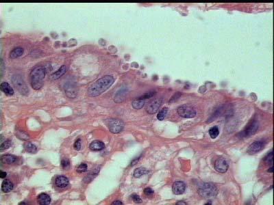
1 HDF Case CRYPTOSPORIDIOSE 45 yo male with severe diarrhea. Known HIV positive. Endoscopic biopsy of duodenum, the colon and ileum.

2 EXUDATIVE CHANGES GRANULAR BASOPHILIC BODIES Colonic biopsy shows a preserved glandular architecture, and inflammatory exudative changes. Glands are formed by hyposecretory columnar epithelium, with rounded granular basophilic bodies on their apical border.

3 MITOTIC FIGURES ROUNDED PARASITES At higher magnification, the reactive hyperplastic epithelium with mitotic figures, is covered by numerous rounded parasites, some are intracytoplasmic.


4 Parasites are basophilic with the Giemsa stain.

5 They are also found on the intestinal mucosa.

6 At higher magnification, they appear embedded in the brush border of enterocytes.
7 DIAGNOSIS: ENTEROCOLITIS DUE TO PARASITIC INFESTATION CONSISTENT WITH CRYPTOSPORIDIOSIS.
8 Cryptosporidium This is a coccidial organism first described in humans in Until the advent of the AIDS epidemic in 1981, only a handful of cases had been reported, and most patients were immunocompromised. With the AIDS epidemic, it became evident that cryptosporidiosis in these patients caused a severe, unrelenting secretory diarrhea. Subsequently, better stool detection techniques became available so that tissue sampling did not constitute the primary mode of diagnosis. With that advance, it became clear that cryptosporidiosis could occur in immunocompetent individuals and was a common cause of sporadic and epidemic gastroenteritis. Among immunocompetent individuals, cryptosporidiosis presents as a self-limited diarrheal illness and many account for 3-5% of all childhood admissions to hospitals for "infectious gastroenteritis."
9 Histologically, the organism may be patchily distributed and thus may be present in random sections, even though there is evidence of substantial infection elsewhere. Cryptosporidium has a characteristic appearance with medium or high power objectives, namely, pinpoint blue dots primarily concentrated at the villous tips or on the upper sides of the villi. This is in contrast to their location in the rectal mucosa, where they tend to cluster in the rectal crypts. The mucosal changes associated with cryptosporidiosis are focally distributed and variable in severity. Even in patients with AIDS who have massive secretory diarrhea, there may only be mild mucosal injury, although some patients do have more severe lesions. Sometimes there is an intense neutrophilic infiltration of the surface epithelium.
10 Medium magnification, glands are separated by an increase of the inflammatory infiltrate of the lamina propria.

11 Higher magnification discloses rounded basophilic structures embedded in the brush border of the enterocytes.
12 The density of these structures vary from an area to another.
13
14 DIAGNOSIS: ENTEROCOLITIS DUE TO PARASITIC INFESTATION CONSISTENT WITH CRYPTOSPORIDIOSIS. DIFFERENTIAL HISTOLOGICAL DIAGNOSIS
15 Some biopsies show basophilic rounded structures on the mucosal surface suggestive of cryptospora.

16 Higher magnification on the previous area.

17 Giemsa stain is negative for microorganism, confirming the mucinous nature of the material observed on the HE.
18 Another colonic biopsy showing heavy basophilic granular material on the surface of the mucosa.

19
20 Giemsa stain

21 Giemsa stain is negative for cryptospora, some nuclear debris are noted. The basophilic granular material on HE is probably due the fixative.
HDF Case Whipple s disease
 HDF Case 952556 Whipple s disease 63 yo female complaining of a diarrhea for 2 months, weigth loss (12 Kg in 3 months), and joint pains. Duodenal biopsy performed. Scanning view, enlarged intestinal villi,
General Structure of Digestive Tract
 Dr. Nabil Khouri General Structure of Digestive Tract Common Characteristics: Hollow tube composed of a lumen whose diameter varies. Surrounded by a wall made up of 4 principal layers: Mucosa Epithelial
Epithelia will be discussed according to the following scheme: Type Number of layers Shape Line drawing. Squamous Cuboidal Columnar
 Epithelia Epithelia will be discussed according to the following scheme: Type Number of layers Shape Line drawing Simple Squamous Cuboidal Columnar Covering and Lining epithelium Pseudostratified Stratified
Small intestine. Small intestine
 General features Tubular organ longest part; 5-6 m most of chemical digestion absorption of nutrients reabsorption of H2O occurs. Two structural features; maximize the lumenal surface area villi microvilli
Supplemental Digital Content 1. Endoscopic and histolological findings in INR and FR study subjects
 Supplemental Digital Content 1. Endoscopic and histolological findings in INR and FR study subjects Patient Group Macroscopic examination Ileum Histology Colon/rectum Histology 1 INR Normal Acute and chronic
Histopathology: gastritis and peptic ulceration
 Histopathology: gastritis and peptic ulceration These presentations are to help you identify, and to test yourself on identifying, basic histopathological features. They do not contain the additional factual
This is the second learning component (Learning Component 2) in our first learning module (Learning Module 1). In this component we review a very
 in our first learning module (Learning Module 1). In this component we review a very") This is the second learning component (Learning Component 2) in our first learning module (Learning Module 1). In this component we review a very basic response to injury inflammation. We ll look at examples
This is the second learning component (Learning Component 2) in our first learning module (Learning Module 1). In this component we review a very basic response to injury inflammation. We ll look at examples
Giardia lamblia (flagellates)
") Giardia lamblia (flagellates) Dr. Hala Al Daghistani Giardia lamblia (Giardia duodenalis or Giardia intestinalis) is the causative agent of giardiasis and is the only common pathogenic protozoan found
Giardia lamblia (flagellates) Dr. Hala Al Daghistani Giardia lamblia (Giardia duodenalis or Giardia intestinalis) is the causative agent of giardiasis and is the only common pathogenic protozoan found
하부위장관비종양성질환의 감별진단 주미인제의대일산백병원
 하부위장관비종양성질환의 감별진단 주미인제의대일산백병원 Solutions for diagnostic problems in Colitis : Please ask yourself five questions Normal or Inflamed? Acute or Chronic? IBD or Other chronic colitis? Ulcerative colitis or
Small Intestine, Large Intestine and anal cannel
 Small Intestine, Large Intestine and anal cannel 32409 Small intestine Large intestine Small intestine General Structure of the Digestive Tract rat 32409 Epithelium with goblet cells and absorptive cells
Domain 1b Appendix Example Stephen Hines
 Domain 1b Appendix Example Stephen Hines The Characterization / Sorting Exercise INSTRUCTIONS: 1. If it s not already done for you, cut along the dotted lines to create 3 sets of colored cards Clinical
5/2/2018. Low Grade Dysplasia of GI Tract. High Grade Dysplasia of GI Tract. Dysplasia in Gastrointestinal Tract: Practical Pearls and Issues
 Dysplasia in Gastrointestinal Tract: Practical Pearls and Issues Arief Suriawinata, M.D. Professor of Pathology and Laboratory Medicine Geisel School of Medicine at Dartmouth Department of Pathology and
Anatomy & Histology of The Small intestine
 Anatomy & Histology of The Small intestine Prof. Abdulameer Al-Nuaimi E-mail: a.al-nuaimi@sheffield.ac.uk E. mail: abdulameerh@yahoo.com Jejunum Ileum Histology: Duodenum, jejunum, and ileum
Unexpected Findings at Endoscopy
 The Endoscopic Incidentaloma: What to Tell Your Patient t with Unexpected Endoscopic Findings: Gastric Intestinal Metaplasia, Silent Ileitis, Carcinoid David Greenwald, MD Montefiore Medical Center Albert
Small Bowel Cases. Introduction. Introduction, Continued 12/7/2011. Lesions Found on endoscopic biopsies Just Like Signing Out
 Small Bowel Cases Lesions Found on endoscopic biopsies Just Like Signing Out Introduction Small intestinal biopsies have a few special pitfalls, for example: Neuroendocrine tumors are readily mistaken
Alimentary Canal (I)
") Alimentary Canal (I) Esophagus and Stomach (Objectives) By the end of this lecture, the student should be able to discuss the microscopic structure in correlation with the function of the following organs:
Alimentary Canal (I) Esophagus and Stomach (Objectives) By the end of this lecture, the student should be able to discuss the microscopic structure in correlation with the function of the following organs:
DIGESTIVE TRACT ESOPHAGUS
 DIGESTIVE TRACT From the lower esophagus to the lower rectum four fundamental layers comprise the wall of the digestive tube: mucosa, submucosa, muscularis propria (externa), and adventitia or serosa (see
VIRAL AGENTS CAUSING GASTROENTERITIS
 VIRAL AGENTS CAUSING GASTROENTERITIS VIRAL AGENTS CAUSING GASTROENTERITIS Pathogens discussed in our lectures 1. Rotavirus 2. Enteric adenoviruses 3. Caliciviruses 4. Astroviruses 5. Toroviruses Viruses
Gynecologic Cytopathology: Glandular lesions
 Gynecologic Cytopathology: Glandular lesions Lin Wai Fung (MSc, MPH, CMIAC) 17/4/2014 Glandular lesions of the uterus Endocervix Endometrium Normal endocervical cells Sheets, strips well-preserved architecture:
IBD. Crohn s. Outline. Ulcerative colitis versus Crohn s disease: is biopsy useful? UC vs. Crohn s? Is it easy? Biopsy settings 21/07/2017 IBD
 Outline Ulcerative colitis versus Crohn s disease: is biopsy useful? Roger Feakins Colorectal biopsies Ileal and upper GI biopsies Special situations New techniques Summary Inflammatory bowel disease (IBD)
3/30/2017. Disclosure of Relevant Financial Relationships. Case 5: Polypoid mass in ulcerative colitis. Case 5. TC Smyrk
 Case 5: Polypoid mass in ulcerative colitis TC Smyrk Disclosure of Relevant Financial Relationships USCAP requires that all faculty in a position to influence or control the content of CME disclose any
TRICHURIASIS : LOCALIZED INFLAMMATORY RESPONSES IN THE COLON
 TRICHURIASIS : LOCALIZED INFLAMMATORY RESPONSES IN THE COLON Gurjeet Kaur 1, S Mahendra Raj 2 and Nyi Nyi Naing 3 Departments of 1 Pathology and 2 Medicine and the 3 Epidemiology and Medical Statistics
A Practical Approach to Small Bowel Biopsies: All that flattens is not sprue
 A Practical Approach to Small Bowel Biopsies: All that flattens is not sprue UCSF Liver and Gastrointestinal Pathology Update Sept. 4, 2009 How to Go Wrong When Evaluating Small Bowel Biopsies, Based on
HISTOLOGY VIRTUAL LABORATORY GASTROINTESTINAL SYSTEM
 HISTOLOGY VIRTUAL LABORATORY GASTROINTESTINAL SYSTEM LIP (Slides GI 1, 2) Identify the outer portion lined by stratified squamous (keratinized) epithelium. Note the hair follicles and sebaceous glands
A adipose cells. B capillary. C epithelium
 EPITHELIA Objective The objective of this class is to observe how different epithelia vary in terms of cell shape, size and number of cell layers enabling them to be well adapted for functions in different
SAM PROVIDER TOOLKIT
 THE AMERICAN BOARD OF PATHOLOGY Maintenance of Certification (MOC) Program SAM PROVIDER TOOLKIT Developing Self-Assessment Modules (SAMs) www.abpath.org The American Board of Pathology (ABP) approves educational
African Trypanosomes
 African Trypanosomes Giemsa-stained blood smear of African trypanosomes viewed under the 100X objective lens. The block arrows denote trypomastigote forms of the African trypanosomes found within the blood
Oncologist-induced Disease of the GI tract: New Developments
 Oncologist-induced Disease of the GI tract: New Developments Jeffrey D Goldsmith, MD Children s Hospital Boston, Beth Israel Deaconess Medical Center, Harvard Medical School Boston, MA Everyone s on drugs
coeliac syndrome per day. Investigations showed a megaloblastic anaemia showed a flat mucosa. ileum were resected and he made an uninterrupted
 Gut, 1965, 6, 466 Post-mortem examination of a small intestine in the coeliac syndrome B. CREAMER AND P. LEPPARD From the Gastro-Intestinal Laboratory, St Thomas's Hospital, London EDITORIAL SYNOPSIS This
Scanning Electron Microscopy of the Small Intestine of a Normal Unsuckled Calf and a Calf with Enteric Colibacillosis
 Vet. Pathol. 15; 400-406 (1978) Scanning Electron Microscopy of the Small Intestine of a Normal Unsuckled Calf and a Calf with Enteric Colibacillosis G. R. PEARSON. E. F. LOGAN and G. P. BRENNAN Departmcnt
What is your diagnosis? a. Lymphocytic colitis. b. Collagenous colitis. c. Common variable immunodeficiency (CVID) associated colitis
 associated colitis") Case History A 24 year old male presented with fatigue, fever, watery diarrhea, and a cough with sputum production for the past three weeks. His past medical history was significant for recurrent bouts
Case History A 24 year old male presented with fatigue, fever, watery diarrhea, and a cough with sputum production for the past three weeks. His past medical history was significant for recurrent bouts
CINtec p16 INK4a Staining Atlas
 CINtec p16 INK4a Staining Atlas Rating Rating Positive The rating positive will be assigned if the p16 INK4a -stained slide shows a continuous staining of cells of the basal and parabasal cell layers of
Rare Case of Chronic Diarrhoea in an Immunocompetent Host
 American Journal of Infectious Diseases and Microbiology, 2018, Vol. 6, No. 2, 46-50 Available online at http://pubs.sciepub.com/ajidm/6/2/2 Science and Education Publishing DOI:10.12691/ajidm-6-2-2 Rare
Lab activity manual - Histology of the digestive system. Lab activity 1: esophagus stomach - small intestines
 Lab activity manual - Histology of the digestive system Jeanne Adiwinata Pawitan Prerequisite: Histology of the 4 basic tissues In this module we learn about the histology of the digestive system, from
Primary mucinous adenocarcinoma developing in an ileostomy stoma
 Gut, 1988, 29, 1607-1612 Primary mucinous adenocarcinoma developing in an ileostomy stoma P J SMART, S SASTRY, AND S WELLS From the Departments of Histopathology and Surgery, Bolton General Hospital, Fan
Histological and immunological characteristics of colitis associated with anti-ctla 4 antibody therapy
 Histological and immunological characteristics of colitis associated with anti-ctla 4 antibody therapy M. Perdiki 2, G. Bamias 1, D. Pouloudi 2, H. Gogas 3, I. Delladetsima 2 1 Academic Dpt. of Gastroenterology,
Human Structure and Function GI Tract Exercises
 GI Tract Exercises Study Exercises. Review of the Elements of the Alimentary Tube. On the following two pages is a chart or matrix of blank spaces. Each space is the intersection of a horizontal row and
Gastrointestinal Pathology of Pigs. Jerome C. Nietfeld, DVM, MS, PhD Kansas State Veterinary Diagnostic Lab Department DMP Kansas State University
 Gastrointestinal Pathology of Pigs Jerome C. Nietfeld, DVM, MS, PhD Kansas State Veterinary Diagnostic Lab Department DMP Kansas State University Neonatal Diarrhea Likely the number 1 killer of neonatal
Pancreatitis: A Potential Pitfall in Endoscopic Ultrasound Guided Pancreatic FNA
 Pancreatitis: A Potential Pitfall in Endoscopic Ultrasound Guided Pancreatic FNA Jack Yang, MD Department of Pathology, Medical University of South Carolina Objectives Understand the indication of EUS
Oesophagus and Stomach update dysplasia and early cancer
 Oesophagus and Stomach update dysplasia and early cancer Dr Tim Bracey STR teaching 13/4/16 Please check pathkids.com for previous talks One of the biggest units in the country (100 major resections per
PATHOLOGY OF NON NEOPLASTIC LESIONS OF THE UPPER GASTROINTESTINAL TRACT.
 PATHOLOGY OF NON NEOPLASTIC LESIONS OF THE UPPER GASTROINTESTINAL TRACT. OESOPHAGEAL LESIONS OESOPHAGITIS AND OTHER NON NEOPLASTIC DISORDERS Corrosive Gastroesophageal reflux (GERD), Pills, Acid intake,
Dr Nadine Gravett School of Anatomical Sciences Room 2B10B
 Dr Nadine Gravett School of Anatomical Sciences Room 2B10B Nadine.Gravett@wits.ac.za Oral cavity Mechanical breakdown Formation of bolus Oesophagus Conduit from mouth to stomach Stomach Digestion Temporary
Prelab #4 BLOOD; BONE MARROW; RESPIRATORY; INTEGUEMENT Page 1
 Prelab #4 BLOOD; BONE MARROW; RESPIRATORY; INTEGUEMENT Page 1 Blood Slide 101 This a classic slide of blood cells using a Wright stain. Inspect red blood cells and their appearance. Note the approximate
What Every Pathologist Wants the GI Nurse to Know (and how you can help us help you)
") What Every Pathologist Wants the GI Nurse to Know (and how you can help us help you) Jonathan N. Glickman MD PhD Director, GI Pathology, Caris Diagnostics, Newton, MA Associate Professor of Pathology,
What Every Pathologist Wants the GI Nurse to Know (and how you can help us help you) Jonathan N. Glickman MD PhD Director, GI Pathology, Caris Diagnostics, Newton, MA Associate Professor of Pathology,
Anatomy of the Intes.ne: Epithelium, Lympha.cs, Vessels and Nerves
 Master Course Gastroenterology 2015 Anatomy of the Intes.ne: Epithelium, Lympha.cs, Vessels and Nerves Dr. Stephanie Ganal Department Klinische Forschung University of Bern stephanie.ganal@dkf.unibe.ch
The doctor mentioned a few things about the esophagus from the previous lecture:
 السالم عليكم [HISOLOGY 2] April 27, 2014 The doctor mentioned a few things about the esophagus from the previous lecture: Esophagus - It is about 25 cm in length (from the incisor it is 45 cm) Histological
Small Intestinal Biopsy in a Patient with Crohn's Disease of the Duodenum
 GASTROENTEROLOGY 76:1009-1014, 1979 Small Intestinal Biopsy in a Patient with Crohn's Disease of the Duodenum The Spectrum of Abnormal Findings in the Absence of Granulomas MICHAEL D. SCHUFFLER and ROBERT
Endometrial Metaplasia, Hyperplasia & Other Cancer Mimics: a Consultant s Experience
 Endometrial Metaplasia, Hyperplasia & Other Cancer Mimics: a Consultant s Experience Pacific Northwest Society of Pathologists Vancouver, B.C. September 26, 2015 Teri A. Longacre, M.D. longacre@stanford.edu
Collagenous gastritis and collagenous colitis: a report with sequential histological and ultrastructural findings
 Gut 1999;44:881 885 881 CASE REPORTS Wellcome Trust Research Laboratory, Christian Medical College and Hospital, Vellore 632004, India A B Pulimood B S Ramakrishna M M Mathan Correspondence to: Dr A B
SAMs Guidelines DEVELOPING SELF-ASSESSMENT MODULES TEST QUESTIONS. Ver. #
 SAMs Guidelines DEVELOPING SELF-ASSESSMENT MODULES TEST Ver. #5-02.12.17 GUIDELINES FOR DEVELOPING SELF-ASSESSMENT MODULES TEST The USCAP is accredited by the American Board of Pathology (ABP) to offer
A HISTOLOGICAL STUDY OF THE EFFECTS OF NONSTEROIDAL ANTI-INFLAMMATORY DRUGS (NSAIDs) ON THE GASTRIC AND DUODENAL MUCOSA
 ON THE GASTRIC AND DUODENAL MUCOSA") A HISTOLOGICAL STUDY OF THE EFFECTS OF NONSTEROIDAL ANTI-INFLAMMATORY DRUGS (NSAIDs) ON THE GASTRIC AND DUODENAL MUCOSA Abstract Pages with reference to book, From 287 To 290 Naheed Moghal, N.A. Jafarey
A HISTOLOGICAL STUDY OF THE EFFECTS OF NONSTEROIDAL ANTI-INFLAMMATORY DRUGS (NSAIDs) ON THE GASTRIC AND DUODENAL MUCOSA Abstract Pages with reference to book, From 287 To 290 Naheed Moghal, N.A. Jafarey
Dissecting Microscope Appearance of
 Arch. Dis. Childh., 1967, 42, 626. Dissecting Microscope Appearance of Small Bowel Mucosa in Children JOHN WALKER-SMITH From Professorial Unit, Royal Alexandra Hospital for Children, Camperdown, Sydney,
Lymphocytic Gastritis, Isolated Type Occurring in Family Members. A Case Report.
 Lymphocytic Gastritis, Isolated Type Occurring in Family Members. A Case Report. Alan Shienbaum, DO; AndriyPavlenko, MD; Jun Liu, MD, PhD; Janusz J Godyn, MD. Pathology Department, Kennedy University Hospitals,
LARYNGEAL DYSPLASIA. Tomas Fernandez M; 3 rd year ENT resident, Son Espases University Hospital
 LARYNGEAL DYSPLASIA Tomas Fernandez M; 3 rd year ENT resident, Son Espases University Hospital INTRODUCTION Laryngeal cancer constitutes 1-2% of all malignancies diagnosed worldwide Survival is related
Colonic Polyp. Najmeh Aletaha. MD
 Colonic Polyp Najmeh Aletaha. MD 1 Polyps & classification 2 Colorectal cancer risk factors 3 Pathogenesis 4 Surveillance polyp of the colon refers to a protuberance into the lumen above the surrounding
Rectal biopsy in patients presenting to an infectious disease unit with diarrhoeal disease
 Gut, 1979, 20, 141-148 Rectal biopsy in patients presenting to an infectious disease unit with diarrhoeal disease R. J. DICKINSON, H. M. GILMOUR, AND D. B. L. McCLELLAND' From the Department of Infectious
GOBLET CELL CARCINOID. Hanlin L. Wang, MD, PhD University of California Los Angeles
 GOBLET CELL CARCINOID Hanlin L. Wang, MD, PhD University of California Los Angeles Disclosure of Relevant Financial Relationships USCAP requires that all planners (Education Committee) in a position to
GOBLET CELL CARCINOID
 GOBLET CELL CARCINOID Hanlin L. Wang, MD, PhD University of California Los Angeles Disclosure of Relevant Financial Relationships USCAP requires that all planners (Education Committee) in a position to
Chronic immune colitis in rabbits
 Chronic immune colitis in rabbits A. S. MEE, J. E. McLAUGHLIN, H. J. F. HODGSON, AND D. P. JEWELL Gut, 1979, 20, 1-5 From the Academic Department of Medicine and Department of Histopathology, Royal Free
A Case of Crohn s Disease with Mesalazine Allergy that was Difficult to Differentiate from Comorbid Ulcerative Colitis
 doi: 10.2169/internalmedicine.1607-18 http://internmed.jp CASE REPORT A Case of Crohn s Disease with Mesalazine Allergy that was Difficult to Differentiate from Comorbid Ulcerative Colitis Rumiko Tsuboi,
Slide 154: Pancreas, H&E
 Slide 154: Pancreas, H&E the pancreas, located adjacent to the duodenum, is a mixed exocrine and endocrine gland; it is usually readily identifiable by the presence of the interspersed endocrine pancreatic
BIOPSY DIAGNOSIS OF COLITIS Common and Unusual Forms of Inflammatory Bowel disease
 BIOPSY DIAGNOSIS OF COLITIS Common and Unusual Forms of Inflammatory Bowel disease David A Owen University of British Columbia CAUSES OF DIARRHEA DIARRHEA COLITIS PRESENT COLITIS ABSENT INFECTIOUS NON-INFECTIOUS
The Third Department of Internal Medicine, Tohoku University School of Medicine, Sendai 980
 Tohoku J. exp. Med., 1977, 123, 197-213 Clinical Observations on Ulcerative Colitis HIKARU WATANABE, NOBUO HIWATASHI and SHOICHI YAMAGATA The Third Department of Internal Medicine, Tohoku University School
Series Editors: Daniel Kamin, MD and Christine Waasdorp Hurtado, MD
 NASPGHAN Physiology Lecture Series GI Physiology Module: Absorption of Water and Ions Jason Soden, MD Reviewers: George Fuchs MD: UAMS College of Medicine / Arkansas Children s Hospital Wayne Lencer MD:
Malabsorption is characterized by defective absorption of: Fats fat- and water-soluble vitamins Proteins Carbohydrates Electrolytes Minerals water
 Malabsorption Malabsorption is characterized by defective absorption of: Fats fat- and water-soluble vitamins Proteins Carbohydrates Electrolytes Minerals water presents most commonly as chronic diarrhea
 SUMMARY Coeliac disease is a common food intolerance in Western populations, in which it has a prevalence of about 1%. In early infancy, when the transition is made to a gluten-containing diet (particularly
Gastric epithelium in the duodenum
 Gastric epithelium in the duodenum A. H. JAMES From the Medical Unit, Cardiff Royal Infirmary, Cardiff, Wales Gut, 1964, 5, 285 EDITORIAL SYNOPSIS It has been found that a 'gastric' type of epithelium
Diffuse Nodular Lymphoid Hyperplasia of the Intestine Caused by Common Variable Immunodeficiency and Refractory Giardiasis
 CASE REPORT Diffuse Nodular Lymphoid Hyperplasia of the Intestine Caused by Common Variable Immunodeficiency and Refractory Giardiasis Jung Hye Choi 1,DongSooHan 1, Jieun Kim 1, Kijong Yi 2, Young-Ha Oh
Done By : shady soghayr
 Done By : shady soghayr Malabsorption Malabsorption is characterized by defective absorption of: Fats fat- and water-soluble vitamins Proteins Carbohydrates Electrolytes Minerals Water presents most commonly
Imaging Evaluation of Polyps. CT Colonography: Sessile Adenoma. Polyps, DALMs & Megacolon Objectives
 Polyps, DALMs & Megacolon: Pathology and Imaging of the Colon and Rectum Angela D. Levy and Leslie H. Sobin Washington, DC Drs. Levy and Sobin have indicated that they have no relationships which, in the
Topics and aims. Introduction. Metabolism and Excretion
 Topics and aims Introduction This section contains instructions that are applicable to all material, irrespective of your specific course. Please take note and make sure to comply. Failure to comply could
Non-Invasive Assessment of Intestinal Function
 Overview Non-Invasive Assessment of Intestinal Function Introduction This paper will demonstrate that the 13 C-sucrose breath test ( 13 C-SBT) determines the health and function of the small intestine.
Figure 2: Lymph node Cortical follicular (F) and paracortical (PC) atrophy, with narrowing of the cortex relative to the medulla (M).
 and paracortical (PC) atrophy, with narrowing of the cortex relative to the medulla (M).") Figure 1: Lymph node Follicular hyperplasia, with expansion of the follicular germinal centres (F) by large blast cells. Paracortical hyperplasia, with expansion of the paracortex (PC) by small lymphocytes.
Figure 1: Lymph node Follicular hyperplasia, with expansion of the follicular germinal centres (F) by large blast cells. Paracortical hyperplasia, with expansion of the paracortex (PC) by small lymphocytes.
3/28/2017. Disclosure of Relevant Financial Relationships. GU Evening Subspecialty Case Conference. Differential Diagnosis:
 GU Evening Subspecialty Case Conference Rajal B. Shah, M.D. VP, Medical Director, Urologic Pathology Miraca Life Sciences, Irving, Texas Clinical Associate Professor of Pathology Baylor College of Medicine,
Short Communication. Cryptosporidium parvum Initiates Inflammatory Bowel Disease in Germfree T Cell Receptor- -Deficient Mice
 American Journal of Pathology, Vol. 153, No. 6, December 1998 Copyright American Society for Investigative Pathology Short Communication Cryptosporidium parvum Initiates Inflammatory Bowel Disease in Germfree
The Digestive System Laboratory
 The Digestive System Laboratory 1 The Digestive Tract The alimentary canal is a continuous tube stretching from the mouth to the anus. Liver Gallbladder Small intestine Anus Parotid, sublingual, and submaxillary
Infiltrative small intestinal disorders of cats are commonly
 J Vet Intern Med 2011;25:1253 1257 Utility of Endoscopic Biopsies of the Duodenum and Ileum for Diagnosis of Inflammatory Bowel Disease and Small Cell Lymphoma in Cats K.D. Scott, D.L. Zoran, J. Mansell,
Fibrin thrombi, a cause of clindamycin-associated
 Gut, 1976, 17, 483487 Fibrin thrombi, a cause of clindamycin-associated colitis? W. V. BOGOMOLETZ From the Department of Pathology, Queen Mary's Hospital, London summary Rectal biopsies from five patients
Correlation between Gastric Mucosal Morphologic Patterns and Histopathological Severity of
 Hindawi Publishing Corporation BioMed Research International Volume 2015, Article ID 808505, 7 pages http://dx.doi.org/10.1155/2015/808505 Research Article Correlation between Gastric Mucosal Morphologic
GUIDE TO: Diagnosing Coccidiosis & Necrotic Enteritis
 GUIDE TO: Diagnosing Coccidiosis & Necrotic Enteritis Site of Infection Species E. acervulina E. brunetti E. maxima E. mivati E. tenella E. necatrix Oocyst Size 2µ{ 18.3 x 14.6 24.6 x 18.8 30.5 x 20.7
11/1/2017. Tetyana Mettler, MD Department of Laboratory Medicine and Pathology University of Minnesota. Cerilli & Greenson
 Tetyana Mettler, MD Department of Laboratory Medicine and Pathology University of Minnesota Acute infectious (self-limited) colitis Focal active colitis Pseudomembranous colitis Ischemic colitis Collagenous
Malignant histiocytosis of the intestine: the early histological lesion
 Gut, 1980, 21, 381-386 Malignant histiocytosis of the intestine: the early histological lesion P ISAACSON From the Department of Pathology, Faculty of Medicine, Southampton University Hospital, Southampton
INTERNATIONAL COURSE ON THE PATHOLOGY OF THE DIGESTIVE SYSTEM VICTOR BABES NATIONAL INSTITUTE OF PATHOLOGY BUCHAREST 7-8 BUCHAREST 2014
 INTERNATIONAL COURSE ON THE PATHOLOGY OF THE DIGESTIVE SYSTEM VICTOR BABES NATIONAL INSTITUTE OF PATHOLOGY BUCHAREST 7-8 BUCHAREST 2014 Endoscopic biopsy samples of naïve colitides patients: Role of basal
1. Esophageal diverticulum located above the upper esophageal sphincter is called
 Test Bank for Robbins Basic Pathology 9th Edition by Kumar Link full download: http://testbankair.com/download/test-bank-for-robbins-basic-pathology-9thedition-by-kumar/ Chapter 14: Oral Cavity and Gastrointestinal
HISTOLOGY. GIT Block 432 Histology Team. Lecture 1: Alimentary Canal (1) (Esophagus & Stomach) Done by: Ethar Alqarni Reviewed by: Ibrahim Alfuraih
 (Esophagus & Stomach) Done by: Ethar Alqarni Reviewed by: Ibrahim Alfuraih") HISTOLOGY Lecture 1: Alimentary Canal (1) (Esophagus & Stomach) Done by: Ethar Alqarni Reviewed by: Ibrahim Alfuraih Color Guide: Black: Slides. Red: Important. Green: Doctor s notes. Blue: Explanation.
HISTOLOGY Lecture 1: Alimentary Canal (1) (Esophagus & Stomach) Done by: Ethar Alqarni Reviewed by: Ibrahim Alfuraih Color Guide: Black: Slides. Red: Important. Green: Doctor s notes. Blue: Explanation.
CMV. Inclusions predominantly in endothelial cells. Immunostaining greater sensitivity than H&E alone.
 CMV Inclusions predominantly in endothelial cells. Immunostaining greater sensitivity than H&E alone. CMV inclusions are often present in a very patchy distribution Carefully examine all levels CMV CMV
Objectives. Atypical Glandular Cells. Atypical Endocervical Cells. Reactive Endocervical Cells
 2013 California Society of Pathologists 66 th Annual Meeting San Francisco, CA Atypical Glandular Cells to Early Invasive Adenocarcinoma: Cervical Cytology and Histology Christina S. Kong, MD Associate
In current practice in surgical pathology, colorectal polyps
 Colorectal Polyps With Extensive Absorptive Enterocyte Differentiation Histologically Distinct Variant of Hyperplastic Polyps Hidejiro Yokoo, MD; M. Irtaza Usman, Bsc(Hons); Susan Wheaton, MD; Patricia
Two Cases of Pediatric Collagenous Gastritis Each Presenting with Refractory Iron Deficiency Anemia and Chronic Diarrhea
 pissn: 2234-8646 eissn: 2234-8840 http://dx.doi.org/10.5223/pghn.2012.15.3.183 Pediatric Gastroenterology, Hepatology & Nutrition 2012 September 15(3):183-187 Case Report PGHN Two Cases of Pediatric Collagenous
ABDOMINAL PAIN AND DIARRHEA - IT S NOT (ALWAYS) WHAT YOU THINK. Yakov Wainer, MD Gastroenterology and Hepatology Meir Medical Center
 WHAT YOU THINK. Yakov Wainer, MD Gastroenterology and Hepatology Meir Medical Center") ABDOMINAL PAIN AND DIARRHEA - IT S NOT (ALWAYS) WHAT YOU THINK Yakov Wainer, MD Gastroenterology and Hepatology Meir Medical Center 1 ST ADMISSION - 2015 38 y/o female Abdominal pain, diarrhea - intermittent
ABDOMINAL PAIN AND DIARRHEA - IT S NOT (ALWAYS) WHAT YOU THINK Yakov Wainer, MD Gastroenterology and Hepatology Meir Medical Center 1 ST ADMISSION - 2015 38 y/o female Abdominal pain, diarrhea - intermittent
Coeliac disease with histological features of peptic duodenitis: Value of assessment of intraepithelial lymphocytes
 420 4 Clin Pathol 1993;46:420-424 Department of Pathology, St James's Hospital and Trinity College, Dublin Ireland M D Jeffers D O'B Hourihane Correspondence to: Professor D O'B Hourihane Accepted for
Common Inflammatory Gastrointestinal Disorders: Endoscopic and Pathologic Correlations
 Common Inflammatory Gastrointestinal Disorders: Endoscopic and Pathologic Correlations Nicole C. Panarelli, M.D. Attending Pathologist Montefiore Medical Center Associate Professor of Pathology - Albert
Cryptosporidium sp. Edinburgh,2 Scotland. lambs, 19 were maintained under gnotobiotic conditions. in plastic isolators and 2 were maintained under
 INFECTION AND IMMUNITY, Aug. 1981, p. 401406 Vol. 33, No. 2 0019-9567/81/080401-06$02.00/0 Diarrhea in Lambs: Experimental Infections with Enterotoxigenic Escherichia coli, Rotavirus, and Cryptosporidium
GASTROENTEROLOGY. Official Publication of the American Gastroenterological Association
 GASTROENTEROLOGY Official Publication of the American Gastroenterological Association @ COPYRIGHT 1975 THE WILLIAMS & WILKINS CO. Vol 68 March 1975 Number 3 ALIMENTARY TRACT STRUCfURE OF THE GASTRIC MUCOSA
Gastric Polyps. Bible class
 Gastric Polyps Bible class 29.08.2018 Starting my training in gastroenterology, some decades ago, my first chief always told me that colonoscopy may seem technically more challenging but gastroscopy has
Endoscopic Characteristics of the Healing Process of Ulcerative Colitis
 Diagnostic and Therapeutic Endoscopy, Vol. 5, pp. 37-48 Reprints available directly from the publisher Photocopying permitted by license only (C) 1998 OPA (Overseas Publishers Association) N.V. Published
SCANNING ELECTRON MICROSCOPY IN CHILDHOOD INFLAMMATORY BOWEL DISEASE
 Scanning Microscopy Vol. 12, No. 3, 1998 (Pages 495-502) 0891-7035/98$5.00+.25 Scanning Microscopy International, Chicago (AMF Intestinal O Hare), mucosa IL 60666 in childhood USA IBD SCANNING ELECTRON
GUIDELINES FOR THE INITIAL BIOPSY DIAGNOSIS OF CHRONIC IDIOPATHIC INFLAMMATORY BOWEL DISEASE A STRUCTURED APPROACH TO COLORECTAL BIOPSY ASSESSMENT
 Guidelines for the Initial Biopsy Diagnosis of Chronic Idiopathic Inflammatory Bowel Disease 1 GUIDELINES FOR THE INITIAL BIOPSY DIAGNOSIS OF CHRONIC IDIOPATHIC INFLAMMATORY BOWEL DISEASE A STRUCTURED
Definition. Celiac disease is an immune-mediated enteropathy caused by a permanent sensitivity to gluten in genetically susceptible individuals.
 Definition 1 Definition Celiac disease is an immune-mediated enteropathy caused by a permanent sensitivity to gluten in genetically susceptible individuals. It occurs in symptomatic subjects with gastrointestinal
Rectal mucosa in cows' milk allergy
 Archives of Disease in Childhood, 1989, 64, 1256-1260 Rectal mucosa in cows' milk allergy N IYNGKARAN,* M YADAVt AND C G BOEYt Departments of *Paediatrics and tpathology, University Hospital, and tdepartment