Anatomy & Histology of The Small intestine
|
|
|
- Griffin Hensley
- 5 years ago
- Views:
Transcription

1 Anatomy & Histology of The Small intestine Prof. Abdulameer Al-Nuaimi E. mail:
2
3
4
5
6
7
8
9
10
11

12 Jejunum Ileum
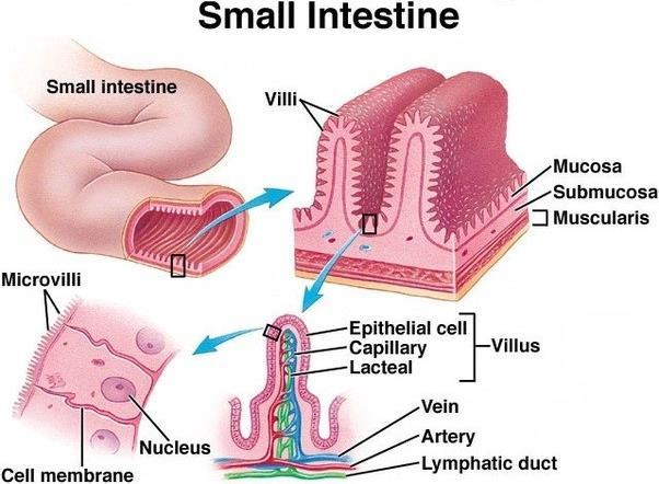
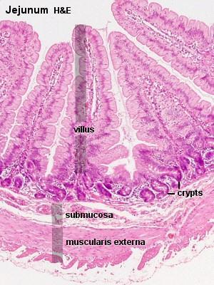
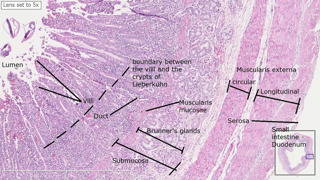
13 Histology: Duodenum, jejunum, and ileum share the same wall structure formed by, a mucosa, a submucosa, a muscularis interna, a muscularis externa, and a serosa. The mucosa of the small intestine, comprising simple columnar epithelium and a lamina propria, forms finger-like projections, villi, which protrude into the lumen, and deep cavities, the crypts of Lieberkühn (intestinal glands) between the villi. The predominant cell in the epithelium is the absorptive enterocyte with microvilli on its apical membrane. Interspersed between the enterocytes are the oval, mucous goblet cells. Deep in the crypts of Lieberkuhn, the epithelium contains entero endocrine cells with granules (secrete hormones).

14 Lamina propria
15 Crypts of Lieberkuhn
16 The lamina propria consists of loose connective tissue. In the lamina propria of each villus there are blood vessels. central lymph vessel, the lacteal. In the submucosa throughout the intestines but mainly in the ileum, there are large lymphoid aggregates Peyer s patches (un encapsulated lymphoid nodules). M cells form part of the epithelium covering the Peyer s patches (they are concerned with immune system of the intestine). The jejunum and ileum are histologically identical, except for their villi and the presence of Paneth cells. The villi of the jejunum are tall and cylindrical, while they are short and cylindrical in the ileum. Paneth cells are especially found in the jejunum, they have eosinophilic cytoplasmic granules and occur in clusters at the bases of crypts. they secrete digestive enzymes
17 Nerve supply of the intestine The myenteric plexus (Auerbach's plexus) provides motor innervation to both layers of the muscular layer of the gut, having both parasympathetic and sympathetic input (Ganglionic cell bodies belong to parasympathetic innervation and fibres from sympathetic innervation). The submucous plexus has only parasympathetic fibres and provides secretomotor innervation to the mucosa nearest the lumen of the gut
18

19 Peyer s patches Ileum

20 Chylomicrons (small fat globule composed of protein and lipid) transport lipids absorbed from the intestine to adipose, cardiac, and skeletal muscle tissue, where their triglyceride components are hydrolyzed by the activity of the lipoprotein lipase, allowing the released free fatty acids to be absorbed by the tissues. Villus
21 CRYPTS OF LIEBERKUHN CONSISTS OF FOLLOWING CELLS, 1. STEM CELLS: ACTIVE,UNDIFFERENTATED CELLS. Histology of Duodenum MUCOSA: LINED BY SIMPLE COLUMNAR EPITHELIUM WITH FINE MICROVILLI and MUCOUS SECRETING GOBLET CELLS. The inside surface of duodenum is thrown into villi. PLYCA CIRCULARIS IS A MUCOSAL FOLD WITH A CORE OF SUBMUCOSA. LAMINA PROPRIA: Contains Crypts of Lieberkühn (TUBULAR INTESTINAL GLANDS)
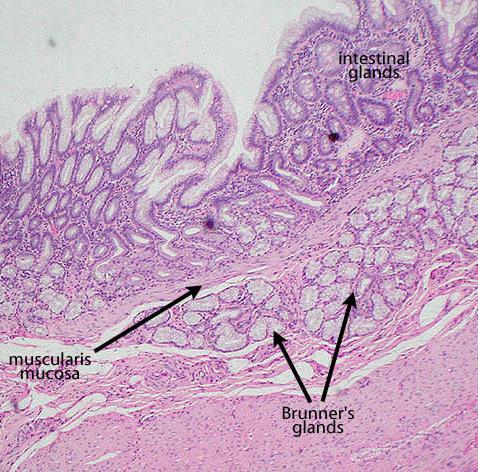
22 2. GOBLET CELLS: Secrete mucous. 3. ENTERO ENDOCRINE CELLS: produce gastrointestinal hormones 4. ARGENTAFFIN CELLS: They produce and release hormones in response to a number of stimuli. Hormones may be distributed as local messengers. They may also stimulate a nervous response. 5. PANETH CELLS: They are ZYMOGENIC CELLS, PRODUCING DIGESTIVE ENZYMES AND LYSOZYMES (an antimicrobial enzyme that forms part of the immune system) MUSCULARIS MUCOSA: Circular muscle layer limits the lower aspect of the mucosa SUBMUCOSA: MADE UP OF LOOSE AREOLAR CONNECTIVE TISSUE. It contains MUCOUS SECRETING BRUNNER S GLANDS.
23 MUSCULARIS EXTERNA: INNER CIRCULAR and OUTER LONGITUDINAL. PARASYMPATHETIC GANGLION CELLS OF MYENTERIC PLEXUS CAN BE SEEN. SEROSA: OUTER MOST LAYER MADE UP OF FEW CONNECTIVE TISSUE CELLS AND FIBRES, COVERED BY MESOTHELIUM OF VISCERAL PERITONEUM. FUNCTIONS: VILLI HAS ABSORPTIVE FUNCTION. MICROVILLI INCREASE THE SURFACE AREA OF ABSORPTION. BRUNNER S GLANDS SECRETE ALKALINE FLUID RICH IN HCO3. MUSCULARIS EXTERNA HELPS IN CHURNING FOOD PARTICLES SEROSA IS SUPPORTIVE AND PROTECTIVE
24 Duodenum
25 Duodenum

26
General Structure of Digestive Tract
 Dr. Nabil Khouri General Structure of Digestive Tract Common Characteristics: Hollow tube composed of a lumen whose diameter varies. Surrounded by a wall made up of 4 principal layers: Mucosa Epithelial
Small intestine. Small intestine
 General features Tubular organ longest part; 5-6 m most of chemical digestion absorption of nutrients reabsorption of H2O occurs. Two structural features; maximize the lumenal surface area villi microvilli
Dana Alrafaiah. Dareen Abu Shalbak. Mohammad Almuhtaseb. 1 P a g e
 2 Dana Alrafaiah Dareen Abu Shalbak Mohammad Almuhtaseb 1 P a g e Esophagus: A muscular tube that is 25 cm long, but if measured from the incisors it would be 45cm long. Extends from C6 of cervical vertebra,
Alimentary Canal (I)
") Alimentary Canal (I) Esophagus and Stomach (Objectives) By the end of this lecture, the student should be able to discuss the microscopic structure in correlation with the function of the following organs:
Alimentary Canal (I) Esophagus and Stomach (Objectives) By the end of this lecture, the student should be able to discuss the microscopic structure in correlation with the function of the following organs:
The doctor mentioned a few things about the esophagus from the previous lecture:
 السالم عليكم [HISOLOGY 2] April 27, 2014 The doctor mentioned a few things about the esophagus from the previous lecture: Esophagus - It is about 25 cm in length (from the incisor it is 45 cm) Histological
DIGESTIVE TRACT ESOPHAGUS
 DIGESTIVE TRACT From the lower esophagus to the lower rectum four fundamental layers comprise the wall of the digestive tube: mucosa, submucosa, muscularis propria (externa), and adventitia or serosa (see
Dr Nadine Gravett School of Anatomical Sciences Room 2B10B
 Dr Nadine Gravett School of Anatomical Sciences Room 2B10B Nadine.Gravett@wits.ac.za Oral cavity Mechanical breakdown Formation of bolus Oesophagus Conduit from mouth to stomach Stomach Digestion Temporary
Small Intestine, Large Intestine and anal cannel
 Small Intestine, Large Intestine and anal cannel 32409 Small intestine Large intestine Small intestine General Structure of the Digestive Tract rat 32409 Epithelium with goblet cells and absorptive cells
Digestive System II - Lower tract Revised
 ANAT D502 Basic Histology Digestive System II - Lower tract Revised 10.12.12 Outline: I. Small intestine II. Enterocyte digestion II. Hepatic portal system IV. Large intestine V. Enteric nervous system
Tissues and organs PART 1
 Tissues and organs PART 1 Animals and plants are multicellular (made of many cells). Cells become specialised according to their function Tissues: Many cells that perform one or several functions; they
HISTOLOGY. GIT Block 432 Histology Team. Lecture 1: Alimentary Canal (1) (Esophagus & Stomach) Done by: Ethar Alqarni Reviewed by: Ibrahim Alfuraih
 (Esophagus & Stomach) Done by: Ethar Alqarni Reviewed by: Ibrahim Alfuraih") HISTOLOGY Lecture 1: Alimentary Canal (1) (Esophagus & Stomach) Done by: Ethar Alqarni Reviewed by: Ibrahim Alfuraih Color Guide: Black: Slides. Red: Important. Green: Doctor s notes. Blue: Explanation.
HISTOLOGY Lecture 1: Alimentary Canal (1) (Esophagus & Stomach) Done by: Ethar Alqarni Reviewed by: Ibrahim Alfuraih Color Guide: Black: Slides. Red: Important. Green: Doctor s notes. Blue: Explanation.
Esophagus. Transport is achieved by peristaltic contractions and relaxation of the esophageal sphincters (upper and lower)
") GI Histology 2 Esophagus is a muscular tube whose function is to transport foodstuffs from the mouth to the stomach and to prevent the retrograde flow of gastric contents Transport is achieved by peristaltic
GI Histology 2 Esophagus is a muscular tube whose function is to transport foodstuffs from the mouth to the stomach and to prevent the retrograde flow of gastric contents Transport is achieved by peristaltic
Gastrointestinal Anatomy and Physiology. Bio 219 Napa Valley College Dr. Adam Ross
 Gastrointestinal Anatomy and Physiology Bio 219 Napa Valley College Dr. Adam Ross Functions of digestive system Digestion Breakdown of food (chemically) using enzymes, acid, and water Absorption Nutrients,
The Digestive System Laboratory
 The Digestive System Laboratory 1 The Digestive Tract The alimentary canal is a continuous tube stretching from the mouth to the anus. Liver Gallbladder Small intestine Anus Parotid, sublingual, and submaxillary
Digestive system L 2. Lecturer Dr. Firdous M. Jaafar Department of Anatomy/Histology section
 Digestive system L 2 Lecturer Dr. Firdous M. Jaafar Department of Anatomy/Histology section objectives 1-Describe the general structure of digestive tract: a-mucosa. b-submucosa. c-muscularis externa d-adventitia
الله الر ح م ن الر ح يم مسب
 بسم رلا هللارلا هللا This is the second histology lecture in the GI system. In this lecture, we will discuss the histology of the esophagus, stomach, and small intestine so prepare yourself.this sheet
(b) Stomach s function 1. Dilution of food materials 2. Acidification of food (absorption of dietary Fe in small intestine) 3. Partial chemical digest
 Stomach s function 1. Dilution of food materials 2. Acidification of food (absorption of dietary Fe in small intestine) 3. Partial chemical digest") (1) General features a) Stomach is widened portion of gut-tube: between tubular and spherical; Note arranged of smooth muscle tissue in muscularis externa. 1 (b) Stomach s function 1. Dilution of food
(1) General features a) Stomach is widened portion of gut-tube: between tubular and spherical; Note arranged of smooth muscle tissue in muscularis externa. 1 (b) Stomach s function 1. Dilution of food
DIGESTIVE. CHAPTER 17 Lecture: Part 1 Part 2 BIO 212: ANATOMY & PHYSIOLOGY II
 BIO 212: ANATOMY & PHYSIOLOGY II CHAPTER 17 Lecture: DIGESTIVE Part 1 Part 2 Dr. Lawrence G. Altman www.lawrencegaltman.com Some illustrations are courtesy of McGraw-Hill. SMALL INTESTINE DUODENUM > JEJUNUM
Lab activity manual - Histology of the digestive system. Lab activity 1: esophagus stomach - small intestines
 Lab activity manual - Histology of the digestive system Jeanne Adiwinata Pawitan Prerequisite: Histology of the 4 basic tissues In this module we learn about the histology of the digestive system, from
The Digestive System and Body Metabolism Premedical Biology
 The Digestive System and Body Metabolism Premedical Biology Copyright 2003 Pearson Education, Inc. publishing as Benjamin Cummings The Digestive System and Body Digestion Metabolism Breakdown of ingested
Chapter 9. The digestive system. Glossary. Louise McErlean
 Chapter 9 The digestive system Louise McErlean Glossary Absorption Process whereby the products of digestion move into the blood or lymph fluid. Acini glands Produce pancreatic juice. Amylase Carbohydrate
HISTOLOGY VIRTUAL LABORATORY GASTROINTESTINAL SYSTEM
 HISTOLOGY VIRTUAL LABORATORY GASTROINTESTINAL SYSTEM LIP (Slides GI 1, 2) Identify the outer portion lined by stratified squamous (keratinized) epithelium. Note the hair follicles and sebaceous glands
Anatomy of the liver and pancreas
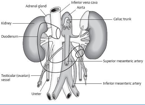
 Anatomy of the liver and pancreas Prof. Abdulameer Al-Nuaimi E-mail: a.al-nuaimi@sheffield.ac.uk abdulameerh@yahoo.com Liver Aorta Pulm. Trunk Rt. At, Duct. Art. Lt. Ven. Rt. Ven. Internal Posterior
International Journal of Science, Environment and Technology, Vol. 7, No 5, 2018,
 International Journal of Science, Environment and Technology, Vol. 7, No 5, 2018, 1608 1614 ISSN 2278-3687 (O) 2277-663X (P) COMPARATIVE HISTOLOGICAL STUDIES OF DUEODENUM IN CATTLE SHEEP AND GOATS Thete
Physiological processes in the GI tract:
 Gastrointestinal physiology for medical students General principal of gastrointestinal function Motility, nervous control and blood circulation Physiological processes in the GI tract: Motility Secretion
Chapter 14: The Digestive System
 Chapter 14: The Digestive System Digestive system consists of Muscular tube (digestive tract) alimentary canal Accessory organs teeth, tongue, glandular organs 6 essential activities 1. 2. 3. 4. 5. 6.
HUMAN NUTRITION: ABSORPTION & ASSIMILATION 14 MAY 2014
 HUMAN NUTRITION: ABSORPTION & ASSIMILATION 14 MAY 2014 In this lesson, we: Absorption Lesson Description Examine and understand absorption Define absorption and describe where it occurs Study the structure
General principles of gastrointestinal motility
 General principles of gastrointestinal motility OBJECTIVES Physiological anatomy General Principles Circulation of blood through the GIT organs Control of all GIT functions by local, nervous, and hormonal
Histology Lab. looking at microscopic pictures of tissues, for more information use Junqueira book and you can use BlueHistolgy website
 Done By: Aseel Twaijer & Laith Sorour Histology Lab *These notes help in differentiating tissues and you must read them while looking at microscopic pictures of tissues, for more information use Junqueira
The stomach is formed of three parts: -
 The stomach is formed of three parts: - (a) CARDIAC STOMACH: - It receives the oesophagus through Cardiac aperture guarded by a cardiac sphincter which prevents regurgitation of food. (b) FUNDIC PART:
The Digestive System and Body Metabolism
 14 PART B The Digestive System and Body Metabolism PowerPoint Lecture Slide Presentation by Jerry L. Cook, Sam Houston University ESSENTIALS OF HUMAN ANATOMY & PHYSIOLOGY EIGHTH EDITION ELAINE N. MARIEB
Slide 154: Pancreas, H&E
 Slide 154: Pancreas, H&E the pancreas, located adjacent to the duodenum, is a mixed exocrine and endocrine gland; it is usually readily identifiable by the presence of the interspersed endocrine pancreatic
Histology 3. We will continue talking about a few things from last lecture, starting with M cells:
 Histology 3 This is the last Histology lecture in the GI system. Enjoy! There are some extra notes listed as footnotes. We will continue talking about a few things from last lecture, starting with M cells:
The Digestive System
 The Digestive System Identify the Structure and Function. Mesentery of the Large Intestine The mesentery functions to connect the visceral organs to the abdominal wall. Identify the Structure. Nasal Cavity
Organs Histology D. Sahar AL-Sharqi. Digestive System
 Digestive System The digestive system consists of the digestive tract oral cavity, esophagus, stomach, small and large intestines, and anus and its associated glands salivary glands, liver, and pancreas.
Overview of the Digestive
 Overview of the Digestive System Bởi: OpenStaxCollege The function of the digestive system is to break down the foods you eat, release their nutrients, and absorb those nutrients into the body. Although
Digestive System 7/15/2015. Outline Digestive System. Digestive System
 Digestive System Biology 105 Lecture 18 Chapter 15 Outline Digestive System I. Functions II. Layers of the GI tract III. Major parts: mouth, pharynx, esophagus, stomach, small intestine, large intestine,
Connective tissue The Digestive System
 Connective tissue The Digestive System Part 1 Structure of digestive system Functions Basic Structure of the Alimentary Canal Wall Tube is made up of four layers: 1. Mucosa 2. Submucosa 3. Muscularis externa
HISTOLOGY OF THE RESPIRATORY SYSTEM I. Introduction A. The respiratory system provides for gas exchange between the environment and the blood. B.
 HISTOLOGY OF THE RESPIRATORY SYSTEM I. Introduction A. The respiratory system provides for gas exchange between the environment and the blood. B. The human respiratory system may be subdivided into two
Digestive Anatomy Lab
 Digestive Anatomy Lab In-Lab Exercises I have included the word list in this document. Any descrepencies between this document and the wordlist, you should default to this document. There is a lot of repetition
Objectives. Describe the cells of the GI tract and their function. Differentiate between different parts of the GI tract
 GI Histology 1 Objectives Describe the cells of the GI tract and their function Describe the histological features of each part of the GI tract. Differentiate between different parts of the GI tract Appreciate
Practical Histology o
 Practical Histology o 1.. Contents: Histology of the : Stomach Esophagus Small intestine Large intestine Liver Gallbladder Exocrine pancreas Spleen GNT Block Things you need to know before the exam : o
/30/17 Ch 8: Muscular System 1. Table of Contents # Date Title Page # 03/13/17 Ch 10: Somatic and Special Senses 53
 Table of Contents # Date Title Page # 1. 01/30/17 Ch 8: Muscular System 1 2. 3. 4. 5. 6. 7. 02/14/17 Ch 9: Nervous System 12 03/13/17 Ch 10: Somatic and Special Senses 53 03/27/17 Ch 11: Endocrine System
1. Approximately 21 ft. long: duodenum (one ft.), jejunum (eight ft.), and ileum (twelve ft.)
, jejunum (eight ft.), and ileum (twelve ft.)") IV. Small Intestines A. General features and functions 1. Approximately 21 ft. long: duodenum (one ft.), jejunum (eight ft.), and ileum (twelve ft.) 2. Functions: move forward chyme, continue digestion,
IV. Small Intestines A. General features and functions 1. Approximately 21 ft. long: duodenum (one ft.), jejunum (eight ft.), and ileum (twelve ft.) 2. Functions: move forward chyme, continue digestion,
Gastrointestinal System!
 Gastrointestinal System! Assoc. Prof. Prasit Suwannalert, Ph.D. (Email: prasit.suw@mahidol.ac.th)! Objectives: After learning, student should be able to describe and discuss in topics of! 1. Anatomical
For more information about how to cite these materials visit
 Author(s): Matthew Velkey, 2009 License: Unless otherwise noted, this material is made available under the terms of the Creative Commons Attribution Non-Commercial Share Alike 3.0 License: http://creativecommons.org/licenses/by-nc-sa/3.0/
Soft palate elevates, closing off the nasopharynx. Hard palate Tongue Bolus Epiglottis. Glottis Larynx moves up and forward.
 The Cephalic Phase Chemical and mechanical digestion begins in the mouth Saliva is an exocrine secretion Salivary secretion is under autonomic control Softens and lubricates food Chemical digestion: salivary
Anatomy of the Intes.ne: Epithelium, Lympha.cs, Vessels and Nerves
 Master Course Gastroenterology 2015 Anatomy of the Intes.ne: Epithelium, Lympha.cs, Vessels and Nerves Dr. Stephanie Ganal Department Klinische Forschung University of Bern stephanie.ganal@dkf.unibe.ch
Section 1.1: What is the function of digestion?
 Section 1.1: What is the function of digestion? When you have completed this section, you should be able to: Describe the overall function of the GI tract. Describe the processes involved in digestion.
Gastric Contrac,le Ac,vity. Regula,on of Gastric Emptying
 Gastric Contrac,le Ac,vity Figure 23.18 Regula,on of Gastric Emptying Gastric emptying is regulated by: Neural enterogastric reflex Hormonal (enterogastrone) mechanisms In the presence of gastric gastrin
Epithelia will be discussed according to the following scheme: Type Number of layers Shape Line drawing. Squamous Cuboidal Columnar
 Epithelia Epithelia will be discussed according to the following scheme: Type Number of layers Shape Line drawing Simple Squamous Cuboidal Columnar Covering and Lining epithelium Pseudostratified Stratified
DIGESTIVE SYSTEM ALIMENTARY CANAL / GI TRACT & ACCESSORY ORGANS. Mar 16 10:34 PM
 DIGESTIVE SYSTEM ALIMENTARY CANAL / GI TRACT & ACCESSORY ORGANS Mar 16 10:34 PM 1 I. Digestive System Functions > Ingestion the taking in of food > Propulsion movement caused by force > Digestion breakdown
Human Structure and Function GI Tract Exercises
 GI Tract Exercises Study Exercises. Review of the Elements of the Alimentary Tube. On the following two pages is a chart or matrix of blank spaces. Each space is the intersection of a horizontal row and
The Digestive System. What is the advantage of a one-way gut? If you swallow something, is it really inside you?
 The Digestive System What is the advantage of a one-way gut?! If you swallow something, is it really inside you? Functions and Processes of the Digestive System: Move nutrients, water, electrolytes from
Includes mouth, pharynx, esophagus, stomach, small intestine, large intestine, rectum, anus. Salivary glands, liver, gallbladder, pancreas
 Chapter 14 The Digestive System and Nutrition Digestive System Brings Nutrients Into the Body The digestive system includes Gastrointestinal (GI) tract (hollow tube) Lumen: space within this tube Includes
Overview of the Digestive System *
 OpenStax-CNX module: m46506 1 Overview of the Digestive System * OpenStax This work is produced by OpenStax-CNX and licensed under the Creative Commons Attribution License 4.0 By the end of this section,
Week 12 - Outline. Outline. Digestive System I Major Organs. Overview of Digestive System
 Outline Week 12 - Digestive System I Major Organs Copyright The McGraw-Hill Companies, Inc. Permission required for reproduction or display. Digestive Tract Function GI Tract Structure Regulation of the
Urinary system. Urinary system
 Distal convoluted tubule (DCT) Highly coiled, ~ 5 mm in length Last part of the nephron. Wall; simple cuboidal epithelium Less metabolically active than the PCT no brush border light eosinophilic cytoplasm
consists of: Muscular, hollow tube (= digestive tract ) + Various accessory organs
 + Various accessory organs") DIGESTIVE SYSTEM consists of: Muscular, hollow tube (= digestive tract ) + Various accessory organs FUNCTION Individual parts function in: ingestion mechanical digestion chemical and enzymatic digestion
DIGESTIVE SYSTEM consists of: Muscular, hollow tube (= digestive tract ) + Various accessory organs FUNCTION Individual parts function in: ingestion mechanical digestion chemical and enzymatic digestion
Al-Mohtaseb. Saba Alfayoumi. Mo Alfarra
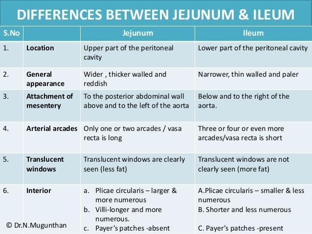
 8 Al-Mohtaseb Saba Alfayoumi Mo Alfarra For the comparison purposes refer to the last page where you can find a table that summarizes them. Enjoy Jejunum and Ileum -They're intraperitoneal and freely mobile
Digestive System. Presented by: Dr M. Arianmanesh PhD in Reproductive and Developmental Biology Dept. of Anatomical Sciences
 Digestive System Presented by: Dr M. Arianmanesh PhD in Reproductive and Developmental Biology Dept. of Anatomical Sciences Today we will discuss: Histological layers of alimentary canal Oral cavity Lip
The Digestive System. Chapter 25
 The Digestive System Chapter 25 Introduction Structure of the digestive system A tube that extends from mouth to anus Accessory organs are attached Functions include Ingestion Movement Digestion Absorption
Sunday 29th January. Day 1: The Digestive Tract. From Anatomy to Treatment. A Nutritional Approach MPS.
 Sunday 29th January Day 1: The Digestive Tract From Anatomy to Treatment A Nutritional Approach MPS www.osteopathicea.com mike@osteopathicea.com AKA Digestive Tract, Alimentary Canal, Enteron, or Gut Body
MICROSTRUCTURES LIPS TOOTH TONGUE OESOPHAGUS STOMACH, CARDIAC, PYLORIC FUNDIC GLANDS
 MICROSTRUCTURES LIPS TOOTH TONGUE OESOPHAGUS STOMACH, CARDIAC, PYLORIC FUNDIC GLANDS HUMAN ANATOMY: MICROSTRUCTURES CLASSIFICATION: LOCATION AND BOUNDARIES, FORM, FUNCTION, MICROSCOPIC STRUCTURE: A hollow
Principles of Anatomy and Physiology
 Principles of Anatomy and Physiology 14 th Edition CHAPTER 24 The Digestive System Introduction The purpose of this chapter is to Identify the anatomical components of the digestive system as well as their
A deep groove encircles the body of the circumvallate papilla. Serous (von Ebner s) glands (serous) drain into the base of this groove.
 glands (serous) drain into the base of this groove.") By Dr. Raja Ali A deep groove encircles the body of the circumvallate papilla. Serous (von Ebner s) glands (serous) drain into the base of this groove. The flow of fluid from these glands serves to wash
By Dr. Raja Ali A deep groove encircles the body of the circumvallate papilla. Serous (von Ebner s) glands (serous) drain into the base of this groove. The flow of fluid from these glands serves to wash
(A) Diarrhea. (B) Stomach cramps. (C) Dehydration due to excess fluid loss. (D) A, B, and C are correct. (E) Only answer B is correct.
 Diarrhea. (B) Stomach cramps. (C) Dehydration due to excess fluid loss. (D) A, B, and C are correct. (E) Only answer B is correct.") Human Anatomy - Problem Drill 21: The Digestive System Question No. 1 of 10 1. A 26-year-old male is treated in the emergency department for severe gastrointestinal disturbance. Which of the following
Human Anatomy - Problem Drill 21: The Digestive System Question No. 1 of 10 1. A 26-year-old male is treated in the emergency department for severe gastrointestinal disturbance. Which of the following
Gastrointestinal Tract
 CTO Lab #5 GI TRACT & GLANDS; ENDOCRINE SYSTEM Page 1 Gastrointestinal Tract Slide 126 This section through the esophagus shows the characteristic layers of the gastrointestinal tract. Examine the non-keratinized
Lab 8: Digestive System
 BIOL 221 A&P II Lab 8: Digestive System Become familiar with the gross anatomy of the digestive system (Exercise 38) using the models, Fig. 38.1 (Activity 1), and the rat. Recognize and know the functions
بسم هللا الرحمن الرحيم
 بسم هللا الرحمن الرحيم Today, we will leave all hormones and start with another topic which is GI system This lecture is talking about general histology of Gastrointestinal system.. The gastrointestinal
Anatomy & Physiology Revealed Instructions. 1. From the Module dropdown menu, chose the 12. Digestive system.
 #10 - Objectives: Examine the histology of selected body organs using Anatomy & Physiology Revealed software and microscope slides. Be able to identify each organ and the specific structures indicated
the serous membranes lining the peritoneal cavity continuously produce what?
 Basic A & P II Dr. L. Bacha Chapter Outline (Martini & Nath 2010) - two groups of organs form the digestive system (see Fig. 22-1): 1. digestive tract what is it also called? list the organs that make
General functions of digestive system. Ch. 15 The Digestive System. General histology of the wall of the digestive tract. Overview of digestive organs
 Overall idea: obtain nutrients from food (for energy and raw materials for synthesis), and defecate the leftover waste 2 types of organs involved: 1. Parts of the digestive tract (= a long muscular tube
Tissues Review 4 type
 Tissues Review 4 type Tissues Definition: a group of closely associated cells that perform related functions and are similar in structure Between cells: nonliving extracellular material Four basic types
Lab activity manual Histology of the digestive system
 Lab activity manual Histology of the digestive system Jeanne Adiwinata Pawitan Prerequisite: Histology of the 4 basic tissues In this module we learn about the histology of the digestive system, from the
BIO 132 Anatomy and Physiology II Spring, 2016 Exam 1 Name: BIO 132 ID Number. Section 1 Answer questions 1 40 on the scan sheet.
 BIO 132 Anatomy and Physiology II Spring, 2016 Exam 1 Name: BIO 132 ID Number Section 1 Answer questions 1 40 on the scan sheet. 1. The homeostatic value of a particular controlled variable in the body
Energy, Chemical Reactions and Enzymes
 Phosphorylation Hydrolysis Energy, Chemical Reactions and Enzymes Chapter 2 (selections) What is Energy? Energy is the capacity to do work Potential Energy Kinetic Energy Chemical Bond Energy Like a rechargeable
458 Essentials of Human Anatomy and Physiology
 458 Essentials of Human Anatomy and Physiology Visceral peritoneum Intrinsic nerve plexuses: Myenteric nerve plexus Submucosal nerve plexus Submucosal glands Mucosa: Surface epithelium Lamina propria Muscle
bolus. The bolus is passed to the pharynx which will convey it to the esophagus, the start of the digestive tube proper. This muscular tube will
 Chapter 13 Digestive System (Oral Cavity and the Alimentary Canal) 13.1. Basic Concepts The digestive system is involved with the intake, mechanical and chemical breakdown, and absorption of food. It also
GI Histology Lab 1. Prepared by: Zeina Kalaji
 GI Histology Lab 1 Prepared by: Zeina Kalaji Lip ORAL MUCOSA -Arrow shows labial salivary glands in the submucosa. VERMILLION transitional zone. SKIN Stratified Squamous epithelium, keratinized -Arrow
The Tissue Level of Organization
 The Tissue Level of Organization Study of this lecture is to be accomplished in conjunction with the Histology Module on the Web!! 1. Introduction Cell Tissue Histology A. General Tissue Types i. Epithelial
Connective tissue The Digestive System
 Connective tissue The Digestive System Part 1 Structure of digestive system Functions Basic Structure of the Alimentary Canal Wall Tube is made up of four layers: 1. Mucosa 2. Submucosa 3. Muscularis externa
Biology. TOPIC : Digestion and Absorption. Marks : 120 mks Time : ½ hr
 TOPIC : Digestion and Absorption Date : Marks : 120 mks Time : ½ hr (1) Match with and select the correct option from the codes given below A Van Kupffer cells (i) Isolets of langerhans B - cells (ii)
Physiology Unit 4 DIGESTIVE PHYSIOLOGY
 Physiology Unit 4 DIGESTIVE PHYSIOLOGY In Physiology Today Functions Motility Ingestion Mastication Deglutition Peristalsis Secretion 7 liters/day! Exocrine/endocrine Digestion Absorption Digestion of
PRACTICAL ROADMAP EPITHELIUM A. JOVANOVIĆ
 PRACTICAL ROADMAP EPITHELIUM A. JOVANOVIĆ Epithelia Simple epithelia Stratified epithelia Simple squamous Simple cuboidal Simple columnar Pseudostratified Stratified squamous - non keratinized - keratinized
Class XI Chapter 16 Digestion and Absorption Biology
 Question 1: Choose the correct answer among the following: (a) Gastric juice contains (i) pepsin, lipase and rennin (ii) trypsin lipase and rennin (iii) trypsin, pepsin and lipase (iv) trypsin, pepsin
A. Incorrect! The esophagus connects the pharynx and the stomach.
 Human Physiology - Problem Drill 19: Digestive Physiology and Nutrition Question No. 1 of 10 Instructions: (1) Read the problem and answer choices carefully, (2) Work the problems on paper as 1. This organ
Digestive Lecture Test Questions Set 4
 Digestive Lecture Test Questions Set 4 1. Which of the following is not associated directly with the small intestine: a. villi b. circular folds c. microvilli d. haustrae e. secretin 2. The largest (longest)
Outline. Bio 105: Tissues Laboratory. Organization of the Human Body. Tissue - Epithelium. Tissues 3/2/ Copyright 2009 Pearson Education, Inc
 Outline Bio 105: Tissues Laboratory Laboratory 5 Reading: Chapter 4 I. Cell to cell contact II. Body Cavities III. Membranes IV. Homeostasis V. Integumentary System I. Includes skin, hair and nails 1 2
Digestion and Absorption
 Digestion and Absorption General Considerations - No absorption in esophagus, little in the stomach and vast majority of absorption occurs in small intestine. - The small intestine has specialized structures
MICROSTRUCTURES SMALL INTESTIN LARGE INTESTIN PANCREAS LIVER GALLBLADDER SALIVARY GLANDS ADRENALS THYROID AND PARATHYROID GLANDS
 MICROSTRUCTURES SMALL INTESTIN LARGE INTESTIN PANCREAS LIVER GALLBLADDER SALIVARY GLANDS ADRENALS THYROID AND PARATHYROID GLANDS HUMAN ANATOMY: MICROSTRUCTURES CLASSIFICATION: LOCATION AND BOUNDARIES,
 Question 1: Choose the correct answer among the following: (a) Gastric juice contains (i) pepsin, lipase and rennin (ii) trypsin lipase and rennin (iii) trypsin, pepsin and lipase (iv) trypsin, pepsin
BIOH122 Human Biological Science 2
 BIOH122 Human Biological Science 2 Session 14 Digestive System 2 Pancreas, Liver, Small Intestine Bioscience Department Endeavour College of Natural Health endeavour.edu.au Session Plan o Pancreas Anatomy
Chapter 26 The Digestive System
 Chapter 26 The Digestive System Digestive System Gastroenterology is the study of the stomach and intestine. Digestion Catabolism Absorption Anabolism The actions of the digestive system are controlled
BIOH122 Human Biological Science 2
 BIOH122 Human Biological Science 2 Session 13 Digestive System 1 Mouth to Stomach Bioscience Department Endeavour College of Natural Health endeavour.edu.au Session Plan o Functions of the digestive system
DIGESTIVE SYSTEM CLASS NOTES. tube along with several
 DIGESTIVE SYSTEM CLASS NOTES Digestion Breakdown of food and the of nutrients in the bloodstream. Metabolism Production of for and cellular activities. The digestive system is composed of the canal which
Stomach. Stomach. Nerve supply. Blood supply. Sympathe0c and parasympathe0c fibers of the autonomic nervous system
 Stomach Nerve supply Sympathe0c and parasympathe0c fibers of the autonomic nervous system Blood supply Celiac trunk, and corresponding veins (part of the hepa0c portal system) Stomach Figure 23.14a Chapter
The Digestive System 1
 The Digestive System 1 Digestion Processing of food Types Mechanical (physical) Chew Tear Grind Mash Mix Chemical Catabolic reactions Enzymatic hydrolysis Carbohydrate Protein Lipid 2 Digestion Phases
The Digestive System. Chapter 16. Introduction. Overview of Digestive System. Histological Organization. Movement and Mixing of Digestive Materials
 The Digestive System Chapter 16 Introduction Structure of the digestive system A tube that extends from mouth to anus Accessory organs are attached Functions include Ingestion Movement Digestion Absorption
Alimentary Canal (I) Salivatory Glands. (Esophagus and Stomach) Color index: Slides.. Important..Notes..Extra..
 Salivatory Glands. (Esophagus and Stomach) Color index: Slides.. Important..Notes..Extra..") Alimentary Canal (I) (Esophagus and Stomach) Salivatory Glands Color index: Slides.. Important..Notes..Extra.. Objectives: 1. By the end of this lecture, the student should be able to discuss the microscopic
Alimentary Canal (I) (Esophagus and Stomach) Salivatory Glands Color index: Slides.. Important..Notes..Extra.. Objectives: 1. By the end of this lecture, the student should be able to discuss the microscopic