MEDICAL IMAGING AND BREAST DISEASE HOW CAN WE HELP YOU?
|
|
|
- Jordan McKinney
- 6 years ago
- Views:
Transcription
1 MEDICAL IMAGING AND BREAST DISEASE HOW CAN WE HELP YOU? Barbara M. Preston, M.D.
2 SCREENING MAMMOGRAPHY AVERAGE RISK PATIENTS KAISER RECOMMENDATION: ALL WOMEN (INCLUDING TRANSGENDER FEMALES) Every years age 40-74
3 SCREENING MAMMOGRAPHY HIGH RISK PATIENTS PERSONAL HISTORY OF BREAST CANCER OR HIGH RISK LESION ADH, ALH, LCIS (INCLUDING MALES WITH BREAST CANCER) FAMILY HISTORY OF FIRST DEGREE RELATIVE(MOTHER, DAUGHTER, SISTER, FATHER, BROTHER) BRCA 1/BRCA 2 GENE CARRIERS KAISER RECOMMENDATION: EVERY YEAR, age35-74
4 SCREENING MAMMOGRAPHY VERY HIGH RISK PATIENTS FAMILY HISTORY OF FIRST DEGREE RELATIVE WITH ONSET UNDER THE AGE OF 40: BEGIN YEARLY SCREENING YEARS BEFORE AGE OF RELATIVE S S CANCER (NO EARLIER THAN 25) MANTLE IRRADIATION AS CHILD OR YOUNG ADULT: BEGIN YEARLY SCREENING 25
5 SCREENING ULTRASOUND AS YET NOT EVIDENCE PROVED EFFICACY (ACRIN 6666 STILL ACCRUING) My patient is requesting a screening breast sonography,; what should I tell her? : : Berg, et al, Am J of Radiology: 180, , 1478, 2003 (
6 SCREENING MRI BRCA1 / BRCA2 GENE CARRIERS NO EVIDENCE PROVED EFFICACY FOR AVERAGE RISK PATIENTS Efficacy or MRI and Mammography for Breast Cancer screening in women w with familial or genetic predisposition, Kriege et al, New England J of Medicine: 351: , 437, (


7
8 DIAGNOSTIC BREAST IMAGING SYMPTOMATIC PATIENTS: PALPABLE MASS AXILLARY LYMPHADENOPATHY (+CXR) SPONTANEOUS OR BLOODY DISCHARGE SKIN / NIPPLE RETRACTION / THICKENING PAIN ALONE IS NOT AN INDICATION FOR MAMMOGRAPHY
9 DIAGNOSTIC BREAST IMAGING SYMPTOMATIC PATIENTS MAMMOGRAPHY ULTRASOUND GALACTOGRAPHY MRI PET BIOPSY REMOVAL / ABLATION
10 DIAGNOSTIC BREAST IMAGING SYMPTOMATIC PATIENTS-MALES IN OUR CENTER, MAMMOGRAMS ARE NOT DONE IN MALES WITH BREAST MASSES UNLESS REQUESTED BY A SURGEON
11 BENIGN MAMMOGRAPHIC FINDINGS-CALCIFICATIONS CALCIFICATIONS- Alert code 2 SECRETORY CALCIFICATION MILK OF CALCIUM DYSTROPHIC VASCULAR CALCIFICATIONS <50 Consider: DM, premature vascular disease, Renal disease, Hyperparathyroid SKIN CALCIFICATION CALCIFIED FIBROADENOMA
12 BENIGN MAMMOGRAPHIC FINDINGS-Alert code 2 FAT CONTAINING MASSES (OIL CYSTS, LIPOMAS, HAMARTOMAS, GALACTOCELES) BENIGN INTRAMAMMARY LYMPH NODES SURGICAL SCARS
13
14 PROBABLY BENIGN MAMMOGRAPHIC FINDINGS- Alert code 3A MASSES SMOOTH SHARP MARGINS AROUND AT LEAST 75% OF CIRCUMFERENCE ROUND OR OVAL ABSENCE OF MALIGNANT FEATURES ULTRASOUND
15 PROBABLY BENIGN FINDINGS- ALERT code 3 A MASSES OR CALCIFICATIONS: LESS THAN 2% CHANCE OF MALIGNANCY- 6 MONTH FOLLOWUP
16 INDETERMINATE MAMMOGRAPHIC FINDINGS- ALERT CODE 4 POWDERY CALCIFICATIONS Sclerosing adenosis vs Low Nuclear Grade DCIS GRANULAR CALCIFICATIONS Crushed stone Calcified fibroadenoma vs fibrocystic change vs High nuclear grade DCIS
17 INDETERMINATE CALCIFICAITONS DCIS can be stable for years; therefore biopsy is almost always recommended OCCASIONALLY 6 MONTH FOLLOWUP IS APPROPRIATE (suspect calcifying fibroadenoma,, fat necrosis)


18 MALIGNANT MAMMOGRAPHIC FINDINGS-- --Alert code 5 CALCIFICATIONS PLEOMORPHIC XYZ USUALLY HIGH NUCLEAR GRADE DCIS


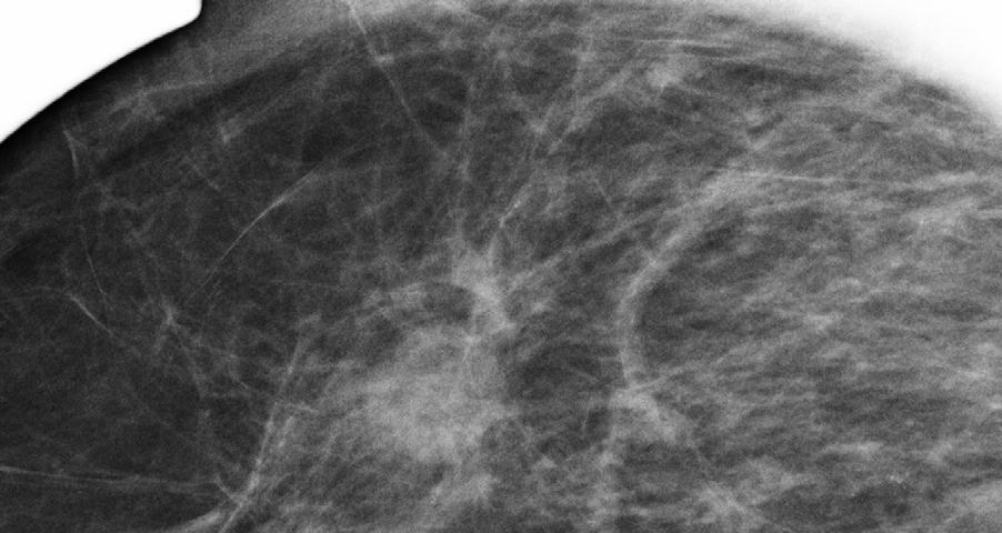
19 MALIGNANT MAMMOGRAPHIC FINDINGS ARCHITECTURAL DISTORTION Nipple
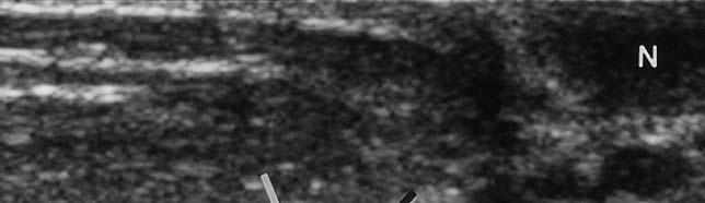
20 ULTRASOUND NOT YOUR MAMA S ULTRASOUND

21 BENIGN ULTRASOUND FINDINGS CYST SMOOTH SHARP BORDERS THROUGH TRANSMISSION ROUND OR OVAL NO INTERNAL ECHOES ( OR MOBILE DEBRIS)
22 A CYST THAT S S NOT A CYST

23 BENIGN ULTRASOUND FINDINGS-Alert code 2 BENIGN SOLID MASSES SMOOTH SHARP MARGINS OVAL ONE/ TWO GENTLE LOBULATIONS NO MALIGNANT FEATURES

24 MALIGNANT ULTRASOUND FINDINGS-Alert code 5 SPICULATTIONS POOR THROUGH TRANSMISSION ANGULAR MARGINS SHADOWING ECHOGENIC FAT NON-COMPRESSIBLE


25 STAGING BREAST CANCER WITH ULTRASOUND DUCT EXTENSION SATELLITE LESIONS


26 STAGING BREAST CANCER WITH ULTRASOUND AXILLARY ADENOPATHY LOOKING FOR INVASION WITHIN DCIS

27 SPECIAL PROBLEMS IMPLANT INTEGRITY MAMMOGRAPHY ULTRASOUND MRI


28


29 SPECIAL PROBLEMS - INFLAMMATORY BREAST CANCER -
30 SPECIAL PROBLEMS AXILLARY LYMPHADENOPATHY WITH A NEGATIVE MAMMOGRAM ULTRASOUND MRI
31 SPECIAL PROBLEMS PAGET S S DISEASE OF THE NIPPLE MAMMOGRAPHY MRI
32 GALACTOGRAPHY FILLING DEFECTS DUCT OBSTRUCTIONS DUCT DISTORTION EXTRAVASATION

33 GALACTOGRAPHY EXCISE ALL LESIONS EXCISE EXTRAVASATION


34 GALACTOGRAPHY MULTIPLE LESIONS LESIONS MAY REQUIRE 2 WIRE NEEDLE LOCALIZATION PERIPHERAL LESIONS MAY REQUIRE NEEDLE LOCALIZATION


35 GALACTOGRAPHY FAILED GALACTOGRAM: TRY ULTRASOUND (MRI?)
Imaging in breast cancer. Mammography and Ultrasound Donya Farrokh.MD Radiologist Mashhad University of Medical Since
 Imaging in breast cancer Mammography and Ultrasound Donya Farrokh.MD Radiologist Mashhad University of Medical Since A mammogram report is a key component of the breast cancer diagnostic process. A mammogram
Lesion Imaging Characteristics Mass, Favoring Benign Circumscribed Margins Intramammary Lymph Node
 Lesion Imaging Characteristics Mass, Favoring Benign Circumscribed Margins Intramammary Lymph Node Oil Cyst Mass, Intermediate Concern Microlobulated Margins Obscured Margins Mass, Favoring Malignant Indistinct
Armed Forces Institute of Pathology.
 Armed Forces Institute of Pathology www.radpath.com Armed Forces Institute of Pathology Breast Disease www.radpath.org Armed Forces Institute of Pathology Interpretation of Breast MRI Leonard M. Glassman
Breast Imaging Lexicon
 9//201 200 BI RADS th Edition 201 BI RADS th Edition Breast Imaging Lexicon Mammographic Pathology and Assessment Categories Deborah Thames, R.T.(R)(M)(QM) The Advanced Health Education Center Nonmember:
ACRIN 6666 IM Additional Evaluation: Additional Views/Targeted US
 Additional Evaluation: Additional Views/Targeted US For revised or corrected form check box and fax to 215-717-0936. Instructions: The form is completed based on recommendations (from ID form) for additional
Leonard M. Glassman MD
 BI-RADS The New BI-RADS Leonard M. Glassman MD FACR Former Chief of Breast Imaging American Institute for Radiologic Pathology Washington Radiology Associates, PC Breast Imaging Reporting and Data System
Amammography report is a key component of the breast
 Review Article Writing a Mammography Report Amammography report is a key component of the breast cancer diagnostic process. Although mammographic findings were not clearly differentiated between benign
Imaging the Symptomatic Patient. Avice M.O Connell MD,FACR,FSBI Professor of Imaging Sciences Director, Women s Imaging University of Rochester
 Imaging the Symptomatic Patient Avice M.O Connell MD,FACR,FSBI Professor of Imaging Sciences Director, Women s Imaging University of Rochester The four most common symptoms Mass Pain Discharge Infection
Ana Sofia Preto 19/06/2013
 Ana Sofia Preto 19/06/2013 Understanding the underlying pathophysiologic processes leading to the various types of calcifications Description and illustration of the several types of calcifications, according
Ultrasound of the Breast BASICS FOR THE ORDERING CLINICIAN
 Ultrasound of the Breast BASICS FOR THE ORDERING CLINICIAN Breast Ultrasound Anatomy Skin Breast Parenchyma Pectoralis Fascia Pectoralis Breast Ultrasound Anatomy Indications for Breast Ultrasound Palpable
Index. C Calcifications fat necrosis 1, 61 fat necrosis 4, 69 nipple/peri-areolar involvement 1, 165
 A ADH. See Atypical ductal hyperplasia (ADH) American College of Radiology (ACR), BI-RADS background parenchymal enhancement, 8, 9, 81, 82 fibroglandular tissue guidelines, 6 American Joint Committee on
RADIOLOGIC EVALUATION OF BREAST CANCER
 RADIOLOGIC EVALUATION OF BREAST CANCER Orsolya Farkas, Gabriella Bodrogi and Gábor Szalai Department of Radiology, Pécs University Orsifarkas@yahoo.com Complex evaluation of the breast Patient history
Leonard M. Glassman MD Analysis of Breast Calcifications
 Importance of Calcification Leonard M. Glassman MD FACR American Institute for Radiologic Pathology Washington Radiology Associates, PC Washington DC 45% of all breast cancers present as calcification
ORIGINAL ARTICLE EVALUATION OF BREAST LESIONS USING X-RAY MAMMOGRAM WITH HISTOPATHOLOGICAL CORRELATION
 Available online at www.journalijmrr.com INTERNATIONAL JOURNAL OF MODERN RESEARCH AND REVIEWS IJMRR ISSN: 2347-8314 Int. J. Modn. Res. Revs. Volume 3, Issue 10, pp 807-814, October, 2015 ORIGINAL ARTICLE
AMSER Case of the Month: September 2018
 AMSER Case of the Month: September 2018 60-year-old woman with a left breast mass noted on screening mammography. Catherine McNulty, MS4 Tulane University School of Medicine Dr. Robin Sobolewski Breast
EARLY DETECTION: MAMMOGRAPHY AND SONOGRAPHY
 EARLY DETECTION: MAMMOGRAPHY AND SONOGRAPHY Elizabeth A. Rafferty, M.D. Avon Comprehensive Breast Center Massachusetts General Hospital Harvard Medical School Breast Cancer Screening Early detection of
EARLY DETECTION: MAMMOGRAPHY AND SONOGRAPHY
 EARLY DETECTION: MAMMOGRAPHY AND SONOGRAPHY Elizabeth A. Rafferty, M.D. Avon Comprehensive Breast Center Massachusetts General Hospital Harvard Medical School Breast Cancer Screening Early detection of
Breast Imaging! Ravi Adhikary, MD!
 Breast Imaging! Ravi Adhikary, MD! ACS Estimated Cancers Statistics 2014! Breast! New Cases in Women! 232,670 (+67,570 in situ)! Deaths in Women! 40,000! Colon! 48,380! 24,040! Cervical! 12,360! 4,020!
Mammographic imaging of nonpalpable breast lesions. Malai Muttarak, MD Department of Radiology Chiang Mai University Chiang Mai, Thailand
 Mammographic imaging of nonpalpable breast lesions Malai Muttarak, MD Department of Radiology Chiang Mai University Chiang Mai, Thailand Introduction Contents Mammographic signs of nonpalpable breast cancer
BI-RADS and Breast MRI. Kathy Borovicka, M.D. Thursday February 15, 2018
 BI-RADS and Breast MRI Kathy Borovicka, M.D. Thursday February 15, 2018 Learning Objectives Be familiar with the Breast Imaging Reporting and Data System (BI-RADS) Understand the components of a breast
Breast Evaluation & Management Guidelines
 Breast Evaluation & Management Guidelines Pamela L. Kurtzhals, M.D. F.A.C.S. Head, Dept. of General Surgery Scripps Clinic, La Jolla Objective Review screening & diagnostic guidelines Focused patient complaints
Case study 1. Rie Horii, M.D., Ph.D. Division of Pathology Cancer Institute Hospital, Japanese Foundation for Cancer Research
 NCCN/JCCNB Seminar in Japan April 15, 2012 Case study 1 Rie Horii, M.D., Ph.D. Division of Pathology Cancer Institute Hospital, Japanese Foundation for Cancer Research Present illness: A 50y.o.premenopausal
CLINICAL SIGNIFICANCE OF BENIGN EPITHELIAL CHANGES
 Papillomas. Papillomas are composed of multiple branching fibrovascular cores, each having a connective tissue axis lined by luminal and myoepithelial cells ( Fig. 23-11 ). Growth occurs within a dilated
S. Murgo, MD. Chr St-Joseph, Mons Erasme Hospital, Brussels
 S. Murgo, MD Chr St-Joseph, Mons Erasme Hospital, Brussels? Introduction Mammography reports are sometimes ambiguous and indecisive. ACR has developped the BIRADS. BIRADS consists of a lexicon in order
Breast Health. Learning Objectives. Breast Anatomy. Poll Question. Breast Anatomy
 Learning Objectives Describe breast anatomy to a patient Breast Health Answer questions about causes of breast pain and masses Explain breast cancer screening/diagnostic modalities Appropriately triage
1/12/2007 Fernald Medical Monitoring Program Sort Code Mammogram Coding
 1/12/2007 Fernald Medical Monitoring Program Sort Code Mammogram Coding Exam (Test) Performed 1 2 Code Description 3 1 Screening mammogram 4 2 Diagnostic mammogram/recall unilateral mammogram/coned magnification
Imaging Guidelines for Breast Cancer Screening
 Imaging Guidelines for Breast Cancer Screening Sarah Colwick, MD Dr. Sarah Colwick was born and raised in Sikeston, MO. She attended college and medical school at the University of Missouri-Kansas City
Pitfalls and Limitations of Breast MRI. Susan Orel Roth, MD Professor of Radiology University of Pennsylvania
 Pitfalls and Limitations of Breast MRI Susan Orel Roth, MD Professor of Radiology University of Pennsylvania Objectives Review the etiologies of false negative breast MRI examinations Discuss the limitations
Breast Disease: What PCPs Need to Know. Eunice Cho MD FACS
 Breast Disease: What PCPs Need to Know Eunice Cho MD FACS New Breast Cancer Screening Guideline for women with average risk Every other year AGE 40 AGE 45 AGE 55 AGE 55 + Talk with your doctor about when
Breast Ultrasound: Improving Your Skills & Patient Care
 Breast Ultrasound: Improving Your Skills & Patient Care Objectives Discuss US techniques available for image optimization. Review & compare the US appearances of benign & malignant masses. Cherie M. Kuzmiak,
Case Scenario 1 History and Physical 3/15/13 Imaging Pathology
 Case Scenario 1 History and Physical 3/15/13 The patient is an 84 year old white female who presented with an abnormal mammogram. The patient has a five year history of refractory anemia with ringed sideroblasts
Breast Cancer. Common kinds of breast cancer are
 Breast Cancer A breast is made up of three main parts: glands, ducts, and connective tissue. The glands produce milk. The ducts are passages that carry milk to the nipple. The connective tissue (which
Armed Forces Institute of Pathology.
 Armed Forces Institute of Pathology www.radpath.com Armed Forces Institute of Pathology Breast Disease www.radpath.org Armed Forces Institute of Pathology Evaluation of Breast Calcifications Leonard M.
The radiologic workup of a palpable breast mass
 Imaging in Practice CME CREDIT EDUCTIONL OJECTIVE: The reader will consider which breast masses require further workup and which imaging study is most appropriate Lauren Stein, MD Imaging Institute, Cleveland
Advanced Course on Multimodality Detection and Diagnosis of Breast Diseases
 BREAST SEMINAR Advanced Course on Multimodality Detection and 3D image of the breast tissue Invited speaker LÁSZLÓ TABÁR, MD,FACR (Hon) Falun, Sweden Nov 22-23, 2014 ATHENS, Greece Royal Olympic Hotel
Here are examples of bilateral analog mammograms from the same patient including CC and MLO projections.
 Good afternoon. It s my pleasure to be discussing Diagnostic Breast Imaging over the next half hour. I m Wei Yang, Professor of Diagnostic Radiology and Chief, the Section of Breast Imaging as well as
BI-RADS Update. Martha B. Mainiero, MD, FACR, FSBI Brown University Rhode Island Hospital
 BI-RADS Update Martha B. Mainiero, MD, FACR, FSBI Brown University Rhode Island Hospital No Disclosures BI-RADS History 1980s Quality Issues ACR Accreditation BI-RADS 1994 2003 4 th Edition MRI, US January
Diagnostic Dilemmas of Breast Imaging
 Diagnostic Dilemmas of Breast Imaging Common Causes of Error in Breast Cancer Detection By: Jason Cord, M.D. Mammography: Initial Imaging The standard for detection of breast cancer Screening mammography
Breast Imaging Update: Old Dog New Tricks
 Breast Imaging Update: Old Dog New Tricks Claire McKay, DO M&S Imaging Assoc. San Antonio, TX cmckayhart@juno.com Goals Describe modalities available, old and new Provide understanding of pros and cons
Mammography Education, Inc.
 Mammography Education, Inc. 2018 3D image of the breast tissue BREAST SEMINAR SERIES Faculty LÁSZLÓ TABÁR, MD, FACR (Hon) Professor emeritus of Radiology Using the Multimodality Approach. A FULLY INTERACTIVE,
Benign, Reactive and Inflammatory Lesions of the Breast
 Benign, Reactive and Inflammatory Lesions of the Breast Marilin Rosa, MD Associate Member Section Head of Breast Pathology Department of Anatomic Pathology Program Director, Breast Pathology Fellowship
Mary Smania, DNP, FNP-BC Clinical Practice Champion Assistant Professor Michigan State University College of Nursing
 Mary Smania, DNP, FNP-BC Clinical Practice Champion Assistant Professor Michigan State University College of Nursing Identify evidence based routine screening guidelines for women of all ages Identify
ROLE OF MRI IN SCREENING, DIAGNOSIS AND MANAGEMENT OF BREAST CANCER. B.Zandi Professor of Radiology
 ROLE OF MRI IN SCREENING, DIAGNOSIS AND MANAGEMENT OF BREAST CANCER B.Zandi Professor of Radiology Introduction In the USA, Breast Cancer is : The Most Common Non-Skin Cancer The Second Leading cause of
Radiology Review Course Hotel del Coronado Coronado, California
 37 th Annual Radiology Review Course Hotel del Coronado Coronado, California Friday, April 21, 2017 - PM TABLE OF CONTENTS Friday, April 21, 2017 - PM SAM Session - Breast Imaging Update 12:45 PM 1:30
Mammography and Other Screening Tests. for Breast Problems
 301.681.3400 OBGYNCWC.COM Mammography and Other Screening Tests What is a screening test? for Breast Problems A screening test is used to find diseases, such as cancer, in people who do not have signs
Breast imaging of benign fat containing lesions
 Breast imaging of benign fat containing lesions Poster No.: C-1870 Congress: ECR 2017 Type: Educational Exhibit Authors: R. Aouini, I. Megdiche, D. Ben Hammadi, N. BEN MAMI, I. Attia, R. Neila, A. Zidi;
Galactography (Ductography)
") Scan for mobile link. Galactography (Ductography) Galactography uses mammography and an injection of contrast material to create pictures of the inside of the breast s milk ducts. It is most commonly used
Scan for mobile link. Galactography (Ductography) Galactography uses mammography and an injection of contrast material to create pictures of the inside of the breast s milk ducts. It is most commonly used
Mousa. Israa Ayed. Abdullah AlZibdeh. 0 P a g e
 1 Mousa Israa Ayed Abdullah AlZibdeh 0 P a g e Breast pathology The basic histological units of the breast are called lobules, which are composed of glandular epithelial cells (luminal cells) resting on
BREAST IMAGING and NEW IMAGING MODALITIES- A Surgeons view
 BREAST IMAGING and NEW IMAGING MODALITIES- A Surgeons view DR CHANTEL THORNTON SPECIALIST BREAST CANCER SURGEON BMSc (hons) MBBS (hons) FRACS Epworth Hospital, Richmond- Agora Centre for Women s Health
Breast imaging in general practice
 Breast series CLINICAL PRACTICE Breast imaging in general practice Nehmat Houssami, MBBS, FAFPHM, FASBP, PhD, is Associate Clinical Director, NSW Breast Cancer Institute, Westmead Hospital, Honorary Senior
Criteria of Malignancy. Evaluation Score
 30 5 Diagnostic Criteria Criteria of Malignancy Table 5.2 lists criteria in contrast-enhancing MR mammography that strongly indicate the presence of malignancy or are unspecific. Unifactorial evaluation
Breast Imaging Essentials
 Breast Imaging Essentials Module 5 Transcript 2016 ASRT. All rights reserved. Breast Imaging Essentials Module 5 Pathology 1. ASRT Animation 2. Welcome Welcome to Module 5 of Breast Imaging Essentials
Mammography Education, Inc.
 Mammography Education, Inc. 2018 3D image of the breast tissue Sept 18-21 Hs-on Screening Course combined with Multimodality Diagnosis of Breast Diseases with Emphasis on Breast MRI SIGTUNA Hotel Kristina
Breast pathology. 2nd Department of Pathology Semmelweis University
 Breast pathology 2nd Department of Pathology Semmelweis University Breast pathology - Summary - Benign lesions - Acute mastitis - Plasma cell mastitis / duct ectasia - Fat necrosis - Fibrocystic change/
Mammography Education, Inc.
 Hands-on Breast Screening and Diagnosis Course * Screening of 315 full field digital mammography cases. * Reading a mixture of normals and proven abnormals at high resolution viewing stations. * Immediate
Diseases of the breast (1 of 2)
") Diseases of the breast (1 of 2) Introduction A histology introduction Normal ducts and lobules of the breast are lined by two layers of cells a layer of luminal cells overlying a second layer of myoepithelial
Diseases of the breast (1 of 2) Introduction A histology introduction Normal ducts and lobules of the breast are lined by two layers of cells a layer of luminal cells overlying a second layer of myoepithelial
Contrast-enhanced Breast MRI RSSA 2013
 Contrast-enhanced Breast MRI RSSA 2013 Prof. dr. Maurice van den Bosch University Medical Center Utrecht, the Netherlands Index 1) Breast cancer 2) Why MRI of the breast 3) Technique 4) Interpretation
Non-mass Enhancement on Breast MRI. Aditi A. Desai, MD Margaret Ann Mays, MD
 Non-mass Enhancement on Breast MRI Aditi A. Desai, MD Margaret Ann Mays, MD Breast MRI Important screening and diagnostic tool, given its high sensitivity for breast cancer detection Breast MRI - Indications
Abid Irshad, MD Director Breast Imaging. Medical University of South Carolina Charleston
 Abid Irshad, MD Director Breast Imaging Medical University of South Carolina Charleston Cases Financial disclosure: I or my family have no financial interest related to the material discussed in this presentation
Ultrasonography. Methods. Brief Description. Indications. Device-related Prerequisites. Technical Requirements. Evaluation Criteria
 1 Ultrasonography Brief Description Imaging modality using sound waves Tissue-specific wave reflection. Indications Evaluation of palpable breast nodules Evaluation of clinically occult mammographic findings
Breast Health and Imaging Glossary
 Contact: Lorna Vaughan HerSpace Breast Imaging & Biopsy Associates 300 State Route 35 South W. Long Branch, NJ 07764 732-571-9100, ext. 104 lorna@breast-imaging.com Breast Health and Imaging Glossary Women
Breast Cancer Screening
 Scan for mobile link. Breast Cancer Screening What is breast cancer screening? Screening examinations are tests performed to find disease before symptoms begin. The goal of screening is to detect disease
Mammography Education, Inc.
 Mammography Education, Inc. 2018 3D image of the breast tissue BREAST SEMINAR SERIES Faculty LÁSZLÓ TABÁR, MD, FACR (Hon) Professor emeritus of Radiology and GILLIAN NEWSTEAD, MD, FACR Professor emeritus
Treatment options for the precancerous Atypical Breast lesions. Prof. YOUNG-JIN SUH The Catholic University of Korea
 Treatment options for the precancerous Atypical Breast lesions Prof. YOUNG-JIN SUH The Catholic University of Korea Not so benign lesions? Imaging abnormalities(10% recall) lead to diagnostic evaluation,
CPC 4 Breast Cancer. Rochelle Harwood, a 35 year old sales assistant, presents to her GP because she has noticed a painless lump in her left breast.
 CPC 4 Breast Cancer Rochelle Harwood, a 35 year old sales assistant, presents to her GP because she has noticed a painless lump in her left breast. 1. What are the most likely diagnoses of this lump? Fibroadenoma
Breast Cancer. Most common cancer among women in the US. 2nd leading cause of death in women. Mortality rates though have declined
 Breast Cancer Most common cancer among women in the US 2nd leading cause of death in women Mortality rates though have declined 1 in 8 women will develop breast cancer Breast Cancer Breast cancer increases
Breast Cancer. Saima Saeed MD
 Breast Cancer Saima Saeed MD Breast Cancer Most common cancer among women in the US 2nd leading cause of death in women 1 in 8 women will develop breast cancer Incidence/mortality rates have declined Breast
A GP S APPROACH TO BREAST LUMPS AND SYMPTOMS DR KK CHEUNG GPGC WORKSHOP
 A GP S APPROACH TO BREAST LUMPS AND SYMPTOMS DR KK CHEUNG GPGC WORKSHOP 18.08.18 HAVE A SYSTEM HISTORY EXAMINATION INVESTIGATION FOLLOW UP BREAST SYMPTOMS HISTORY DON T FORGET SKIN CHANGES AND NIPPLE CHANGES
Intracystic papillary carcinoma of the breast
 Intracystic papillary carcinoma of the breast Poster No.: C-1932 Congress: ECR 2011 Type: Educational Exhibit Authors: V. Dimarelos, F. TZIKOS, N. Kotziamani, G. Rodokalakis, 1 2 3 1 1 1 2 T. MALKOTSI
Mammography Education, Inc.
 Mammography Education, Inc. 2018 3D image of the breast tissue BREAST SEMINAR SERIES Faculty LÁSZLÓ TABÁR, MD, FACR (Hon) Professor emeritus of Radiology The Critical Role of the Breast Imaging Technologists
UNC Breast Imaging Division July 2018
 UNC Breast Imaging Division July 2018 This module is to educate residents on ACR BI-RADS Atlas 2 nd edition Ultrasound Sonomammographic anatomy Sonographic features most associated with malignancy Clinical
Papillary lesions of the breast - Imaging findings and diagnostic challenges
 Papillary lesions of the breast - Imaging findings and diagnostic challenges Poster No.: R-0146 Congress: RANZCR-AOCR 2012 Type: Educational Exhibit Authors: P. Jagmohan, F. J. Pool Keywords: Breast, Mammography,
Scottsdale, AZ Plaza Hotel, 7200 N. Scottsdale Rd
 Mammography Education, Inc. 2014 3D image of the breast tissue LÁSZLÓ TABÁR, M.D.,F.A.C.R (Hon) and STAMATIA DESTOUNIS, M.D., F.A.C.R. The normal TDLUs have bud-like acini Multimodality Approach to Detection
Mammographic evaluation of palpable breast masses with pathological correlation: a tertiary care centre study in Nepal
 Original article 21 Mammographic evaluation of palpable breast masses with pathological correlation: a tertiary care centre study in Nepal G. Gurung, R. K. Ghimire, B. Lohani Department of Radiology and
BREAST PATHOLOGY. Fibrocystic Changes
 BREAST PATHOLOGY Lesions of the breast are very common, and they present as palpable, sometimes painful, nodules or masses. Most of these lesions are benign. Breast cancer is the 2 nd most common cause
Mammography Education, Inc.
 Hands-on Breast Screening and Diagnosis Course * Screening of 315 full field digital mammography cases. * Reading a mixture of normals and proven abnormals at high resolution viewing stations. * Immediate
Journal of Medical Imaging and Radiation Oncology
 Journal of Medical Imaging and Radiation Oncology 60 (2016) 506 513 MEDICAL IMAGING PICTORIAL ESSAY Malignant hyperechoic breast lesions at ultrasound: A pictorial essay Stephen Tiang, 1 Cecily Metcalf,
Fat Necrosis: A Grand Imposter
 Fat Necrosis: A Grand Imposter Poster No.: C-0751 Congress: ECR 2015 Type: Educational Exhibit Authors: L. C. Flores Salinas, Y. A. Ramirez Galvan, A. Garza Báez, C. M. Ferrara Chapa; Monterrey/MX Keywords:
FY16 BCCS Reimbursement Rates and Billing Guidelines Appendix B 2
 FY16 BCCS Reimbursement Rates and Billing Guidelines Appendix B 2 77053 Mammary ductogram or galactogram, single duct, Global Fee $59.05 May be billed with 77055, G0206, 77056, G0204, 76641, 76642 Billable
ISSN X (Print) Research Article. *Corresponding author Dr. Amlendu Nagar
 Research Article. *Corresponding author Dr. Amlendu Nagar") Scholars Journal of Applied Medical Sciences (SJAMS) Sch. J. App. Med. Sci., 2015; 3(3A):1069-1073 Scholars Academic and Scientific Publisher (An International Publisher for Academic and Scientific Resources)
Scholars Journal of Applied Medical Sciences (SJAMS) Sch. J. App. Med. Sci., 2015; 3(3A):1069-1073 Scholars Academic and Scientific Publisher (An International Publisher for Academic and Scientific Resources)
Benign Breast Disease. David Anderson, MD Assistant Professor of Clinical Surgery
 Benign Breast Disease David Anderson, MD Assistant Professor of Clinical Surgery Overview Nipple Discharge Breast infection Breast Pain Gynecomastia Fibroepithelial lesions High Risk Lesions-Papilloma,
Evaluation of Abnormal Screening Mammograms
 342 Evaluation of Abnormal Screening Mammograms Ellen Shaw de Paredes, M.D. The purpose of routine screening mammography is to detect unsuspected cancer that has the potential to be cured. Abnormalities
Breast Cancer Imaging
 Breast Cancer Imaging I. Policy University Health Alliance (UHA) will cover breast imaging when such services meet the medical criteria guidelines (subject to limitations and exclusions) indicated below.
Primary Breast Liposarcoma
 Primary Breast Liposarcoma Bhagyam Nagarajan 1*, GayatriAutkar 1, Keyuri Patel 1, Meghal Sanghvi 1 1. Department of Radiology, Wockhardt Hospital, Mumbai, India * Correspondence: Dr Bhagyam Nagarajan,
AMSER Case of the Month: November 2018
 AMSER Case of the Month: November 2018 42 year old with right breast mass Rina Kiyota Petek Lake Erie College of Osteopathic Medicine, OMS-III Kossivi Dantey, MD Bibianna Klepchick, MD Matthew Hartman,
Breast Pathology. Breast Development
 Breast Pathology Lecturer: Hanina Hibshoosh, M.D. Reading: Kumar, Cotran, Robbins, Basic Pathology, 6th Edition, pages 623-635 Breast Development 5th week - thickening of the epidermis - milk line 5th
Automated Breast Volume Scanner: 3D-Ultrasound of breast lesions
 Automated Breast Volume Scanner: 3D-Ultrasound of breast lesions Poster No.: C-2167 Congress: ECR 2011 Type: Educational Exhibit Authors: T. Fassaert, I. Dubelaar, M. D. F. de Jong, M. Rutten ; 's- 1 1
Wellness Along the Cancer Journey: Cancer Types Revised October 2015 Chapter 2: Breast Cancer
 Wellness Along the Cancer Journey: Cancer Types Revised October 2015 Chapter 2: Breast Cancer Cancer Types Rev. 10.20.15 Page 19 Breast Cancer Group Discussion True False Not Sure 1. Breast cancer is not
Automated Breast Volume Scanner: 3D-Ultrasound of breast lesions
 Automated Breast Volume Scanner: 3D-Ultrasound of breast lesions Poster No.: C-2167 Congress: ECR 2011 Type: Educational Exhibit Authors: T. Fassaert, I. Dubelaar, M. D. F. de Jong, M. Rutten ; 's- 1 1
Breast Cancer Screening with Mammography
 Progress in Public Health Breast Cancer Screening with Mammography JMAJ 44(7): 318 324, 2001 Tokiko ENDO Director, Department of Radiology, National Nagoya Hospital Abstract: Breast cancer has been increasing
Cystic Masses of the Breast
 Residents Section Pattern of the Month Eisenberg ystic Masses of the reast Residents Section Pattern of the Month Residents inradiology Neely Hines 1 Priscilla J. Slanetz Ronald L. Eisenberg Hines N, Slanetz
LYMPHATIC DRAINAGE AXILLARY (MOSTLY) INTERNAL MAMMARY SUPRACLAVICULAR
 INTERNAL MAMMARY SUPRACLAVICULAR") BREAST LYMPHATIC DRAINAGE AXILLARY (MOSTLY) INTERNAL MAMMARY SUPRACLAVICULAR HISTOLOGY LOBE: (10 in whole breast) LOBULE: (many per lobe) ACINUS/I, aka ALVEOLUS/I: (many per lobule) DUCT(S): INTRA- or
BREAST LYMPHATIC DRAINAGE AXILLARY (MOSTLY) INTERNAL MAMMARY SUPRACLAVICULAR HISTOLOGY LOBE: (10 in whole breast) LOBULE: (many per lobe) ACINUS/I, aka ALVEOLUS/I: (many per lobule) DUCT(S): INTRA- or
RSNA, /radiol Appendix E1. Methods
 RSNA, 2016 10.1148/radiol.2016151097 Appendix E1 Methods US and Near-infrared Data Acquisition Four optical wavelengths (740 nm, 780 nm, 808 nm, and 830 nm) were used to sequentially deliver the light
Microcalcifications detected on mammography classified as BIRADS 4 and 5 and their correlations with histopatologic findigns
 Microcalcifications detected on mammography classified as BIRADS 4 and 5 and their correlations with histopatologic findigns Poster No.: C-0401 Congress: ECR 2010 Type: Educational Exhibit Topic: Breast
Mammography Education, Inc.
 Mammography Education, Inc. 2018 3D image of the breast tissue BREAST SEMINAR SERIES Faculty LÁSZLÓ TABÁR, MD, FACR (Hon) Professor emeritus of Radiology Using the Multimodality Approach A FULLY INTERACTIVE,
Breast Update Therese Cusick MS MD FACS
 Breast Update 2017 Therese Cusick MS MD FACS Conflict of Interest Disclosure Nothing to disclose Sources Adapted from SESAP- Surgical Education and Self-Assessment program American College of Surgeons
UW Radiology Review Course Breast Calcifications. BI-RADS 5 th Edition
 UW Radiology Review Course Breast Calcifications Grace Kalish, MD Vantage Radiology BI-RADS 5 th Edition Benign Skin Vascular Large rod like Coarse popcorn Suspicious Amorphous Coarse heterogenous Fine
Alena Levit MD Avice O Connell MD University of Rochester, Rochester, NY
 Alena Levit MD Avice O Connell MD University of Rochester, Rochester, NY Purpose Review imaging spectrum of both common benign and malignant breast lesions Describe and demonstrate CT features with mammogram,
2017BREAST SEMINAR SERIES
 Hands-on Breast Screening and Diagnosis Course * Screening of 510 full field digital mammography cases. * Reading a mixture of normals and proven abnormals at high resolution viewing stations. * Immediate
Mimickers of breast malignancy
 Mimickers of breast malignancy Poster No.: C-0477 Congress: ECR 2010 Type: Educational Exhibit Topic: Breast Authors: S. H. Park, H.-Y. Choi; Incheon/KR Keywords: mimickers, breast malignancy, ultrasound