Chest imaging III: Nodular pulmonary disease. Ádám Domonkos Tárnoki, MD, PhD Assistant professor Department of Radiology, Semmelweis University 1
|
|
|
- Janis Morris
- 5 years ago
- Views:
Transcription


1 Chest imaging III: Nodular pulmonary disease Ádám Domonkos Tárnoki, MD, PhD Assistant professor Department of Radiology, Semmelweis University 1

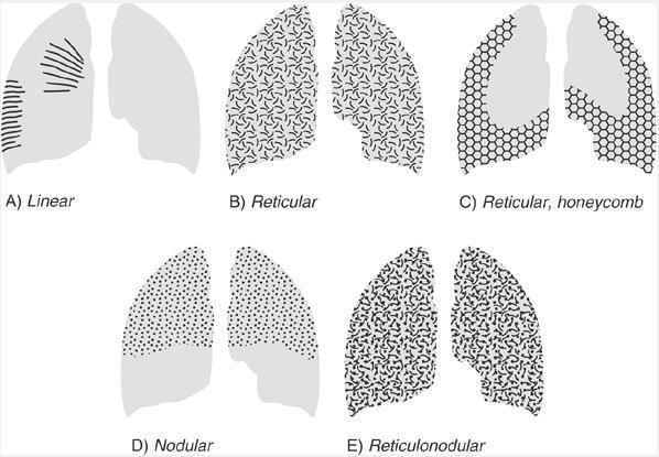
2 Pattern 2

3 Nodular pattern Several round opacity, typically 1 mm - 1 cm in diameter Forms: miliary (1-2 mm), small, medium or large, as the size of opacity increases When the nodular patterns are located in predominant distribution, may refer to specific differential diagnoses 3

4 The morphology of nodular pulmonary abnormalities Solitary lung nodules: Tuberculoma, bronchial carcinoma, metastasis, hamartoma, abscess, aspergilloma, adenoma, round atelectasis, AV shunt, bronchogenic cyst, sequestration, echinococcal cyst, infarction Multiple lung nodules can be cathegorized based on size: Miliary: TB, sarcoidosis, histiocytosis, silicosis, metastases Medium (sub-miliary): bronchogenic TB, metastases, peripheral Kaposi's sarcoma Large: metastases, Wegener's disease, lymphoma 4
5 Differentialdiagnosis of nodules Group Tumor Examples for some diseases metastases multiple primary tumor Kaposi's sarcoma Inflammation miliary tbc multiple lung abscess, septic embolism histoplasmosis, blastomycosis, candidiasis aspergillosis, varicella-zoster virus Connective tissue disorders and vasculitis Rheumatoid arthritis Wegener granulomatosis Pneumoconioses Vascular diseases Hypersensitive and idiopathic diseases silicosis pneumoconiosis of coal workers Multiplex AV malformations hemosiderosis sarcoidosis Langerhans cell histiocystosis hypersensitive pneumonitis 5
 The secondary")

6 Multiple lung nodules (<3 cm) The secondary pulmonary lobule 6


7 Differentialdiagnosis of lung nodules Bronchiolar disease pulmonary edema vasculitis Webb HR: High resolution CT of the Lung sarcoidosis silicosis Lymphangiosis cc. Miliary tbc Miliary fungal infection Miliary viral infection metastases 7


8 Centrilobular nodular pattern Webb HR: High resolution CT of the Lung 8

")

9 Perilymphatic nodules Silicosis Sarcoidosis Lymphangitis job history upper lobe pseudo plaques calcified hilar lymph nodes Upper lobe Symmetric lymphadenopathy (BHL) Peribronchovascular nodules (irregular) nodular thickening of the interlobular septa pleural effusion 9
 Fungi (blastomycosis, histoplasmosis) Metastases Thyroid cancer Adenocarcinoma")
10 Differential diagnosis of the random distribution of the lung nodules Inflammatory tuberculosis Virus (CMV, flu, chickenpox) Fungi (blastomycosis, histoplasmosis) Metastases Thyroid cancer Adenocarcinoma 10
11 The solitary pulmonary nodules (<3 cm) Fleischner Radiology 2008 Round or oval, it is surrounded by the lung parenchyma Benign or malignant? 1. Density 2. Growth rate 3. Wind, shape 4. Cavitation 5. Air 6. Calcification 7. Size 11
 nodule Clear ground glass (GG) (nonsolid)")
12 The three types of lung nodules (<3 cm) Fleischner Radiology 2008 Solid nodule Partsolid (semisolid) nodule Clear ground glass (GG) (nonsolid) nodule 12
13 The solitary pulmonary nodules (<3 cm) Fleischner Radiology 2008 Round or oval, it is surrounded by the lung parenchyma Benign or malignant? 1. Density 2. Growth rate 3. Wind, shape 4. Cavitation 5. Air 6. Calcification 7. Size 13
14 The solitary pulmonary nodules (<3 cm) Volume doubling time (VDT): Volume doubling over time = 26% diameter increase Solid nodules VDT: days: most likely malignant VDT: <20 or >400 days: likely benign There is no growth in 2 years: the most reliable sign of benign behavior Subsolid nodules: they can grow very slowly! Hartmann I. ECR
15 The solitary pulmonary nodules (<3 cm) Fleischner Radiology 2008 Round or oval, it is surrounded by the lung parenchyma Benign or malignant? 1. Density 2. Growth rate 3. Wind, shape 4. Cavitation 5. Air 6. Calcification 7. Size 15
 Sharp, spherical: 20-30%")

16 The solitary pulmonary nodules (<3 cm) Sharp, spherical: 20-30% malignant Lobulated, spiculated, irregular: % malignant Hartmann I. ECR
17 The solitary pulmonary nodules (<3 cm) Fleischner Radiology 2008 Round or oval, it is surrounded by the lung parenchyma Benign or malignant? 1. Density 2. Growth rate 3. Wind, shape 4. Cavitation 5. Air 6. Calcification 7. Size 17
 Air and / or liquefaction Often air-fluid level Hartmann I.")
18 The solitary pulmonary nodules (<3 cm) Especially squamous cell carcinoma Any size often eccentric Often thick, irregular wall (=> 5 mm) Air and / or liquefaction Often air-fluid level Hartmann I. ECR
19 The solitary pulmonary nodules (<3 cm) Fleischner Radiology 2008 Round or oval, it is surrounded by the lung parenchyma Benign or malignant? 1. Density 2. Growth rate 3. Wind, shape 4. Cavitation 5. Air 6. Calcification 7. Size 19
 air")


20 The solitary pulmonary nodules (<3 cm) air bronchogram Cyst-like hypodensities ("soap bubbles") Most commonly adenocarcinoma Hartmann I. ECR
21 The solitary pulmonary nodules (<3 cm) Fleischner Radiology 2008 Round or oval, it is surrounded by the lung parenchyma Benign or malignant? 1. Density 2. Growth rate 3. Wind, shape 4. Cavitation 5. Air 6. Calcification 7. Size 21

22 The solitary pulmonary nodules (<3 cm) Benign pattern Malignant pattern B. Ericsoussi Hartmann I. ECR
23 The solitary pulmonary nodules (<3 cm) Fleischner Radiology 2008 Round or oval, it is surrounded by the lung parenchyma Benign or malignant? 1. Density 2. Growth rate 3. Wind, shape 4. Cavitation 5. Air 6. Calcification 7. Size 23
")
24 Follow-up of lung nodules (Fleischner, Radiology 2017) 24
25 The solitary pulmonary nodules (<3 cm) Fleischner Radiology 2008 Round or oval, it is surrounded by the lung parenchyma Benign or malignant? 1. Density 2. Growth rate 3. Wind, shape 4. Cavitation 5. Air 6. Calcification 7. Size 25
26 Node detection: computed aided diagnosis (CAD) Computer-Diagnose (CAD): hilary nodules better than the peripheral Diagnosis by a radiologist (RAD): subpleural nodules better than the central CAD + RAD is better than 2 radiologists 26
27 Primary tumors Benign tumors (hamartoma, chondroma, lipoma): rare Semimalign (adenoma, hamartoma, carcinoid) X-ray: Round or lobulated node with well demarcated boundary, calcifications CT: small hilar lymphadenopathy, possible metastases Malignant bronchial carcinoma Central: in hilus area, unclear hilary extension Peripheral: in the lung parenchyma or along the thoracic wall Pancoast tumor: in the lung apex and grows transpleural through the chest wall, infiltrating the cervical sympathetic ganglia BAC (bronchoalveolar carcinoma) spreads into the alveoli, it is manifested by multinodular, infiltrative condensations in the peripheral pulmonary parenchyma, but can also appear in the form of round condensations 27
28 Carcinoid central lesion well circumscribed, round or oval ~ 2-5 cm homogeneous contrast enhancement Calcification (eccentric) 28

29 Bronchial carcinoma Adenocarcinoma Squamous cell Large cell Small cell SCLC NSCLC 29
30 Bronchioloalveolar carcinoma (BAC) 1-9% of the lung tumors Subtype of NSCLC (adenocarcinoma) 50% asymptomatic The terminal bronchioles, alveoli are affected Slowly progredient, "non-invasive" growth (the blood and lymph vessels, surrounding lung parenchyma are not affected) "lepidic" growth "Pre-invasive" malignant lesions (in situ carcinoma) invasive adenocarcinoma Non-smoker, women, Asia 30
")
 31")

31 Small growth (BAC) (alveolar collapse) 31 GGO PS PS PS S Vall d Hebron Hospital, Barcelona
32 IASLC/ATS/ERS classification of lung adenocarcinoma (2013) Preinvasiv lesions (formerly BAC) Atypical adenomatous hyperplasia (AAH) Adenocarcinoma In Situ (AIS) Minimally invasive lesions Minimally invasive Adenocarcinoma (MIA) Invasive ADC Predominantly lepidic ADC (LPA) Predominantly acinar Predominantly papillary Predominantly micropapillary Predominantly solid BAC stopped! Instead: AIS / MIA Instead of mucinous BAC: invasive mucinous ADC 32
 Takashima AJR 2003 33 Gandara J Tho Oncol")
33 IASLC/ATS/ERS classification of lung adenocarcinoma (2013) Takashima AJR Gandara J Tho Oncol 2006

34 CT and histopathological correlation in ADC Lederlin M Diagn Interv Imaging Lee HY AJR 2014

35 Lung cancer (mass: > 3cm): staging 5 yr survival Rami-Porta et al. Ann Thorac Cardiovasc Surg


36 Hematogenic spread: Metastases breast, prostate, kidney, thyroid, cervical, testicular, bone, melanoma, gastrointestinal and pancreatic tumors Lymphatic spread: breast, Bronchial carcinoma Lymphangitis carcinomatosa 36
37 DDD (detection, delineation, differentation) Nodules: >8-10 mm - to do! TNM Staging T: CT, MRI assessment of some cases (resectability) N: PET-CT M: PET-CT or MRI Staging CT-Screening has sence 37
38 CT-Screening of lung cancers (2017) Callister, Baldwin, Akram et al: British Thoracic Society guidelines for the investigation and management of pulmonary nodules: accredited by NICE Low dose CT Volumetry CAD? Double reporting? 38
39 CT-Screening of lung cancers (2017) Callister, Baldwin, Akram et al: British Thoracic Society guidelines for the investigation and management of pulmonary nodules: accredited by NICE ADVANTAGES There is 1 death associated death with radiation exposure versus 22 rescued patients fear-induced anxiety (2-6 months) smoking cessation Cost-effective: Screening + intensive smoking cessation + intervention DISADVANTAGES Overdiagnoses False positive cases Reduced by categorization of nodules Volumetry for an accurate assessment of growth Resection through benign disease Patz EF Jr et al. Overdiagnosis in low-dose computed tomography screening for lung cancer. JAMA Intern Med Feb 1;174(2): de Koning HJ et al. Benefits and harms of computed tomography lung cancer screening strategies: a comparative modeling study for the U.S. Preventive Services Task Force. Ann Intern Med Mar 4;160(5):

40 Always look at the "hidden" areas on the x-ray film: Hilus Apex Behind the diaphragm and cardiac shadow
41 Summary Solitary/multiple lung nodules Multiple DD: localization: centrilobular, perilymphatic, random Solitary DD: solid, partsolid (semisolid), clear ground glass (GG) (nonsolid) nodules Density, growth rate, wind, shape, cavitation, air, calcification, size Nodules follow-up: Fleischner (2017) Primary tumors BAC! Lung cancer CT-Screening: preferred Staging (TNM): CT/MRT/PET-CT 41

42 THANK YOU FOR YOUR ATTENTION 42
Thoracic CT pattern in lung cancer: correlation of CT and pathologic diagnosis
 19 th Congress of APSR PG of Lung Cancer (ESAP): Update of Lung Cancer Thoracic CT pattern in lung cancer: correlation of CT and pathologic diagnosis Kazuma Kishi, M.D. Department of Respiratory Medicine,
I appreciate the courtesy of Kusumoto at NCC for this presentation. What is Early Lung Cancers. Early Lung Cancers. Early Lung Cancers 18/10/55
 I appreciate the courtesy of Kusumoto at NCC for this presentation. Dr. What is Early Lung Cancers DEATH Early period in its lifetime Curative period in its lifetime Early Lung Cancers Early Lung Cancers
Pulmonary Nodules & Masses
 Pulmonary Nodules & Masses A Diagnostic Approach Heber MacMahon The University of Chicago Department of Radiology Disclosure Information Consultant for Riverain Technology Minor equity in Hologic Royalties
LUNG NODULES: MODERN MANAGEMENT STRATEGIES
 Department of Radiology LUNG NODULES: MODERN MANAGEMENT STRATEGIES Christian J. Herold M.D. Department of Biomedical Imaging and Image-guided Therapy Medical University of Vienna Vienna, Austria Pulmonary
HRCT in Diffuse Interstitial Lung Disease Steps in High Resolution CT Diagnosis. Where are the lymphatics? Anatomic distribution
 Steps in High Resolution CT Diagnosis Pattern of abnormality Distribution of disease Associated findings Clinical history Tomás Franquet MD What is the diagnosis? Hospital de Sant Pau. Barcelona Secondary
The Spectrum of Management of Pulmonary Ground Glass Nodules
 The Spectrum of Management of Pulmonary Ground Glass Nodules Stanley S Siegelman CT Society 10/26/2011 No financial disclosures. Noguchi M et al. Cancer 75: 2844-2852, 1995. 236 surgically resected peripheral
OBJECTIVES. Solitary Solid Spiculated Nodule. What would you do next? Case Based Discussion: State of the Art Management of Lung Nodules.
 Organ Imaging : September 25 2015 OBJECTIVES Case Based Discussion: State of the Art Management of Lung Nodules Dr. Elsie T. Nguyen Dr. Kazuhiro Yasufuku 1. To review guidelines for follow up and management
PULMONARY NODULES AND MASSES : DIAGNOSTIC APPROACH AND NEW MANAGEMENT GUIDELINES. https://tinyurl.com/hmpn2018
 PULMONARY NODULES AND MASSES : DIAGNOSTIC APPROACH AND NEW MANAGEMENT GUIDELINES Heber MacMahon MB, BCh Department of Radiology The University of Chicago https://tinyurl.com/hmpn2018 Disclosures Consultant
Approach to Pulmonary Nodules
 Approach to Pulmonary Nodules Edwin Jackson, Jr., DO Assistant Professor-Clinical Director, James Early Detection Clinic Department of Internal Medicine Division of Pulmonary, Allergy, Critical Care and
How to Analyse Difficult Chest CT
 How to Analyse Difficult Chest CT Complex diseases are:- - Large lesion - Unusual or atypical pattern - Multiple discordant findings Diffuse diseases are:- - Numerous findings in both sides 3 basic steps
Rodney C Richie MD FACP FCCP DBIM Texas Life and EMSI
 Rodney C Richie MD FACP FCCP DBIM Texas Life and EMSI Pulmonary Nodules Well-circumscribed, radiographic opacities measuring 3 cm in diameter Surrounded by aerated lung Not associated with atelectesis
CT findings in multifocal or diffuse non-mucinous bronchioloalveolar carcinoma (BAC)
") CT findings in multifocal or diffuse non-mucinous bronchioloalveolar carcinoma (BAC) Poster No.: C-2192 Congress: ECR 2014 Type: Educational Exhibit Authors: I. Sandu, A. R. Popita, I.-A. Brumboiu; Cluj-Napoca/RO
CT findings in multifocal or diffuse non-mucinous bronchioloalveolar carcinoma (BAC) Poster No.: C-2192 Congress: ECR 2014 Type: Educational Exhibit Authors: I. Sandu, A. R. Popita, I.-A. Brumboiu; Cluj-Napoca/RO
Case 1 : Question. 1.1 What is the intralobular distribution? 1. Centrilobular 2. Perilymphatic 3. Random
 Interesting case Case 1 Case 1 : Question 1.1 What is the intralobular distribution? 1. Centrilobular 2. Perilymphatic 3. Random Case 1: Answer 1.1 What is the intralobular distribution? 1. Centrilobular
GUIDELINES FOR PULMONARY NODULE MANAGEMENT : RECENT CHANGES AND UPDATES
 Venice 2017 GUIDELINES FOR PULMONARY NODULE MANAGEMENT : RECENT CHANGES AND UPDATES Heber MacMahon MB, BCh Department of Radiology The University of Chicago Disclosures Consultant for Riverain Medical
CT Signs of Solitary Pulmonary Lesions: Revisited
 CT Signs of Solitary Pulmonary Lesions: Revisited Poster No.: C-1764 Congress: ECR 2015 Type: Educational Exhibit Authors: H. Hayashi, K. Ashizawa, Y. Ogihara, A. Nishida, T. Tanaka, 1 1 2 1 1 1 1 1 2
Chief Complain. For chemotherapy
 Chief Complain For chemotherapy Present Illness 93.12 Progressive weakness of R t arm for 1 year X-ray: peneative lesion over right proximal humorous Bone scan: multiple increased intake Biopsy of distal
Chest XRay interpretation INTERPRETATIONS Identifications: Name & Date Technical evaluation Basic Interpretations
 Chest XRay interpretation INTERPRETATIONS Identifications: Name & Date Technical evaluation Basic Interpretations TECHNICAL EVALUATION 1. Projection: AP/PA view To differentiate between AP & PA films,
The 2015 World Health Organization Classification for Lung Adenocarcinomas: A Practical Approach
 The 2015 World Health Organization Classification for Lung Adenocarcinomas: A Practical Approach Dr. Carol Farver Director, Pulmonary Pathology Pathology and Laboratory Medicine Institute Objectives Discuss
Interstitial syndrome
 Interstitial syndrome Ground-glass attenuation Miliary and nodular images linear images Etienne Leroy Terquem Pierre L Her SPI / ISP Soutien Pneumologique International / International Support for Pulmonology
Lung Cancer Imaging. Terence Z. Wong, MD,PhD. Department of Radiology Duke University Medical Center Durham, NC 9/9/09
 Lung Cancer Imaging Terence Z. Wong, MD,PhD Department of Radiology Duke University Medical Center Durham, NC 9/9/09 Acknowledgements Edward F. Patz, Jr., MD Jenny Hoang, MD Ellen L. Jones, MD, PhD Lung
Interstitial Syndrome Ground glass attenuation miliary and nodular images Linear images
 Interstitial Syndrome Ground glass attenuation miliary and nodular images Linear images Dr Etienne Leroy-Terquem Centre hospitalier de Meulan les Mureaux. France French-cambodian association for pneumology
Chest Radiology Interpretation: Findings of Tuberculosis
 Chest Radiology Interpretation: Findings of Tuberculosis Get out your laptops, smart phones or other devices pollev.com/chestradiology Case #1 1 Plombage Pneumonia Cancer 2 Reading the TB CXR Be systematic!
Lung Cancer-a primer. Sai Yendamuri, MD Professor and Chair, Dept of Thoracic Surgery,RPCI,Buffalo
 Lung Cancer-a primer Sai Yendamuri, MD Professor and Chair, Dept of Thoracic Surgery,RPCI,Buffalo CLINICAL CATEGORIES THE SOLITARY PULMONARY NODULE MULTIPLE PULMONARY NODULES Differential Diagnosis Malignant
Joseph Garland, HMS IV Gillian Lieberman, MD. Round Pneumonia. Joseph Garland, HMS IV Gillian Lieberman, MD
 Round Pneumonia Joseph Garland, HMS IV Case 1: Mr. H Mr. H is a 45-year-old man who presents with a 4 day history of full-body myalgias, headaches and fever to 103 F. He also complains of sharp leftsided
Ground Glass Opacities
 Ground Glass Opacities A pathologist s perspective Marie-Christine Aubry, M.D. Professor of Pathology Mayo Clinic Objectives Discuss the proposed new pathologic classification of adenocarcinoma with historical
Low-dose CT Lung Cancer Screening Guidelines for Pulmonary Nodules Management Version 2
 Low-dose CT Lung Cancer Screening Guidelines for Pulmonary Nodules Management Version 2 The Committee for Management of CT-screening-detected Pulmonary Nodules 2009-2011 The Japanese Society of CT Screening
The revised lung adenocarcinoma classification an imaging guide
 Review Article The revised lung adenocarcinoma classification an imaging guide Natasha Gardiner 1, Sanjay Jogai 2, Adam Wallis 3 1 Specialty Registrar in Clinical Radiology, Wessex Deanery, UK; 2 Consultant
Pulmonary Nodules. Michael Morris, MD
 Pulmonary Nodules Michael Morris, MD Case 45 year old healthy male Smokes socially Normal physical exam Pre-employment screening remote +PPD screening CXR nodular opacity Case 45 year old healthy male
Lung Cancer Diagnosis for Primary Care
 Lung Cancer Diagnosis for Primary Care Daniel Nader, DO, FCCP Cancer Treatment Center of America Case 1 In which of the following situations would the U.S. Preventive Services Task Force (USPSTF) recommend
Evaluation of the chest Part II.
 Evaluation of the chest Part II. Nagy Endre SZEGEDI TUDOMÁNYEGYETEM ÁOK, RADIOLÓGIAI KLINIKA, SZEGED ANATOMY parenchyma: alveoloacinar system, pulmonary arteries and veins interstitium: connective tissues
CT findings of high-attenuation pulmonary abnormalities
 Insights Imaging (2010) 1:287 292 DOI 10.1007/s13244-010-0039-2 PICTORIAL REVIEW CT findings of high-attenuation pulmonary abnormalities Naim Ceylan & Selen Bayraktaroglu & Recep Savaş & Hudaver Alper
Acute and Chronic Lung Disease
 KATHOLIEKE UNIVERSITEIT LEUVEN Faculty of Medicine Acute and Chronic Lung Disease W De Wever, JA Verschakelen Department of Radiology, University Hospitals Leuven, Belgium Clinical utility of HRCT To detect
Radiologic-pathologic correlation of pulmonary diseases
 The 1578 th Chest Conference/ 3 rd Biennial Clinical- Radiologic-Pathologic Correlation Radiologic-pathologic correlation of pulmonary diseases Harumi Itoh, M.D. University of Fukui, Japan Centriacinar
Radiologists toolbox to differentiate alveolar versus interstitial lung diseases
 Radiologists toolbox to differentiate alveolar versus interstitial lung diseases Dr Sumer Shikhare, Dr Trishna Shimpi, Dr Ashish Chawla Khoo Teck Puat Hospital Singapore. Relevant financial disclosures
Lecture Goals. Lung (Bronchogenic) Cancer. Causes of Lung Cancer. Elizabeth Weihe, MD Assistant Professor of Radiology Director of UCSD RECIST clinic
 Cancer. Causes of Lung Cancer. Elizabeth Weihe, MD Assistant Professor of Radiology Director of UCSD RECIST clinic") Lecture Goals Origin of Lung Cancer Subtypes New Treatment Paradigms in Lung Cancer Overview of Lung Cancer Elizabeth Weihe, MD Assistant Professor of Radiology Director of UCSD RECIST clinic Lung (Bronchogenic)
Lecture Goals Origin of Lung Cancer Subtypes New Treatment Paradigms in Lung Cancer Overview of Lung Cancer Elizabeth Weihe, MD Assistant Professor of Radiology Director of UCSD RECIST clinic Lung (Bronchogenic)
objectives Pitfalls and Pearls in PET/CT imaging Kevin Robinson, DO Assistant Professor Department of Radiology Michigan State University
 objectives Pitfalls and Pearls in PET/CT imaging Kevin Robinson, DO Assistant Professor Department of Radiology Michigan State University To determine the regions of physiologic activity To understand
Xiaohuan Pan 1,2 *, Xinguan Yang 1,2 *, Jingxu Li 1,2, Xiao Dong 1,2, Jianxing He 2,3, Yubao Guan 1,2. Original Article
 Original Article Is a 5-mm diameter an appropriate cut-off value for the diagnosis of atypical adenomatous hyperplasia and adenocarcinoma in situ on chest computed tomography and pathological examination?
Interesting Cases. Pulmonary
 Interesting Cases Pulmonary 54M with prior history of COPD, hep B/C, and possible history of TB presented with acute on chronic dyspnea, and productive cough Hazy opacity overlying the left hemithorax
Radiological staging of lung cancer. Shukri Loutfi,MD,FRCR Consultant Thoracic Radiologist KAMC-Riyadh
 Radiological staging of lung cancer Shukri Loutfi,MD,FRCR Consultant Thoracic Radiologist KAMC-Riyadh Bronchogenic Carcinoma Accounts for 14% of new cancer diagnoses in 2012. Estimated to kill ~150,000
Correlation in histological subtypes with high resolution computed tomography signatures of early stage lung adenocarcinoma
 Original Article Correlation in histological subtypes with high resolution computed tomography signatures of early stage lung adenocarcinoma Yingying Miao 1,2 *, Jianya Zhang 1,2 *, Jiawei Zou 1,2, Qingqing
Update on 2015 WHO Classification of Lung Adenocarcinoma 1/3/ Mayo Foundation for Medical Education and Research. All rights reserved.
 1 Our speaker for this program is Dr. Anja Roden, an associate professor of Laboratory Medicine and Pathology at Mayo Clinic as well as consultant in the Anatomic Pathology Laboratory and co-director of
Lung Neoplasia II Resection specimens Pathobasic. Lukas Bubendorf Pathology
 Lung Neoplasia II Resection specimens Pathobasic Lukas Bubendorf Pathology Agenda Preneoplastic lesions Histological subtypes of lung cancer Histological patterns of AC Cells of origin and characteristic
Lung Cancer Screening: To Screen or Not to Screen?
 Lung Cancer Screening: To Screen or Not to Screen? Lorriana Leard, MD Co-Director of UCSF Lung Cancer Screening Program Vice Chief of Clinical Activities UCSF Pulmonary, Critical Care, Allergy & Sleep
Pneumocystis jirovecci pneumonia: from mild disease to a real disaster. A pictorial review of the different radiologic patterns in acute settings
 Pneumocystis jirovecci pneumonia: from mild disease to a real disaster. A pictorial review of the different radiologic patterns in acute settings Poster No.: C-1425 Congress: ECR 2017 Type: Educational
8/22/2016. Major risk factors for the development of lung cancer are: Outline
 Carcinomas of the Lung: Changes in Staging, Adenocarcinoma Classification and Genetics Grace Y. Lin, M.D., Ph.D. Outline Background Staging of Lung Cancer: Review of the 2010 7 th Edition of the AJCC Cancer
An Introduction to Radiology for TB Nurses
 An Introduction to Radiology for TB Nurses Garold O. Minns, MD September 14, 2017 TB Nurse Case Management September 12 14, 2017 EXCELLENCE EXPERTISE INNOVATION Garold O. Minns, MD has the following disclosures
Micronodular Lung Disease an algorithm
 Micronodular Lung Disease an algorithm H. Page McAdams, MD Department of Radiology Duke University Medical Center Durham, NC USA page.mcadams@duke.edu Question Which of the following lung diseases is MOST
Case 1: Question. 1.1 What is the main pattern of this HRCT? 1. Intralobular line 2. Groundglass opacity 3. Perilymphatic nodule
 HRCT WORK SHOP Case 1 Case 1: Question 1.1 What is the main pattern of this HRCT? 1. Intralobular line 2. Groundglass opacity 3. Perilymphatic nodule Case 1: Question 1.2 What is the diagnosis? 1. Hypersensitivity
PULMONARY TUBERCULOSIS RADIOLOGY
 PULMONARY TUBERCULOSIS RADIOLOGY RADIOLOGICAL MODALITIES Medical radiophotography Radiography Fluoroscopy Linear (conventional) tomography Computed tomography Pulmonary angiography, bronchography Ultrasonography,
Financial disclosure COMMON DIAGNOSES IN HRCT. High Res Chest HRCT. HRCT Pre test. I have no financial relationships to disclose. Anatomy Nomenclature
 Financial disclosure I have no financial relationships to disclose. Douglas Johnson D.O. Cardiothoracic Imaging Gaston Radiology COMMON DIAGNOSES IN HRCT High Res Chest Anatomy Nomenclature HRCT Sampling
Diagnosis of TB: Radiology David Finlay, MD
 TB Intensive Tyler, Texas June 2-4, 2010 Diagnosis of TB: Radiology David Finlay, MD June 3, 2010 2stages stages- Tuberculosis 1. primary infection 2. reactivation, or post primary disease 2 1 Primary
The small subsolid pulmonary nodules. What radiologists need to know.
 The small subsolid pulmonary nodules. What radiologists need to know. Poster No.: C-1250 Congress: ECR 2016 Type: Educational Exhibit Authors: L. Fernandez Rodriguez, A. Martín Díaz, A. Linares Beltrán,
Use of Integrated PET CT in the Clinical Staging of Non Small Cell Lung Cancer
 November 2010 Use of Integrated PET CT in the Clinical Staging of Non Small Cell Lung Cancer Laura Myers, Harvard Medical School, Year III Clinical Presentation 79yo woman with cough productive of green
Evidence based approach to incidentally detected subsolid pulmonary nodule. DM SEMINAR July 27, 2018 Harshith Rao
 Evidence based approach to incidentally detected subsolid pulmonary nodule DM SEMINAR July 27, 2018 Harshith Rao Outline Definitions Etiologies Risk evaluation Clinical features Radiology Approach Modifications:
Guidelines for the Management of Pulmonary Nodules Detected by Low-dose CT Lung Cancer Screening
 Guidelines for the Management of Pulmonary Nodules Detected by Low-dose CT Lung Cancer Screening 1. Introduction In January 2005, the Committee for Preparation of Clinical Practice Guidelines for the Management
TB Intensive Houston, Texas
 TB Intensive Houston, Texas October 15-17, 17 2013 Diagnosis of TB: Radiology Rosa M Estrada-Y-Martin, MD MSc FCCP October 16, 2013 Rosa M Estrada-Y-Martin, MD MSc FCCP, has the following disclosures to
Steering Committee. Waiting on photo. Paul A. Bunn, Jr., MD Kavita Garg, MD Kim Geisinger, MD Fred R. Hirsch, Gregory Riely, MD, PhD.
 Steering Committee Paul A. Bunn, Jr., MD Kavita Garg, MD Kim Geisinger, MD Fred R. Hirsch, Gregory Riely, MD, PhD MD, PhD Waiting on photo Paul Van Schil, MD, PhD William D. Travis, MD Ming-Sound Tsao,
Lung. 10/24/13 Chest X-ray: 2.9 cm mass like density in the inferior lingular segment worrisome for neoplasm. Malignancy cannot be excluded.
 Lung Case Scenario 1 A 54 year white male presents with a recent abnormal CT of the chest. The patient has a history of melanoma, kidney, and prostate cancers. 10/24/13 Chest X-ray: 2.9 cm mass like density
Stage I synchronous multiple primary non-small cell lung cancer: CT findings and the effect of TNM staging with the 7th and 8th editions on prognosis
 Original Article Stage I synchronous multiple primary non-small cell lung cancer: CT findings and the effect of TNM staging with the 7th and 8th editions on prognosis Jingxu Li, Xinguan Yang, Tingting
PET/CT in lung cancer
 PET/CT in lung cancer Andrei Šamarin North Estonia Medical Centre 3 rd Baltic Congress of Radiology 08.10.2010 Imaging in lung cancer Why do we need PET/CT? CT is routine imaging modality for staging of
Learning Objectives. 1. Identify which patients meet criteria for annual lung cancer screening
 Disclosure I, Taylor Rowlett, DO NOT have a financial interest /arrangement or affiliation with one or more organizations that could be perceived as a real or apparent conflict of interest in the context
Radiology Pathology Conference
 Radiology Pathology Conference Nadia F. Yusaf, M.D. PGY-3 1/29/2010 Presentation material is for education purposes only. All rights reserved. 2010 URMC Radiology Page 1 of 90 Case 1 60 year- old man presents
Micronodular lung pattern - Differential diagnosis
 Micronodular lung pattern - Differential diagnosis Poster No.: P-0074 Congress: ESTI 2015 Type: Educational Poster Authors: P. Ninitas, F. Marinho, P. Campos, I. Távora ; Lisbon/PT, 1 2 2 3 1 1 3 Funchal/PT,
CT Screening for Lung Cancer: Frequency and Significance of Part-Solid and Nonsolid Nodules
 Claudia I. Henschke 1 David F. Yankelevitz 1 Rosna Mirtcheva 1 Georgeann McGuinness 2 Dorothy McCauley 1 0lli S. Miettinen 3 for the ELCAP Group Received June 19, 2001; accepted after revision November
OFF THE BEATEN TRACK: A Pictorial Review of Atypical Features of Pulmonary Metastases
 OFF THE BEATEN TRACK: A Pictorial Review of Atypical Features of Pulmonary Metastases Megan Hora, MD Chi Wan Koo, MD Christian Cox, MD Department of Diagnostic Radiology Thoracic Imaging Section Mayo Clinic
TB Radiology for Nurses Garold O. Minns, MD
 TB Nurse Case Management Salina, Kansas March 31-April 1, 2010 TB Radiology for Nurses Garold O. Minns, MD April 1, 2010 TB Radiology for Nurses Highway Patrol Training Center Salina, KS April 1, 2010
FDG PET/CT in Lung Cancer Read with the experts. Homer A. Macapinlac, M.D.
 FDG PET/CT in Lung Cancer Read with the experts Homer A. Macapinlac, M.D. Patient with suspected lung cancer presents with left sided chest pain T3 What is the T stage of this patient? A) T2a B) T2b C)
Lung tumors & pleural lesions
 Lung tumors & pleural lesions A brief introduction 95% of lung tumors are carcinomas Among the remaining 5%, we will discuss: -Hamartoma the most common benign lung tumor spherical, coin lesion on x-rays
SCBT-MR 2015 Incidentaloma on Chest CT
 SCBT-MR 2015 Incidentaloma on Chest CT Reginald F. Munden MD, DMD, MBA I have no conflicts of interest to report Incidentaloma Pulmonary Nodule Mediastinal Lymph Node Coronary Artery Calcium Incidental
Objectives. Why? Why? Background 11/5/ % incurable disease at presentation Locally advanced disease Metastasis. 14% 5 year survival
 Objectives Appraise lung cancer screening trials results Review screening guidelines Lung Cancer Screening: Past, Present and Future Chi Wan Koo, MD Koo.chiwan@mayo.edu Discuss recommendations essential
Manish Powari Regional Training Day 10/12/2014
 Manish Powari Regional Training Day 10/12/2014 Large number of different types of Interstitial Lung Disease (ILD). Most are very rare Most patients present with one of a smaller number of commoner diseases
INDEX. surgpath.theclinics.com. Note: Page numbers of article titles are in boldface type. diffuse pleural fibrosis, pleural plaques,
 INDEX Note: Page numbers of article titles are in boldface type. A Adenocarcinoma, minimally invasive. See Minimally invasive adenocarcinoma (MIA). Airway-centered interstitial fibrosis, 183 184 ALK (anaplastic
Histopathological and CT Imaging Correlation of Various Primary Lung Carcinoma
 IOSR Journal of Dental and Medical Sciences (IOSR-JDMS) e-issn: 2279-0853, p-issn: 2279-0861.Volume 15, Issue 3 Ver. VII (Mar. 2016), PP 104-110 www.iosrjournals.org Histopathological and CT Imaging Correlation
An Image Repository for Chest CT
 An Image Repository for Chest CT Francesco Frajoli for the Chest CT in Antibody Deficiency Group An Image Repository for Chest CT he Chest CT in Antibody Deficiency Group is an international and interdisciplinary
Respiratory Interactive Session. Elaine Borg
 Respiratory Interactive Session Elaine Borg Case 1 Respiratory Cytology 55 year old gentleman Anterior mediastinal mass EBUS FNA Case 1 Respiratory Cytology 55 year old gentleman with anterior mediastinal
Immunocompromised patients. Immunocompromised patients. Immunocompromised patients
 Value of CT in Early Pneumonia in Immunocompromised Patients Nantaka Kiranantawat, PSU Preventative Factors Phagocyts Cellular immunity Humoral immunity Predisposing Factors Infection, Stress, Poor nutrition,
100 Chest X Rays for Study Group. by Dr. Suneet Khurana
 100 Chest X Rays for Study Group by Dr. Suneet Khurana Approach to - Chest X Ray (shadow of the viscera on a photographic plate) Gas appears Black Fat appears Dark Grey Water Appears as Light Grey Bone
Cancer in the United States, 2004
 Cancer in the United States, 2004 Five-year Cancer Survival Rates (%) US 1974-1998 100 90 80 70 60 50 40 30 20 10 0 1974-1976 1983-1985 1992-1998 Prostate Lung Colon Breast Source: CA Cancer J Clin 2000;50:7-33
Lung Metastases Imaging
 Lung Metastases Imaging Updated: Oct 23, 2015 Author: Tanay Patel, MD; Chief Editor: Eugene C Lin, MD more... OVERVIEW Overview Pulmonary metastasis is seen in 20-54% of extrathoracic malignancies. [1]
Histopathology of NSCLC, IHC markers and ptnm classification
 ESMO Preceptorship on Non-Small Cell Lung Cancer November 15 th & 16 th 2017 Singapore Histopathology of NSCLC, IHC markers and ptnm classification Prof Keith M Kerr Department of Pathology, Aberdeen University
CT Screening for Lung Cancer for High Risk Patients
 CT Screening for Lung Cancer for High Risk Patients The recently published National Lung Cancer Screening Trial (NLST) showed that low-dose CT screening for lung cancer reduces mortality in high-risk patients
Typical and atypical findings of pulmonary sarcoidosis at high resolution CT
 Typical and atypical findings of pulmonary sarcoidosis at high resolution CT Poster No.: C-0169 Congress: ECR 2013 Type: Educational Exhibit Authors: L. Raposo Rodríguez, C. Mejía, B. Escobar Mallada,
Pictorial essay of unusual radiologic manifestations of pulmonary and airway metastasis at initial presentation of lung cancer
 Pictorial essay of unusual radiologic manifestations of pulmonary and airway metastasis at initial presentation of lung cancer Poster No.: C-2297 Congress: ECR 2012 Type: Educational Exhibit Authors: Y.
PET CT for Staging Lung Cancer
 PET CT for Staging Lung Cancer Rohit Kochhar Consultant Radiologist Disclosures Neither I nor my immediate family members have financial relationships with commercial organizations that may have a direct
Imaging in breast cancer. Mammography and Ultrasound Donya Farrokh.MD Radiologist Mashhad University of Medical Since
 Imaging in breast cancer Mammography and Ultrasound Donya Farrokh.MD Radiologist Mashhad University of Medical Since A mammogram report is a key component of the breast cancer diagnostic process. A mammogram
What to Do with Small Lung Nodules Hanh Vu Nghiem, MD William Beaumont Hospital Royal Oak, Michigan
 What to Do with Small Lung Nodules Hanh Vu Nghiem, MD William Beaumont Hospital Royal Oak, Michigan Small Lung Nodules What to do with small lung nodules? We biopsy them when requested What are our accuracy
Web Chapter 3. Image Gallery: Lesion detection on low dose chest CT
 Web Chapter 3 Image Gallery: Lesion detection on low dose chest CT Sarabjeet Singh, MD Mannudeep K. Kalra, MD *Eugene J. Mark, MD *James Stone, MD James H. Thrall, MD Department of Radiology and *Department
Adam J. Hansen, MD UHC Thoracic Surgery
 Adam J. Hansen, MD UHC Thoracic Surgery Sometimes seen on Chest X-ray (CXR) Common incidental findings on computed tomography (CT) chest and abdomen done for other reasons Most lung cancers discovered
Impact of immunostaining of pulmonary and mediastinal cytology
 Impact of immunostaining of pulmonary and mediastinal cytology Harman Sekhon MD, PhD Director of Cytopathology Head of Ottawa-site Ontario Tumour Bank June 20, 2014 Disclaimer Pfizer: Honorarium-Advisory
Lung Cancer Risks. Cancer in the United States, Cancer Death Rates, US The Scheme: From Nicotine Addiction to Lung Cancer
 Cancer in the United States, 2004 Lung Cancer Risks Cigarette Smoking Environmental Tobacco Smoke Other Carcinogens Asbestos, Arsenic, Radon, Bis(chloromethyl) ether, Chromium, Foundry fumes, nickel, mustard
Uses, limitations and interpretation of CT in pulmonary infections: A practical approach
 Uses, limitations and interpretation of CT in pulmonary infections: A practical approach Canadian Association of Radiologists 2013 DISCLOSURES Speakers honorarium, Siemens Canada Objectives 1. Recognize
Bronkhorst colloquium Interstitiële longziekten. Katrien Grünberg, klinisch patholoog
 Bronkhorst colloquium 2013-2014 Interstitiële longziekten De pathologie achter de CT Katrien Grünberg, klinisch patholoog K.grunberg@vumc.nl Preparing: introduction and 3 cases The introduction on microscopic
HRCT features distinguishing minimally invasive adenocarcinomas from invasive adenocarcinomas appearing as mixed ground-glass nodules
 Original Article HRCT features distinguishing minimally invasive adenocarcinomas from invasive adenocarcinomas appearing as mixed ground-glass nodules Wei Yu 1, Zhaoyu Wang 2, Liyong Qian 2, Shanjun Wang
Spectrum of Radiological Findings in Bronchogenic Carcinoma A Retrospective Study
 IOSR Journal of Dental and Medical Sciences (IOSR-JDMS) e-issn: 2279-0853, p-issn: 2279-0861.Volume 17, Issue 01 Ver. VIII January. (2018), PP 43-59 www.iosrjournals.org Spectrum of Radiological Findings
Imaging Decisions Start Here SM
 Owing to its high resolution and wide anatomic coverage, dynamic first-pass perfusion 320-detector-row CT outperforms PET/CT for distinguishing benign from malignant lung nodules, researchers from Japan
Radiology Pathology Conference
 Radiology Pathology Conference Sharlin Johnykutty,, MD, Cytopathology Fellow Sara Majewski, MD, Radiology Resident Friday, August 28, 2009 Presentation material is for education purposes only. All rights
Lung Cancer - Suspected
 Lung Cancer - Suspected Shared Decision Making Lung Cancer: http://www.enhertsccg.nhs.uk/ Patient presents with abnormal CXR Lung cancer - clinical presentation History and Examination Incidental finding