10 years of NordiQC Why are 30% of labs still getting it wrong?
|
|
|
- Hilary Gilmore
- 5 years ago
- Views:
Transcription


1 Mogens Vyberg & Søren Nielsen NordiQC Institute of Pathology Aalborg University Hospital Aalborg, Denmark May 29th years of NordiQC Why are 30% of labs still getting it wrong?
2 Nothing to declare

3 Nordic immunohistochemical Quality Control 3 International organization for quality assurance of IHC Founded 2003 by Nordic pathologists Independent, scientific, not-for-profit organisation Institute of Pathology, Aalborg University Hospital, DK General module: 3 runs/year different marker challenges Breast cancer IHC module: 2 runs/y HER-2, ER/PR, Ki67/E-Cad HER-2 ISH module: 2 runs/year BRISH, FISH

4 4
5 5
6 6

7 Test material Multi-tissue FFPE blocks 10% NBF h (ASCO/CAP guidelines ) Normal and clinically relevant tumour tissues Different levels of antigen expression high, moderate, low, none 2 unstained slides for each marker send to the participants 1 stained slide returned for central assessment

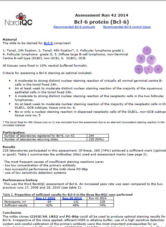
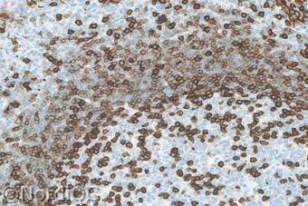
8 Test material 8 The slide to be stained for Bcl-6 comprised: 1. Tonsil, 24 h. 2. Tonsil, 48 h. 3. Follicular lymphoma, grade I 4. Follicular lymphoma, grade II 5. Diffuse large B-cell lymphoma HE NE Tissue selection High Low None Expressor LE
9
10 Nordic immunohistochemical Quality Control Participants


11 ~ 90 Markers in NordiQC runs Tested 1-15 times
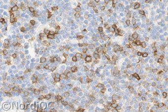
12 NordiQC test - haempath 1x 2x 3x 4x 5x CD8, CD14, CD19, CD163, IgL, MUM1 X ALK, BSAP, Bcl2, CD45, CD56, CD138, p53, TdT X Bcl6, CD4, CD20, CD34, CD68, CD79a, IgK, Ki67 X CD3, CD10, CD23, CD30, Cyclin D1, IgM X CD5, CD15 X
 Insufficient 32% 21% 11% 33% 35% Optimal Good Borderline")
13 NordiQC assessment results General module ~ 20,000 slides ( ~ core sections) Insufficient 32% 21% 11% 33% 35% Optimal Good Borderline Poor
 Insufficient 21% 9% 12% 21% 58% Optimal Good")
14 NordiQC assessment results Breast cancer module ~ 9,000 slides (~35,000 core sections) Insufficient 21% 9% 12% 21% 58% Optimal Good Borderline Poor

15 Publications AJCP 2005,124:782 AIMM 2011, 19:437 AIMM 2013, 21:64 AIMM 2014, 22:449
16 Publications AIMM 2015, 23:1 AIMM 2014, 22:241
17 Serial sections stained for Estrogen receptor Lab. A Lab. B ER in ductal breast carcinoma
18 Serial sections stained for Estrogen receptor Lab. A Lab. B False neg.
19 Serial sections stained for Estrogen receptor Control: uterine cervix Lab. A Lab. B False neg. 19


20 Serial sections stained for Estrogen receptor Control: uterine cervix Clone SP1/EP1/1D5 in 225 labs Clone 6F11 in 15/37 labs False pos. 20


21 NordiQC runs for HER2 IHC 21 CK7 Optimal Ampl. 3+ Ampl. 2+ Unampl. 2+ Unampl. 0 Poor Ampl. 3+ Ampl. 1+ Unampl. 1+ Unampl. 0 21


22 NordiQC runs for HER2 IHC 22 CK7 Optimal Ampl. 3+ Ampl. 2+ Unampl. 2+ Unampl. 0 Poor Ampl. 3+ Ampl. 2+ Unampl. 3+ Unampl. 1 22
23 NordiQC general results Optimal: 36% Good: 33% Borderline: Poor: } 31% { too weak / false neg.: ~ 90% over-stained / false pos.: ~ 10% 23
24 NordiQC general results Major causes of insufficient stains in ~ 9,000 slides Less successful antibodies/rtus 17 % Inappropriate antibody dilution 20 % Inappropriate epitope retrieval 27 % Inappropriate detection kit 19 % Other inappropriate lab. performance 17 % Endogenous biotin reaction (EBR) Section drying-out after HIER Technical platform error.... Unexplained 24
25 NordiQC general results Less successful antibodies 17 % Poor antibodies Poor ready-to-use formats Less robust antibodies Platform dependent antibodies Other error-prone antibodies Lot-to-lot variation Mouse-anti-Golgi (MAG) reaction Poor cocktail composition NordiQC. regrets any offence caused to laboratories and companies 25
26 Poor antibodies 26
27 Poor antibodies (few examples) Antigen Clone High expressor Low expressor Non expressor CD5 CD5/54/F6 FN CD23 MHM6 FN CD31 1A10 ( ) FN CD31 SP38 * ( ) FN CD138 5F7 ( ) FN CDX2 SP54 * ( ) FN FP CDX2 CDX2-88 FN FP CEA TF-3H8-1 FP CGA DAK. A3 FN PR SP2 * FP SYP SY38 FN 27
28 Poor antibodies: CD5 CD5 N Sufficient* Optimal* 4C7 conc % 49% SP19 conc 11 91% 46% CD5/54/F6 conc 28 4% 0% * With optimal protocol settings 28

29 Poor antibodies: CD5 SP19 TP Tonsil B-CLL CD5/54/F6 FN TP FN 29

30 Poor antibodies: CD31 JC70A 1A10 Optimal (16%)

31 Poor antibodies: CD31 JC70A 1A10 Optimal (16%)
")
32 Poor antibodies: CD31 JC70A 1A10 Optimal (16%) Haemangiosarcoma
33 NordiQC performance Dako Not Dako


34 Poor antibodies MLH1 MLH1 clone ES05 MLH1 clone EPR3894
35 Poor RTU formats 35
36 NordiQC performance Ready-To-Use system potential: Replace laboratory developed assays Reduce workload to calibrate and validate IHC assays Reduce need for technical competences in IHC Optimize workflow / reduce protocol variables Add value to inter- and intra-laboratory consistency Requires a correctly calibrated system Requires precise information on protocol and control

37 NordiQC performance Proportion of protocols based on concentrates vs. RTU formats in NordiQC AMACR CD10 CK LMW CK HMW MLA BCL2 BCL6 BSAP CD99 EMA WT1 BCL6 CD15 GLP3 MLA PAX8


38 NordiQC assessment CD45 RTU format Vendor protocol Off-label protocol Epitope retrieval None HIER CC1, 64 min. Primary Ab inc. 16 min. 16 min. Detection system 2-step multimer 3-step multimer : HIER Control Tonsil Tonsil + liver Reaction pattern Lymphocytes Lymphocytes + macroph. Assessment score Borderline False neg. Optimal Tonsil + liver Plasmacytoma
39 Poor RTU formats: CD5 CD5 Run 24 N Sufficient* Optimal* SP19 conc 11 91% 46% SP19 RTU Dako 3 100% 100% SP19 RTU VMS 14 79% 14% CD5 Run 34 N Sufficient* Optimal* SP19 RTU VMS 33 97% 97% * With optimal protocol settings FN


40 Poor RTU formats: CGA Medullary carcinoma LK2H10 REF pab RTU Company 1 mab LK2H10 RTU Company 2 mab LK2H10 RTU Company 3


41 Poor RTU formats: CGA Small cell carcinoma LK2H10 REF pab RTU Company 1 mab LK2H10 RTU Company 2 mab LK2H10 RTU Company 3
42 Platform dependant antibodies 42
43 Platform dependent antibodies Antigen Clone XT / Ultra automated Bond-max automated Autostainer semiautomated CD4 1F6 FN Weak SP35 CD56 123C3 FN Weak MRQ-42? CD79a JCB117 Weak SP18 BSAP/Pax5 24 FN Weak SP34 BCL6 PG-B6p FN Weak GI191E/A8 SYP 27G12 Weak MRQ-40 43

44 Platform dependent antibodies: PAX5 Hodgkin lymphoma NS clone SP34 RTU VMS/CM x200 clone 24 RTU VMS/CM x200
45 Inappropriate antibody dilution 45


46 Inappropriate antibody dilution Ig light chains IgK: Dako pab A0191 ~1:300 ~1:3.000 ~1:30.000
47 Inappropriate antibody dilution Ig light chains 239 IgK tests, 12 Abs: 12% optimal Dako pab A0191: 17% optimal +TRS/Ci : 29 % optimal All other Abs: 0% optimal
48 Inappropriate visualization system 48

49 NordiQC run 41/ MMR 1 generation 3-step multimer, VMS 2 generation 3-step multimer, VMS MMR MLH1 mab clone ES05, 1:20 Leica UltraView + Amplification OptiView + Amplification (Tyr.)


50 NordiQC run PMS2 131 labs Optimal: 47% Insufficient: 15% NO mutation
51 NordiQC run PMS2 131 labs Optimal: 47% Insufficient: 15% Mutation
52 NordiQC run PMS2 131 labs Optimal: 47% Insufficient: 15% Mutation Too dilute Ab Insufficient HIER Insensitive viz system
53 Inappropriate epitope retrieval & Misleading data sheets 53
")

54 Inappropriate retrieval (31%) AE1/AE3 + HIER TP Liver RCC FN AE1/AE3 + proteolysis TP 54 FN

55 Misleading datasheets Giving false negative results when only LMW-CKs are present 55

56 Misleading datasheets improved information

57 IHC - NordiQC 2014 AE1/AE3 : Optimal results only obtained by HIER in NordiQC runs Dako: RTU HIER Leica: RTU Proteolysis Thermo: VMS: RTU - Proteolysis Conc: Proteolysis or HIER Conc: HIER Conc: HIER Quanto Proteolysis UltraVision Misleading data sheets + Wrong control material used 57

58 Improved datasheets By 17 th October 2014
59 NordiQC run ECAD 271 labs Fra: Galloway, Mary Sendt: 13. november :14 Til: Søren Nielsen / Region Nordjylland Emne: RE: Changes Made to Package Inserts Sören, Thanks for identifying and alerting us to the issues with anti-pan Keratin. The package inserts are now changed (see links below). I hope we can continue to learn of any future staining problems you may uncover. False positive: EP700Y Much appreciated! Mary RCC TP FN

60 Misleading datasheets Antigen Clone Company Datasheet Result CGA Lk2H10 VMS No retrieval FN CK8 5D3 Leica RTU: HIER Conc: proteolysis Confound FN CK19 RCK108 BioGenex Proteolysis FN CK19 B170 Leica Proteolysis FN CKPan AE1/AE3 VMS/Dako Proteolysis FN CD34 QBEnd 10 Leica RTU: HIER Conc: proteolysis CD34 QBEnd 10 VMS Changed from no retrieval to HIER Confound FN CD68 KP1 Thermo Proteolysis FN DES DE-R-11 Cell Marque Proteolysis FN PLAP PL8-F6 BioGenex No retrieval FN VIM 3B4 VMS Proteolysis FN FN / OK WT1 6F-H2 Dako RTU: HIER Confound 60


61 Misleading datasheets Giving false negative results in low expressing cells and tumours
62 Tailored NordiQC recommendations 62
63 Tailored recommendations Replace less successful antibodies (conc./rtu) Calibrate the antibody concentration Use HIER (instead of proteolysis or no retrieval) Increase HIER time / temperature / buffer ph For 95% of epitopes ph 8-9 is preferable to ph 6 Use a non-biotin based viz. system Use FDA approved kits instead of home-brews..... Improve the internal QC: Identify the right controls 63 Select well defined normal low expressor cells/tissues
64 Results of NordiQC recommendations 419 advices for 11 markers No. Improved % Positive Negative
65 NordiQC EQA: Estrogen Receptor % % B1 B3 B5 B7 B8 B10 B11 B13 B15 B17 PASS RATE (%)
66 NordiQC EQA: Estrogen Receptor % 70 Participants % B1 B3 B5 B7 B8 B10 B11 B13 B15 B17 PASS RATE (%)

67 HER-2 staining results in 17 runs

68 Roche NordiQC joint venture Normalized to the American breast cancer population * * The large majority due to 1+ reactions in amplified 2+ tumours

69 Roche NordiQC joint venture Normalized to the American breast cancer population
70 Roche NordiQC joint venture For each 1$ saved by the pathology lab by usage of cheaper reagents, the healthcare system is ultimately burdened with ~ 6$ Immunohistochemical expression of HER2 in breast cancer: Socioeconomic impact of inaccurate tests Vyberg M, Nielsen S, Røge R, Sheppard B, Ranger-Moore J, Walk E, Gartemann J, Rohr UP, Teichgräber V NordiQC, Aalborg, DK, Ventana Medical Systems Inc, Tucson, AZ, F. Hoffmann-La Roche Ltd, Basel, Switzerland Submitted for publication
71 Perspective Almost 1/3 of all IHC stains produced by NordiQC participants are still insufficient! New labs New antibodies, techniques, platforms Increasing demands How many IHC stains produced by labs not participating in an EQA scheme are insufficient? How many scientific publications are based on insufficient IHC stains? What are the consequences for the patients?
72 Conclusion External Quality Assurance (EQA) Provides objective evidence of lab performance Identifies methodological errors Provides directions for improvements & controls The results of the NordiQC work indicate that Improvement of IHC is strongly needed EQA schemes, industry and KOL must align - describing the requirements for optimal IHC performance. 72
73 73

74 Thank you for your attention! 74
Quality Assurance in Immunohistochemistry: Experiences from NordiQC
 Nordic immunohistochemical Quality Control 2 Quality Assurance in Immunohistochemistry: Experiences from NordiQC Prof. Mogens Vyberg NordiQC Institute of Pathology Aalborg University Hospital Aalborg,
NordiQC External Quality Assurance in Immunohistochemistry
 NordiQC External Quality Assurance in Immunohistochemistry Mogens Vyberg Professor of Clinical Pathology Director of NordiQC Aalborg University Hospital, Aalborg, Denmark AALBORG (~ 200.000 inhabitants)
The impact of proficiency testing on lab immunoassays
 The impact of proficiency testing on lab immunoassays Mogens Vyberg Professor of Clinical Pathology Director of NordiQC Aalborg University Hospital, Aalborg, Denmark Nordic Immunohistochemical Quality
The unknown primary tumour: IHC classification part I, the primary panel - Antibody selection, protocol optimization, controls and EQA
 The unknown primary tumour: IHC classification part I, Mogens Vyberg Professor of Clinical Pathology Director of NordiQC Aalborg University Hospital, Aalborg, Denmark the primary panel - Antibody selection,
NordiQC - update
 NordiQC - update 00-0 EQUALIS Uppsala 0 Tomas Seidal NordiQC participants NordiQC participants n:30 S DK N 6 F Ice Bel 54 NL 4 Ger 6 Aust USA 0 It 8 Argent 8.. 96% participation in S,DK & N ~ 60% in Finland
The unkown primary tumour: IHC Classification, antibody selection, protocol optimization, controls and EQA (part I)
") The unkown primary tumour: IHC Classification, antibody selection, protocol optimization, Mogens Vyberg Professor of Clinical Pathology Director of NordiQC Aalborg University Hospital, Aalborg, Denmark
The unkown primary tumour: IHC Classification, antibody selection, protocol optimization, Mogens Vyberg Professor of Clinical Pathology Director of NordiQC Aalborg University Hospital, Aalborg, Denmark
Breast cancer: Antibody selection, protocol optimzation controls and EQA
 Breast cancer: Antibody selection, protocol optimzation controls and EQA Workshop in Diagnostic Immunohistochemistry Oud St. Jan/ Old St. John Brugge (Bruges), Belgium June 13th 15nd 2018 Rasmus Røge,
Optimization of antibodies, selection, protocols and controls Breast tumours
 Optimization of antibodies, selection, protocols and controls Breast tumours Søren Nielsen Project coordinator & Scheme Manager NordiQC Aalborg University Hospital, Denmark Breast panel: GCDFP-15 Mammaglobin
External Quality Assessment of Breast Marker Analysis. NordiQC data
 External Quality Assessment of Breast Marker Analysis NordiQC data Søren Nielsen Scheme Manager NordiQC Aalborg University Hospital, Denmark Aalborg 12.06 2015 Markers assessed in NordiQC Predictive markers
Assessment Run GATA3
 Assessment Run 44 2015 GATA3 Material The slide to be stained for GATA3 comprised: 1. Tonsil 2. Kidney, 3. Urothelial carcinoma, 4. Breast ductal carcinoma, 5. Colon adenocarcinoma All tissues were fixed
Assessment Run B HER2 IHC
 Assessment Run B26 208 HER2 IHC Material The slide to be stained for HER2 comprised the following 5 materials: IHC: HER2 Score* (0, +, 2+, 3+) FISH: HER2 gene/chr 7 ratio**. Breast carcinoma, no. 2+..3
Nordic Immunohistochemical Quality Control
 Nordic Immunohistochemical Quality Control Immunohistochemistry in the classifiation of neoplasias of the alimentary tract & External Quality Assurance of Immunohistochemistry for GI cancer markers Mogens
Assessment Run B HER2 IHC
 Assessment Run B24 2017 HER2 IHC Material The slide to be stained for HER2 comprised the following 5 materials: IHC: HER2 Score* (0, 1+, 2+, 3+) FISH: HER2 gene/chr 17 ratio** 1. Breast carcinoma, no.
SMH (Myosin, smooth muscle heavy chain)
") Material The slide to be stained for SMH comprised: Assessment Run 50 2017 SMH (Myosin, smooth muscle heavy chain) 1.Tonsil, 2. Esophagus, 3. Breast hyperplasia, 4. Breast ductal carcinoma in situ (DCIS),
Material The slide to be stained for SMH comprised: Assessment Run 50 2017 SMH (Myosin, smooth muscle heavy chain) 1.Tonsil, 2. Esophagus, 3. Breast hyperplasia, 4. Breast ductal carcinoma in situ (DCIS),
Assessment Run B HER-2 IHC. HER-2/chr17 ratio**
 Assessment Run B2 20 HER-2 IHC Material The slide to be stained for HER-2 comprised the following 5 tissues: IHC HER-2 Score* (0, +, 2+,3+) FISH HER-2/chr7 ratio**. Breast ductal carcinoma 0..3 2. Breast
Estrogen receptor (ER)
") Assessment Run B7 204 Estrogen receptor (ER) Material The slide to be stained for ER comprised: No. Tissue ER-positivity* ER-intensity*. Uterine cervix 80-90% Moderate to strong 2. Breast carcinoma 0%
Assessment Run B7 204 Estrogen receptor (ER) Material The slide to be stained for ER comprised: No. Tissue ER-positivity* ER-intensity*. Uterine cervix 80-90% Moderate to strong 2. Breast carcinoma 0%
Carcinoembryonic antigen (CEA)
") Assessment Run 37 2013 Carcinoembryonic antigen (CEA) Material The slide to be stained for CEA comprised: 1. Appendix, 2. Liver, 3-4. Colon adenocarcinoma, 5. Urothelial carcinoma All tissues were fixed
Assessment Run 37 2013 Carcinoembryonic antigen (CEA) Material The slide to be stained for CEA comprised: 1. Appendix, 2. Liver, 3-4. Colon adenocarcinoma, 5. Urothelial carcinoma All tissues were fixed
Sal-like protein 4 (SALL4)
") Assessment Run 43 205 Sal-like protein 4 (SALL4) The slide to be stained for SALL4 comprised:. Appendix, 2. Testis, 3. Renal clear cell carcinoma, 4. Seminoma, 5. Intratubular germ cell neoplasia (IGCN),
Assessment Run 43 205 Sal-like protein 4 (SALL4) The slide to be stained for SALL4 comprised:. Appendix, 2. Testis, 3. Renal clear cell carcinoma, 4. Seminoma, 5. Intratubular germ cell neoplasia (IGCN),
Cytokeratin 19 (CK19)
") Assessment Run 34 202 Cytokeratin 9 (CK9) Material The slide to be stained for CK9 comprised:. Thyroid gland, 2. Appendix, 3. Esophagus, 4. Papillary thyroid carcinoma, 5 & 6. Pancreatic neuroendocrine
Assessment Run 34 202 Cytokeratin 9 (CK9) Material The slide to be stained for CK9 comprised:. Thyroid gland, 2. Appendix, 3. Esophagus, 4. Papillary thyroid carcinoma, 5 & 6. Pancreatic neuroendocrine
Estrogen receptor (ER)
") Material The slide to be stained for ER comprised: Assessment Run B26 2018 Estrogen receptor (ER) No. Tissue ER-positivity* ER-intensity* 1. Uterine cervix 80-90% Moderate to strong 2. Tonsil 1-5% Weak
Material The slide to be stained for ER comprised: Assessment Run B26 2018 Estrogen receptor (ER) No. Tissue ER-positivity* ER-intensity* 1. Uterine cervix 80-90% Moderate to strong 2. Tonsil 1-5% Weak
Estrogen receptor (ER)
") Material The slide to be stained for ER comprised: Assessment B25 208 Estrogen receptor (ER) No. Tissue ER-positivity* ER-intensity*. Uterine cervix 80-90% Moderate to strong 2. Tonsil < 2-5% Weak to strong
Material The slide to be stained for ER comprised: Assessment B25 208 Estrogen receptor (ER) No. Tissue ER-positivity* ER-intensity*. Uterine cervix 80-90% Moderate to strong 2. Tonsil < 2-5% Weak to strong
Thyroid transcription factor-1 (TTF1) Assessment run
 Assessment run") Thyroid transcription factor- (TTF) Assessment run 39 203 The slide to be stained for TTF comprised:. Thyroid gland, 2. Liver, 3. Normal lung, 4. Lung adenocarcinoma 5. Colon adenocarcinoma, 6 & 7. Lung
Thyroid transcription factor- (TTF) Assessment run 39 203 The slide to be stained for TTF comprised:. Thyroid gland, 2. Liver, 3. Normal lung, 4. Lung adenocarcinoma 5. Colon adenocarcinoma, 6 & 7. Lung
HER2 ISH (BRISH or FISH)
") Assessment Run H14 2018 HER2 ISH (BRISH or FISH) Material Table 1. Content of the multi-block used for the NordiQC HER2 ISH assessment, run H14 HER2 IHC* IHC score Dual - SISH** FISH*** FISH*** HER2/chr17
Assessment Run H14 2018 HER2 ISH (BRISH or FISH) Material Table 1. Content of the multi-block used for the NordiQC HER2 ISH assessment, run H14 HER2 IHC* IHC score Dual - SISH** FISH*** FISH*** HER2/chr17
Assessment Run C1 2017
 Assessment Run C1 2017 PD-L1 The first assessment in this new NordiQC Companion module C1 focused on the accuracy of the PD-L1 IHC assays performed by the participating laboratories to identify patients
Assessment Run NKX3.1 (NKX3.1)
") Assessment Run 49 2017 NKX3.1 (NKX3.1) Material The slide to be stained for NKX3.1 comprised: 1. Testis 2. Appendix 3-4. Prostate adenocarcinoma 5. Prostate hyperplasia All tissues were fixed in 10% neutral
Assessment Run 49 2017 NKX3.1 (NKX3.1) Material The slide to be stained for NKX3.1 comprised: 1. Testis 2. Appendix 3-4. Prostate adenocarcinoma 5. Prostate hyperplasia All tissues were fixed in 10% neutral
Assessment Run C3 2018
 Assessment Run C3 2018 PD-L1 Amended version May 14 th 2018 The third assessment in NordiQC Companion module C3 focused on the accuracy of the PD-L1 IHC assays performed by the participating laboratories
Lung Anaplastic Lymphoma Kinase (lu-alk)
") Assessment Run 5 207 Lung Anaplastic Lymphoma Kinase (lu-alk) Material The slide to be stained for lu-alk comprised:. Appendix, 2. Tonsil, 3. Merkel cell carcinoma, 4. Anaplastic large cell lymphoma with
Assessment Run 5 207 Lung Anaplastic Lymphoma Kinase (lu-alk) Material The slide to be stained for lu-alk comprised:. Appendix, 2. Tonsil, 3. Merkel cell carcinoma, 4. Anaplastic large cell lymphoma with
Assessment Run
 Assessment Run 50 2017 S100 Material The slide to be stained for S100 comprised: 1. Appendix, 2. Tonsil, 3. Schwannoma, 4-5. Malignant melanoma, 6. Colon adenocarcinoma. All tissues were fixed in 10% neutral
Assessment Run CK19
 Assessment Run 29 200 CK9 The slide to be stained for CK9 comprised:. Appendix, 2. Thyroid gland, 3. Pancreas, 4. Ductal breast carcinoma, 5. Esophagus, 6. Papillary thyroid carcinoma. All tissues were
Assessment Run B HER-2
 Assessment Run B1 2006 HER-2 The slide to be stained for HER-2 comprised: 1. Cell line JIMT-1 (Amplified)* 2. Cell line MDA-453 (Amplified) 3. Cell line MCF-7 (Not amplified) 4. Cell line BT474 (Amplified)
IHC Stainer platforms. Overview, pros and cons
 IHC Stainer platforms Overview, pros and cons Bart De Wiest Quality manager IHC OLV Hospital, Aalst, Belgium Donald Van Hecke Lab & Quality manager AZ St-Lucas, Brugge, Belgium Goal of this lecture: to
Immunohistochemistry. Potential and challenges To be or not to be
 Immunohistochemistry Potential and challenges To be or not to be Søren Nielsen Scheme Manager NordiQC Aalborg University Hospital, Denmark Vårmöte 19.05.2016 Karlstad Overview IHC project coordinator at
Epithelial cell-cell adhesion molecule (Ep-CAM)
") Assessment Run 3 011 Epithelial cell-cell adhesion molecule (Ep-CAM) Material The slide to be stained for Ep-CAM comprised: 1. Appendix,. Kidney, 3. Adrenal gland, 4. Lung carcinoid, 5 & 6. Renal clear
Assessment Run 3 011 Epithelial cell-cell adhesion molecule (Ep-CAM) Material The slide to be stained for Ep-CAM comprised: 1. Appendix,. Kidney, 3. Adrenal gland, 4. Lung carcinoid, 5 & 6. Renal clear
Image analysis in IHC overview, considerations and applications
 Image analysis in IHC overview, considerations and applications Rasmus Røge, MD, Institute of Pathology, Aalborg University Hospital NordiQC workshop September 2016 Aalborg, Denmark Outline Theory Image
Immunohistochemical principles The technical test approach. Pre-analytical parametres
 Immunohistochemical principles The technical test approach Pre-analytical parametres Søren Nielsen Global Pathology Manager Agilent Technologies (Former Scheme Manager, NordiQC) 2 IHC project coordinator
External Quality Assessment of melanocytic marker analyses NordiQC experience
 External Quality Assessment of melanocytic marker analyses NordiQC experience Jan Klos MD, Department of Pathology Stavanger University Hospital Norway 1 Content 18 Runs = 2112 submissions between 2001-2014
Product Introduction. Product Codes: HCL029, HCL030 and HCL031. Issue
 Product Introduction Product Codes: HCL029, HCL030 and HCL031 Issue 1. 180510 Contents Introduction to Estrogen Receptor 2 ER immunohistochemistry 3 Quality control 5 Cell lines as controls 6 Estrogen
Assessment performed on Tuesday, July 29, 2014, at Lions Gate Hospital, North Vancouver
 Assessors report for ciqc Run 37: BRAF V600E (April 2014) Assessors: B Gilks, R Wolber, K Ung, P Tavassoli, J Garratt and J Won (recorder) Assessment performed on Tuesday, July 29, 2014, at Lions Gate
Breast cancer: IHC classification. Mogens Vyberg Professor of Clinical Pathology Director of NordiQC Aalborg University Hospital, Aalborg, Denmark
 Breast cancer: IHC classification Mogens Vyberg Professor of Clinical Pathology Director of NordiQC Aalborg University Hospital, Aalborg, Denmark http://upload.wikimedia.org/wikipedia/commons/1/1a/breast.svg
Protocols for Zytomed Systems antibodies on fully automated IHC staining systems date of issue: September 20, 2012
 Protocols for Zytomed Systems antibodies on fully automated IHC staining systems date of issue: September 20, 2012 These protocols were provided by customers. Under no circumstances shall Zytomed Systems
Immunohistochemical classification of the unknown primary tumour (UPT) Part I. Prof. Mogens Vyberg NordiQC Institute of Pathology Aalborg, Denmark
 Part I. Prof. Mogens Vyberg NordiQC Institute of Pathology Aalborg, Denmark") Immunohistochemical classification of the unknown primary tumour (UPT) Part I Prof. Mogens Vyberg NordiQC Institute of Pathology Aalborg, Denmark Tumours of unknown origin: Histology Brain tumour - biopsy
Immunohistochemical classification of the unknown primary tumour (UPT) Part I Prof. Mogens Vyberg NordiQC Institute of Pathology Aalborg, Denmark Tumours of unknown origin: Histology Brain tumour - biopsy
Breast cancer diagnostic solutions Deliver diagnostic confidence
 Breast cancer diagnostic solutions Deliver diagnostic confidence 2 Breast cancer diagnostic solutions Roche Tissue Diagnostics is committed to improving outcomes in breast cancer Breast cancer...the most
Quality Control/Quality Assurance in Diagnostic Immunohistochemistry
 CIHRT Exhibit P- Page Quality Control/Quality Assurance in Diagnostic Immunohistochemistry Emina Torlakovic, MD, PhD College of Medicine University of Saskatchewan Emina Emilia Torlakovic, MD, PhD Associate
The unkown primary tumour: IHC Classification, antibody selection, protocol optimization, controls and EQA (part II)
") The unkown primary tumour: IHC Classification, antibody selection, protocol optimization, Mogens Vyberg Professor of Clinical Pathology Director of NordiQC Aalborg University Hospital, Aalborg, Denmark
The unkown primary tumour: IHC Classification, antibody selection, protocol optimization, Mogens Vyberg Professor of Clinical Pathology Director of NordiQC Aalborg University Hospital, Aalborg, Denmark
HistoCyte Laboratories Ltd
 HistoCyte Laboratories Ltd Progesterone Receptor: The neglected breast receptor! Dr Ian Milton & Colin Tristram November 2018 UKNEQAS Autumn meeting Introduction Progesterone is an important prognostic
PD-L1 Analyte Control DR
 Quality in Control PD-L1 Analyte Control DR PD-L1_PI_v2 Product Codes: HCL019, HCL020 and HCL021 Contents PD-L1 Analyte Control DR 2 What is PD-L1? 3 The Role of PD-L1 in Cancer 3 PD-L1 Assessment 4 PD-L1
Schedule of Accreditation issued by United Kingdom Accreditation Service 2 Pine Trees, Chertsey Lane, Staines-upon-Thames, TW18 3HR, UK
 2 Pine Trees, Chertsey Lane, Staines-upon-Thames, TW18 3HR, UK UCL-Advanced Diagnostics 1st Floor, Rockefeller Building 21 University Street London WC1E 6JJ Contact: David Allen Tel: +44 (0)20 7679 6912
Diagnostic IHC in lung and pleura pathology
 Diagnostic IHC in lung and pleura pathology Mogens Vyberg Professor of Clinical Pathology Director of NordiQC Aalborg University Hospital, Aalborg, Denmark WHO 2004 and Web Malignant mesothelioma Epithelioid
Single and Multiplex Immunohistochemistry
 Single and Multiplex Immunohistochemistry Steve Westra, BS Reagent Product Specialist Leica Biosystems IHC Theory Polyclonal vs Monoclonal Polyclonal reagents Detect a multitude of epitopes Batch to batch
Product Introduction
 Product Introduction Product Codes: HCL026, HCL027 and HCL028 Contents Introduction to HER2 2 HER2 immunohistochemistry 3 Cell lines as controls 5 HER2 Analyte Control DR IHC 7 HER2 Analyte Control DR
Assessment performed on Friday, September 18, 2015, at Vancouver General Hospital
 Assessors report for ciqc Run 49: ATRX (June 2015) Assessors: S Yip and J Won (recorder) Assessment performed on Friday, September 18, 2015, at Vancouver General Hospital Background The combined application
Immunohistochemical classification of lung carcinomas and mesotheliomas. Prof. Mogens Vyberg NordiQC Institute of Pathology Aalborg, Denmark
 Immunohistochemical classification of lung carcinomas and mesotheliomas Prof. Mogens Vyberg NordiQC Institute of Pathology Aalborg, Denmark Endobronchial ultrasound guided transbronchial needle biopsy
Thermo Scientific UltraVision Quanto for Immunohistochemistry The New Generation Micro-Polymer Detection System
 Thermo Scientific for Immunohistochemistry The New Generation Micro-Polymer Detection System highest sensitivity sharp crisp clear shorter incubation times UltraVision Quanto the new Micro-Polymer System
The role of immunohistochemistry in surgical pathology of the uterine corpus and cervix
 The role of immunohistochemistry in surgical pathology of the uterine corpus and cervix Prof. Ben Davidson, MD PhD Department of Pathology, Norwegian Radium Hospital, Oslo University Hospital, Oslo, Norway
Classification of the unknown primary tumour: the primary IHC panel
 CIQC/CAP-ACP SEMINAR 2013: DIAGNOSTIC IHC AND MOLECULAR PATHOLOGY Classification of the unknown primary tumour: the primary IHC panel Aalborg University Hospital Denmark Tumours of unknown origin: Histology
Supplementary Online Content
 Supplementary Online Content Rimm DL, Han G, Taube JM, et al. A prospective, multi-institutional, pathologistbased assessment of 4 immunohistochemistry assays for PD-L1 expression in non small cell lung
Simultaneous de-waxing and standardisation of antigen retrieval in immunohistochemistry using commercially available equipment
 Reprinted by permission of UK NEQAS Immunocytochemistry and David S. Gray Kind thanks to David S. Gray for allowing ThermoFisher Scientific, Lab Vision Products, to distribute this article. Immunocytochemistry
DOUBLE STAINS. Toll-Free: Direct:
 DOUBLE STAINS CD61 + CD71 DAB Brown: CD61 Alk. Phos. Red: CD71 Bone Marrow DAB Brown: Megakaryocytes Alk. Phos. Red: Erythroid Precursors 400x CD61 (2f2) 0.1 ml concentrate............. 161M-14 0.5 ml
HPV/p16 Analyte Control
 HPV/p16 Analyte Control Utility review and ring study results Colin Tristram, Director 2017 HPV/p16 Analyte Control Collaboration: Dr Max Robinson at Newcastle University a leading pathologist in head
The Challenges of Implementing a PD-L1 Proficiency Testing Program in Australia
 VASCULAR CELL OPEN ACCESS ORIGINAL RESEARCH The Challenges of Implementing a PD-L1 Proficiency Testing Program in Australia Pagliuso Julia, Parry Suzanne, Haffajee Zenobia, Badrick Tony, Miller Keith,
Quality assurance and quality control in pathology in breast disease centers
 Quality assurance and quality control in pathology in breast disease centers Judith Sandbank M.D. Pathology Assaf-Harofeh Medical Center ISRAEL jsandbank@asaf.health.gov.il 1 st IBDC, 28 th January, 2011
# Best Practices for IHC Detection and Interpretation of ER, PR, and HER2 Protein Overexpression in Breast Cancer
 #1034 - Best Practices for IHC Detection and Interpretation of ER, PR, and HER2 Protein Overexpression in Breast Cancer Richard W. Cartun, MS, PhD Andrew Ricci, Jr, MD Department of Pathology Hartford
IDH1 R132H/ATRX Immunohistochemical validation
 IDH1 R132H/ATRX Immunohistochemical validation CIQC/DSM 2016 12 June 2016 0835-0905 Stephen Yip, M.D., Ph.D., FRCPC University of British Columbia Disclosure Statement I have nothing to disclose I will
In Situ Hybridization Immunohistochemistry
 2013-2014 In Situ Hybridization Immunohistochemistry A History of Delivering Superior Products Genemed Biotechnologies, Inc. was founded in 1987 and is located in the center of the South San Francisco
Workflow. Connecting the Pieces For Total Patient Care
 Workflow Connecting the Pieces For Total Patient Care Biocare provides a full line of IHC and molecular pathology products for cancer and infectious disease diagnosis. From a full range of equipment: including
COMPUTER-AIDED HER-2/neu EVALUATION IN EXTERNAL QUALITY ASSURANCE (EQA) OF BREAST CANCER SCREENING PROGRAMME
 OF BREAST CANCER SCREENING PROGRAMME") COMPUTER-AIDED HER-2/neu EVALUATION IN EXTERNAL QUALITY ASSURANCE (EQA) OF BREAST CANCER SCREENING PROGRAMME Maria Lunardi MD Anatomic Pathology Fracastoro Hospital San Bonifacio, Verona -Italy HER2-neu
COMPUTER-AIDED HER-2/neu EVALUATION IN EXTERNAL QUALITY ASSURANCE (EQA) OF BREAST CANCER SCREENING PROGRAMME Maria Lunardi MD Anatomic Pathology Fracastoro Hospital San Bonifacio, Verona -Italy HER2-neu
Quality Indicators - Anatomic Pathology- HSC/STC Jul-Sep 2 nd Qtr. Apr-Jun 1 st Qtr
 Eastern Health Volume 86 Page 001 CIHRT Exhibit P-3595 Page 1 INDICATOR Financial Overtime Hours / FTE Workload Increase - FTE equivalent Workload Quality Indicators - Anatomic Pathology- HSC/STC TOTAL
Quality in Control ALK-Lung Analyte Control (EML4-ALK) ALK-Lymphoma Analyte Control (NPM-ALK)
 ALK-Lymphoma Analyte Control (NPM-ALK)") Quality in Control ALK-Lung Analyte Control (EML4-ALK) ALK-Lymphoma Analyte Control (NPM-ALK) 10 Product Codes: HCL007, HCL008 and HCL009 HCL010, HCL011 and HCL012 Page 1 of Contents 1. What is ALK?...
Quality in Control ALK-Lung Analyte Control (EML4-ALK) ALK-Lymphoma Analyte Control (NPM-ALK) 10 Product Codes: HCL007, HCL008 and HCL009 HCL010, HCL011 and HCL012 Page 1 of Contents 1. What is ALK?...
In Situ Hybridization: Market Strategies and Forecasts, US,
 In Situ Hybridization: Market Strategies and Forecasts, US, 2018-2024 Table of Contents In Situ Hybridization: Executive Summary The study is designed to give a comprehensive overview of the In Situ Hybridization
Results you can trust
 PRODUCT I NF OR MAT ION pharmdx Results you can trust The first and only FDA-approved PD-L1 test to assess the magnitude of treatment effect on progression-free survival in melanoma patients from OPDIVO
Milestones in Her 2 Testing
 Human Epidermal Growth Factor Receptor 2 (HER2) Testing - Validation, Application and Correlation Her2 is encoded by the C-erbB2 gene and is one of four oncoproteins belonging to the Human Epidermal Growth
Immunocytochemistry. Run 119/48. Improving Immunocytochemistry for Over 25 Years Results - Summary Graphs - Pass Rates Best Methods - Selected Images
 g Run 119/48 Immunocytochemistry Modules General Pathology: TTF-1& p63 2-11 Breast Pathology: PR 12-19 Breast Pathology: HER2 IHC 2-26 Immunocytochemistry Improving Immunocytochemistry for Over 25 Years
Roche receives FDA clearance for the VENTANA MMR IHC Panel for patients diagnosed with colorectal cancer
 Media Release Roche receives FDA clearance for the VENTANA MMR IHC Panel for patients diagnosed with colorectal cancer The VENTANA MMR IHC Panel 1 helps differentiate between sporadic colorectal cancer
Speaking to you. This statement by Dr. Rodney T. Miller, Director of
 Zyto_Facts 1-2013 News for pathology and immunohistochemistry +++Newsflash +++ Newsflash++ Speaking to you IHC algorithm poster now available in English. You can download the poster directly from our homepage
Immunohistochemistry on Fluid Specimens: Technical Considerations
 Immunohistochemistry on Fluid Specimens: Technical Considerations Blake Gilks Dept of Pathology University of British Columbia, Vancouver, BC, Canada Disclosures None Learning Objectives At the end of
Immunotherapy in NSCLC Pathologist role
 Immunotherapy in NSCLC Pathologist role Pimpin Incharoen, M.D. Assistant Professor, Thoracic Pathology Department of Pathology, Ramathibodi Hospital Genetic alterations in NSCLC Khono et al, Trans Lung
EQA SCHEME CIRCULATION 33 EDUCATIONAL SLIDES DR GRAEME SMITH MONKLANDS DGH
 EQA SCHEME CIRCULATION 33 EDUCATIONAL SLIDES DR GRAEME SMITH MONKLANDS DGH CASE E1 M: 68 yrs Left destructive sinonasal lesion.?lymphoma?adenocarcinoma CD20 CD10 BCL6 MIB1 Answers Diffuse large B cell
Applications of IHC. Determination of the primary site in metastatic tumors of unknown origin
 Applications of IHC Determination of the primary site in metastatic tumors of unknown origin Classification of tumors that appear 'undifferentiated' by standard light microscopy Precise classification
EQA for PD-L1 IHC staining: is it a conundrum? Keith Miller
 EQA for PD-L1 IHC staining: is it a conundrum? Keith Miller Director UK National External Quality Assessment Scheme for Immunohistochemistry & In-situ Hybridisation The dangers of using laboratory developed
Quality in Control. ROS1 Analyte Control. Product Codes: HCL022, HCL023 and HCL024
 Quality in Control ROS1 Analyte Control Product Codes: HCL022, HCL023 and HCL024 Contents What is ROS1? 2 The Role of ROS1 in Cancer 3 ROS1 Assessment 3 ROS1 Analyte Control Product Details 4 ROS1 Analyte
Schedule of Accreditation issued by United Kingdom Accreditation Service 2 Pine Trees, Chertsey Lane, Staines-upon-Thames, TW18 3HR, UK
 Schedule of ccreditation United Kingdom ccreditation Service 2 Pine Trees, Chertsey Lane, Staines-upon-Thames, TW18 3HR, UK External Quality ssessment Services for Cancer Diagnostics CIC Issue No: 005
A National Quality Assurance program for breast immunohistochemistry: an Italian perspective
 PATHOLOGICA 208;0:83-9 Original article A National Quality Assurance program for breast immunohistochemistry: an Italian perspective E. GUADAGNO, G. DE ROSA *, O. NAPPI 2 Department of Advanced Biomedical
NHL DLBCL PDX Models. Evaluate novel therapies and combination regimens in PDX models fully characterized for DLBCL related genes
 PDX Models Evaluate novel therapies and combination regimens in PDX models fully characterized for related genes Accelerate your targeted agent and combination therapy drug discovery programs with CrownBio
Predictive markers for treatment with Immune checkpoint inhibitors - PD-L1 et al -
 Predictive markers for treatment with Immune checkpoint inhibitors - PD-L1 et al - Lukas Bubendorf Pathology Improved overall survival as a result of combination therapy Predictive biomarkers for the treatment
VENTANA hematopathology solutions. Deliver diagnostic confidence
 VENTANA hematopathology solutions Deliver diagnostic confidence 2 Hematopathology diagnostic solutions Contents VENTANA hematopathology assays 3 Detecting and subtyping hematological cancers 4 The importance
Interpretation Guide. Product Name: ALK Cell Line Analyte Control Product Codes: ALK2/CS and ALK2/CB. Page 1 of 9
 Interpretation Guide Product Name: ALK Cell Line Analyte Control Product Codes: ALK2/CS and ALK2/CB ALK2/CS/CB_IG_V_001 www.histocyte.com Page 1 of 9 Contents 1. What is ALK?... 2 2. Role of ALK in Cancer...
IHC Reagents & Kits. CE/IVD Certified
 IHC Reagents & Kits CE/IVD Certified BioSiteHisto Nordic BioSite wants to make diagnostic work easier. For this purpose, we have selected a number of markers, which are frequently needed for diagnostics
Biomarcatori per la immunoterapia: cosa e come cercare Paolo Graziano
 Biomarcatori per la immunoterapia: cosa e come cercare Paolo Graziano Unit of Pathology Fondazione IRCCS Casa Sollievo della Sofferenza San Giovanni Rotondo, Foggia,Italy p.graziano@operapadrepio.it Disclosure
ADVANCED STAINING PRODUCT CATALOG. In Situ Hybridization Probes Immunohistochemistry Antibodies Detection Systems Ancillary Reagents
 2017 ADVANCED STAINING PRODUCT CATALOG In Situ Hybridization Probes Immunohistochemistry Antibodies Detection Systems Ancillary Reagents A History of Delivering Superior Products Genemed a wholly-owned
Kristen E. Muller, DO, Jonathan D. Marotti, MD, Vincent A. Memoli, MD, Wendy A. Wells, MD, and Laura J. Tafe, MD
 AJCP / Original Article Impact of the 2013 ASCO/CAP HER2 Guideline Updates at an Academic Medical Center That Performs Primary HER2 FISH Testing Increase in Equivocal Results and Utility of Reflex Immunohistochemistry
HSL-Advanced Diagnostics 2018 / 19 Test & Service List
 HSL-Advanced Diagnostics 2018 / 19 Test & Service List 2018/19 TEST & SERVICE LIST Haematoxylin & Eosin H&E H&E per slide Routine Immunohistochemistry Immunohistochemical demonstration of an antigen in
Digital Pathology and CAP Guidelines
 Digital Pathology and CAP Guidelines Frequently asked questions The VENTANA family of digital pathology products empowers you with the convenience of a comprehensive image and workflow solution. When used
Novocastra Liquid Mouse Monoclonal Antibody Myeloperoxidase
 Novocastra Liquid Mouse Monoclonal Antibody Myeloperoxidase Product Code: NCL-L-MYELO Leica Biosystems Newcastle Ltd Balliol Business Park West Benton Lane Newcastle Upon Tyne NE12 8EW United Kingdom (
Supplemental Data Table 1 Characteristics of the MHH BC cohort number percent cases histology IDBC ILBC others 6 3 pt status pt1
 Supplemental Data Table 1 Characteristics of the MHH BC cohort number percent cases 183 100 histology IDBC 128 70 ILBC 49 27 others 6 3 pt status pt1 98 54 pt2 56 31 pt3 14 8 pt4 14 8 ptx 1 1 pn status
Welcome! HER2 TESTING DIAGNOSTIC ACCURACY 4/11/2016
 HER2 TESTING DIAGNOSTIC ACCURACY Can t We Finally Get It Right? Allen M. Gown, M.D. Medical Director and Chief Pathologist PhenoPath Laboratories Seattle, Washington Clinical Professor of Pathology University
Immunopathology of Lymphoma
 Immunopathology of Lymphoma Noraidah Masir MBBCh, M.Med (Pathology), D.Phil. Department of Pathology Faculty of Medicine Universiti Kebangsaan Malaysia Lymphoma classification has been challenging to pathologists.
CIHRT Exhibit P-1830 Page 1
 Message CIHRT Exhibit P-1830 Page 1 Page 1 of 1 Wpc1 From: Sent: To: Cc: Subject: Mendes, Maria Friday, April 20, 2007 3:43 PM Mullen, Dr. Brendan Kuruzar, Gordana; Ni, Ruoyu; D Mello, Vince ER/PR Newfoundland
IMMUNOPROFILES OF THE MAJOR RENAL NEOPLASMS (%staining)
") Stain Clear Cell Papillary IMMUNOPROFILES OF THE MAJOR RENAL NEOPLASMS (%staining) Chromophobe Collecting Duct Carcinom a Sarcomatoid Xp11 Translocat ion Dr Jon Oxley See also www.jonoxley.com Page 1 MTSCC
Stain Clear Cell Papillary IMMUNOPROFILES OF THE MAJOR RENAL NEOPLASMS (%staining) Chromophobe Collecting Duct Carcinom a Sarcomatoid Xp11 Translocat ion Dr Jon Oxley See also www.jonoxley.com Page 1 MTSCC
Immune Cell Phenotyping in Solid Tumors using Quantitative Pathology
 Immune Cell Phenotyping in Solid Tumors using Quantitative Pathology James R. Mansfield Director of Quantitative Pathology Applications 2009 PerkinElmer What is Quantitative Pathology? Quantitative Pathology