Nordic Immunohistochemical Quality Control
|
|
|
- Emery Morgan Collins
- 5 years ago
- Views:
Transcription
1 Nordic Immunohistochemical Quality Control Immunohistochemistry in the classifiation of neoplasias of the alimentary tract & External Quality Assurance of Immunohistochemistry for GI cancer markers Mogens Vyberg, director, NordiQC


2 Carcinoma or GIST/sarcoma? Primary or secondary? GI tumours of uncertain origin
3 Nordic Immunohistochemical Quality Control Important IHC markers of the alimentary tract Epithelial markers CK-pan, CK20, CK7, CK5, EpCAM, Claudin4 Lower GI tract markers CEA, CDX2, SATB2, Cadherin 17 MMR-proteins pmlh1, pmsh2, pmsh6, ppms2 Neuroendocrine markers SYP, CGA Hepatocellular differentiation markers Glypican 3, can. CD66a/CD10, Arginase, GS6 Mesenchymal markers CD117, DOG1, SOX10, β-catenin
4
5
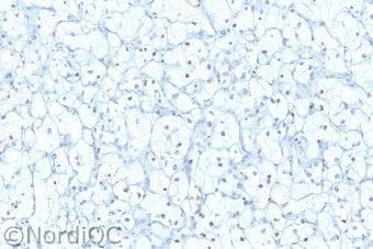
6 PAN-CK Inappropriate retrieval AE1/AE3 + HIER TP Liver RCC FN AE1/AE3 + proteolysis TP 6 FN

7 IHC - NordiQC 2014 AE1/AE3 : Optimal results only obtained by HIER in NordiQC runs Dako: RTU HIER Leica: RTU Proteolysis Thermo: VMS: RTU Proteolysis Misleading data sheets + Wrong control material used Conc: Proteolysis or HIER Conc: HIER Conc: HIER Quanto Proteolysis UltraVision Proteolysis + HIER 7
 Adenocarcinomas (of most types) Neuroendocrine")
8 EpCAM (Epithelial cell adhesion molecule, EP4, MOC31, ) +(-) Adenocarcinomas (of most types) Neuroendocrine neoplasms

9 EpCAM (Epithelial cell adhesion molecule, EP4, MOC31, ) -/+ Lobular breast carcinoma Hepatocellular carcinoma Squamous cell carcinoma Renal cell carcinoma Embryonal carcinoma Epithelioid mal. mesothelioma Adrenal cortical carcinoma Choroid plexus carcinoma


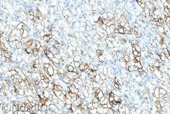
10 EpCAM protocol Colon adenocarcinoma Lab A optimal with EP4 Retrieval: TRS ph 6.1 DAKO Diva2 ph 6.2 Thermo CC1+Proteolysis VMS Clone BS14: TRIS-EDTA a.o. Lab B insufficient with EP4 Retrieval: Any other buffer without concomitant proteolysis

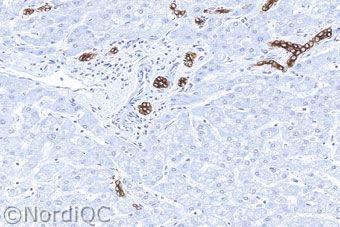
11 EpCAM antibodies renal cell carcinoma SPM491 Ber-EP4 Ab71916 with optimal protocols

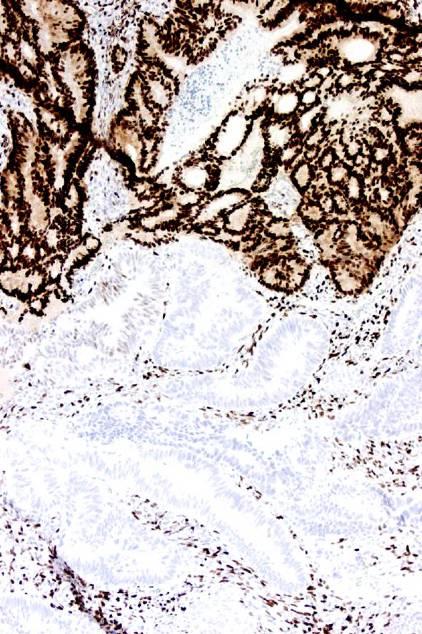
12 Claudin 4 Integral membrane protein, which belongs to the claudin family. The protein is a component of tight junction. Never found in mesothelioma Claudin 4 EpCAM Tonsil

13 Claudin 4 vs. EpCAM Mal. mesothelioma Claudin 4 Mal. mesothelioma EpCAM


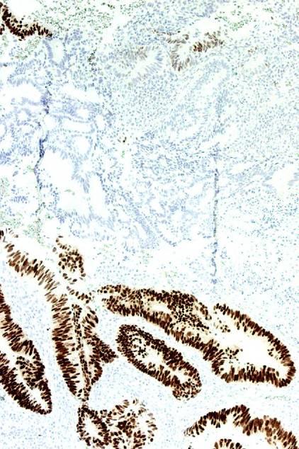
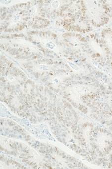
14 CDX-2 protein Drosophila caudal related homeobox gene 2 product: Nuclear transcription factor for intestinal differentiation Intestine all cell types incl. endocrine Intestinal metaplasia chronic gastritis Barrett s esophagus Pancreas/bil.tract colon pancreas

15 CDX-2 protein in adenocarcinoma Colorectum +( ) Mucinous ovar. +( ) Esoph./Stom. +/ Mucinous lung +/ Pancr./biliary /+ Prostate (+) Urothelial (+) Endometrioid (+) Yolk sac tumour +
16 ER CDX-2 Endometrioid carcinoma: ER & CDX-2
17 Quality assurance of immunohistochemical Cdx2 detection in carcinomas Results Mean H-score and proportion of positives N EPR* CONC EPR* RTU DAK- CDX2 AMT- 28 CDX2-88 High Ex % 100% 100% 98% 96% Low Ex rmab EPR2764Y (Ventana, CellMarque) 95% 48% 58% 19% 13%


18 CDX2 Normal colon 1 EPRCON EPRRTU DAK AMT 88


19 CDX2 Colon adenocarc. Optimized protocols EPRCON EPRRTU DAK AMT 88
")
 Pancreas/biliary tract")
20 Cadherin 17 Calcium dependent adhesion molecules CAD17 = Liver-Intestine (LI-) Cadherin Regulated by CDX2 Intestine (uniform) Pancreas/biliary tract (heterogenous)
21 Cadherin 17 + Adenocarcinoma, colon (incl. medullary MMR prot. defic.) Endocrine neoplasm of small intestine +/- or -/+ Adenocarcinomas of esophagus, stomach, pancreas / biliary tract -(+) Adenocarcinomas of lung, endometrium, ovary, breast Endocrine neoplasms of lung and pancreas Squamous cell carcinoma


22 CAD17 and CDX2 Colon adenocarcinoma Cadherin 17: rmab SP183, CM, 1:50, CC1M/16M/UV CDX2: rmab EPR2764Y, CM


23 CAD17 and CDX2 Colon adenocarcinoma Cadherin 17: rmab SP183, CM, 1:50, CC1M/16M/UV CDX2: rmab EPR2764Y, CM
 Neuroendocrine neoplasms usually")
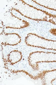
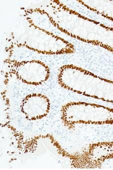
24 SATB2 rmab SP281 CM Special Adenine Thymine-rich sequence-binding protein 2 Nuclear matrix-associated transcription factor of intestine, neurons and osteoblasts Colorectal adenocarcinomas positive in 90% Renal cell carcinoma positive in 30% Other carcinomas usually negative (10-20% focal) Neuroendocrine neoplasms usually negative


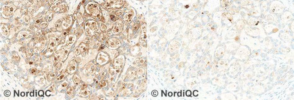
25 Colon adenocarcinoma rmab SP281, CM, 1:200 CC1_32M/16M/OP

26 Carcinoembryonic antigen (CD66e) Adhesion molecule espc. associated with intestine

27 Carcinoembryonic antigen (CD66e) in adenocarcinomas Colorectal + Medull. thyroid + Pancreas/biliary tract +/ Stomach +/ Lung +/ Ovary, mucinous +/ Ovary, non-muc. /+ Prostate Kidney Liver (!) Mesothelioma (!) metast. colon adenoc

28 Carcinoembryonic antigen which antibody?

29 Carcinoembryonic antigen which antibody? Normal liver Clone II-7 Clone TF3H8-1


30 Carcinoembryonic antigen which antibody? Mal. mesothelioma mab II-7 pab or TF3H8-1
31 MLH1 3-step polymer 2-step polymer Improper calibration of Ab titre Less succesful Ab clone G (8/9 insuff.) Less sensitive detection systems:


32 3-step polymer 2-step polymer MLH1 MLH1 loss False neg. internal control

33 MSH2 Improper calibration of Ab titre Poor Ab clone 25D12 (17/17 insuff.) Insufficient HIER 25D12 combined false negative and positive


34 Poor RTU formats: Chromogranin A Well diff endocrine carcinoma LK2H10 with optimized protocvol pab RTU Company 1 mab LK2H10 RTU Company 2 mab LK2H10 RTU Company 3


35 Poor RTU formats: Chromogranin A Small cell carcinoma LK2H10 REF pab RTU Company 1 mab LK2H10 RTU Company 2 mab LK2H10 RTU Company 3


36 Liver tumour of unknown origin hepatocellular carcinoma? Atrophic pancreas
 Ovarian serous carcinoma (+) Liposarcoma +/- HCC")
37 Glypican 3 Hepatocellular carcinoma +/- Yolk sac tumour + Chorionic carcinoma + Merkel cell carcinoma +/- Colorectal adenocarcinoma -/+ Gastric adenocarcinoma -/+ Ovarian clear cell carcinoma -/+ Emb. carcinoma (+) Ovarian serous carcinoma (+) Liposarcoma +/- HCC

38 Glypican 3
39 Poor calibration of conc.: Glypican 3
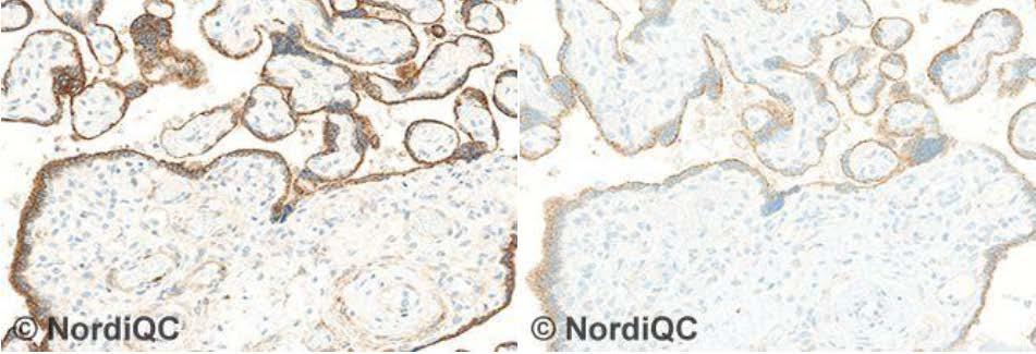
 Bile canaliculi Many epithelia")
40 CD66a (biliary glycoprotein-1) CEA-like cell adhesion molecule ( pcea ) Bile canaliculi Many epithelia Trophoblast
")

41 cancd66a (biliary glycoprotein-1) Hepatocelullar carcinoma ~70% canalicular staining HCC HCC 41


42 cancd10 ~70% pos. HCC HCC 42

43 Nordic Immunohistochemical Quality Control Immunohistochemistry in the classifiation of neoplasias of the alimentary tract & External Quality Assurance of Immunohistochemistry for GI cancer markers Mogens Vyberg, director, NordiQC
The unkown primary tumour: IHC Classification, antibody selection, protocol optimization, controls and EQA (part II)
") The unkown primary tumour: IHC Classification, antibody selection, protocol optimization, Mogens Vyberg Professor of Clinical Pathology Director of NordiQC Aalborg University Hospital, Aalborg, Denmark
The unkown primary tumour: IHC Classification, antibody selection, protocol optimization, Mogens Vyberg Professor of Clinical Pathology Director of NordiQC Aalborg University Hospital, Aalborg, Denmark
Diagnostic IHC in lung and pleura pathology
 Diagnostic IHC in lung and pleura pathology Mogens Vyberg Professor of Clinical Pathology Director of NordiQC Aalborg University Hospital, Aalborg, Denmark WHO 2004 and Web Malignant mesothelioma Epithelioid
The impact of proficiency testing on lab immunoassays
 The impact of proficiency testing on lab immunoassays Mogens Vyberg Professor of Clinical Pathology Director of NordiQC Aalborg University Hospital, Aalborg, Denmark Nordic Immunohistochemical Quality
Breast cancer: IHC classification. Mogens Vyberg Professor of Clinical Pathology Director of NordiQC Aalborg University Hospital, Aalborg, Denmark
 Breast cancer: IHC classification Mogens Vyberg Professor of Clinical Pathology Director of NordiQC Aalborg University Hospital, Aalborg, Denmark http://upload.wikimedia.org/wikipedia/commons/1/1a/breast.svg
The unkown primary tumour: IHC Classification, antibody selection, protocol optimization, controls and EQA (part I)
") The unkown primary tumour: IHC Classification, antibody selection, protocol optimization, Mogens Vyberg Professor of Clinical Pathology Director of NordiQC Aalborg University Hospital, Aalborg, Denmark
The unkown primary tumour: IHC Classification, antibody selection, protocol optimization, Mogens Vyberg Professor of Clinical Pathology Director of NordiQC Aalborg University Hospital, Aalborg, Denmark
The unknown primary tumour: IHC classification part I, the primary panel - Antibody selection, protocol optimization, controls and EQA
 The unknown primary tumour: IHC classification part I, Mogens Vyberg Professor of Clinical Pathology Director of NordiQC Aalborg University Hospital, Aalborg, Denmark the primary panel - Antibody selection,
Quality Assurance in Immunohistochemistry: Experiences from NordiQC
 Nordic immunohistochemical Quality Control 2 Quality Assurance in Immunohistochemistry: Experiences from NordiQC Prof. Mogens Vyberg NordiQC Institute of Pathology Aalborg University Hospital Aalborg,
Carcinoembryonic antigen (CEA)
") Assessment Run 37 2013 Carcinoembryonic antigen (CEA) Material The slide to be stained for CEA comprised: 1. Appendix, 2. Liver, 3-4. Colon adenocarcinoma, 5. Urothelial carcinoma All tissues were fixed
Assessment Run 37 2013 Carcinoembryonic antigen (CEA) Material The slide to be stained for CEA comprised: 1. Appendix, 2. Liver, 3-4. Colon adenocarcinoma, 5. Urothelial carcinoma All tissues were fixed
10 years of NordiQC Why are 30% of labs still getting it wrong?
 Mogens Vyberg & Søren Nielsen NordiQC Institute of Pathology Aalborg University Hospital Aalborg, Denmark May 29th 2015 10 years of NordiQC Why are 30% of labs still getting it wrong? Nothing to declare
Immunohistochemical classification of the unknown primary tumour (UPT) Part I. Prof. Mogens Vyberg NordiQC Institute of Pathology Aalborg, Denmark
 Part I. Prof. Mogens Vyberg NordiQC Institute of Pathology Aalborg, Denmark") Immunohistochemical classification of the unknown primary tumour (UPT) Part I Prof. Mogens Vyberg NordiQC Institute of Pathology Aalborg, Denmark Tumours of unknown origin: Histology Brain tumour - biopsy
Immunohistochemical classification of the unknown primary tumour (UPT) Part I Prof. Mogens Vyberg NordiQC Institute of Pathology Aalborg, Denmark Tumours of unknown origin: Histology Brain tumour - biopsy
Cytokeratin 19 (CK19)
") Assessment Run 34 202 Cytokeratin 9 (CK9) Material The slide to be stained for CK9 comprised:. Thyroid gland, 2. Appendix, 3. Esophagus, 4. Papillary thyroid carcinoma, 5 & 6. Pancreatic neuroendocrine
Assessment Run 34 202 Cytokeratin 9 (CK9) Material The slide to be stained for CK9 comprised:. Thyroid gland, 2. Appendix, 3. Esophagus, 4. Papillary thyroid carcinoma, 5 & 6. Pancreatic neuroendocrine
Assessment Run GATA3
 Assessment Run 44 2015 GATA3 Material The slide to be stained for GATA3 comprised: 1. Tonsil 2. Kidney, 3. Urothelial carcinoma, 4. Breast ductal carcinoma, 5. Colon adenocarcinoma All tissues were fixed
Classification of the unknown primary tumour: the primary IHC panel
 CIQC/CAP-ACP SEMINAR 2013: DIAGNOSTIC IHC AND MOLECULAR PATHOLOGY Classification of the unknown primary tumour: the primary IHC panel Aalborg University Hospital Denmark Tumours of unknown origin: Histology
Differential diagnosis of HCC
 Hepatocellular Carcinoma Quest for an Ideal Immunohistochemical Panel Sanjay Kakar, MD UCSF Differential diagnosis of HCC Hepatocellular lesions Adenoma, FNH, HG dysplasia Adenocarcinoma CholangioCA, metastasis
Optimization of antibodies, selection, protocols and controls Breast tumours
 Optimization of antibodies, selection, protocols and controls Breast tumours Søren Nielsen Project coordinator & Scheme Manager NordiQC Aalborg University Hospital, Denmark Breast panel: GCDFP-15 Mammaglobin
Epithelial cell-cell adhesion molecule (Ep-CAM)
") Assessment Run 3 011 Epithelial cell-cell adhesion molecule (Ep-CAM) Material The slide to be stained for Ep-CAM comprised: 1. Appendix,. Kidney, 3. Adrenal gland, 4. Lung carcinoid, 5 & 6. Renal clear
Assessment Run 3 011 Epithelial cell-cell adhesion molecule (Ep-CAM) Material The slide to be stained for Ep-CAM comprised: 1. Appendix,. Kidney, 3. Adrenal gland, 4. Lung carcinoid, 5 & 6. Renal clear
Immunohistochemical classification of lung carcinomas and mesotheliomas. Prof. Mogens Vyberg NordiQC Institute of Pathology Aalborg, Denmark
 Immunohistochemical classification of lung carcinomas and mesotheliomas Prof. Mogens Vyberg NordiQC Institute of Pathology Aalborg, Denmark Endobronchial ultrasound guided transbronchial needle biopsy
Breast cancer: Antibody selection, protocol optimzation controls and EQA
 Breast cancer: Antibody selection, protocol optimzation controls and EQA Workshop in Diagnostic Immunohistochemistry Oud St. Jan/ Old St. John Brugge (Bruges), Belgium June 13th 15nd 2018 Rasmus Røge,
The Panel Approach to Diagnostics. Lauren Hopson International Product Specialist Cell Marque Corporation
 The Panel Approach to Diagnostics Lauren Hopson International Product Specialist Cell Marque Corporation Cell Marque Rocklin, California About Cell Marque: IVD primary antibody manufacturer Distributors
SMH (Myosin, smooth muscle heavy chain)
") Material The slide to be stained for SMH comprised: Assessment Run 50 2017 SMH (Myosin, smooth muscle heavy chain) 1.Tonsil, 2. Esophagus, 3. Breast hyperplasia, 4. Breast ductal carcinoma in situ (DCIS),
Material The slide to be stained for SMH comprised: Assessment Run 50 2017 SMH (Myosin, smooth muscle heavy chain) 1.Tonsil, 2. Esophagus, 3. Breast hyperplasia, 4. Breast ductal carcinoma in situ (DCIS),
Estrogen receptor (ER)
") Assessment Run B7 204 Estrogen receptor (ER) Material The slide to be stained for ER comprised: No. Tissue ER-positivity* ER-intensity*. Uterine cervix 80-90% Moderate to strong 2. Breast carcinoma 0%
Assessment Run B7 204 Estrogen receptor (ER) Material The slide to be stained for ER comprised: No. Tissue ER-positivity* ER-intensity*. Uterine cervix 80-90% Moderate to strong 2. Breast carcinoma 0%
Assessment Run B HER-2 IHC. HER-2/chr17 ratio**
 Assessment Run B2 20 HER-2 IHC Material The slide to be stained for HER-2 comprised the following 5 tissues: IHC HER-2 Score* (0, +, 2+,3+) FISH HER-2/chr7 ratio**. Breast ductal carcinoma 0..3 2. Breast
Assessment Run NKX3.1 (NKX3.1)
") Assessment Run 49 2017 NKX3.1 (NKX3.1) Material The slide to be stained for NKX3.1 comprised: 1. Testis 2. Appendix 3-4. Prostate adenocarcinoma 5. Prostate hyperplasia All tissues were fixed in 10% neutral
Assessment Run 49 2017 NKX3.1 (NKX3.1) Material The slide to be stained for NKX3.1 comprised: 1. Testis 2. Appendix 3-4. Prostate adenocarcinoma 5. Prostate hyperplasia All tissues were fixed in 10% neutral
Thyroid transcription factor-1 (TTF1) Assessment run
 Assessment run") Thyroid transcription factor- (TTF) Assessment run 39 203 The slide to be stained for TTF comprised:. Thyroid gland, 2. Liver, 3. Normal lung, 4. Lung adenocarcinoma 5. Colon adenocarcinoma, 6 & 7. Lung
Thyroid transcription factor- (TTF) Assessment run 39 203 The slide to be stained for TTF comprised:. Thyroid gland, 2. Liver, 3. Normal lung, 4. Lung adenocarcinoma 5. Colon adenocarcinoma, 6 & 7. Lung
CARCINOMA OF UNKNOWN PRIMARY: DIAGNOSTIC APPROACH USING IMMUNOHISTOCHEMISTRY
 CARCINOMA OF UNKNOWN PRIMARY: DIAGNOSTIC APPROACH USING IMMUNOHISTOCHEMISTRY Jason L Hornick, MD, PhD Director of Surgical Pathology Director of Immunohistochemistry Brigham and Women s Hospital Associate
Assessment Run CK19
 Assessment Run 29 200 CK9 The slide to be stained for CK9 comprised:. Appendix, 2. Thyroid gland, 3. Pancreas, 4. Ductal breast carcinoma, 5. Esophagus, 6. Papillary thyroid carcinoma. All tissues were
Estrogen receptor (ER)
") Material The slide to be stained for ER comprised: Assessment Run B26 2018 Estrogen receptor (ER) No. Tissue ER-positivity* ER-intensity* 1. Uterine cervix 80-90% Moderate to strong 2. Tonsil 1-5% Weak
Material The slide to be stained for ER comprised: Assessment Run B26 2018 Estrogen receptor (ER) No. Tissue ER-positivity* ER-intensity* 1. Uterine cervix 80-90% Moderate to strong 2. Tonsil 1-5% Weak
Assessment Run
 Assessment Run 50 2017 S100 Material The slide to be stained for S100 comprised: 1. Appendix, 2. Tonsil, 3. Schwannoma, 4-5. Malignant melanoma, 6. Colon adenocarcinoma. All tissues were fixed in 10% neutral
NordiQC - update
 NordiQC - update 00-0 EQUALIS Uppsala 0 Tomas Seidal NordiQC participants NordiQC participants n:30 S DK N 6 F Ice Bel 54 NL 4 Ger 6 Aust USA 0 It 8 Argent 8.. 96% participation in S,DK & N ~ 60% in Finland
NordiQC External Quality Assurance in Immunohistochemistry
 NordiQC External Quality Assurance in Immunohistochemistry Mogens Vyberg Professor of Clinical Pathology Director of NordiQC Aalborg University Hospital, Aalborg, Denmark AALBORG (~ 200.000 inhabitants)
Estrogen receptor (ER)
") Material The slide to be stained for ER comprised: Assessment B25 208 Estrogen receptor (ER) No. Tissue ER-positivity* ER-intensity*. Uterine cervix 80-90% Moderate to strong 2. Tonsil < 2-5% Weak to strong
Material The slide to be stained for ER comprised: Assessment B25 208 Estrogen receptor (ER) No. Tissue ER-positivity* ER-intensity*. Uterine cervix 80-90% Moderate to strong 2. Tonsil < 2-5% Weak to strong
Lung Anaplastic Lymphoma Kinase (lu-alk)
") Assessment Run 5 207 Lung Anaplastic Lymphoma Kinase (lu-alk) Material The slide to be stained for lu-alk comprised:. Appendix, 2. Tonsil, 3. Merkel cell carcinoma, 4. Anaplastic large cell lymphoma with
Assessment Run 5 207 Lung Anaplastic Lymphoma Kinase (lu-alk) Material The slide to be stained for lu-alk comprised:. Appendix, 2. Tonsil, 3. Merkel cell carcinoma, 4. Anaplastic large cell lymphoma with
Assessment Run B HER2 IHC
 Assessment Run B24 2017 HER2 IHC Material The slide to be stained for HER2 comprised the following 5 materials: IHC: HER2 Score* (0, 1+, 2+, 3+) FISH: HER2 gene/chr 17 ratio** 1. Breast carcinoma, no.
Sal-like protein 4 (SALL4)
") Assessment Run 43 205 Sal-like protein 4 (SALL4) The slide to be stained for SALL4 comprised:. Appendix, 2. Testis, 3. Renal clear cell carcinoma, 4. Seminoma, 5. Intratubular germ cell neoplasia (IGCN),
Assessment Run 43 205 Sal-like protein 4 (SALL4) The slide to be stained for SALL4 comprised:. Appendix, 2. Testis, 3. Renal clear cell carcinoma, 4. Seminoma, 5. Intratubular germ cell neoplasia (IGCN),
Assessment Run C3 2018
 Assessment Run C3 2018 PD-L1 Amended version May 14 th 2018 The third assessment in NordiQC Companion module C3 focused on the accuracy of the PD-L1 IHC assays performed by the participating laboratories
Assessment Run C1 2017
 Assessment Run C1 2017 PD-L1 The first assessment in this new NordiQC Companion module C1 focused on the accuracy of the PD-L1 IHC assays performed by the participating laboratories to identify patients
External Quality Assessment of melanocytic marker analyses NordiQC experience
 External Quality Assessment of melanocytic marker analyses NordiQC experience Jan Klos MD, Department of Pathology Stavanger University Hospital Norway 1 Content 18 Runs = 2112 submissions between 2001-2014
Malignant neoplasms of the gastrointestinal (GI) tract,
 tract,") Special Section First Chinese American Pathologists Association Diagnostic Pathology Course, Part II Practical Immunohistochemistry in Neoplastic Pathology of the Gastrointestinal Tract, Liver, Biliary
Special Section First Chinese American Pathologists Association Diagnostic Pathology Course, Part II Practical Immunohistochemistry in Neoplastic Pathology of the Gastrointestinal Tract, Liver, Biliary
Reporting of carcinoma of unknown primary tumour (CUP)
") Reporting of carcinoma of unknown primary tumour (CUP) Prof John Schofield Kent Oncology Centre with grateful thanks to Dr Karin Oien University of Glasgow Royal College of Pathologists Cancer datasets
Reporting of carcinoma of unknown primary tumour (CUP) Prof John Schofield Kent Oncology Centre with grateful thanks to Dr Karin Oien University of Glasgow Royal College of Pathologists Cancer datasets
The role of immunohistochemistry in surgical pathology of the uterine corpus and cervix
 The role of immunohistochemistry in surgical pathology of the uterine corpus and cervix Prof. Ben Davidson, MD PhD Department of Pathology, Norwegian Radium Hospital, Oslo University Hospital, Oslo, Norway
Fast, automated, precise
 Thermo Scientific B R A H M S / NSE Immunodiagnostic Assays Fast, automated, precise Neuroendocrine tumor markers on KRYPTOR Systems First and only fully automated CgA assay worldwide Shortest time to
The clinically challenging entity of liver metastasis from tumors of unknown primary
 The clinically challenging entity of liver metastasis from tumors of unknown primary Xuchen Zhang, MD, PhD Associate Professor of Pathology Department of Pathology Yale University School of Medicine Liver
Cancers of unknown primary : Knowing the unknown. Prof. Ahmed Hossain Professor of Medicine SSMC
 Cancers of unknown primary : Knowing the unknown Prof. Ahmed Hossain Professor of Medicine SSMC Definition Cancers of unknown primary site (CUPs) Represent a heterogeneous group of metastatic tumours,
Immunohistochemistry on Fluid Specimens: Technical Considerations
 Immunohistochemistry on Fluid Specimens: Technical Considerations Blake Gilks Dept of Pathology University of British Columbia, Vancouver, BC, Canada Disclosures None Learning Objectives At the end of
Assessment Run B HER2 IHC
 Assessment Run B26 208 HER2 IHC Material The slide to be stained for HER2 comprised the following 5 materials: IHC: HER2 Score* (0, +, 2+, 3+) FISH: HER2 gene/chr 7 ratio**. Breast carcinoma, no. 2+..3
Assessment Run B HER-2
 Assessment Run B1 2006 HER-2 The slide to be stained for HER-2 comprised: 1. Cell line JIMT-1 (Amplified)* 2. Cell line MDA-453 (Amplified) 3. Cell line MCF-7 (Not amplified) 4. Cell line BT474 (Amplified)
8 years later! Next Generation Sequencing. Pathogenic Findings: HNF1A c.864delinscc, p.g292rfs*25 (NM_ ) (VAF: 59%) HNF1A Loss
 (VAF: 59%) HNF1A Loss") 8 years later! Next Generation Sequencing Pathogenic Findings: HNF1A c.864delinscc, p.g292rfs*25 (NM_000545.6) (VAF: 59%) HNF1A Loss Interpretation HNF1A c.864delinscc, p.g292rfs*25 (NM_000545.6) This
8 years later! Next Generation Sequencing Pathogenic Findings: HNF1A c.864delinscc, p.g292rfs*25 (NM_000545.6) (VAF: 59%) HNF1A Loss Interpretation HNF1A c.864delinscc, p.g292rfs*25 (NM_000545.6) This
Histopathological diagnosis of CUP
 Histopathological diagnosis of CUP Dr Karin Oien karin.oien@glasgow.ac.uk Disclosure slide Dr Karin Oien has no financial interests in any company mentioned in this presentation. Dr Karin Oien is conducting
New Developments in Immunohistochemistry for Gynecologic Pathology
 New Developments in Immunohistochemistry for Gynecologic Pathology Michael T. Deavers, M.D. Professor, Departments of Pathology and Gynecologic Oncology Immunohistochemistry in Gynecologic Pathology Majority
I. Diagnosis of the cancer type in CUP
 Latest Research: USA I. Diagnosis of the cancer type in CUP II. Outcomes of site-specific therapy of the cancer type in CUP a. Prospective clinical trial b. Retrospective clinical trials 1 Latest Research:
Expression of Cytokeratin 5/6 in Epithelial Neoplasms: An Immunohistochemical Study of 509 Cases
 Expression of Cytokeratin 5/6 in Epithelial Neoplasms: An Immunohistochemical Study of 509 Peiguo G. Chu, M.D., Ph.D., Lawrence M. Weiss, M.D. Department of Pathology, City of Hope National Medical Center,
Cutaneous metastases. Thaddeus Mully. University of California, San Francisco Professor, Departments of Pathology and Dermatology
 Cutaneous metastases Thaddeus Mully University of California, San Francisco Professor, Departments of Pathology and Dermatology DISCLOSURE OF RELATIONSHIPS WITH INDUSTRY Thaddeus Mully Course C005 Essential
Tissue-Specific Cadherin CDH17 Is a Useful Marker of Gastrointestinal Adenocarcinomas With Higher Sensitivity Than CDX2
 Anatomic Pathology / CDH17 in Gastrointestinal Carcinomas Tissue-Specific Cadherin CDH17 Is a Useful Marker of Gastrointestinal Adenocarcinomas With Higher Sensitivity Than CDX2 Nicole C. Panarelli, MD,
ISSN X (Print) Original Research Article. DOI: /sjams
 Original Research Article. DOI: /sjams") DOI: 10.21276/sjams.2016.4.7.33 Scholars Journal of Applied Medical Sciences (SJAMS) Sch. J. App. Med. Sci., 2016; 4(7C):2468-2473 Scholars Academic and Scientific Publisher (An International Publisher
DOI: 10.21276/sjams.2016.4.7.33 Scholars Journal of Applied Medical Sciences (SJAMS) Sch. J. App. Med. Sci., 2016; 4(7C):2468-2473 Scholars Academic and Scientific Publisher (An International Publisher
Tumour Markers. For these reasons, only a handful of tumour markers are commonly used by most doctors.
 Tumour Markers What are Tumour Markers? Tumour markers are substances that can be found in the body when cancer is present. They are usually found in the blood or urine. They can be products of cancer
Enterprise Interest Nothing to declare
 Enterprise Interest Nothing to declare Update of mixed tumours of the GI tract, the pancreas and the liver Introduction to the concept of mixed tumours and clinical implication Jean-Yves SCOAZEC Surgical
Immunohistochemical principles The technical test approach. Pre-analytical parametres
 Immunohistochemical principles The technical test approach Pre-analytical parametres Søren Nielsen Global Pathology Manager Agilent Technologies (Former Scheme Manager, NordiQC) 2 IHC project coordinator
External Quality Assessment of Breast Marker Analysis. NordiQC data
 External Quality Assessment of Breast Marker Analysis NordiQC data Søren Nielsen Scheme Manager NordiQC Aalborg University Hospital, Denmark Aalborg 12.06 2015 Markers assessed in NordiQC Predictive markers
Carcinoma of unknown primary origin (CUP) is defined
 is defined") REVIEW ARTICLE Metastatic Carcinoma of Unknown Primary: Diagnostic Approach Using Immunohistochemistry James R. Conner, MD, PhD and Jason L. Hornick, MD, PhD Abstract: Carcinoma of unknown primary origin
REVIEW ARTICLE Metastatic Carcinoma of Unknown Primary: Diagnostic Approach Using Immunohistochemistry James R. Conner, MD, PhD and Jason L. Hornick, MD, PhD Abstract: Carcinoma of unknown primary origin
Tissue-based Immunohistochemical Biomarker Expression in Malignant Glandular Lesions of the Uterine Cervix: a Systematic Review
 Tissue-based Immunohistochemical Biomarker Expression in Malignant Glandular Lesions of the Uterine Cervix: a Systematic Review Sandra Lee MD, FRCPC 1 *, Vikrant V. Sahasrabuddhe, MBBS, DrPH 2 *, Diana
Immunohistochemistry. Potential and challenges To be or not to be
 Immunohistochemistry Potential and challenges To be or not to be Søren Nielsen Scheme Manager NordiQC Aalborg University Hospital, Denmark Vårmöte 19.05.2016 Karlstad Overview IHC project coordinator at
Liver Specialty Evening Conference. Matthew M. Yeh, MD, PhD Professor of Pathology Adjunct Professor of Medicine University of Washington, Seattle
 Liver Specialty Evening Conference Matthew M. Yeh, MD, PhD Professor of Pathology Adjunct Professor of Medicine University of Washington, Seattle Case History A 65 year-old man presents with abdominal
Thermo Scientific UltraVision Quanto for Immunohistochemistry The New Generation Micro-Polymer Detection System
 Thermo Scientific for Immunohistochemistry The New Generation Micro-Polymer Detection System highest sensitivity sharp crisp clear shorter incubation times UltraVision Quanto the new Micro-Polymer System
How to Recognize Gynecologic Cancer Cells from Pelvic Washing and Ascetic Specimens
 How to Recognize Gynecologic Cancer Cells from Pelvic Washing and Ascetic Specimens Wenxin Zheng, M.D. Professor of Pathology and Gynecology University of Arizona zhengw@email.arizona.edu http://www.zheng.gynpath.medicine.arizona.edu/index.html
Speaking to you. This statement by Dr. Rodney T. Miller, Director of
 Zyto_Facts 1-2013 News for pathology and immunohistochemistry +++Newsflash +++ Newsflash++ Speaking to you IHC algorithm poster now available in English. You can download the poster directly from our homepage
Carcinoma of Unknown Primary (CUP)
") Metasta c Carcinoma of Unknown Primary: Diagnos c Approach Using Immunohistochemistry James R. Conner, MD, PhD Mount Sinai Hospital Toronto, ON Carcinoma of Unknown Primary (CUP) 3-5% of all new malignant
Metasta c Carcinoma of Unknown Primary: Diagnos c Approach Using Immunohistochemistry James R. Conner, MD, PhD Mount Sinai Hospital Toronto, ON Carcinoma of Unknown Primary (CUP) 3-5% of all new malignant
Protocols for Zytomed Systems antibodies on fully automated IHC staining systems date of issue: September 20, 2012
 Protocols for Zytomed Systems antibodies on fully automated IHC staining systems date of issue: September 20, 2012 These protocols were provided by customers. Under no circumstances shall Zytomed Systems
IHC And Special Stains In Daily Practice. Dr Ian Brown Envoi Pathology Brisbane, Australia
 IHC And Special Stains In Daily Practice Dr Ian Brown Envoi Pathology Brisbane, Australia Why do special stains? Tumour Classify disease Prognostication Predict response to therapy Identify an inherited
List of Available TMAs in the PRN
 TMA RPCI_BrainCa01 RPCI_BrCa03 RPCI_BrCa04 RPCI_BrCa05 RPCI_BrCa0 RPCI_BrCa07 RPCI_BrCa08 RPCI_BrCa15 RPCI_BrCa1 RPCI_BrCa17 RPCI_BrCa18 RPCI_BrCa19 RPCI_BrCa20 RPCI_BrCa21 RPCI_BrCa24 RPCI_BrCa25 RPCI_BrCa2
Supplementary Online Content
 Supplementary Online Content Chacón MR, Enrico DH, Burton J, Waisberg FD, Videla VM. Incidence of placebo adverse events in randomized clinical trials of targeted and immunotherapy cancer drugs in the
Schedule of Accreditation issued by United Kingdom Accreditation Service 2 Pine Trees, Chertsey Lane, Staines-upon-Thames, TW18 3HR, UK
 Schedule of ccreditation United Kingdom ccreditation Service 2 Pine Trees, Chertsey Lane, Staines-upon-Thames, TW18 3HR, UK External Quality ssessment Services for Cancer Diagnostics CIC Issue No: 005
Immunohistochemical Expression of Cytokeratin 5/6 in Gynaecological Tumors.
 ISPUB.COM The Internet Journal of Pathology Volume 13 Number 2 Immunohistochemical Expression of Cytokeratin 5/6 in Gynaecological Tumors. A Baghla, S Choudhry, A Kataria Citation A Baghla, S Choudhry,
ACCURACY OF IMMUNOHISTOCHEMISTRY IN EVALUATION
 POL J PATHOL 2011; 2: 95-100 ACCURACY OF IMMUNOHISTOCHEMISTRY IN EVALUATION OF MALIGNANT PLEURAL AND PERITONEAL EFFUSIONS FERESHTEH ENSANI, FARNAZ NEMATIZADEH, GITI IRVANLOU Department of Cytology, Cancer
Zyto-Facts Editorial. News for Pathology and Immunohistochemistry. +++ Newsflash +++ Newsflash +++
 Zyto-Facts 1-2016 News for Pathology and Immunohistochemistry Editorial The primary site of a tumor is not known in about 3 % to 5 % of all cancer diagnoses. Patients, whose primary site could be identified,
Technology from Abcam
 CD2 (EP222) CD2 is one of the earliest T-cell lineage restricted antigens to appear during T-cell differentiation and only rare CD2+ cells can be found in the bone marrow. Anti-CD2 is a pan-t-cell antigen
Single and Multiplex Immunohistochemistry
 Single and Multiplex Immunohistochemistry Steve Westra, BS Reagent Product Specialist Leica Biosystems IHC Theory Polyclonal vs Monoclonal Polyclonal reagents Detect a multitude of epitopes Batch to batch
Outline. Hepatocellular Carcinoma Histologic variants. HCC: Histologic variants
 2018 Park City AP Update Hepatocellular Carcinoma Histologic variants Sanjay Kakar, MD University of California, San Francisco Outline Histologic variants of HCC Morphologic and Immunohistochemical pitfalls
NEW IHC A n t i b o d i e s
 NEW IHC Antibodies TABLE OF CONTENTS NEW IHC ANTIBODIES from Cell Marque CITED1 (5H6).... 1 Claudin 7 (5D10F3).... 1 GATA1 (4F5).... 1 Transgelin (2A10C2).... 1 NEW IHC ANTIBODIES using RabMAb Technology
Charles Halsey, DVM, PhD, DACVP Pfizer, Inc. IHC Resources
 Charles Halsey, DVM, PhD, DACVP Pfizer, Inc. IHC Resources 1 IHC Identification Targets Specimens Controls 2 Tissue controls Trouble Spots 3 The Key to Description IHC Description 4 Intermediate Filaments
Breast - ductal carcinoma CK7 ER PR GATA3 Mammaglobin (50-70%) GCDFP-15 (50-70%) E-cadherin HMWCK CK20 PAX2 ER/PR/HER2 on all newly diagnosed cases
 GCDFP-15 (50-70%) E-cadherin HMWCK CK20 PAX2 ER/PR/HER2 on all newly diagnosed cases") Adrenal cortical carcinoma Inhibin Synap Melan-A Calretinin Vimentin Chromogr CK7 CK20 Breast - ductal carcinoma CK7 ER PR GATA3 Mammaglobin (50-70%) GCDFP-15 (50-70%) E-cadherin HMWCK CK20 PAX2 ER/PR/HER2
Adrenal cortical carcinoma Inhibin Synap Melan-A Calretinin Vimentin Chromogr CK7 CK20 Breast - ductal carcinoma CK7 ER PR GATA3 Mammaglobin (50-70%) GCDFP-15 (50-70%) E-cadherin HMWCK CK20 PAX2 ER/PR/HER2
NEUROENDOCRINE DIFFERENTIATION IN EPITHELIAL TUMORS Marco Volante
 NEUROENDOCRINE DIFFERENTIATION IN EPITHELIAL TUMORS Marco Volante University of Turin, San Luigi Hospital, Orbassano, Turin, Italy marco.volante@unito.it pure NE tum..a grey zone pure non-ne ca. 0% NE
Video Microscopy Tutorial 8
 Video Microscopy Tutorial 8 Common and Uncommon Lesions of the Liver Gladwyn Leiman, MD There are no disclosures necessary. Common and Uncommon Lesions in Liver FNA Gladwyn Leiman University of Vermont
Insulinoma-associated protein (INSM1) is a sensitive and specific marker for lung neuroendocrine tumors in cytologic and surgical specimens
 is a sensitive and specific marker for lung neuroendocrine tumors in cytologic and surgical specimens") Insulinoma-associated protein (INSM1) is a sensitive and specific marker for lung neuroendocrine tumors in cytologic and surgical specimens Kartik Viswanathan, M.D., Ph.D New York Presbyterian - Weill
Insulinoma-associated protein (INSM1) is a sensitive and specific marker for lung neuroendocrine tumors in cytologic and surgical specimens Kartik Viswanathan, M.D., Ph.D New York Presbyterian - Weill
Mesothelioma: diagnostic challenges from a pathological perspective. Naseema Vorajee August 2016
 Mesothelioma: diagnostic challenges from a pathological perspective Naseema Vorajee August 2016 Naseema.vorajee@nhls.ac.za Pleural diseases (whether neoplastic, reactive or infective) may have similar
Neuroendocrine Carcinoma. Lebanon Neuroendocrine Neoplasms of H&N Nov /7/2011. Broad Classification:
 H&N Neuroendocrine Neoplasms: Classification and Diagnostic Considerations Adel K. El-Naggar, M.D., Ph.D. The University of Texas MD Anderson Cancer Center, Houston, Texas Broad Classification: A. Epithelial:
CEA (CARCINOEMBRYONIC ANTIGEN)
") (CARCINOEMBRYONIC ANTIGEN) 428 C15.3 Malignant neoplasm of upper third of esophagus C15.4 Malignant neoplasm of middle third of esophagus C15.5 Malignant neoplasm of lower third of esophagus C15.8 Malignant
(CARCINOEMBRYONIC ANTIGEN) 428 C15.3 Malignant neoplasm of upper third of esophagus C15.4 Malignant neoplasm of middle third of esophagus C15.5 Malignant neoplasm of lower third of esophagus C15.8 Malignant
MT09 - Normal Human Tissue Microarray, FDA
 Reveal Biosciences offers Histochemical Staining, Immunohistochemistry (IHC), In Situ Hybridization (ISH), Whole Slide Imaging, and Quantitative Image Analysis on any TMA MT09 - Normal Human Tissue Microarray,
Cytokeratin 7 and Cytokeratin 20 Expression in Epithelial Neoplasms: A Survey of 435 Cases
 Cytokeratin 7 and Cytokeratin 20 Expression in Epithelial Neoplasms: A Survey of 435 Cases Peiguo Chu, M.D., Ph.D., Emerald Wu, B.S., Lawrence M Weiss, M.D. Division of Pathology, City of Hope National
Carcinoembryonic Antigen
 Other Names/Abbreviations CEA 190.26 - Carcinoembryonic Antigen Carcinoembryonic antigen (CEA) is a protein polysaccharide found in some carcinomas. It is effective as a biochemical marker for monitoring
Case Presentation Diana Lim, MBBS, FRCPA, FRCPath Senior Consultant Department of Pathology, National University Health System, Singapore Assistant Pr
 Case Presentation Diana Lim, MBBS, FRCPA, FRCPath Senior Consultant Department of Pathology, National University Health System, Singapore Assistant Professor Yong Loo Lin School of Medicine, National University
CASE 4 21/07/2017. Ectopic Prostatic Tissue in Cervix. Female 31. LLETZ for borderline nuclear abnormalities
 Female 31 CASE 4 LLETZ for borderline nuclear abnormalities PSA Ectopic Prostatic Tissue in Cervix AJSP 2006;30;209-215 usually incidental microscopic finding usually in ectocervical stroma? developmental
Immunohistochemistry and Gastrointestinal Carcinomas
 Cell Marque Tissue Diagnostics Immunohistochemistry and Gastrointestinal Carcinomas Mike Lacey, M.D. Gastrointestinal (GI) Pathology The life science business of Merck KGaA, Darmstadt, Germany operates
Hereditary Non Polyposis Colorectal Cancer(HNPCC) From clinic to genetics
 From clinic to genetics") From clinic to genetics Question 1) Clinical pattern of inheritance of the HNPCC-Syndrome? Question 1) Clinical pattern of inheritance of the HNPCC-Syndrome? Autosomal dominant Question 2) Incidence of
From clinic to genetics Question 1) Clinical pattern of inheritance of the HNPCC-Syndrome? Question 1) Clinical pattern of inheritance of the HNPCC-Syndrome? Autosomal dominant Question 2) Incidence of
WT1, Estrogen Receptor, and Progesterone Receptor as Markers for Breast or Ovarian Primary Sites in Metastatic Adenocarcinoma to Body Fluids
 Anatomic Pathology / WT1, ESTROGEN RECEPTOR, AND PROGESTERONE RECEPTOR IN CYTOLOGY OF BODY FLUIDS WT1, Estrogen Receptor, and Progesterone Receptor as Markers for Breast or Ovarian Primary Sites in Metastatic
Serous effusion Objectives. Cytology of Serous Effusions From basics to challenges
 Cytology of Serous Effusions From basics to challenges Cytology of Serous Effusions From basics to challenges Pınar Fırat, MD, MIAC Department of Pathology, İstanbul University, İstanbul Faculty of Medicine,
Effusion Cytology: Diagnostic Challenges
 Effusion Cytology: Diagnostic Challenges Tarik M. Elsheikh, MD Professor and Medical Director, Anatomic Pathology Cleveland Clinic Outside Consult Case 45 year old woman, presented with nausea, dyspnea,
4/12/2018. MUSC Pathology Symposium Kiawah Island April 18, Jesse K. McKenney, MD
 MUSC Pathology Symposium Kiawah Island April 18, 2018 Jesse K. McKenney, MD 1 Urothelial Carcinoma with Alternative Differentiation 2 Urothelial Carcinoma with Alternative Differentiation Recognition as
HER2 ISH (BRISH or FISH)
") Assessment Run H14 2018 HER2 ISH (BRISH or FISH) Material Table 1. Content of the multi-block used for the NordiQC HER2 ISH assessment, run H14 HER2 IHC* IHC score Dual - SISH** FISH*** FISH*** HER2/chr17
Assessment Run H14 2018 HER2 ISH (BRISH or FISH) Material Table 1. Content of the multi-block used for the NordiQC HER2 ISH assessment, run H14 HER2 IHC* IHC score Dual - SISH** FISH*** FISH*** HER2/chr17
Neoplasms of the Canine, Feline and Lemur Liver:
 Neoplasms of the Canine, Feline and Lemur Liver: Classification and Prognosis Annual Seminar of the French Society of Veterinary Pathology John M. Cullen VMD PhD DACVP North Carolina State University Primary