The unkown primary tumour: IHC Classification, antibody selection, protocol optimization, controls and EQA (part II)
|
|
|
- Annabel Eaton
- 5 years ago
- Views:
Transcription

1 The unkown primary tumour: IHC Classification, antibody selection, protocol optimization, Mogens Vyberg Professor of Clinical Pathology Director of NordiQC Aalborg University Hospital, Aalborg, Denmark controls and EQA (part II)
2 Secondary panels for carcinoma identification/subclassification Cell adhesion molecules EpCAM Claudin 4 Cytokeratin subtypes Oncofetal proteins Transcription factors Neuroendocrine proteins Hormone receptors Secretory proteins... GI-markers Fem.gen.tract markers Liver cell markers Neuroendocrine cell markers Breast markers Lung markers Urinary tract markers Prostate markers Squamous cell markers Mesothelial markers Adrenal cortical markers Germinal cell markers
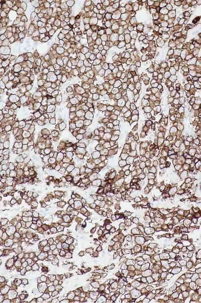
3 General epithelial markers: Epithelial cell adhesion molecule (Ep-CAM) Claudin 4
 hepatocytes (pos. in reactive) renal proximal tubular cells gastric parietal cells some endocrine cells Also negative mesothelial cells (pos.")
4 Ep-CAM Epithelial specific antigen : Glycoproteins located on the cell membrane surface (preferentially basolaterally) and in the cytoplasm of virtually all epithelial cells with the exception of squamous epithelia (mucosae and reactive focal pos.) hepatocytes (pos. in reactive) renal proximal tubular cells gastric parietal cells some endocrine cells Also negative mesothelial cells (pos. in reactive) mesenchymal cells germ cells neural crest cells (except. olfactory neurons) Liver
 Synovial sarcoma Brenner tumour Desmoplastic small cell tumour Olfactory neuroblastoma SYP EpCAM Olfactory")
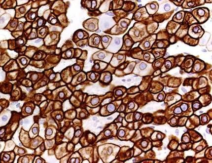
5 Ep-CAM Often upgraded in malignancies +(-) Adenocarcinomas (of most types) Neuroendocrine neoplasms +/- Germinal cell tumours (seminoma weak) Synovial sarcoma Brenner tumour Desmoplastic small cell tumour Olfactory neuroblastoma SYP EpCAM Olfactory neuroblastoma
")
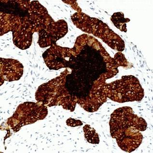
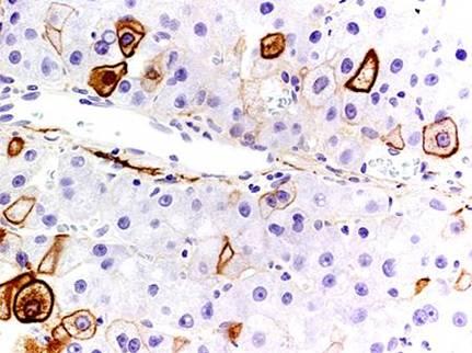
6 Ep-CAM -/+ Lobular breast carcinoma Hepatocellular carcinoma Squamous cell carcinoma Renal cell carcinoma Malignant mesothelioma - Adrenal cortical carcinoma Choroid plexus carcinoma Sarcomas (apart from synovial) Lymphomas HCC

7 Ep-CAM Undiff carcinoma

8 Ep-CAM run DAKO TRS Low 3-step polymer Ventana CC1 2-step multimer


9 Diva2 ph6.2 Thermo RCC DAKO TRS Low 3-step polymer Ventana CC1 2-step multimer

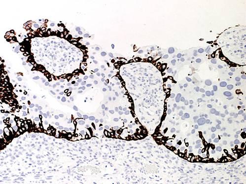
10 Claudin 4 Integral membrane protein, which belongs to the claudin family. The protein is a component of tight junction strands and may play a role in internal organ development and function. Claudin 4 Ep-CAM Tonsil

11 Claudin 4 vs. Ep-CAM Mesothelioma Claudin 4 Ep-CAM
12 Cytokeratins a multigene family Highly complex family of intermediate filaments > 50 distinct types (excl. trichocytic keratins) Types CK1-20 diagnostically most relevant: Class I (type A - Acidic): CK9 (64 kda) - CK20 (40 kda) Class II (type B - Basic/neutral): CK1 (68 kda) - CK8 (52.5 kda)

13 Pairing of cytokeratins Modified from: Lane & Alexander Semin Cancer Biol 1990, 1:165 One CK class I and one CK class II always paired CK class I in a pair ~ 8 kda smaller than class II
14 Cytokeratin types and cell types Neutral/Basic (B, class II) Acidic (A, class I) Squamous epithelia: - suprabasal, keratinizing (+) suprabasal, non-keratinizing (+) basal cells ( 1 tonsil, 2 mucosa) (+) 1 (++) 12 (+) 1 - (+) 1 Transit. epith.: superficial cells intermed. / 3 basal cells - (+) (+++) 3 - (++) (+) +++ Mesothelium Bronchus, breast, prost., cerv.: - basal/myoepithelial cells luminal cells Biliary/pancr. ducts, lung alv., endometr., renal collect. ducts Stomach (foveola), intestine (+) Hepatocytes, pancr. acini, prox. renal tubules Endocrine cells ( 5 Merkel, 6 thyr.) (+++) 5 (++) 6 (+++) Smooth muscle (vasc., myom.), myofibrobl., 7 sm.ves.endothelia (++)
15 HMW Cytokeratin types - squamous epithelia Neutral/Basic (B, class II) Acidic (A, class I) Squamous epithelia: - suprabasal, keratinizing (+) suprabasal, non-keratinizing (+) basal cells ( 1 tonsil, 2 mucosa) (+) 1 (++) 12 (+) 1 - (+) 1 Transit. epith.: superficial cells intermed. / 3 basal cells - (+) (+++) 3 - (++) (+) +++ Mesothelium Bronchus, breast, prost., cerv.: - basal/myoepithelial cells luminal cells Biliary/pancr. ducts, lung alv., endometr., renal collect. ducts Stomach (foveola), intestine (+) Hepatocytes, pancr. acini, prox. renal tubules Endocrine cells ( 5 Merkel, 6 thyr.) (+++) 5 (++) 6 (+++) Smooth muscle (vasc., myom.), myofibrobl., 7 sm.ves.endothelia (++)


16 HMW Cytokeratin types - squamous epithelia CK5: esophagus CK4: esophagus
17 Cytokeratin types - urothelium Neutral/Basic (B, class II) Acidic (A, class I) Squamous epithelia: - suprabasal, keratinizing (+) suprabasal, non-keratinizing (+) basal cells ( 1 tonsil, 2 mucosa) (+) 1 (++) 12 (+) 1 - (+) 1 Transit. epith.: superficial cells intermed. / 3 basal cells - (+) (+++) 3 - (++) (+) +++ Mesothelium Bronchus, breast, prost., cerv.: - basal/myoepithelial cells luminal cells Biliary/pancr. ducts, lung alv., endometr., renal collect. ducts Stomach (foveola), intestine (+) Hepatocytes, pancr. acini, prox. renal tubules Endocrine cells ( 5 Merkel, 6 thyr.) (+++) 5 (++) 6 (+++) Smooth muscle (vasc., myom.), myofibrobl., 7 sm.ves.endothelia (++)

")
18 Cytokeratin types - urothelium CK5 (+CK13) CK20
19 Cytokeratin types - mesothelium Neutral/Basic (B, class II) Acidic (A, class I) Squamous epithelia: - suprabasal, keratinizing (+) suprabasal, non-keratiniz.ep (+) basal cells ( 1 tonsil, 2 mucosa) (+) 1 (++) 12 (+) 1 - (+) 1 Transit. epith.: superficial cells intermed. / 3 basal cells - (+) (+++) 3 - (++) (+) +++ Mesothelium Bronchus, breast, prost., cerv.: - basal/myoepithelial cells luminal cells Biliary/pancr. ducts, lung alv., endometr., renal collect. ducts Stomach (foveola), intestine (+) Hepatocytes, pancr. acini, prox. renal tubules Endocrine cells ( 5 Merkel, 6 thyr.) (+++) 5 (++) 6 (+++) Smooth muscle (vasc., myom.), myofibrobl., 7 sm.ves.endothelia (++)


20 Cytokeratin types - mesothelium CK19: mesothelium CK5: mesothelium (adenocarcinoma neg.)
21 Cytokeratin types - complex epithelia Neutral/Basic (B, class II) Acidic (A, class I) Squamous epithelia: - suprabasal, keratinizing (+) suprabasal, non-keratinizing (+) basal cells ( 1 tonsil, 2 mucosa) (+) 1 (++) 12 (+) 1 - (+) 1 Transit. epith.: superficial cells intermed. / 3 basal cells - (+) (+++) 3 - (++) (+) +++ Mesothelium Bronchus, breast, prost., cerv.: - basal/myoepithelial cells luminal cells Biliary/pancr. ducts, lung alv., endometr., renal collect. ducts Stomach (foveola), intestine (+) Hepatocytes, pancr. acini, prox. renal tubules Endocrine cells ( 5 Merkel, 6 thyr.) (+++) 5 (++) 6 (+++) Smooth muscle (vasc., myom.), myofibrobl., 7 sm.ves.endothelia (++)

: breast")
22 Cytokeratin types - complex epithelia CK5 (14,17): breast CK18 (8,7): breast
23 Cytokeratin types - simple epithelia Neutral/Basic (B, class II) Acidic (A, class I) Squamous epithelia: - suprabasal, keratinizing (+) suprabasal, non-keratinizing (+) basal cells ( 1 tonsil, 2 mucosa) (+) 1 (++) 12 -(+) 1 - -(+) 1 Transit. epith.: superficial cells intermed. / 3 basal cells - (+) (+++) 3 - (++) (+) +++ Mesothelium Bronchus, breast, prost., cerv.: - basal/myoepithelial cells luminal cells Biliary/pancr. ducts, lung alv., endometr., renal collect. ducts Stomach (foveola), intestine (+) Hepatocytes, pancr. acini, prox. renal tubules Endocrine cells ( 5 Merkel, 6 thyr.) (+++) 5 (++) 6 (+++) Smooth muscle (vasc., myom.), myofibrobl., 7 sm.ves.endothelia (++)

: bile ducts")
24 Cytokeratin types - simple epithelia (liver, kidney) CK7(19): bile ducts CK7(19): renal col.
25 Cytokeratin types - simple epithelia Neutral/Basic (B, class II) Acidic (A, class I) Squamous epithelia: - suprabasal, keratinizing (+) suprabasal, non-keratinizing (+) basal cells ( 1 tonsil, 2 mucosa) (+) 1 (++) 12 -(+) 1 - -(+) 1 Transit. epith.: superficial cells intermed. / 3 basal cells - (+) (+++) 3 - (++) (+) +++ Mesothelium Bronchus, breast, prost., cerv.: - basal/myoepithelial cells luminal cells Biliary/pancr. ducts, lung alv., endometr., renal collect. ducts Stomach (foveola), intestine (+) Hepatocytes, pancr. acini, prox. renal tubules Endocrine cells ( 5 Merkel, 6 thyr.) (+++) 5 (++) 6 (+++) Smooth muscle (vasc., myom.), myofibrobl., 7 sm.ves.endothelia (++)


26 Cytokeratin types - simple epithelia CK20: gastric foveolae CK20: colon
27 Cytokeratin types - simple epithelia Neutral/Basic (B, class II) Acidic (A, class I) Squamous epithelia: - suprabasal, keratinizing (+) suprabasal, non-keratinizing (+) basal cells ( 1 tonsil, 2 mucosa) (+) 1 (++) 12 -(+) 1 - -(+) 1 Transit. epith.: superficial cells intermed. / 3 basal cells - (+) (+++) 3 - (++) (+) +++ Mesothelium Bronchus, breast, prost., cerv.: - basal/myoepithelial cells luminal cells Biliary/pancr. ducts, lung alv., endometr., renal collect. ducts Stomach (foveola), intestine (+) Hepatocytes, pancr. acini, prox. renal tubules Endocrine cells ( 5 Merkel, 6 thyr.) (+++) 5 (++) 6 (+++) Smooth muscle (vasc., myom.), myofibrobl., 7 sm.ves.endothelia (++)


28 Cytokeratin types - simple epithelia CK8: liver CK8: kidney
29 Cytokeratin types - simple epithelia Neutral/Basic (B, class II) Acidic (A, class I) Squamous epithelia: - suprabasal, keratinizing (+) suprabasal, non-keratinizing (+) basal cells ( 1 tonsil, 2 mucosa) (+) 1 (++) 12 -(+) 1 - -(+) 1 Transit. epith.: superficial cells intermed. / 3 basal cells - (+) (+++) 3 - (++) (+) +++ Mesothelium Bronchus, breast, prost., cerv.: - basal/myoepithelial cells luminal cells Biliary/pancr. ducts, lung alv., endometr., renal collect. ducts Stomach (foveola), intestine (+) Hepatocytes, pancr. acini, prox. renal tubules Endocrine cells ( 5 Merkel, 6 thyr.) (+++) 5 (++) 6 (+++) Smooth muscle (vasc., myom.), myofibrobl., 7 sm.ves.endothelia (++)


30 Cytokeratin types - simple epithelia CK20: Merkel cells CK8: Pancreas Islet of Langerhans
31 Cytokeratin types - mesenchymal cells Neutral/Basic (B, class II) Acidic (A, class I) Squamous epithelia: - suprabasal, keratinizing (+) suprabasal, non-keratinizing (+) basal cells ( 1 tonsil, 2 mucosa) (+) 1 (++) 12 -(+) 1 - -(+) 1 Transit. epith.: superficial cells intermed. / 3 basal cells - (+) (+++) 3 - (++) (+) +++ Mesothelium Bronchus, breast, prost., cerv.: - basal/myoepithelial cells luminal cells Biliary/pancr. ducts, lung alv., endometr., renal collect. ducts Stomach (foveola), intestine (+) Hepatocytes, pancr. acini, prox. renal tubules Endocrine cells ( 5 Merkel, 6 thyr.) (+++) 5 (++) 6 (+++) Smooth muscle (vasc., myom.), myofibrobl., 7 sm.ves.endothelia (++)


32 Cytokeratin types - mesenchymal cells CK7: endothelium CK8: lymph node fibroblastic reticulum cells
33 Cytokeratins in epithelial neoplasias Neutral/Basic (B, class II) Acidic (A, class I) Squamous cell carcinoma (+) (+) (+) (+) - (+) Transitional cell tumour Malignant mesothelioma Adenocarcinoma: complex epithelia (lung, breast) (+) (+) (+) Adenocarc.: biliary tract, pancr., endom., ovary (+) (+) (+) (+) ++ Adenocarc.: stomach Adenocarc.: intestine Hepatocellular carcinoma Renal cell carcinoma Endocrine tumours: carcinoids Merkel cell carcinoma Thyroid carcinoma
34 Cytokeratins in squamous cell carcinoma Neutral/Basic (B, class II) Acidic (A, class I) Squamous cell carcinoma Transitional cell tumour Malignant mesothelioma Adenocarcinoma: complex epithelia (lung, breast) (+) (+) (+) Adenocarc.: biliary tract, pancr., endom., ovary (+) (+) (+) (+) ++ Adenocarc.: stomach Adenocarc.: intestine Hepatocellular carcinoma Renal cell carcinoma Endocrine tumours: carcinoids Merkel cell carcinoma Thyroid carcinoma


35 Cytokeratins in squamous cell carcinoma CK5 CK4 esophagus
36 Cytokeratins in urothelial carcinoma Neutral/Basic (B, class II) Acidic (A, class I) Squamous cell carcinoma Transitional cell tumour ! +! Malignant mesothelioma Adenocarcinoma: complex epithelia (lung, breast) (+) (+) (+) Adenocarc.: biliary tract, pancr., endom., ovary (+) (+) (+) (+) ++ Adenocarc.: stomach Adenocarc.: intestine Hepatocellular carcinoma Renal cell carcinoma Endocrine tumours: carcinoids Merkel cell carcinoma Thyroid carcinoma
37 Cytokeratins in urothelial carcinoma CK20 CK5 CK20 CK5
38 Cytokeratins in malignant mesothelioma Neutral/Basic (B, class II) Acidic (A, class I) Squamous cell carcinoma (+) (+) (+) (+) - (+) Transitional cell tumour Malignant mesothelioma Adenocarcinoma: complex epithelia (lung, breast) (+) (+) (+) Adenocarc.: biliary tract, pancr., endom., ovary (+) (+) (+) (+) ++ Adenocarc.: stomach Adenocarc.: intestine Hepatocellular carcinoma Renal cell carcinoma Endocrine tumours: carcinoids Merkel cell carcinoma Thyroid carcinoma
39 Cytokeratins in malignant mesothelioma CK5 CK5
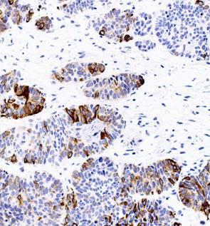
40 Cytokeratins in adenocarcinomas Neutral/Basic (B, class II) Acidic (A, class I) Squamous cell carcinoma (+) (+) (+) (+) - (+) Transitional cell tumour Malignant mesothelioma Adenocarcinoma: complex epith. (lung, breast) (+) (+) (+) Adenocarc.: biliary tract, pancr., endom., ovary (+) (+) (+) (+) ++ Adenocarc.: stomach Adenocarc.: intestine Hepatocellular carcinoma Renal cell carcinoma Endocrine tumours: carcinoids Merkel cell carcinoma Thyroid carcinoma


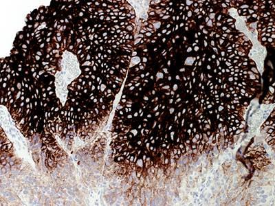
41 Cytokeratins in adenocarcinomas CK5: Breast lob. carcinoma CK5: Breast duct. carc.
42


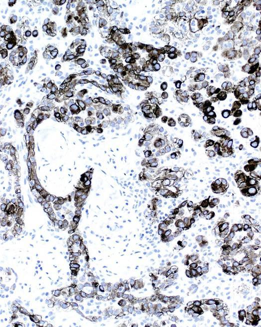
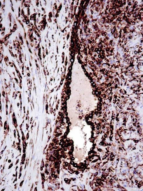
43 Cytokeratins in adenocarcinomas CK5: Prostate Breast lob. hyperplasia carcinoma CK5: prostate adenocarc.
44 Cytokeratins in adenocarcinomas Neutral/Basic (B, class II) Acidic (A, class I) Squamous cell carcinoma (+) (+) (+) (+) - (+) Transitional cell tumour Malignant mesothelioma Adenocarcinoma: complex epith. (lung, breast) (+) (+) (+) Adenocarc.: biliary tract, pancr., endom., ovary (+)! (+)! (+)! (+)! ++ Adenocarc.: stomach Adenocarc.: intestine Hepatocellular carcinoma Renal cell carcinoma Endocrine tumours: carcinoids Merkel cell carcinoma Thyroid carcinoma


45 Cytokeratins in adenocarcinomas CK5: Serous ovarian carcinoma CK17 CK20: Gall bladder adenocarcinoma
46 Cytokeratins in adenocarcinomas Neutral/Basic (B, class II) Acidic (A, class I) Squamous cell carcinoma (+) (+) (+) (+) - (+) Transitional cell tumour Malignant mesothelioma Adenocarcinoma: complex epith. (lung, breast) (+) (+) (+) Adenocarc.: biliary tract, pancr., endom., ovary (+) (+) (+) (+) ++ Adenocarc.: stomach Adenocarc.: intestine ! Hepatocellular carcinoma Renal cell carcinoma Endocrine tumours: carcinoids Merkel cell carcinoma Thyroid carcinoma
47 Cytokeratins in adenocarcinomas CK20 CK20 CK7 Colon: Typical Colon: Deviant CK7
48 Cytokeratins in adenocarcinomas Neutral/Basic (B, class II) Acidic (A, class I) Squamous cell carcinoma (+) (+) (+) (+) - (+) Transitional cell tumour Malignant mesothelioma Adenocarcinoma: complex epith. (lung, breast) (+) (+) (+) Adenocarc.: biliary tract, pancr., endom., ovary (+) (+) (+) (+) ++ Adenocarc.: stomach Adenocarc.: intestine Hepatocellular carcinoma Renal cell carcinoma Endocrine tumours: carcinoids Merkel cell carcinoma Thyroid carcinoma
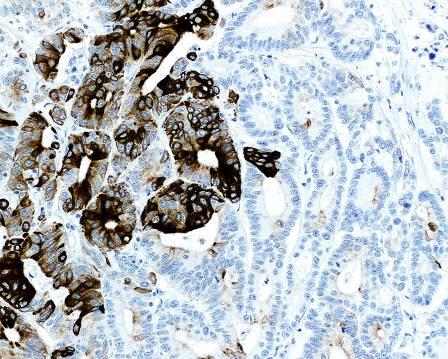
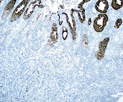
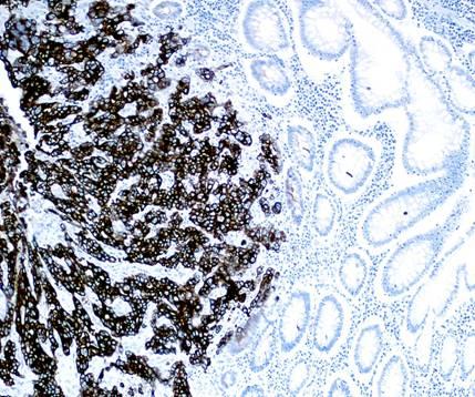
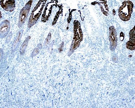
49 Cytokeratins in adenocarcinomas HCC: typical CK7 CK7 HCC: deviant CK7 Mallory body CK20
50 Cytokeratins in endocrine tumours Neutral/Basic (B, class II) Acidic (A, class I) Squamous cell carcinoma (+) (+) (+) (+) - (+) Transitional cell tumour Malignant mesothelioma Adenocarcinoma: complex epith. (lung, breast) (+) (+) (+) Adenocarc.: biliary tract, pancr., endom., ovary (+) (+) (+) (+) ++ Adenocarc.: stomach Adenocarc.: intestine Hepatocellular carcinoma Renal cell carcinoma Endocr. tumours: carcinoids Merkel cell carcinoma Thyroid carcinoma


51 Cytokeratins in endocrine tumours CK20: Merkel cell carc. CK19: Thyr. papill. carc.

52 Cytokeratins: Selection of antibodies optimal too diluted HIER Prot. CK5: XM-26 CK5/6: clone D5/16 B4 34BE12: CK5/14/1/10/19?


53 Cytokeratins: Selection of antibodies CK5: XM-26 34BE12: CK5/6: clone D5/16 B4 CK5/14/1/10/19?
54 NORDIQC RESULTS DEC JULY 2017
55 Breast dc Renal ccc K8: EP17 (Epitomics) K8/18: EP17/EP30 (Agilent) K8/18: B22.1/B23.1 (Roche, CM) K18: DC10 (several) K8(7): CAM5.2 (several) K8: 35βH11 (Roche)
 K8/18:")

 K8(7): CAM5.")
56 Breast dc Renal ccc K8: EP17 (Epitomics) K8/18: EP17/EP30 (Agilent) K8/18: B22.1/B23.1 (Roche, CM) K18: DC10 (several) K8(7): CAM5.2 (several) K8: 35βH11
 and Ventana BenchMark (B) to detect LMW Ks in serial sections of")
57 NORDIQC RESULTS DEC JULY 2017 Comparison of clone 5D3 used with optimized protocols on two platforms: Leica Bond (A) and Ventana BenchMark (B) to detect LMW Ks in serial sections of liver (1) and breast ductal carcinoma (2). Clone 5D3 gives strong reactions on Leica Bond but consistently poor reactions when used on BenchMark.


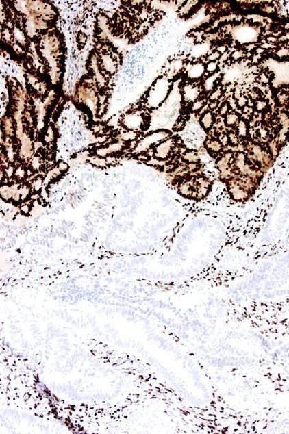
58 Cytokeratins: Selection of antibodies optimal Esophagus CK19: clone A53-B/A2.26 Papillary thyroid carcinoma CK19: clone RCK108 After proteolysis as recommended by the vendor
59 Secondary panels for carcinoma identification/subclassification - Apart from cytokeratins and general epithelial markers (EpCAM, Cl4) GI-markers CDX2, CAD17, SATB2, SMAD4, CEA Fem.gen.tract markers CA125, PAX8, WT1, ER, p53 Liver cell markers Neuroendocr. markers Breast markers Arginase, Glypican3, GlutSynt, cancd66a, cancd10, AFP Synaptophysin, Chromogranin GATA3, ER, GCDFP15, Mam.glob.
60 Secondary panels for carcinoma identification/subclassification Lung markers TTF-1, Napsin, p40 Mesothelioma makers Calretinin, Popoplanin, WT1, CA125 Adrenal cortic.markers Inhibin, Melan A, Synaptophysin Germinal cell markers Prostate markers Urinary tract markers SALL4, OCT3/4, PLAP, AFP, CD117, CD30 NKX3.1, PSA, Prostein, AMACR, ERG Uroplakin II, GATA3, (CK5, CK20)
61 GI markers CDX2 CEA CAD17 SATB2 SMAD4


62 CDX-2 protein Drosophila caudal related homeobox gene 2 product: Nuclear transcription factor for intestinal differentiation Intestine all cell types incl. endocrine Intestinal metaplasia chronic gastritis Barrett s esophagus Pancreas/bil.tract heterogenous colon pancreas
 Yolk sac tumour + Esoph./Stom. +/ Pancr.")
63 Colon adenocarcinoma CDX-2 protein in adenocarcinoma Colorectum +( ) Mucinous ovar. +( ) Yolk sac tumour + Esoph./Stom. +/ Pancr./biliary /+ Lung /+ Prostate (+) Urothelial (+) Endometrioid (+)
 Mucinous")
 Urothelial (+)")
64 Colon adenosquamous carc. CDX-2 protein in adenocarcinoma Colorectum +( ) Mucinous ovar. +( ) Yolk sac tumour + Esoph./Stom. +/ Pancr./biliary /+ Lung /+ Prostate (+) Urothelial (+) Endometrioid (+) Colon adenoc. medullary adenoc.
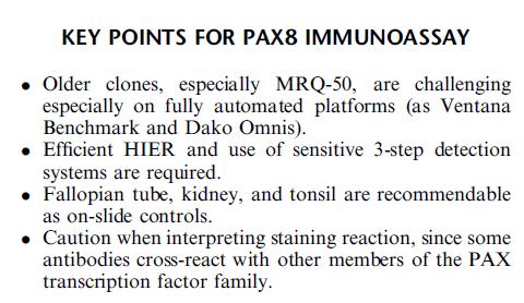
65 ER CDX-2 Endometrioid carcinoma: ER & CDX-2

66 (Appl Immunohistochem Mol Morphol 2013;21:64 72)


67 CDX2 Normal colon 1 Optimized protocols EPR = EPR2764Y EPR-CON EPR-RTU DAK-CDX2 AMT28 CDX2-88


68 CDX2 Normal colon 2 Optimized protocols EPR = EPR2764Y EPR-CON EPR-RTU DAK-CDX2 AMT28 CDX2-88


69 CDX2 Normal pancreas Optimized protocols EPR = EPR2764Y EPR-CON EPR-RTU DAK-CDX2 AMT28 CDX2-88


70 CDX2 Colon adenocarc.1 Optimized protocols EPR = EPR2764Y EPR-CON EPR-RTU DAK-CDX2 AMT28 CDX2-88

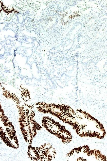
71 Carcinoembryonic antigen (CD66e) Adhesion molecule espc. associated with intestine

72 metast. colon adenoc Carcinoembryonic antigen (CD66e) in adenocarcinomas Colorectal + Medull. thyroid + Pancreas/biliary tract +/ Stomach +/ Lung +/ Ovary, mucinous +/ Ovary, non-muc. /+ Prostate Kidney Liver (!)


73 Medul. thyroid carc. Breast ductal carc. Carcinoembryonic antigen
74


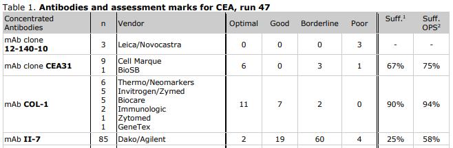
75 CEA31 LD II-7 RTU Low express. carcinoma

76 Normal liver II-7 TF3H8-1
77 Female genital tract markers PAX8 CA125 WT1 p53 ER
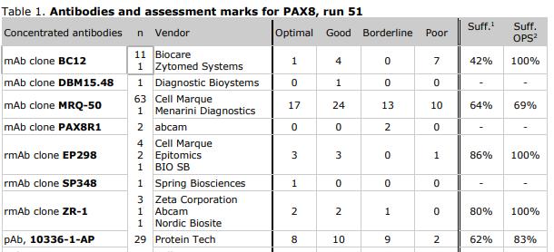
78 PAX8 Crucial to the organogenesis and development of Urogenital tract Adult genital tract Thyroid Neuroendocrine system
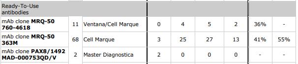
79 PAX8 Ovary - serous carcinoma LG + - serous carcinoma HG / clear cell + - endometrioid carcinoma +/- - mucinous carcinoma -(+) Kidney - clear cell / papillary carcinoma + - chromophobe / collect. duct carc. + - RCC, sarcomatoid +/- NET - pancreas, duodenum, rectum -(+)? Thyroid carcinoma + Other carcinomas -(+) Malignant mesothelioma -


80 Ovarian serous carcinoma PAX8


81 PAX8 in clear cell renal cell carcinoma
82
83

84 Optimal Too weak Fallopian tube Kidney Ovarian serous carcinoma Clear cell renal cell carcinoma False negative
85 The unkown primary tumour: IHC Classification, antibody selection, protocol optimization, Mogens Vyberg Professor of Clinical Pathology Director of NordiQC Aalborg University Hospital, Aalborg, Denmark controls and EQA (part II)
Nordic Immunohistochemical Quality Control
 Nordic Immunohistochemical Quality Control Immunohistochemistry in the classifiation of neoplasias of the alimentary tract & External Quality Assurance of Immunohistochemistry for GI cancer markers Mogens
Diagnostic IHC in lung and pleura pathology
 Diagnostic IHC in lung and pleura pathology Mogens Vyberg Professor of Clinical Pathology Director of NordiQC Aalborg University Hospital, Aalborg, Denmark WHO 2004 and Web Malignant mesothelioma Epithelioid
Breast cancer: IHC classification. Mogens Vyberg Professor of Clinical Pathology Director of NordiQC Aalborg University Hospital, Aalborg, Denmark
 Breast cancer: IHC classification Mogens Vyberg Professor of Clinical Pathology Director of NordiQC Aalborg University Hospital, Aalborg, Denmark http://upload.wikimedia.org/wikipedia/commons/1/1a/breast.svg
Immunohistochemical classification of lung carcinomas and mesotheliomas. Prof. Mogens Vyberg NordiQC Institute of Pathology Aalborg, Denmark
 Immunohistochemical classification of lung carcinomas and mesotheliomas Prof. Mogens Vyberg NordiQC Institute of Pathology Aalborg, Denmark Endobronchial ultrasound guided transbronchial needle biopsy
Immunohistochemical classification of the unknown primary tumour (UPT) Part I. Prof. Mogens Vyberg NordiQC Institute of Pathology Aalborg, Denmark
 Part I. Prof. Mogens Vyberg NordiQC Institute of Pathology Aalborg, Denmark") Immunohistochemical classification of the unknown primary tumour (UPT) Part I Prof. Mogens Vyberg NordiQC Institute of Pathology Aalborg, Denmark Tumours of unknown origin: Histology Brain tumour - biopsy
Immunohistochemical classification of the unknown primary tumour (UPT) Part I Prof. Mogens Vyberg NordiQC Institute of Pathology Aalborg, Denmark Tumours of unknown origin: Histology Brain tumour - biopsy
The unkown primary tumour: IHC Classification, antibody selection, protocol optimization, controls and EQA (part I)
") The unkown primary tumour: IHC Classification, antibody selection, protocol optimization, Mogens Vyberg Professor of Clinical Pathology Director of NordiQC Aalborg University Hospital, Aalborg, Denmark
The unkown primary tumour: IHC Classification, antibody selection, protocol optimization, Mogens Vyberg Professor of Clinical Pathology Director of NordiQC Aalborg University Hospital, Aalborg, Denmark
The unknown primary tumour: IHC classification part I, the primary panel - Antibody selection, protocol optimization, controls and EQA
 The unknown primary tumour: IHC classification part I, Mogens Vyberg Professor of Clinical Pathology Director of NordiQC Aalborg University Hospital, Aalborg, Denmark the primary panel - Antibody selection,
CARCINOMA OF UNKNOWN PRIMARY: DIAGNOSTIC APPROACH USING IMMUNOHISTOCHEMISTRY
 CARCINOMA OF UNKNOWN PRIMARY: DIAGNOSTIC APPROACH USING IMMUNOHISTOCHEMISTRY Jason L Hornick, MD, PhD Director of Surgical Pathology Director of Immunohistochemistry Brigham and Women s Hospital Associate
Classification of the unknown primary tumour: the primary IHC panel
 CIQC/CAP-ACP SEMINAR 2013: DIAGNOSTIC IHC AND MOLECULAR PATHOLOGY Classification of the unknown primary tumour: the primary IHC panel Aalborg University Hospital Denmark Tumours of unknown origin: Histology
Differential diagnosis of HCC
 Hepatocellular Carcinoma Quest for an Ideal Immunohistochemical Panel Sanjay Kakar, MD UCSF Differential diagnosis of HCC Hepatocellular lesions Adenoma, FNH, HG dysplasia Adenocarcinoma CholangioCA, metastasis
Histopathological diagnosis of CUP
 Histopathological diagnosis of CUP Dr Karin Oien karin.oien@glasgow.ac.uk Disclosure slide Dr Karin Oien has no financial interests in any company mentioned in this presentation. Dr Karin Oien is conducting
The Panel Approach to Diagnostics. Lauren Hopson International Product Specialist Cell Marque Corporation
 The Panel Approach to Diagnostics Lauren Hopson International Product Specialist Cell Marque Corporation Cell Marque Rocklin, California About Cell Marque: IVD primary antibody manufacturer Distributors
Assessment Run GATA3
 Assessment Run 44 2015 GATA3 Material The slide to be stained for GATA3 comprised: 1. Tonsil 2. Kidney, 3. Urothelial carcinoma, 4. Breast ductal carcinoma, 5. Colon adenocarcinoma All tissues were fixed
Carcinoma of Unknown Primary (CUP)
") Metasta c Carcinoma of Unknown Primary: Diagnos c Approach Using Immunohistochemistry James R. Conner, MD, PhD Mount Sinai Hospital Toronto, ON Carcinoma of Unknown Primary (CUP) 3-5% of all new malignant
Metasta c Carcinoma of Unknown Primary: Diagnos c Approach Using Immunohistochemistry James R. Conner, MD, PhD Mount Sinai Hospital Toronto, ON Carcinoma of Unknown Primary (CUP) 3-5% of all new malignant
Reporting of carcinoma of unknown primary tumour (CUP)
") Reporting of carcinoma of unknown primary tumour (CUP) Prof John Schofield Kent Oncology Centre with grateful thanks to Dr Karin Oien University of Glasgow Royal College of Pathologists Cancer datasets
Reporting of carcinoma of unknown primary tumour (CUP) Prof John Schofield Kent Oncology Centre with grateful thanks to Dr Karin Oien University of Glasgow Royal College of Pathologists Cancer datasets
The impact of proficiency testing on lab immunoassays
 The impact of proficiency testing on lab immunoassays Mogens Vyberg Professor of Clinical Pathology Director of NordiQC Aalborg University Hospital, Aalborg, Denmark Nordic Immunohistochemical Quality
The clinically challenging entity of liver metastasis from tumors of unknown primary
 The clinically challenging entity of liver metastasis from tumors of unknown primary Xuchen Zhang, MD, PhD Associate Professor of Pathology Department of Pathology Yale University School of Medicine Liver
Optimization of antibodies, selection, protocols and controls Breast tumours
 Optimization of antibodies, selection, protocols and controls Breast tumours Søren Nielsen Project coordinator & Scheme Manager NordiQC Aalborg University Hospital, Denmark Breast panel: GCDFP-15 Mammaglobin
New Developments in Immunohistochemistry for Gynecologic Pathology
 New Developments in Immunohistochemistry for Gynecologic Pathology Michael T. Deavers, M.D. Professor, Departments of Pathology and Gynecologic Oncology Immunohistochemistry in Gynecologic Pathology Majority
Cytokeratin 19 (CK19)
") Assessment Run 34 202 Cytokeratin 9 (CK9) Material The slide to be stained for CK9 comprised:. Thyroid gland, 2. Appendix, 3. Esophagus, 4. Papillary thyroid carcinoma, 5 & 6. Pancreatic neuroendocrine
Assessment Run 34 202 Cytokeratin 9 (CK9) Material The slide to be stained for CK9 comprised:. Thyroid gland, 2. Appendix, 3. Esophagus, 4. Papillary thyroid carcinoma, 5 & 6. Pancreatic neuroendocrine
Cancers of unknown primary : Knowing the unknown. Prof. Ahmed Hossain Professor of Medicine SSMC
 Cancers of unknown primary : Knowing the unknown Prof. Ahmed Hossain Professor of Medicine SSMC Definition Cancers of unknown primary site (CUPs) Represent a heterogeneous group of metastatic tumours,
I. Diagnosis of the cancer type in CUP
 Latest Research: USA I. Diagnosis of the cancer type in CUP II. Outcomes of site-specific therapy of the cancer type in CUP a. Prospective clinical trial b. Retrospective clinical trials 1 Latest Research:
Carcinoma of unknown primary origin (CUP) is defined
 is defined") REVIEW ARTICLE Metastatic Carcinoma of Unknown Primary: Diagnostic Approach Using Immunohistochemistry James R. Conner, MD, PhD and Jason L. Hornick, MD, PhD Abstract: Carcinoma of unknown primary origin
REVIEW ARTICLE Metastatic Carcinoma of Unknown Primary: Diagnostic Approach Using Immunohistochemistry James R. Conner, MD, PhD and Jason L. Hornick, MD, PhD Abstract: Carcinoma of unknown primary origin
Carcinoembryonic antigen (CEA)
") Assessment Run 37 2013 Carcinoembryonic antigen (CEA) Material The slide to be stained for CEA comprised: 1. Appendix, 2. Liver, 3-4. Colon adenocarcinoma, 5. Urothelial carcinoma All tissues were fixed
Assessment Run 37 2013 Carcinoembryonic antigen (CEA) Material The slide to be stained for CEA comprised: 1. Appendix, 2. Liver, 3-4. Colon adenocarcinoma, 5. Urothelial carcinoma All tissues were fixed
SHN-1 Human Digestive Panel Test results
 SHN-1 Human Digestive Panel Test results HN-30 tongue HN-24 salivary gland HN-12 larynx HN-28 esophagus HN-29 stomach HN-20 pancreas HN-13 liver HN-14 gall bladder HN-27-1 duodenum HN-27-2 ileum HN-27-3
Assessment Run CK19
 Assessment Run 29 200 CK9 The slide to be stained for CK9 comprised:. Appendix, 2. Thyroid gland, 3. Pancreas, 4. Ductal breast carcinoma, 5. Esophagus, 6. Papillary thyroid carcinoma. All tissues were
The role of immunohistochemistry in surgical pathology of the uterine corpus and cervix
 The role of immunohistochemistry in surgical pathology of the uterine corpus and cervix Prof. Ben Davidson, MD PhD Department of Pathology, Norwegian Radium Hospital, Oslo University Hospital, Oslo, Norway
Charles Halsey, DVM, PhD, DACVP Pfizer, Inc. IHC Resources
 Charles Halsey, DVM, PhD, DACVP Pfizer, Inc. IHC Resources 1 IHC Identification Targets Specimens Controls 2 Tissue controls Trouble Spots 3 The Key to Description IHC Description 4 Intermediate Filaments
NEW IHC A n t i b o d i e s
 NEW IHC Antibodies TABLE OF CONTENTS NEW IHC ANTIBODIES from Cell Marque CITED1 (5H6).... 1 Claudin 7 (5D10F3).... 1 GATA1 (4F5).... 1 Transgelin (2A10C2).... 1 NEW IHC ANTIBODIES using RabMAb Technology
How to Recognize Gynecologic Cancer Cells from Pelvic Washing and Ascetic Specimens
 How to Recognize Gynecologic Cancer Cells from Pelvic Washing and Ascetic Specimens Wenxin Zheng, M.D. Professor of Pathology and Gynecology University of Arizona zhengw@email.arizona.edu http://www.zheng.gynpath.medicine.arizona.edu/index.html
Breast - ductal carcinoma CK7 ER PR GATA3 Mammaglobin (50-70%) GCDFP-15 (50-70%) E-cadherin HMWCK CK20 PAX2 ER/PR/HER2 on all newly diagnosed cases
 GCDFP-15 (50-70%) E-cadherin HMWCK CK20 PAX2 ER/PR/HER2 on all newly diagnosed cases") Adrenal cortical carcinoma Inhibin Synap Melan-A Calretinin Vimentin Chromogr CK7 CK20 Breast - ductal carcinoma CK7 ER PR GATA3 Mammaglobin (50-70%) GCDFP-15 (50-70%) E-cadherin HMWCK CK20 PAX2 ER/PR/HER2
Adrenal cortical carcinoma Inhibin Synap Melan-A Calretinin Vimentin Chromogr CK7 CK20 Breast - ductal carcinoma CK7 ER PR GATA3 Mammaglobin (50-70%) GCDFP-15 (50-70%) E-cadherin HMWCK CK20 PAX2 ER/PR/HER2
Epithelial cell-cell adhesion molecule (Ep-CAM)
") Assessment Run 3 011 Epithelial cell-cell adhesion molecule (Ep-CAM) Material The slide to be stained for Ep-CAM comprised: 1. Appendix,. Kidney, 3. Adrenal gland, 4. Lung carcinoid, 5 & 6. Renal clear
Assessment Run 3 011 Epithelial cell-cell adhesion molecule (Ep-CAM) Material The slide to be stained for Ep-CAM comprised: 1. Appendix,. Kidney, 3. Adrenal gland, 4. Lung carcinoid, 5 & 6. Renal clear
Cutaneous metastases. Thaddeus Mully. University of California, San Francisco Professor, Departments of Pathology and Dermatology
 Cutaneous metastases Thaddeus Mully University of California, San Francisco Professor, Departments of Pathology and Dermatology DISCLOSURE OF RELATIONSHIPS WITH INDUSTRY Thaddeus Mully Course C005 Essential
Expression of Cytokeratin 5/6 in Epithelial Neoplasms: An Immunohistochemical Study of 509 Cases
 Expression of Cytokeratin 5/6 in Epithelial Neoplasms: An Immunohistochemical Study of 509 Peiguo G. Chu, M.D., Ph.D., Lawrence M. Weiss, M.D. Department of Pathology, City of Hope National Medical Center,
10 years of NordiQC Why are 30% of labs still getting it wrong?
 Mogens Vyberg & Søren Nielsen NordiQC Institute of Pathology Aalborg University Hospital Aalborg, Denmark May 29th 2015 10 years of NordiQC Why are 30% of labs still getting it wrong? Nothing to declare
Quality Assurance in Immunohistochemistry: Experiences from NordiQC
 Nordic immunohistochemical Quality Control 2 Quality Assurance in Immunohistochemistry: Experiences from NordiQC Prof. Mogens Vyberg NordiQC Institute of Pathology Aalborg University Hospital Aalborg,
C.L. Davis Foundation Descriptive Veterinary Pathology Course
 C.L. Davis Foundation 2015 Descriptive Veterinary Pathology Course IHC Resources IHC Identification Targets Antibodies Antibodies 1 Antibodies Specimens Antigen Retrieval Unmasks antigen epitopes Methods
ISSN X (Print) Original Research Article. DOI: /sjams
 Original Research Article. DOI: /sjams") DOI: 10.21276/sjams.2016.4.7.33 Scholars Journal of Applied Medical Sciences (SJAMS) Sch. J. App. Med. Sci., 2016; 4(7C):2468-2473 Scholars Academic and Scientific Publisher (An International Publisher
DOI: 10.21276/sjams.2016.4.7.33 Scholars Journal of Applied Medical Sciences (SJAMS) Sch. J. App. Med. Sci., 2016; 4(7C):2468-2473 Scholars Academic and Scientific Publisher (An International Publisher
SMH (Myosin, smooth muscle heavy chain)
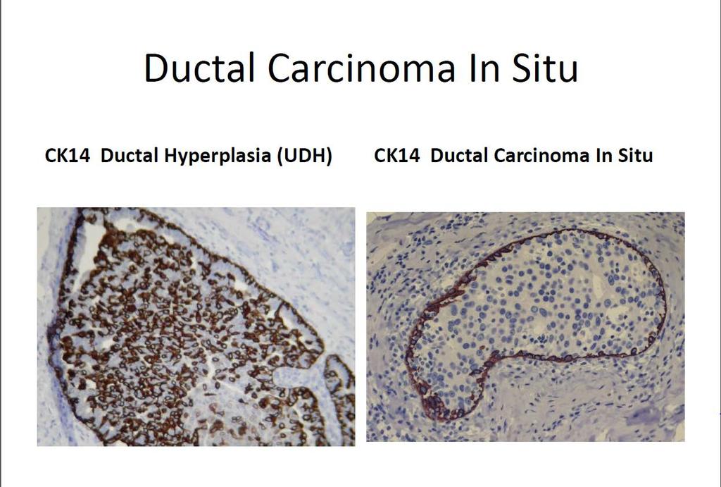
") Material The slide to be stained for SMH comprised: Assessment Run 50 2017 SMH (Myosin, smooth muscle heavy chain) 1.Tonsil, 2. Esophagus, 3. Breast hyperplasia, 4. Breast ductal carcinoma in situ (DCIS),
Material The slide to be stained for SMH comprised: Assessment Run 50 2017 SMH (Myosin, smooth muscle heavy chain) 1.Tonsil, 2. Esophagus, 3. Breast hyperplasia, 4. Breast ductal carcinoma in situ (DCIS),
Thyroid transcription factor-1 (TTF1) Assessment run
 Assessment run") Thyroid transcription factor- (TTF) Assessment run 39 203 The slide to be stained for TTF comprised:. Thyroid gland, 2. Liver, 3. Normal lung, 4. Lung adenocarcinoma 5. Colon adenocarcinoma, 6 & 7. Lung
Thyroid transcription factor- (TTF) Assessment run 39 203 The slide to be stained for TTF comprised:. Thyroid gland, 2. Liver, 3. Normal lung, 4. Lung adenocarcinoma 5. Colon adenocarcinoma, 6 & 7. Lung
Applications of IHC. Determination of the primary site in metastatic tumors of unknown origin
 Applications of IHC Determination of the primary site in metastatic tumors of unknown origin Classification of tumors that appear 'undifferentiated' by standard light microscopy Precise classification
Assessment Run NKX3.1 (NKX3.1)
") Assessment Run 49 2017 NKX3.1 (NKX3.1) Material The slide to be stained for NKX3.1 comprised: 1. Testis 2. Appendix 3-4. Prostate adenocarcinoma 5. Prostate hyperplasia All tissues were fixed in 10% neutral
Assessment Run 49 2017 NKX3.1 (NKX3.1) Material The slide to be stained for NKX3.1 comprised: 1. Testis 2. Appendix 3-4. Prostate adenocarcinoma 5. Prostate hyperplasia All tissues were fixed in 10% neutral
List of Available TMAs in the PRN
 TMA RPCI_BrainCa01 RPCI_BrCa03 RPCI_BrCa04 RPCI_BrCa05 RPCI_BrCa0 RPCI_BrCa07 RPCI_BrCa08 RPCI_BrCa15 RPCI_BrCa1 RPCI_BrCa17 RPCI_BrCa18 RPCI_BrCa19 RPCI_BrCa20 RPCI_BrCa21 RPCI_BrCa24 RPCI_BrCa25 RPCI_BrCa2
Technology from Abcam
 CD2 (EP222) CD2 is one of the earliest T-cell lineage restricted antigens to appear during T-cell differentiation and only rare CD2+ cells can be found in the bone marrow. Anti-CD2 is a pan-t-cell antigen
IHC Panels as an Aid in Diagnostic Decision Making
 IHC Antibody Test Selection Using a Panel Approach Steven Westra B.S. Reagent Product Specialist Leica Biosystems IHC Panels as an Aid in Diagnostic Decision Making Diagnostic Use of Tumors Using Algorithms
What I Learned from 3 Cases and 3 Antibodies
 What I Learned from 3 Cases and 3 Antibodies Melinda Sanders, M.D Vanderbilt University Medical Center Professor of Pathology Consultant in Breast Pathology Disclosure of Relevant Financial Relationships
Tumour Markers. For these reasons, only a handful of tumour markers are commonly used by most doctors.
 Tumour Markers What are Tumour Markers? Tumour markers are substances that can be found in the body when cancer is present. They are usually found in the blood or urine. They can be products of cancer
External Quality Assessment of melanocytic marker analyses NordiQC experience
 External Quality Assessment of melanocytic marker analyses NordiQC experience Jan Klos MD, Department of Pathology Stavanger University Hospital Norway 1 Content 18 Runs = 2112 submissions between 2001-2014
8 years later! Next Generation Sequencing. Pathogenic Findings: HNF1A c.864delinscc, p.g292rfs*25 (NM_ ) (VAF: 59%) HNF1A Loss
 (VAF: 59%) HNF1A Loss") 8 years later! Next Generation Sequencing Pathogenic Findings: HNF1A c.864delinscc, p.g292rfs*25 (NM_000545.6) (VAF: 59%) HNF1A Loss Interpretation HNF1A c.864delinscc, p.g292rfs*25 (NM_000545.6) This
8 years later! Next Generation Sequencing Pathogenic Findings: HNF1A c.864delinscc, p.g292rfs*25 (NM_000545.6) (VAF: 59%) HNF1A Loss Interpretation HNF1A c.864delinscc, p.g292rfs*25 (NM_000545.6) This
Coordinate Expression of Cytokeratins 7 and 20 in Prostate Adenocarcinoma and Bladder Urothelial Carcinoma
 Anatomic Pathology / CYTOKERATINS 7 AND 20 IN PROSTATE AND BLADDER CARCINOMAS Coordinate Expression of Cytokeratins 7 and 20 in Prostate Adenocarcinoma and Bladder Urothelial Carcinoma Nader H. Bassily,
Assessment Run
 Assessment Run 50 2017 S100 Material The slide to be stained for S100 comprised: 1. Appendix, 2. Tonsil, 3. Schwannoma, 4-5. Malignant melanoma, 6. Colon adenocarcinoma. All tissues were fixed in 10% neutral
Sal-like protein 4 (SALL4)
") Assessment Run 43 205 Sal-like protein 4 (SALL4) The slide to be stained for SALL4 comprised:. Appendix, 2. Testis, 3. Renal clear cell carcinoma, 4. Seminoma, 5. Intratubular germ cell neoplasia (IGCN),
Assessment Run 43 205 Sal-like protein 4 (SALL4) The slide to be stained for SALL4 comprised:. Appendix, 2. Testis, 3. Renal clear cell carcinoma, 4. Seminoma, 5. Intratubular germ cell neoplasia (IGCN),
Tissue-Specific Cadherin CDH17 Is a Useful Marker of Gastrointestinal Adenocarcinomas With Higher Sensitivity Than CDX2
 Anatomic Pathology / CDH17 in Gastrointestinal Carcinomas Tissue-Specific Cadherin CDH17 Is a Useful Marker of Gastrointestinal Adenocarcinomas With Higher Sensitivity Than CDX2 Nicole C. Panarelli, MD,
Fast, automated, precise
 Thermo Scientific B R A H M S / NSE Immunodiagnostic Assays Fast, automated, precise Neuroendocrine tumor markers on KRYPTOR Systems First and only fully automated CgA assay worldwide Shortest time to
Estrogen receptor (ER)
") Material The slide to be stained for ER comprised: Assessment B25 208 Estrogen receptor (ER) No. Tissue ER-positivity* ER-intensity*. Uterine cervix 80-90% Moderate to strong 2. Tonsil < 2-5% Weak to strong
Material The slide to be stained for ER comprised: Assessment B25 208 Estrogen receptor (ER) No. Tissue ER-positivity* ER-intensity*. Uterine cervix 80-90% Moderate to strong 2. Tonsil < 2-5% Weak to strong
BEST PRACTICES IN THE APPLICATION OF IMMUNOHISTOCHEMISTRY TO DIAGNOSTIC UROLOGIC PATHOLOGY: LESSONS FROM USES & ABUSES
 BEST PRACTICES IN THE APPLICATION OF IMMUNOHISTOCHEMISTRY TO DIAGNOSTIC UROLOGIC PATHOLOGY: LESSONS FROM USES & ABUSES Mahul B. Amin Professor & Chairman Department of Pathology & Laboratory Medicine Cedars-Sinai
Estrogen receptor (ER)
") Material The slide to be stained for ER comprised: Assessment Run B26 2018 Estrogen receptor (ER) No. Tissue ER-positivity* ER-intensity* 1. Uterine cervix 80-90% Moderate to strong 2. Tonsil 1-5% Weak
Material The slide to be stained for ER comprised: Assessment Run B26 2018 Estrogen receptor (ER) No. Tissue ER-positivity* ER-intensity* 1. Uterine cervix 80-90% Moderate to strong 2. Tonsil 1-5% Weak
FNA Cytology of Metastatic Malignancies of Unknown Primary Site
 FNA Cytology of Metastatic Malignancies of Unknown Primary Site Tarik M. Elsheikh Cleveland Clinic Jan F. Silverman Alleghany Hospitals Pathologic Diagnosis of Metastasis Smaller specimens, less invasive
FNA Cytology of Metastatic Malignancies of Unknown Primary Site
 FNA Cytology of Metastatic Malignancies of Unknown Primary Site Tarik M. Elsheikh Jan F. Silverman Pathologic Diagnosis of Metastasis Smaller specimens, less invasive techniques FNA cytology is highly
IMMUNOPROFILES OF THE MAJOR RENAL NEOPLASMS (%staining)
") Stain Clear Cell Papillary IMMUNOPROFILES OF THE MAJOR RENAL NEOPLASMS (%staining) Chromophobe Collecting Duct Carcinom a Sarcomatoid Xp11 Translocat ion Dr Jon Oxley See also www.jonoxley.com Page 1 MTSCC
Stain Clear Cell Papillary IMMUNOPROFILES OF THE MAJOR RENAL NEOPLASMS (%staining) Chromophobe Collecting Duct Carcinom a Sarcomatoid Xp11 Translocat ion Dr Jon Oxley See also www.jonoxley.com Page 1 MTSCC
NordiQC External Quality Assurance in Immunohistochemistry
 NordiQC External Quality Assurance in Immunohistochemistry Mogens Vyberg Professor of Clinical Pathology Director of NordiQC Aalborg University Hospital, Aalborg, Denmark AALBORG (~ 200.000 inhabitants)
Estrogen receptor (ER)
") Assessment Run B7 204 Estrogen receptor (ER) Material The slide to be stained for ER comprised: No. Tissue ER-positivity* ER-intensity*. Uterine cervix 80-90% Moderate to strong 2. Breast carcinoma 0%
Assessment Run B7 204 Estrogen receptor (ER) Material The slide to be stained for ER comprised: No. Tissue ER-positivity* ER-intensity*. Uterine cervix 80-90% Moderate to strong 2. Breast carcinoma 0%
Serous effusion Objectives. Cytology of Serous Effusions From basics to challenges
 Cytology of Serous Effusions From basics to challenges Cytology of Serous Effusions From basics to challenges Pınar Fırat, MD, MIAC Department of Pathology, İstanbul University, İstanbul Faculty of Medicine,
CEA (CARCINOEMBRYONIC ANTIGEN)
") (CARCINOEMBRYONIC ANTIGEN) 428 C15.3 Malignant neoplasm of upper third of esophagus C15.4 Malignant neoplasm of middle third of esophagus C15.5 Malignant neoplasm of lower third of esophagus C15.8 Malignant
(CARCINOEMBRYONIC ANTIGEN) 428 C15.3 Malignant neoplasm of upper third of esophagus C15.4 Malignant neoplasm of middle third of esophagus C15.5 Malignant neoplasm of lower third of esophagus C15.8 Malignant
Ovarian Clear Cell Carcinoma
 Ovarian Clear Cell Carcinoma Rouba Ali-Fehmi, MD Professor of Pathology The Karmanos Cancer Institute, Wayne State University School of Medicine 50 year old woman with chief complaint of shortness of breath
Urinary Bladder: WHO Classification and AJCC Staging Update 2017
 Urinary Bladder: WHO Classification and AJCC Staging Update 2017 Houston Society of Clinical Pathologists 58 th Annual Spring Symposium Houston, TX April 8, 2017 Jesse K. McKenney, MD Classification
Tumor Immunology. Tumor (latin) = swelling
 = swelling") Tumor Immunology Tumor (latin) = swelling benign tumor malignant tumor Tumor immunology : the study of the types of antigens that are expressed by tumors how the immune system recognizes and responds to
Tumor Immunology Tumor (latin) = swelling benign tumor malignant tumor Tumor immunology : the study of the types of antigens that are expressed by tumors how the immune system recognizes and responds to
Pathology Mystery and Surprise
 Pathology Mystery and Surprise Tim Smith, MD Director Anatomic Pathology Medical University of South Carolina Disclosures No conflicts to declare Some problem cases Kidney tumor Scalp tumor Bladder tumor
Immunohistochemistry on Fluid Specimens: Technical Considerations
 Immunohistochemistry on Fluid Specimens: Technical Considerations Blake Gilks Dept of Pathology University of British Columbia, Vancouver, BC, Canada Disclosures None Learning Objectives At the end of
Effusion Cytology: Diagnostic Challenges
 Effusion Cytology: Diagnostic Challenges Tarik M. Elsheikh, MD Professor and Medical Director, Anatomic Pathology Cleveland Clinic Outside Consult Case 45 year old woman, presented with nausea, dyspnea,
Immunohistochemistry. Potential and challenges To be or not to be
 Immunohistochemistry Potential and challenges To be or not to be Søren Nielsen Scheme Manager NordiQC Aalborg University Hospital, Denmark Vårmöte 19.05.2016 Karlstad Overview IHC project coordinator at
Neuroendocrine Carcinoma. Lebanon Neuroendocrine Neoplasms of H&N Nov /7/2011. Broad Classification:
 H&N Neuroendocrine Neoplasms: Classification and Diagnostic Considerations Adel K. El-Naggar, M.D., Ph.D. The University of Texas MD Anderson Cancer Center, Houston, Texas Broad Classification: A. Epithelial:
Schedule of Accreditation issued by United Kingdom Accreditation Service 2 Pine Trees, Chertsey Lane, Staines-upon-Thames, TW18 3HR, UK
 United Kingdom Accreditation Service 2 Pine Trees, Chertsey Lane, Staines-upon-Thames, TW18 3HR, UK North Tyneside General Hospital Rake Lane North Shields Tyne & Wear NE29 8NH Contact: Ian Taylor Tel:
incidence rate x 100,000/year
 Tier R=rare C=common Cancer Entity European crude and age adjusted incidence by cancer, years of diagnosis 2000 and 2007 Analisys based on 83 population-based cancer registries * applying the European
ICD-O Morphology code. R=Rare Tier Tumour ICD-O Topography code C30.0, C31
 R=Rare Tier Tumour ICD-O Topography code ICD-O Morphology code EPITHELIAL TUMOURS OF NASAL CAVITY AND SINUSES R 2 Squamous cell carcinoma with variants of nasal cavity and sinuses C30.0, C3 C30.0, C3 8000,
4/12/2018. MUSC Pathology Symposium Kiawah Island April 18, Jesse K. McKenney, MD
 MUSC Pathology Symposium Kiawah Island April 18, 2018 Jesse K. McKenney, MD 1 Urothelial Carcinoma with Alternative Differentiation 2 Urothelial Carcinoma with Alternative Differentiation Recognition as
Malignant neoplasms of the gastrointestinal (GI) tract,
 tract,") Special Section First Chinese American Pathologists Association Diagnostic Pathology Course, Part II Practical Immunohistochemistry in Neoplastic Pathology of the Gastrointestinal Tract, Liver, Biliary
Special Section First Chinese American Pathologists Association Diagnostic Pathology Course, Part II Practical Immunohistochemistry in Neoplastic Pathology of the Gastrointestinal Tract, Liver, Biliary
Tissue: The Living Fabric: Part A
 PowerPoint Lecture Slides prepared by Janice Meeking, Mount Royal College C H A P T E R 4 Tissue: The Living Fabric: Part A Tissues Groups of cells similar in structure and function Types of tissues Epithelial
Tissue-based Immunohistochemical Biomarker Expression in Malignant Glandular Lesions of the Uterine Cervix: a Systematic Review
 Tissue-based Immunohistochemical Biomarker Expression in Malignant Glandular Lesions of the Uterine Cervix: a Systematic Review Sandra Lee MD, FRCPC 1 *, Vikrant V. Sahasrabuddhe, MBBS, DrPH 2 *, Diana
Single and Multiplex Immunohistochemistry
 Single and Multiplex Immunohistochemistry Steve Westra, BS Reagent Product Specialist Leica Biosystems IHC Theory Polyclonal vs Monoclonal Polyclonal reagents Detect a multitude of epitopes Batch to batch
Adnexal primary or Melanocy+c prolifera+ons in sundamaged metastatic carcinoma?
 Adnexal primary or Melanocy+c prolifera+ons in sundamaged metastatic carcinoma? skin Jane L. Messina, MD Interna0onal Melanoma Pathology Working Group 4 th annual mee0ng Tampa, Florida November 14, 2011
Neoplasms of the Canine, Feline and Lemur Liver:
 Neoplasms of the Canine, Feline and Lemur Liver: Classification and Prognosis Annual Seminar of the French Society of Veterinary Pathology John M. Cullen VMD PhD DACVP North Carolina State University Primary
NordiQC - update
 NordiQC - update 00-0 EQUALIS Uppsala 0 Tomas Seidal NordiQC participants NordiQC participants n:30 S DK N 6 F Ice Bel 54 NL 4 Ger 6 Aust USA 0 It 8 Argent 8.. 96% participation in S,DK & N ~ 60% in Finland
Immunohistochemistry and Bladder Tumours
 Immunohistochemistry and Bladder Tumours Dr. Andrew J. Evans MD PhD FRCPC Consultant in Genitourinary Pathology University Health Network Toronto, ON Objec ves Review markers of urothelial differen a on
Lung Anaplastic Lymphoma Kinase (lu-alk)
") Assessment Run 5 207 Lung Anaplastic Lymphoma Kinase (lu-alk) Material The slide to be stained for lu-alk comprised:. Appendix, 2. Tonsil, 3. Merkel cell carcinoma, 4. Anaplastic large cell lymphoma with
Assessment Run 5 207 Lung Anaplastic Lymphoma Kinase (lu-alk) Material The slide to be stained for lu-alk comprised:. Appendix, 2. Tonsil, 3. Merkel cell carcinoma, 4. Anaplastic large cell lymphoma with
Ascitic Fluid and Use of Immunocytochemistry. Mercè Jordà, University of Miami
 Ascitic Fluid and Use of Immunocytochemistry Mercè Jordà, University of Miami Is It Malignant? Yes? No Ascitic Fluid Cytomorphologic Useful Findings Tight clusters with smooth borders Cellular and nuclear
Other Sites. Table 2 Continued. MPH Rules 11/8/07. NAACCR Webinar Series 1
 MPH s 11/8/07 Other s 1 Table 2 Continued Use this two-page table to select combination histology codes. Compare the terms in the diagnosis to the terms in Columns 1 and 2. If the terms match, code the
2. Epithelial Tissues Dr. Manal Othman
 Biology-232 GENERAL HISTOLOGY 2. Epithelial Tissues Dr. Manal Othman Anatomy Department CMMS, AGU HISTOLOGY: w Study of the structure and function of tissues and organs at the microscopic levels. w Tissues
Radiology Pathology Conference
 Radiology Pathology Conference Nadia F. Yusaf, M.D. PGY-3 1/29/2010 Presentation material is for education purposes only. All rights reserved. 2010 URMC Radiology Page 1 of 90 Case 1 60 year- old man presents
Testicular Germ Cell Tumors; A Simplistic Approach
 Testicular Germ Cell Tumors; A Simplistic Approach Merce Jorda, MD, PhD, MBA Professor and Vice Chair, Director of Anatomic Pathology Director of Genitourinary Pathology Service Interim Director of Cytopathology
Enterprise Interest Nothing to declare
 Enterprise Interest Nothing to declare Update of mixed tumours of the GI tract, the pancreas and the liver Introduction to the concept of mixed tumours and clinical implication Jean-Yves SCOAZEC Surgical
Case 18. M75. Excision of mass on scalp. Clinically SCC. The best diagnosis is:
 Case 18 M75. Excision of mass on scalp. Clinically SCC. The best diagnosis is: A. Pilomatrical carcinoma B. Adnexal carcinoma NOS C. Metastatic squamous cell carcinoma D.Primary squamous cell carcinoma
Section 14 Other Cancers. Cancer of Unknown 113 Primary Site INTRODUCTION PATHOLOGIC EVALUATION
 Section 14 Other Cancers Cancer of Unknown 113 Primary Site F. Anthony Greco and John D. Hainsworth INTRODUCTION Cancer of unknown primary (CUP) site is a clinical syndrome that includes many types of
Breast cancer: Antibody selection, protocol optimzation controls and EQA
 Breast cancer: Antibody selection, protocol optimzation controls and EQA Workshop in Diagnostic Immunohistochemistry Oud St. Jan/ Old St. John Brugge (Bruges), Belgium June 13th 15nd 2018 Rasmus Røge,
INTERPRETATION OF IMMUNOHISTOCHEMICAL STAINS - DIFFICULTIES AND PITFALLS. Gabor Fischer Diagnostic Services Manitoba University of Manitoba
 INTERPRETATION OF IMMUNOHISTOCHEMICAL STAINS - DIFFICULTIES AND PITFALLS Gabor Fischer Diagnostic Services Manitoba University of Manitoba IHC INTERPRETATIONS LOCAL DATA Diagnostic Services Manitoba Number
Recent advances in breast cancers
 Recent advances in breast cancers Breast cancer is a hetrogenous disease due to distinct genetic alterations. Similar morphological subtypes show variation in clinical behaviour especially in response
Cytokeratin 7 and Cytokeratin 20 Expression in Epithelial Neoplasms: A Survey of 435 Cases
 Cytokeratin 7 and Cytokeratin 20 Expression in Epithelial Neoplasms: A Survey of 435 Cases Peiguo Chu, M.D., Ph.D., Emerald Wu, B.S., Lawrence M Weiss, M.D. Division of Pathology, City of Hope National
Immunohistochemical Expression of Cytokeratin 5/6 in Gynaecological Tumors.
 ISPUB.COM The Internet Journal of Pathology Volume 13 Number 2 Immunohistochemical Expression of Cytokeratin 5/6 in Gynaecological Tumors. A Baghla, S Choudhry, A Kataria Citation A Baghla, S Choudhry,
Tissues. tissue = many cells w/ same structure and function. cell shape aids function tissue shape aids function. Histology = study of tissues
 Tissues tissue = many cells w/ same structure and function cell shape aids function tissue shape aids function Histology = study of tissues 4 types of tissues Epithelial coverings contact openings Connective