Immunohistochemistry. Potential and challenges To be or not to be
|
|
|
- Augustine Allen
- 5 years ago
- Views:
Transcription


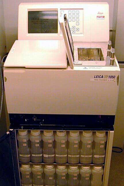
1 Immunohistochemistry Potential and challenges To be or not to be Søren Nielsen Scheme Manager NordiQC Aalborg University Hospital, Denmark Vårmöte Karlstad


2 Overview IHC project coordinator at Institute of Pathology, Aalborg, Denmark & Scheme manager NordiQC > IHC slides annually BenchMark Ultra, Ventana Autostainer Link 48, Dako Omnis, Dako Bond III, Leica IHC cooperation partners Biocare Cell Marque Dako / Agilent Leica Thermo Fisher Ventana / Roche + Ad hoc projects/partners 2

 HER-2 ISH module: 2 runs/year BRISH, FISH (breast cancer) Pilot runs ongoing ALK (lung), PD-L1 (lung) www.")
3 Perspective International academic IHC proficiency testing program Founded 2003 by Nordic pathologists Independent non-profit organisation Institute of Pathology, Aalborg University Hospital, DK General module: 3 runs/year different markers Breast cancer IHC module: 2 runs/y 3-5 different markers (HER2, ER, PR,..) HER-2 ISH module: 2 runs/year BRISH, FISH (breast cancer) Pilot runs ongoing ALK (lung), PD-L1 (lung) 3


4 Albert Coons American pathologist and immunologist

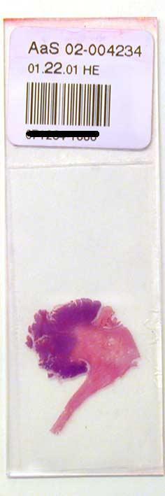
5 IHC is a result to be interpreted in cells/tissue heavily being submitted various chemical and physical interactions HE
6 Hyperplasia or In-situ CK5, CK14, Heavy chain myosin, p63 In-situ or invasive CK5, CK14, Heavy chain myosin, p63 Lobular or ductal lesion E-cadherin, p120 Predictive - Prognostic ER, PR, HER2, Ki67 Intrinsic subtype PAM50 ER, PR, HER2, Ki67, CK5 6

7 Original nomenclature and grouping of IHC tests: Class I IHC tests: Interpreted in the context of histo- or cytomorphologic and clinical data. Results interpreted and used by pathologists. E.g. CD45, TTF1, SOX10, CDX2, p40 etc Class II IHC tests: Stand-alone tests being interpreted (largely) to provide predictive and prognostic information. Results interpreted by pathologists and used by clinicians to give tailored treatment. E.g. ER, PR, HER2, CD117 etc. Am J Clin Pathol 2010;133:
8 Class II (Class III, US), IHC companion diagnostics: IHC test Demonstration Application ER Estrogen receptor protein Breast cancer HER2 Overexpression of HER2 protein Breast cancer, gastric cancer CD117 Protein second to gene mutation GIST EGFR Overexpression of HER1 protein Colorectal cancer ALK Fusion protein second to gene rearrangement NSCLC PD-L1 PD-L1 protein expression NSCLC, Melanoma,..

9 In practice more and more IHC tests become Class II tests: Directly indicated Area Class I Class II Comment CD20 Lymphoma B-cell origin Mabthera Evaluation of theraphy CD30 Lymphoma HL, ALCL Brentuximab CD56 Carcinoma Neuroendo. Lorvotuzumab Class II: Lung SCLC ALK Lymphoma ALCL Crizotinib Class II: Lung NSCLC Indirectly indicated typically due to personalized treatment e.g. Area Class I Class II Comment p40 - lung Carcinoma Squamous TTF1- lung Carcinoma Adeno Crizotinib,. ALK, EGFR, ROS1
10 IHC as a surrogate test to molecular based tests for gene disorders; IHC test Demonstration Application MMR proteins ROS-1 ALK BRAF V600E Detects DNA repair proteins Detects over-expressed gene fusion product Detects gene translocation protein Detects specific mutant protein Screen for inherited cancer syndrome (Lynch) Seen in <2% of NSCLC, predictive of TKI response Seen in <5% of NSCLC, predictive of crizotinib response Predictive maker for melanoma, screen for colon cancer MMR IHC p16 Detects over-expressed protein Surrogate marker for HPV INI-1 Detects normal INI-1 suppressor protein Diagnostic value in rhabdoid tumors of CNS, kidney, soft tissue HER-2 Detects over-expressed protein Screen in breast cancer
11 IHC testing - general Molecular testing - general Morphology YES NO Cell location YES NO Minimal sample YES NO Archival FFPE YES YES/NO Multiplex test NO YES Quantifiable NO/YES YES Standardized NO YES/NoO Simple, cost eff. YES YES


12 To IHC or not to IHC..

13 1. IHC to identify and sub-type cancers 1. E.g. TTF1, Napsin A, CK5 & p40 for lung NSCLC 2. IHC to guide treatment 1. E.g. evaluation of level of protein level immunotherapy 3. IHC to screen for gene disorders 1. Cheap, fast 4. IHC to visualize heterogenous cancers 1. Architecture intact facilities the identification IHC is an open window for the identification of molecular changes and related theraphies Essential for precision medicine

14
15 Major Genetic Alterations in Cancer Mutation Mutant Protein Translocation Loss of Expression Deletion Abnormal Localization Amplification Overexpression Methylation Expression of Fusion Proteins
16 Major Genetic Alterations in Cancer Mutation Translocation Deletion Amplification Methylation Mutant Protein IDH1 Loss of Expression MMR Abnormal Localization ß-catenin Overexpression p53 Expression of Fusion Proteins

17 An anti-idh1-r132h-specific monoclonal antibody, IMab-1, is useful for detecting IDH1-R132H in IHC and predicted time to progression in grade III anaplastic astrocytomas.
")
, or MSH6 (~10%) Accounts for 2-5% of")

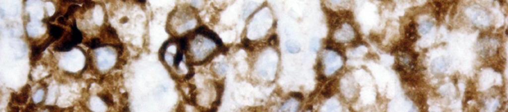
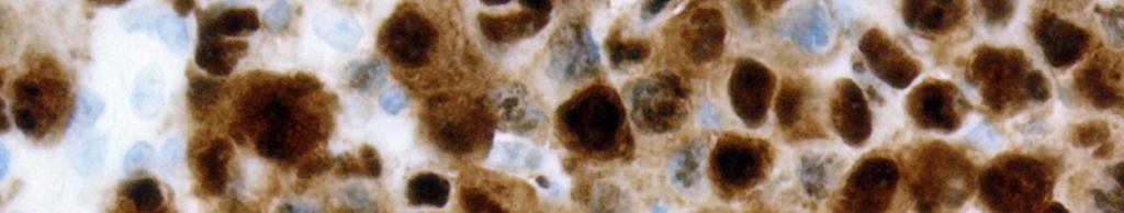
18 Mismatch repair gene mutations Lynch syndrome Hereditary non-polyposis colorectal cancers (HNPCC) Autosomal dominant Mutation in MLH1 (~60%), MSH2 (~30%), or MSH6 (~10%) Accounts for 2-5% of colorectal adenocarcinoma Tumors develop at early age, usually found on right side Also develop endometrial adenocarcinoma


19 Mismatch repair gene mutations Lynch syndrome IHC negative result indicates further analysis


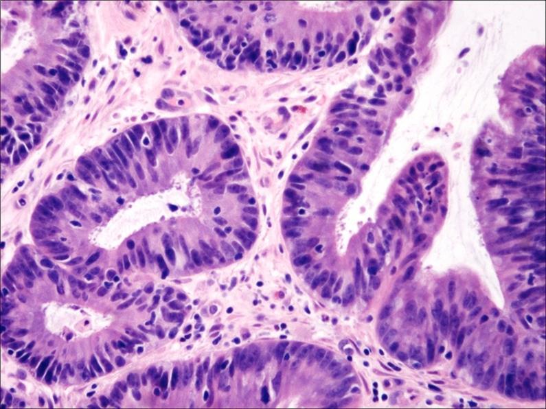
20 Mismatch repair gene mutations Lynch syndrome MLH1 MSH2 PMS2 MSH6
21 Mismatch repair gene mutations Lynch syndrome MMR MSI testing IHC Mol test Cost Low High Analyte Protein DNA How much intact tumor material is required Very little Very little Requirements Tumor only Tumor + normal Possibility of contamination by normal No Yes Turnaround Next day 2-7 days Identifies involved gene Yes No Assay sensitive to fixation Yes No

22 J Clin Oncol 28: , 2010 n = 457 Outcome of Patients with Stage III Colorectal Adenocarcinoma Treated with Adjuvant 5-FU dmmr pmmr IHC for MMR : Identification of Lynch syndrome Prognosis Treatment tree for patients


23 β-catenin β-catenin is a dual function protein, regulating the coordination of cell cell adhesion and gene transcription.


24 β-catenin B-catenin is partner with APC gene / protein If APC gene is mutated, colon cancer may develop APC gene will not keep B-catenin out of nucleus B-catenin inside nucleus bind to e.g. SMADs etc Uncontrolled proliferation and risk of cancer


25 β-catenin Desmoid type fibromatosis (71%) Modern Pathology 18:68-74, 2005 Solitary fibrous tumor (40%) Desmoid Endometrial stromal sarcoma (40%) Synovial sarcoma (28%) GIST
26 Major Genetic Alterations in Cancer Mutation Mutant Protein Translocation Loss of Expression Deletion Abnormal Localization Amplification Overexpression Methylation Expression of Fusion Proteins
(p11.2;q11.")
27 Tumor Translocation Fusion Generated IHC Target PNET/ES t(11;22)(q24;q12) EWSR1-FLI1 FLI1 ALCL t(2;5)(p23;q35) NPM-ALK ALK ASPS der(17)t(x;17)(p11;q25) ASPL-TFE3 TFE3 Synovial sarcoma t(x;18)(p11.2;q11.2) SYT-SSX1 TLE-1 DSRCT t(11;22)(q11;q12) EWSR1-WT1 WT-1 AML t(8;21)(q22;q22) AML1-ETO AML1-ETO Lung cancer Chromosme 2 inversion EML4-ALK ALK


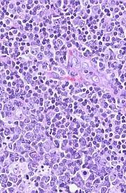
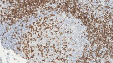
28 ALCL NordiQC CD20 CD3 CD30 ALK

29 29
30 30
31 ~ 90 IHC markers in NordiQC Runs Tested 1-20 times Markers highlighted Mol. derived targets Alpha-methylacyl-CoA racemase Cyclin D1 MLH1 Alpha-smooth muscle actin Cytokeratin 5 MSH2 Anaplastic lymphoma kinase Cytokeratin 7 MSH6 B-cell specific activator protein Cytokeratin 19 Multiple myeloma oncogene 1 bcl-2 protein Cytokeratin 20 Myosin, smooth muscle heavy chain bcl-6 protein Cytokeratin, high molecular weight Napsin A Calretinin Cytokeratin, low molecular weight Neurofilament protein Cancer antigen 125 Cytokeratin, pan- Octamer transcription factor-3/4 Carcinoembryonic antigen Desmin p16 ink4a CD3 Detected on GIST-1 p40 CD4 E-cadherin p53 CD5 Epithelial cell adhesion molecule p57 CD8 Epithelial membrane antigen p63 CD10 Estrogen receptor alpha Paired box gene-2 protein CD14 Factor VIII related antigen Paired box gene-8 protein CD15 GATA3 Placental alkaline phosphatase CD19 Glial fibrillary acidic protein PMS2 CD20 Glypican 3 Podoplanin CD23 Gross cystic disease fluid protein-15 Prostate specific acid phosphatase CD30 HER-2 Prostate specific antigen CD31 Hepatocyte antigen Prostein CD34 Human chorionic gonadotropin Progesterone receptor CD45 Immunoglobulin kappa S-100 protein beta CD56 Immunoglobulin lambda Sal-like protein 4 CD68 Immunoglobubin M SOX10 CD79a Ki-67 Synaptophysin CD99 Mammaglobin Terminal deoxynucl. transferase CD117 Melan-A Vimentin Chromogranin Melanosoma specific antigen Wilm's tumour-1 protein 31
32 The biomarker protocol trap Caution: not for faint-hearted lab personel!!!!! Decalcification Preparation Tissue Type, Dimension, Laser resection, De-differentiation With 3 choices for 5 variables in each phase = > 4 million protocols. Controls Quantification Reporting Fixation Time, Type, Volume Preanalytic Postanalytic Section Thickness Storage Drying Pre-treatment Manual Stainer Visualization Sensitivity, Specificity Primary antibody Clone, Dilution Buffer, Time, Temp Analytic Interpretation Localization Positive/Negative - cut-off level Development Sensitivity, Localization

33 IHC Quality Problem 9 nordic labs

34 IHC Quality Problem 9 nordic labs


35 IHC test: Fit for purpose All IHC tests both laboratory developed assays and RTU systems must be calibrated for the diagnostic use E.g. IHC assays for mismatch repair proteins (MMR) Purpose Diagnostic utility Tool Application Disease screening of patients with Lynch syndrome IHC results have been shown to have high concordance to mutation analysis IHC panel for 4 MMR proteins; MLH1, MSH2, MSH6 & PMS2 Identification of a reliable IHC protocol and interpretation guidelines for the pathologist Diagnostic relevant Diagnostic validity Technically possible Diagnostic possible 35
36 Pass rates (scores; optimal and good) and proportion of optimal scores for MMR in the latest NordiQC assessment run Laboratory developed tests* Company X Ready-To-Use system** Pass rate Optimal Pass rate Optimal MLH % (n=44/66) 36% (n=24/66) 89% (n=23/26) 69% (n=18/26) MSH % (n=23/57) 12% (n=7/57) 96% (n=22/23) 87% (n=20/23) MSH % (n=46/72) 43% (n=31/72) 91% (n=32/35) 69% (n=24/35) PMS % (n=42/47) 45% (n=22/47) 96% (n=24/25) 60% (n=15/25) * Using a concentrated primary antibody by a laboratory calibrated system ** Using company X Ready-To-Use system Number of laboratories / protocols

37 Proportion of protocols based on concentrates and RTU formats in NordiQC AMACR CD10 CK LMW CK HMW MLA BCL2 BCL6 BSAP CD99 EMA WT1 BCL6 CD15 GLP3 MLA PAX8
38 Proportion of protocols based on concentrates and RTU formats in NordiQC ER HER2 ER HER2 ER HER2
39

40 Clone Efficient HIER Titre Detection kit with high sensitivity (FLEX+ Refine OptiView+A)

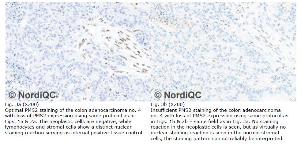
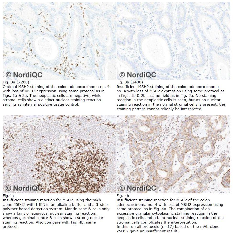
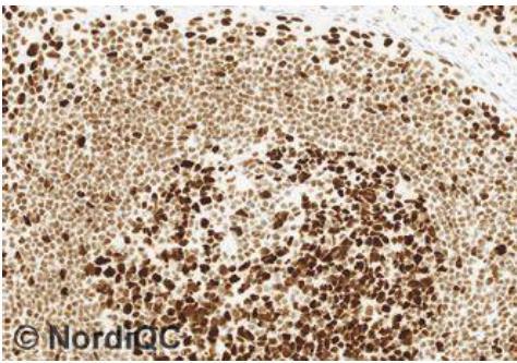
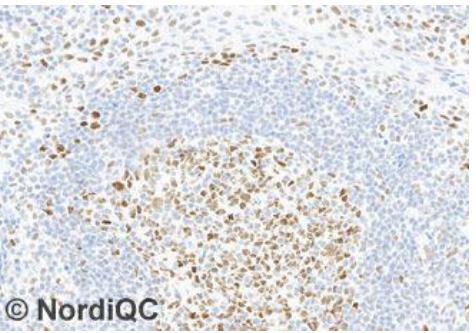
41 Interpretation based on internal tissue control. Negative IHC result in neoplastic cells must be confirmed by identification of stromal cells being positive!
42
43

44 Detection kit with high sensitivity (FLEX+ Refine OptiView+A) Modified
45
46
47 The biomarker protocol trap Caution: not for faint-hearted lab personel!!!!! Decalcification Preparation Tissue Type, Dimension, Laser resection, De-differentiation With 3 choices for 5 variables in each phase = > 4 million protocols. Controls Quantification Reporting Fixation Time, Type, Volume Preanalytic Postanalytic Section Thickness Storage Drying Pre-treatment Manual Stainer Visualization Sensitivity, Specificity Primary antibody Clone, Dilution Buffer, Time, Temp Analytic Interpretation Localization Positive/Negative - cut-off level Development Sensitivity, Localization

48 Colon: S100, polyclonal Pathos 3h NBF, 6h prog. Pathos 24h NBF, 6h prog. Pathos 48h NBF, 6h prog. Pathos 168h NBF, 6h prog. 48

49 Tonsil: S100, polyclonal S100 = Soluble in 100% alcohol Pathos 3h NBF, 2h prog. Pathos 24h NBF, 2h prog. Pathos 48h NBF, 2h prog. Pathos 168h NBF, 2h prog. 49

50 Colon: MLH1, ES05 (same for EPR3947 for PMS2) Pathos 3h NBF, 6h prog. Pathos 24h NBF, 6h prog. Pathos 48h NBF, 6h prog. Pathos 168h NBF, 6h prog.


51 Ref.: Ole Nielsen EPR3947 EP51 Similar
52

53 Clone Titre RTU > In-house
54
55

56 Choice of clone..


57 MSH6 issues : mab clone 44 used Difficult to calibrate EP49

58 Colon: MSH2, G (same for EPR3945 for MSH6) Pathos 3h NBF, 6h prog. Pathos 24h NBF, 6h prog. Pathos 48h NBF, 6h prog. Pathos 168h NBF, 6h prog.
59

60 Clone Detection system CALIBRATION PURPOSE OF TEST


61 EQA Program Class I IHC result and score is related to 1. Technical quality Class II 2. Calibration level and precision can the test be applied for the intended purpose?

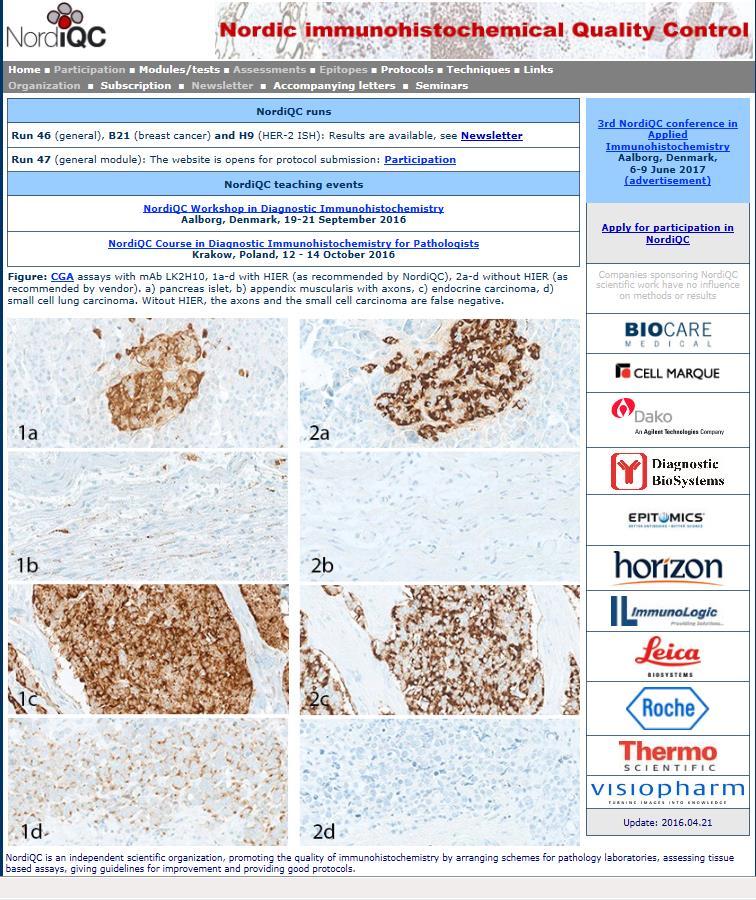
62 100% pass rate ALK in ALCL 67% pass rate ALK in lung cancer
63 Issues to be adressed : 1. Calibration of IHC assay and identification of best practice protocol clone, titre, retrieval etc 2. Evaluation of the robustness of the IHC assay impact on pre-analytics 3. Evaluation of the analytical sensitivity/specificity 4. Identification of most robust controls providing information that the established level of detection is obtained in each test performed in daily practice. Tissue controls are key element 63
64 Analytical validation Laboratory developed tests (concentrates and RTU formats being applied modified to official protocol) Non-predictive markers (- ER, PR, HER-2..) CLSI: 20 cases per entity relevant (pos, neg) CAP: 10 positive, 10 negative The validation set should include high and low expressors for positive cases when appropriate and should span the expected range of clinical results (expression levels) for markers that are reported quantitatively. Ad-Hoc: 10 strongly pos, 10 interm. to low, 5 neg. Number less important compared to use of tissue with full range of expression patterns reflecting the diagnostic use

65 Mantle cell lymphoma Mantle cell lymphoma Pancreas ad. carc. CD5 - rmab SP19 SP54 Pancreas ad. carc. CD5 - mab 4C7


66 Challenge: Rare in cancers and/or in benign cells ALK, ROS1, PD-L1 etc and many molecular derived targets Needed to verify IHC method is working ALK lung; 30 cancers used to find 1 pos case.. ALK Appendix / Colon: Peripheral nerves axons and ganglion cells PD-L1 Tonsil: Germinal centre macrophages Precision and metrics of test to be confirmed
67 Issues to be adressed : 1. Calibration of IHC assay and identification of best practice protocol clone, titre, retrieval etc 2. Evaluation of the robustness of the IHC assay impact on pre-analytics 3. Evaluation of the analytical sensitivity/specificity 4. Identification of most robust controls providing information that the established level of detection is obtained in each test performed in daily practice. Tissue controls Performance controls 67
68 Appl Immunohistochem Mol Morphol. Volume 22, Number 4, October 2014
69 IHC Critical Assay Performance Controls (icapcs) Which tissues are recommended? What is the expected staining pattern? Which tissues / cells are critical? Right antibody Appropriate level of sensitivity Guidance level of specificity

 No")

70 Examples for 17 markers Generel expected patterns High expression (Right antibody) Low expression (Appropriate sensitivity) No expression (Appropriate specificity) Which tissue Which cells Which extension Which intensity
71 IHC is still and will be a valueable tool for many years Both Class I and Class II tests Many new Class II assays to come to guide treatment Will be used together with molecular based tests For precision medicine we need precise IHC Attention on calibration, validation and consistency


72 Questions and answers Thank You for the attendance. Questions??
The impact of proficiency testing on lab immunoassays
 The impact of proficiency testing on lab immunoassays Mogens Vyberg Professor of Clinical Pathology Director of NordiQC Aalborg University Hospital, Aalborg, Denmark Nordic Immunohistochemical Quality
NordiQC External Quality Assurance in Immunohistochemistry
 NordiQC External Quality Assurance in Immunohistochemistry Mogens Vyberg Professor of Clinical Pathology Director of NordiQC Aalborg University Hospital, Aalborg, Denmark AALBORG (~ 200.000 inhabitants)
Immunohistochemical principles The technical test approach. Pre-analytical parametres
 Immunohistochemical principles The technical test approach Pre-analytical parametres Søren Nielsen Global Pathology Manager Agilent Technologies (Former Scheme Manager, NordiQC) 2 IHC project coordinator
Breast cancer: Antibody selection, protocol optimzation controls and EQA
 Breast cancer: Antibody selection, protocol optimzation controls and EQA Workshop in Diagnostic Immunohistochemistry Oud St. Jan/ Old St. John Brugge (Bruges), Belgium June 13th 15nd 2018 Rasmus Røge,
Optimization of antibodies, selection, protocols and controls Breast tumours
 Optimization of antibodies, selection, protocols and controls Breast tumours Søren Nielsen Project coordinator & Scheme Manager NordiQC Aalborg University Hospital, Denmark Breast panel: GCDFP-15 Mammaglobin
10 years of NordiQC Why are 30% of labs still getting it wrong?
 Mogens Vyberg & Søren Nielsen NordiQC Institute of Pathology Aalborg University Hospital Aalborg, Denmark May 29th 2015 10 years of NordiQC Why are 30% of labs still getting it wrong? Nothing to declare
Quality Assurance in Immunohistochemistry: Experiences from NordiQC
 Nordic immunohistochemical Quality Control 2 Quality Assurance in Immunohistochemistry: Experiences from NordiQC Prof. Mogens Vyberg NordiQC Institute of Pathology Aalborg University Hospital Aalborg,
SMH (Myosin, smooth muscle heavy chain)
") Material The slide to be stained for SMH comprised: Assessment Run 50 2017 SMH (Myosin, smooth muscle heavy chain) 1.Tonsil, 2. Esophagus, 3. Breast hyperplasia, 4. Breast ductal carcinoma in situ (DCIS),
Material The slide to be stained for SMH comprised: Assessment Run 50 2017 SMH (Myosin, smooth muscle heavy chain) 1.Tonsil, 2. Esophagus, 3. Breast hyperplasia, 4. Breast ductal carcinoma in situ (DCIS),
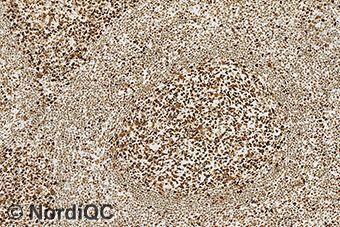
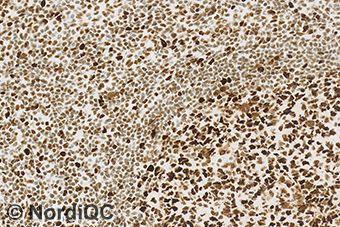
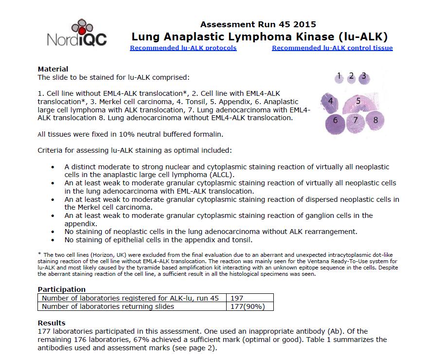
Lung Anaplastic Lymphoma Kinase (lu-alk)
") Assessment Run 5 207 Lung Anaplastic Lymphoma Kinase (lu-alk) Material The slide to be stained for lu-alk comprised:. Appendix, 2. Tonsil, 3. Merkel cell carcinoma, 4. Anaplastic large cell lymphoma with
Assessment Run 5 207 Lung Anaplastic Lymphoma Kinase (lu-alk) Material The slide to be stained for lu-alk comprised:. Appendix, 2. Tonsil, 3. Merkel cell carcinoma, 4. Anaplastic large cell lymphoma with
Assessment Run GATA3
 Assessment Run 44 2015 GATA3 Material The slide to be stained for GATA3 comprised: 1. Tonsil 2. Kidney, 3. Urothelial carcinoma, 4. Breast ductal carcinoma, 5. Colon adenocarcinoma All tissues were fixed
Estrogen receptor (ER)
") Material The slide to be stained for ER comprised: Assessment Run B26 2018 Estrogen receptor (ER) No. Tissue ER-positivity* ER-intensity* 1. Uterine cervix 80-90% Moderate to strong 2. Tonsil 1-5% Weak
Material The slide to be stained for ER comprised: Assessment Run B26 2018 Estrogen receptor (ER) No. Tissue ER-positivity* ER-intensity* 1. Uterine cervix 80-90% Moderate to strong 2. Tonsil 1-5% Weak
External Quality Assessment of Breast Marker Analysis. NordiQC data
 External Quality Assessment of Breast Marker Analysis NordiQC data Søren Nielsen Scheme Manager NordiQC Aalborg University Hospital, Denmark Aalborg 12.06 2015 Markers assessed in NordiQC Predictive markers
Assessment Run
 Assessment Run 50 2017 S100 Material The slide to be stained for S100 comprised: 1. Appendix, 2. Tonsil, 3. Schwannoma, 4-5. Malignant melanoma, 6. Colon adenocarcinoma. All tissues were fixed in 10% neutral
Assessment Run C1 2017
 Assessment Run C1 2017 PD-L1 The first assessment in this new NordiQC Companion module C1 focused on the accuracy of the PD-L1 IHC assays performed by the participating laboratories to identify patients
Nordic Immunohistochemical Quality Control
 Nordic Immunohistochemical Quality Control Immunohistochemistry in the classifiation of neoplasias of the alimentary tract & External Quality Assurance of Immunohistochemistry for GI cancer markers Mogens
Estrogen receptor (ER)
") Material The slide to be stained for ER comprised: Assessment B25 208 Estrogen receptor (ER) No. Tissue ER-positivity* ER-intensity*. Uterine cervix 80-90% Moderate to strong 2. Tonsil < 2-5% Weak to strong
Material The slide to be stained for ER comprised: Assessment B25 208 Estrogen receptor (ER) No. Tissue ER-positivity* ER-intensity*. Uterine cervix 80-90% Moderate to strong 2. Tonsil < 2-5% Weak to strong
Assessment Run B HER2 IHC
 Assessment Run B24 2017 HER2 IHC Material The slide to be stained for HER2 comprised the following 5 materials: IHC: HER2 Score* (0, 1+, 2+, 3+) FISH: HER2 gene/chr 17 ratio** 1. Breast carcinoma, no.
Current and future applications of Molecular Pathology. Kathy Walsh Clinical Scientist NHS Lothian
 Current and future applications of Molecular Pathology Kathy Walsh Clinical Scientist NHS Lothian Molecular Pathology in Solid tumours Cancer type Genes tested Purpose Associated treatments Non small cell
Estrogen receptor (ER)
") Assessment Run B7 204 Estrogen receptor (ER) Material The slide to be stained for ER comprised: No. Tissue ER-positivity* ER-intensity*. Uterine cervix 80-90% Moderate to strong 2. Breast carcinoma 0%
Assessment Run B7 204 Estrogen receptor (ER) Material The slide to be stained for ER comprised: No. Tissue ER-positivity* ER-intensity*. Uterine cervix 80-90% Moderate to strong 2. Breast carcinoma 0%
Assessment Run C3 2018
 Assessment Run C3 2018 PD-L1 Amended version May 14 th 2018 The third assessment in NordiQC Companion module C3 focused on the accuracy of the PD-L1 IHC assays performed by the participating laboratories
NordiQC - update
 NordiQC - update 00-0 EQUALIS Uppsala 0 Tomas Seidal NordiQC participants NordiQC participants n:30 S DK N 6 F Ice Bel 54 NL 4 Ger 6 Aust USA 0 It 8 Argent 8.. 96% participation in S,DK & N ~ 60% in Finland
Protocols for Zytomed Systems antibodies on fully automated IHC staining systems date of issue: September 20, 2012
 Protocols for Zytomed Systems antibodies on fully automated IHC staining systems date of issue: September 20, 2012 These protocols were provided by customers. Under no circumstances shall Zytomed Systems
Carcinoembryonic antigen (CEA)
") Assessment Run 37 2013 Carcinoembryonic antigen (CEA) Material The slide to be stained for CEA comprised: 1. Appendix, 2. Liver, 3-4. Colon adenocarcinoma, 5. Urothelial carcinoma All tissues were fixed
Assessment Run 37 2013 Carcinoembryonic antigen (CEA) Material The slide to be stained for CEA comprised: 1. Appendix, 2. Liver, 3-4. Colon adenocarcinoma, 5. Urothelial carcinoma All tissues were fixed
Schedule of Accreditation issued by United Kingdom Accreditation Service 2 Pine Trees, Chertsey Lane, Staines-upon-Thames, TW18 3HR, UK
 Schedule of ccreditation United Kingdom ccreditation Service 2 Pine Trees, Chertsey Lane, Staines-upon-Thames, TW18 3HR, UK External Quality ssessment Services for Cancer Diagnostics CIC Issue No: 005
HistoCyte Laboratories Ltd
 HistoCyte Laboratories Ltd Progesterone Receptor: The neglected breast receptor! Dr Ian Milton & Colin Tristram November 2018 UKNEQAS Autumn meeting Introduction Progesterone is an important prognostic
Breast cancer: IHC classification. Mogens Vyberg Professor of Clinical Pathology Director of NordiQC Aalborg University Hospital, Aalborg, Denmark
 Breast cancer: IHC classification Mogens Vyberg Professor of Clinical Pathology Director of NordiQC Aalborg University Hospital, Aalborg, Denmark http://upload.wikimedia.org/wikipedia/commons/1/1a/breast.svg
The unkown primary tumour: IHC Classification, antibody selection, protocol optimization, controls and EQA (part I)
") The unkown primary tumour: IHC Classification, antibody selection, protocol optimization, Mogens Vyberg Professor of Clinical Pathology Director of NordiQC Aalborg University Hospital, Aalborg, Denmark
The unkown primary tumour: IHC Classification, antibody selection, protocol optimization, Mogens Vyberg Professor of Clinical Pathology Director of NordiQC Aalborg University Hospital, Aalborg, Denmark
Applications of IHC. Determination of the primary site in metastatic tumors of unknown origin
 Applications of IHC Determination of the primary site in metastatic tumors of unknown origin Classification of tumors that appear 'undifferentiated' by standard light microscopy Precise classification
Assessment Run NKX3.1 (NKX3.1)
") Assessment Run 49 2017 NKX3.1 (NKX3.1) Material The slide to be stained for NKX3.1 comprised: 1. Testis 2. Appendix 3-4. Prostate adenocarcinoma 5. Prostate hyperplasia All tissues were fixed in 10% neutral
Assessment Run 49 2017 NKX3.1 (NKX3.1) Material The slide to be stained for NKX3.1 comprised: 1. Testis 2. Appendix 3-4. Prostate adenocarcinoma 5. Prostate hyperplasia All tissues were fixed in 10% neutral
NEW IHC A n t i b o d i e s
 NEW IHC Antibodies TABLE OF CONTENTS NEW IHC ANTIBODIES from Cell Marque CITED1 (5H6).... 1 Claudin 7 (5D10F3).... 1 GATA1 (4F5).... 1 Transgelin (2A10C2).... 1 NEW IHC ANTIBODIES using RabMAb Technology
Thyroid transcription factor-1 (TTF1) Assessment run
 Assessment run") Thyroid transcription factor- (TTF) Assessment run 39 203 The slide to be stained for TTF comprised:. Thyroid gland, 2. Liver, 3. Normal lung, 4. Lung adenocarcinoma 5. Colon adenocarcinoma, 6 & 7. Lung
Thyroid transcription factor- (TTF) Assessment run 39 203 The slide to be stained for TTF comprised:. Thyroid gland, 2. Liver, 3. Normal lung, 4. Lung adenocarcinoma 5. Colon adenocarcinoma, 6 & 7. Lung
The unknown primary tumour: IHC classification part I, the primary panel - Antibody selection, protocol optimization, controls and EQA
 The unknown primary tumour: IHC classification part I, Mogens Vyberg Professor of Clinical Pathology Director of NordiQC Aalborg University Hospital, Aalborg, Denmark the primary panel - Antibody selection,
Assessment Run B HER-2 IHC. HER-2/chr17 ratio**
 Assessment Run B2 20 HER-2 IHC Material The slide to be stained for HER-2 comprised the following 5 tissues: IHC HER-2 Score* (0, +, 2+,3+) FISH HER-2/chr7 ratio**. Breast ductal carcinoma 0..3 2. Breast
Single and Multiplex Immunohistochemistry
 Single and Multiplex Immunohistochemistry Steve Westra, BS Reagent Product Specialist Leica Biosystems IHC Theory Polyclonal vs Monoclonal Polyclonal reagents Detect a multitude of epitopes Batch to batch
Schedule of Accreditation issued by United Kingdom Accreditation Service 2 Pine Trees, Chertsey Lane, Staines-upon-Thames, TW18 3HR, UK
 2 Pine Trees, Chertsey Lane, Staines-upon-Thames, TW18 3HR, UK UCL-Advanced Diagnostics 1st Floor, Rockefeller Building 21 University Street London WC1E 6JJ Contact: David Allen Tel: +44 (0)20 7679 6912
Assessment Run B HER2 IHC
 Assessment Run B26 208 HER2 IHC Material The slide to be stained for HER2 comprised the following 5 materials: IHC: HER2 Score* (0, +, 2+, 3+) FISH: HER2 gene/chr 7 ratio**. Breast carcinoma, no. 2+..3
Product Introduction. Product Codes: HCL029, HCL030 and HCL031. Issue
 Product Introduction Product Codes: HCL029, HCL030 and HCL031 Issue 1. 180510 Contents Introduction to Estrogen Receptor 2 ER immunohistochemistry 3 Quality control 5 Cell lines as controls 6 Estrogen
Immunohistochemical classification of lung carcinomas and mesotheliomas. Prof. Mogens Vyberg NordiQC Institute of Pathology Aalborg, Denmark
 Immunohistochemical classification of lung carcinomas and mesotheliomas Prof. Mogens Vyberg NordiQC Institute of Pathology Aalborg, Denmark Endobronchial ultrasound guided transbronchial needle biopsy
LUNG CANCER. pathology & molecular biology. Izidor Kern University Clinic Golnik, Slovenia
 LUNG CANCER pathology & molecular biology Izidor Kern University Clinic Golnik, Slovenia 1 Pathology and epidemiology Small biopsy & cytology SCLC 14% NSCC NOS 4% 70% 60% 50% 63% 62% 61% 62% 59% 54% 51%
PD-L1 Analyte Control DR
 Quality in Control PD-L1 Analyte Control DR PD-L1_PI_v2 Product Codes: HCL019, HCL020 and HCL021 Contents PD-L1 Analyte Control DR 2 What is PD-L1? 3 The Role of PD-L1 in Cancer 3 PD-L1 Assessment 4 PD-L1
Cytokeratin 19 (CK19)
") Assessment Run 34 202 Cytokeratin 9 (CK9) Material The slide to be stained for CK9 comprised:. Thyroid gland, 2. Appendix, 3. Esophagus, 4. Papillary thyroid carcinoma, 5 & 6. Pancreatic neuroendocrine
Assessment Run 34 202 Cytokeratin 9 (CK9) Material The slide to be stained for CK9 comprised:. Thyroid gland, 2. Appendix, 3. Esophagus, 4. Papillary thyroid carcinoma, 5 & 6. Pancreatic neuroendocrine
The Panel Approach to Diagnostics. Lauren Hopson International Product Specialist Cell Marque Corporation
 The Panel Approach to Diagnostics Lauren Hopson International Product Specialist Cell Marque Corporation Cell Marque Rocklin, California About Cell Marque: IVD primary antibody manufacturer Distributors
DOUBLE STAINS. Toll-Free: Direct:
 DOUBLE STAINS CD61 + CD71 DAB Brown: CD61 Alk. Phos. Red: CD71 Bone Marrow DAB Brown: Megakaryocytes Alk. Phos. Red: Erythroid Precursors 400x CD61 (2f2) 0.1 ml concentrate............. 161M-14 0.5 ml
Product Introduction
 Product Introduction Product Codes: HCL026, HCL027 and HCL028 Contents Introduction to HER2 2 HER2 immunohistochemistry 3 Cell lines as controls 5 HER2 Analyte Control DR IHC 7 HER2 Analyte Control DR
Immunohistochemical classification of the unknown primary tumour (UPT) Part I. Prof. Mogens Vyberg NordiQC Institute of Pathology Aalborg, Denmark
 Part I. Prof. Mogens Vyberg NordiQC Institute of Pathology Aalborg, Denmark") Immunohistochemical classification of the unknown primary tumour (UPT) Part I Prof. Mogens Vyberg NordiQC Institute of Pathology Aalborg, Denmark Tumours of unknown origin: Histology Brain tumour - biopsy
Immunohistochemical classification of the unknown primary tumour (UPT) Part I Prof. Mogens Vyberg NordiQC Institute of Pathology Aalborg, Denmark Tumours of unknown origin: Histology Brain tumour - biopsy
Evolution of Pathology
 1 Traditional pathology Molecular pathology 2 Evolution of Pathology Gross Pathology Cellular Pathology Morphologic Pathology Molecular/Predictive Pathology Antonio Benivieni (1443-1502): First autopsy
Epithelial cell-cell adhesion molecule (Ep-CAM)
") Assessment Run 3 011 Epithelial cell-cell adhesion molecule (Ep-CAM) Material The slide to be stained for Ep-CAM comprised: 1. Appendix,. Kidney, 3. Adrenal gland, 4. Lung carcinoid, 5 & 6. Renal clear
Assessment Run 3 011 Epithelial cell-cell adhesion molecule (Ep-CAM) Material The slide to be stained for Ep-CAM comprised: 1. Appendix,. Kidney, 3. Adrenal gland, 4. Lung carcinoid, 5 & 6. Renal clear
Disclosures. An update on ancillary techniques in the diagnosis of soft tissue tumors. Ancillary techniques. Introduction
 Disclosures An update on ancillary techniques in the diagnosis of soft tissue tumors. I have nothing to disclose. Andrew Horvai, MD, PhD Clinical Professor, Pathology Introduction Ancillary techniques
The role of immunohistochemistry in surgical pathology of the uterine corpus and cervix
 The role of immunohistochemistry in surgical pathology of the uterine corpus and cervix Prof. Ben Davidson, MD PhD Department of Pathology, Norwegian Radium Hospital, Oslo University Hospital, Oslo, Norway
Technology from Abcam
 CD2 (EP222) CD2 is one of the earliest T-cell lineage restricted antigens to appear during T-cell differentiation and only rare CD2+ cells can be found in the bone marrow. Anti-CD2 is a pan-t-cell antigen
Quality in Control. ROS1 Analyte Control. Product Codes: HCL022, HCL023 and HCL024
 Quality in Control ROS1 Analyte Control Product Codes: HCL022, HCL023 and HCL024 Contents What is ROS1? 2 The Role of ROS1 in Cancer 3 ROS1 Assessment 3 ROS1 Analyte Control Product Details 4 ROS1 Analyte
The unkown primary tumour: IHC Classification, antibody selection, protocol optimization, controls and EQA (part II)
") The unkown primary tumour: IHC Classification, antibody selection, protocol optimization, Mogens Vyberg Professor of Clinical Pathology Director of NordiQC Aalborg University Hospital, Aalborg, Denmark
The unkown primary tumour: IHC Classification, antibody selection, protocol optimization, Mogens Vyberg Professor of Clinical Pathology Director of NordiQC Aalborg University Hospital, Aalborg, Denmark
Breast - ductal carcinoma CK7 ER PR GATA3 Mammaglobin (50-70%) GCDFP-15 (50-70%) E-cadherin HMWCK CK20 PAX2 ER/PR/HER2 on all newly diagnosed cases
 GCDFP-15 (50-70%) E-cadherin HMWCK CK20 PAX2 ER/PR/HER2 on all newly diagnosed cases") Adrenal cortical carcinoma Inhibin Synap Melan-A Calretinin Vimentin Chromogr CK7 CK20 Breast - ductal carcinoma CK7 ER PR GATA3 Mammaglobin (50-70%) GCDFP-15 (50-70%) E-cadherin HMWCK CK20 PAX2 ER/PR/HER2
Adrenal cortical carcinoma Inhibin Synap Melan-A Calretinin Vimentin Chromogr CK7 CK20 Breast - ductal carcinoma CK7 ER PR GATA3 Mammaglobin (50-70%) GCDFP-15 (50-70%) E-cadherin HMWCK CK20 PAX2 ER/PR/HER2
Assessment Run CK19
 Assessment Run 29 200 CK9 The slide to be stained for CK9 comprised:. Appendix, 2. Thyroid gland, 3. Pancreas, 4. Ductal breast carcinoma, 5. Esophagus, 6. Papillary thyroid carcinoma. All tissues were
Sal-like protein 4 (SALL4)
") Assessment Run 43 205 Sal-like protein 4 (SALL4) The slide to be stained for SALL4 comprised:. Appendix, 2. Testis, 3. Renal clear cell carcinoma, 4. Seminoma, 5. Intratubular germ cell neoplasia (IGCN),
Assessment Run 43 205 Sal-like protein 4 (SALL4) The slide to be stained for SALL4 comprised:. Appendix, 2. Testis, 3. Renal clear cell carcinoma, 4. Seminoma, 5. Intratubular germ cell neoplasia (IGCN),
Instant Quality FISH. The name says it all.
 COMPANION DIAGNOSTICS Instant Quality FISH Instant Quality FISH. The name says it all. IQ: Instant Quality every time. Breast carcinoma stained with : Triple filter showing Blue DAPI colors nuclei, FITC
HSL-Advanced Diagnostics 2018 / 19 Test & Service List
 HSL-Advanced Diagnostics 2018 / 19 Test & Service List 2018/19 TEST & SERVICE LIST Haematoxylin & Eosin H&E H&E per slide Routine Immunohistochemistry Immunohistochemical demonstration of an antigen in
Diagnostic IHC in lung and pleura pathology
 Diagnostic IHC in lung and pleura pathology Mogens Vyberg Professor of Clinical Pathology Director of NordiQC Aalborg University Hospital, Aalborg, Denmark WHO 2004 and Web Malignant mesothelioma Epithelioid
Assessment performed on Tuesday, July 29, 2014, at Lions Gate Hospital, North Vancouver
 Assessors report for ciqc Run 37: BRAF V600E (April 2014) Assessors: B Gilks, R Wolber, K Ung, P Tavassoli, J Garratt and J Won (recorder) Assessment performed on Tuesday, July 29, 2014, at Lions Gate
IHC Stainer platforms. Overview, pros and cons
 IHC Stainer platforms Overview, pros and cons Bart De Wiest Quality manager IHC OLV Hospital, Aalst, Belgium Donald Van Hecke Lab & Quality manager AZ St-Lucas, Brugge, Belgium Goal of this lecture: to
Tissue-based Immunohistochemical Biomarker Expression in Malignant Glandular Lesions of the Uterine Cervix: a Systematic Review
 Tissue-based Immunohistochemical Biomarker Expression in Malignant Glandular Lesions of the Uterine Cervix: a Systematic Review Sandra Lee MD, FRCPC 1 *, Vikrant V. Sahasrabuddhe, MBBS, DrPH 2 *, Diana
Classification of the unknown primary tumour: the primary IHC panel
 CIQC/CAP-ACP SEMINAR 2013: DIAGNOSTIC IHC AND MOLECULAR PATHOLOGY Classification of the unknown primary tumour: the primary IHC panel Aalborg University Hospital Denmark Tumours of unknown origin: Histology
Serrated Polyps and a Classification of Colorectal Cancer
 Serrated Polyps and a Classification of Colorectal Cancer Ian Chandler June 2011 Structure Serrated polyps and cancer Molecular biology The Jass classification The familiar but oversimplified Vogelsteingram
Breast cancer diagnostic solutions Deliver diagnostic confidence
 Breast cancer diagnostic solutions Deliver diagnostic confidence 2 Breast cancer diagnostic solutions Roche Tissue Diagnostics is committed to improving outcomes in breast cancer Breast cancer...the most
External Quality Assessment of melanocytic marker analyses NordiQC experience
 External Quality Assessment of melanocytic marker analyses NordiQC experience Jan Klos MD, Department of Pathology Stavanger University Hospital Norway 1 Content 18 Runs = 2112 submissions between 2001-2014
VENTANA MMR IHC Panel Interpretation Guide for Staining of Colorectal Tissue
 VENTANA MMR IHC Panel Interpretation Guide for Staining of Colorectal Tissue VENTANA anti-mlh1 (M1) Mouse Monoclonal Primary Antibody VENTANA anti-pms2 (A16-4) Mouse Monoclonal Primary Antibody VENTANA
El contexto molecular de la sobreexpresión de PD-L1 Esther Conde Gallego, MD, PhD
 El contexto molecular de la sobreexpresión de PD-L1 Esther Conde Gallego, MD, PhD Laboratorio de Dianas Terapéuticas Hospital Universitario HM Sanchinarro Madrid, Spain Contents Background PD-L1 expression
Molecular Testing Updates. Karen Rasmussen, PhD, FACMG Clinical Molecular Genetics Spectrum Medical Group, Pathology Division Portland, Maine
 Molecular Testing Updates Karen Rasmussen, PhD, FACMG Clinical Molecular Genetics Spectrum Medical Group, Pathology Division Portland, Maine Keeping Up with Predictive Molecular Testing in Oncology: Technical
Guidelines for the assessment of mismatch repair (MMR) status in Colorectal Cancer
 status in Colorectal Cancer") Guidelines for the assessment of mismatch repair (MMR) status in Colorectal Cancer Start date: May 2015 Review date: April 2018 1 Background Mismatch repair (MMR) deficiency is seen in approximately 15%
Guidelines for the assessment of mismatch repair (MMR) status in Colorectal Cancer Start date: May 2015 Review date: April 2018 1 Background Mismatch repair (MMR) deficiency is seen in approximately 15%
# Best Practices for IHC Detection and Interpretation of ER, PR, and HER2 Protein Overexpression in Breast Cancer
 #1034 - Best Practices for IHC Detection and Interpretation of ER, PR, and HER2 Protein Overexpression in Breast Cancer Richard W. Cartun, MS, PhD Andrew Ricci, Jr, MD Department of Pathology Hartford
Development of Carcinoma Pathways
 The Construction of Genetic Pathway to Colorectal Cancer Moriah Wright, MD Clinical Fellow in Colorectal Surgery Creighton University School of Medicine Management of Colon and Diseases February 23, 2019
Disclosures. An update on ancillary techniques in the diagnosis of soft tissue tumors. Ancillary techniques. Introduction
 Disclosures An update on ancillary techniques in the diagnosis of soft tissue tumors. I have nothing to disclose. Andrew Horvai, MD, PhD Clinical Professor, Pathology Introduction Ancillary techniques
Present Role of Immunohistochemistry in the. Subtypes. Beppe Viale European Institute of Oncology University of Milan Milan-Italy
 Present Role of Immunohistochemistry in the Classification of Molecular Subtypes Beppe Viale European Institute of Oncology University of Milan Milan-Italy We know it is many diseases Breast cancer is
Current Status of Biomarkers (including DNA Tumor Markers and Immunohistochemistry in the Laboratory Diagnosis of Tumors)
") Current Status of Biomarkers (including DNA Tumor Markers and Immunohistochemistry in the Laboratory Diagnosis of Tumors) Kael Mikesell, DO McKay-Dee Hospital May 14, 2015 Outline Update to DNA Testing
Current Status of Biomarkers (including DNA Tumor Markers and Immunohistochemistry in the Laboratory Diagnosis of Tumors) Kael Mikesell, DO McKay-Dee Hospital May 14, 2015 Outline Update to DNA Testing
Immunohistochemical classification of breast tumours
 Immunohistochemical classification of breast tumours Workshop in Diagnostic Immunohistochemistry September 19 th - 21 th 2018 Anne-Vibeke Lænkholm Department of Surgical Pathology, Zealand University Hospital,
Out-Patient Billing CPT Codes
 Out-Patient Billing CPT Codes Updated Date: August 3, 08 Client Billed Molecular Tests HPV DNA Tissue Testing 8764 No Medicare Billed - Molecular Tests NeoARRAY NeoARRAY SNP/Cytogenetic No 89 NeoLAB NeoLAB
The clinically challenging entity of liver metastasis from tumors of unknown primary
 The clinically challenging entity of liver metastasis from tumors of unknown primary Xuchen Zhang, MD, PhD Associate Professor of Pathology Department of Pathology Yale University School of Medicine Liver
In Situ Hybridization Immunohistochemistry
 2013-2014 In Situ Hybridization Immunohistochemistry A History of Delivering Superior Products Genemed Biotechnologies, Inc. was founded in 1987 and is located in the center of the South San Francisco
ADVANCED STAINING PRODUCT CATALOG. In Situ Hybridization Probes Immunohistochemistry Antibodies Detection Systems Ancillary Reagents
 2017 ADVANCED STAINING PRODUCT CATALOG In Situ Hybridization Probes Immunohistochemistry Antibodies Detection Systems Ancillary Reagents A History of Delivering Superior Products Genemed a wholly-owned
Mesothelioma Pathobasic. Lukas Bubendorf Pathology
 Mesothelioma Pathobasic Lukas Bubendorf Pathology Mechanisms of Asbestos Carcinogenesis in Mesothelioma Asprin High-mobility group protein B1 master switch HMGB1 Initiation/ perpetuation of inflamm. response
Assessment Run B HER-2
 Assessment Run B1 2006 HER-2 The slide to be stained for HER-2 comprised: 1. Cell line JIMT-1 (Amplified)* 2. Cell line MDA-453 (Amplified) 3. Cell line MCF-7 (Not amplified) 4. Cell line BT474 (Amplified)
HER2 ISH (BRISH or FISH)
") Assessment Run H14 2018 HER2 ISH (BRISH or FISH) Material Table 1. Content of the multi-block used for the NordiQC HER2 ISH assessment, run H14 HER2 IHC* IHC score Dual - SISH** FISH*** FISH*** HER2/chr17
Assessment Run H14 2018 HER2 ISH (BRISH or FISH) Material Table 1. Content of the multi-block used for the NordiQC HER2 ISH assessment, run H14 HER2 IHC* IHC score Dual - SISH** FISH*** FISH*** HER2/chr17
ACCME/Disclosures. Diagnosing Mesothelioma in Limited Tissue Samples. Papanicolaou Society of Cytopathology Companion Meeting March 12 th, 2016
 Diagnosing Mesothelioma in Limited Tissue Samples Papanicolaou Society of Cytopathology Companion Meeting March 12 th, 2016 Sanja Dacic, MD, PhD University of Pittsburgh ACCME/Disclosures GENERAL RULES
COLORECTAL PATHWAY GROUP, MANCHESTER CANCER. Guidelines for the assessment of mismatch. Colorectal Cancer
 COLORECTAL PATHWAY GROUP, MANCHESTER CANCER Guidelines for the assessment of mismatch repair (MMR) status in Colorectal Cancer March 2017 1 Background Mismatch repair (MMR) deficiency is seen in approximately
Bihong Zhao, M.D, Ph.D Department of Pathology
 Bihong Zhao, M.D, Ph.D Department of Pathology 04-28-2009 Is tumor self or non-self? How are tumor antigens generated? What are they? How does immune system respond? Introduction Tumor Antigens/Categories
Transform genomic data into real-life results
 CLINICAL SUMMARY Transform genomic data into real-life results Biomarker testing and targeted therapies can drive improved outcomes in clinical practice New FDA-Approved Broad Companion Diagnostic for
Next Generation Sequencing in Clinical Practice: Impact on Therapeutic Decision Making
 Next Generation Sequencing in Clinical Practice: Impact on Therapeutic Decision Making November 20, 2014 Capturing Value in Next Generation Sequencing Symposium Douglas Johnson MD, MSCI Vanderbilt-Ingram
Assessment performed on Friday, September 18, 2015, at Vancouver General Hospital
 Assessors report for ciqc Run 49: ATRX (June 2015) Assessors: S Yip and J Won (recorder) Assessment performed on Friday, September 18, 2015, at Vancouver General Hospital Background The combined application
COLORECTAL PATHWAY GROUP, MANCHESTER CANCER. Guidelines for the assessment of mismatch. Colorectal Cancer
 COLORECTAL PATHWAY GROUP, MANCHESTER CANCER Guidelines for the assessment of mismatch repair (MMR) status in Colorectal Cancer January 2015 1 Background Mismatch repair (MMR) deficiency is seen in approximately
AllinaHealthSystems 1
 Overview Biology and Introduction to the Genetics of Cancer Denise Jones, MS, CGC Certified Genetic Counselor Virginia Piper Cancer Service Line I. Our understanding of cancer the historical perspective
NeoTYPE Cancer Profiles
 NeoTYPE Cancer Profiles 30+ Multimethod Assays for Hematologic Diseases and Solid Tumors Molecular FISH Anatomic Pathology The next generation of diagnostic, prognostic, and therapeutic assessment What
HER2 status assessment in breast cancer. Marc van de Vijver Academic Medical Centre (AMC), Amsterdam
, Amsterdam") HER2 status assessment in breast cancer Marc van de Vijver Academic Medical Centre (AMC), Amsterdam 13e Bossche Mamma Congres 17 th June 2015 Modern cancer therapies are based on sophisticated molecular
HER2 status assessment in breast cancer Marc van de Vijver Academic Medical Centre (AMC), Amsterdam 13e Bossche Mamma Congres 17 th June 2015 Modern cancer therapies are based on sophisticated molecular
Predictive biomarker profiling of > 1,900 sarcomas: Identification of potential novel treatment modalities
 Predictive biomarker profiling of > 1,900 sarcomas: Identification of potential novel treatment modalities Sujana Movva 1, Wenhsiang Wen 2, Wangjuh Chen 2, Sherri Z. Millis 2, Margaret von Mehren 1, Zoran
Disclosure. Relevant Financial Relationship(s) None. Off Label Usage None MFMER slide-1
 None. Off Label Usage None MFMER slide-1") Disclosure Relevant Financial Relationship(s) None Off Label Usage None 2013 MFMER slide-1 Case Presentation A 43 year old male, with partial nephrectomy for a right kidney mass 2013 MFMER slide-2 2013
Disclosure Relevant Financial Relationship(s) None Off Label Usage None 2013 MFMER slide-1 Case Presentation A 43 year old male, with partial nephrectomy for a right kidney mass 2013 MFMER slide-2 2013
Diagnostic & Predictive Immunohistochemistry in Lung Carcinomas
 Diagnostic & Predictive Immunohistochemistry in Lung Carcinomas Lynette M. Sholl, M.D. Associate Pathologist, Brigham and Women s Hospital Associate Professor, Harvard Medical School Boston, MA Disclosures
NeoTYPE Cancer Profiles
 NeoTYPE Cancer Profiles Multimethod Analysis of 25+ Hematologic Diseases and Solid Tumors Anatomic Pathology FISH Molecular The next generation of diagnostic, prognostic, and therapeutic assessment NeoTYPE
Biomarcatori per la immunoterapia: cosa e come cercare Paolo Graziano
 Biomarcatori per la immunoterapia: cosa e come cercare Paolo Graziano Unit of Pathology Fondazione IRCCS Casa Sollievo della Sofferenza San Giovanni Rotondo, Foggia,Italy p.graziano@operapadrepio.it Disclosure
IHC Panels as an Aid in Diagnostic Decision Making
 IHC Antibody Test Selection Using a Panel Approach Steven Westra B.S. Reagent Product Specialist Leica Biosystems IHC Panels as an Aid in Diagnostic Decision Making Diagnostic Use of Tumors Using Algorithms
Cancer genetics
 Cancer genetics General information about tumorogenesis. Cancer induced by viruses. The role of somatic mutations in cancer production. Oncogenes and Tumor Suppressor Genes (TSG). Hereditary cancer. 1