The impact of proficiency testing on lab immunoassays
|
|
|
- Lucas Simmons
- 5 years ago
- Views:
Transcription


1 The impact of proficiency testing on lab immunoassays Mogens Vyberg Professor of Clinical Pathology Director of NordiQC Aalborg University Hospital, Aalborg, Denmark

2 Nordic Immunohistochemical Quality Control Denmark
3 Denmark
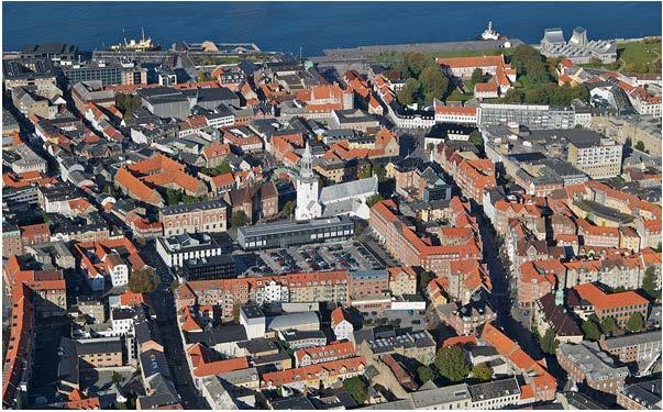
4 AALBORG

5

6
7


8
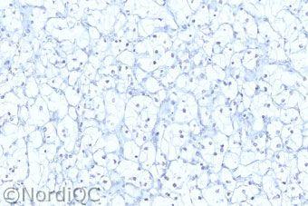
9 Serial sections stained for Estrogen receptor Lab. A Lab. B Optimally processed ductal breast carcinoma tissue
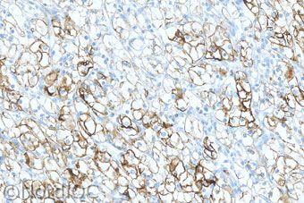
10 Serial sections stained for Estrogen receptor High expressor Lab. A Lab. B Low expressor False neg.
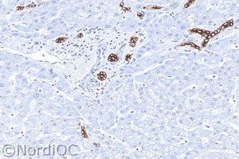
11 Serial sections stained for Estrogen receptor Control: uterine cervix Lab. A Lab. B False neg. 11


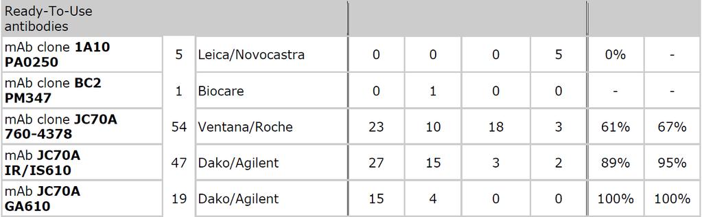
12 Serial sections stained for Estrogen receptor Control: uterine cervix Clone SP1/EP1/1D5 in 225 labs Clone 6F11 in 15/37 labs External Quality Assurance! False pos. 12

13 IHC Biomarker controls Decalcification Preparation Tissue Type, Dimension, Laser resection, De-differentiation With 3 choices for 5 variables in each phase = > 4 million protocols. Controlment Quantification Reporting Fixation Time, Type, Volume Preanalytic Postanalytic Section Thickness Storage Drying Pre-treatment Manual Stainer Visualization Sensitivity, Specificity Primary antibody Clone, Dilution Buffer, Time, Temp Analytic Interpretation Localization Positive/Negative - cut-off level Development Sensitivity, Localization

14 The challenge of IHC Suboptimal IHC assays may be due to: Preanalytical issues Fixation too short, too late, decalcification too soon Analytical issues: Less successful or too dilute antibody clones/rtus Insufficient epitope retrieval Insensitive visualization systems Platform problems Post-analytical issues Should be identified with proper external on-slide controls Interpretation criteria, interobserver variation
15


16 Cold spots a problem with ~30% of all slides

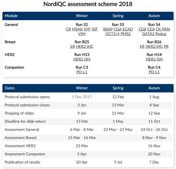
17 Nordic immunohistochemical Quality Control 17 International organization for proficiency testing of IHC Founded 2003 by Nordic pathologists Independent, scientific, not-for-profit organisation Institute of Pathology, Aalborg University Hospital, DK General module: 3 runs/year different marker challenges Breast cancer IHC module: 2 runs/y HER-2, ER/PR, Ki67/E-Cad HER-2 ISH module: 2 runs/year BRISH, FISH Companion module: 2 runs/year PD-L1
18 ~ 90 IHC markers in NordiQC Runs Tested 1-15 times Alpha-methylacyl-CoA racemase CyclinD1 MLH1 Alpha-smooth muscle actin Cytokeratin 5 MSH2 Anaplastic lymphoma kinase Cytokeratin 7 MSH6 B-cell specific activator protein Cytokeratin 19 Multiple myeloma oncogene 1 bcl-2protein Cytokeratin 20 Myosin, smooth muscle heavy chain bcl-6protein Cytokeratin, high molecular weight Napsin A Calretinin Cytokeratin, low molecular weight Neurofilament protein Cancer antigen 125 Cytokeratin, pan- Octamer transcription factor-3/4 Carcinoembryonic antigen Desmin p16 ink4a CD3 Detected on GIST-1 p40 CD4 E-cadherin p53 CD5 Epithelial cell adhesion molecule p57 CD8 Epithelial membrane antigen p63 CD10 Estrogen receptor alpha Paired box gene-2 protein CD14 Factor VIII related antigen Paired box gene-8 protein CD15 GATA3 Placental alkaline phosphatase CD19 Glial fibrillary acidic protein PMS2 CD20 Glypican 3 Podoplanin CD23 Gross cystic disease fluid protein-15 Prostate specific acid phosphatase CD30 HER-2 Prostate specific antigen CD31 Hepatocyte antigen Prostein CD34 Human chorionic gonadotropin Progesterone receptor CD45 Immunoglobulin kappa S-100 protein beta CD56 Immunoglobulin lambda Sal-like protein 4 CD68 Immunoglobubin M SOX10 CD79a Ki-67 Synaptophysin CD99 Mammaglobin Terminal deoxynucl. transferase CD117 Melan-A Vimentin Chromogranin Melanosoma specific antigen Wilm's tumour-1 protein
19 Nordic immunohistochemical Quality Control NordiQC Participants Nordic labs
20 Nordic immunohistochemical Quality Control Free PMC Article

21 FREE ACCESS
22 22
23
24
25

26 Participant site 26

27 Test material Multi-tissue FFPE blocks 10% NBF h (ASCO/CAP guidelines ) Normal and clinically relevant tumour tissues Different levels of antigen expression high, moderate, low, none 2 unstained slides for each marker send to the participants 1 stained slide returned for central assessment

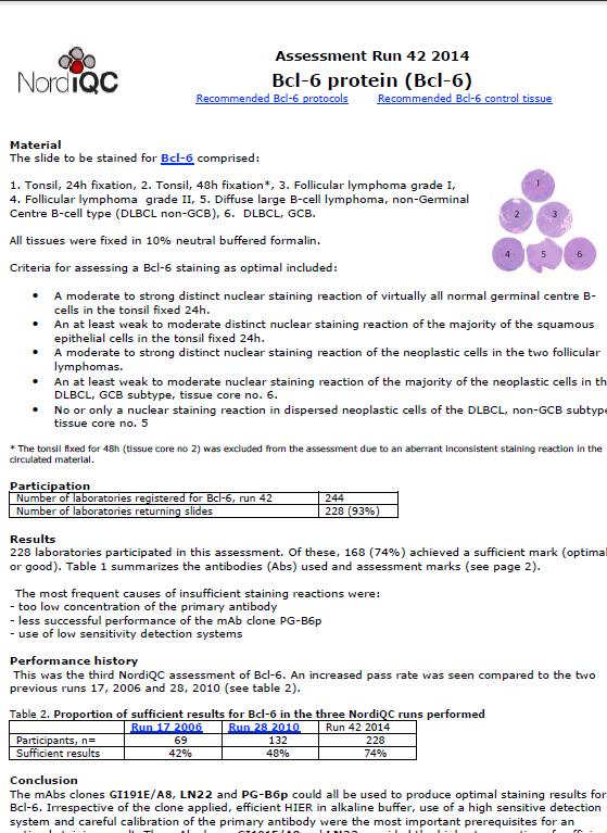
28 Test material 28 The slide to be stained for Bcl-6 comprised: 1-2. Tonsils, 24 h/48 h 3. Follicular lymphoma, grade I 4. Follicular lymphoma, grade II 5. Diffuse large B-cell lymphoma HE NE LE Tissue selection: High Expressor to confirm antibody Low Expressor to ensure sensitivity No-Expressor to ensure specificity

29 Open website


30 PDF file ed to participants with assessment marks and when needed explanations and recommendations 30
 Insufficient 32% 21% 11% 33% 35% Optimal Good Borderline Poor { too weak / false neg.")
31 NordiQC assessment results General module ~ 20,000 slides ( ~ core sections) Insufficient 32% 21% 11% 33% 35% Optimal Good Borderline Poor { too weak / false neg.: ~ 90% Insuff. over-stained / false pos.: ~ 10%

32 NordiQC assessment results Breast cancer module ~ 9,000 slides (~35,000 core sections) Insufficient 21% 9% 12% 21% 58% Optimal Good Borderline Poor { too weak / false neg.: ~ 90% Insuff. over-stained / false pos.: ~ 10%
33 NordiQC general results Major causes of insufficient stains in ~ 9,000 slides Less successful antibodies/rtus 17 % Inappropriate antibody dilution 20 % Inappropriate epitope retrieval 27 % Inappropriate detection kit 19 % Other inappropriate lab. performance 17 % Endogenous biotin reaction Section drying-out after HIER Technical platform error.... Unexplained 33
34 IHC Biomarker controls Go for Low antigen expressors ~ Critical Stain Quality Indicators (CSQI) essential to evaluate sensitivity essential to assure consistency Normal tissues when ever possible - easier to recognize and ensure the quality 90 % of insufficient staining results in EQA are caused by weak/false negative results often related to the use of inappropriate positive tissue controls...
35 Publications AIMM 2014, 22:241 AIMM 2015, 23:1

36 AIMM
37 NordiQC EQA: Estrogen Receptor in 13 runs % 70 Participants % B1 B3 B5 B7 B8 B10 B11 B13 B15 B17 PASS RATE (%)
38 NordiQC EQA ER Estrogen receptor Antibody clone selection SP1 6F11 1D5 EP


39 IHC Optimal performance ER 1D5 1:100 HIER Ci ph 6 2-step polymer 3-step polymer
40 Results of NordiQC recommendations Pass rate (optimal + good) by participant status Estrogen receptor New participants Old participants Run 10, % 71% Run B15, % 86% Run B19, % 73% Average 59% 77%


41 NordiQC runs for HER2 IHC 41 CK7 * * Optimal Ampl. 3+ Ampl. 2+ Unampl. 2+ Unampl. 0 Poor Ampl. 3+ Ampl. 1+ Unampl. 1+ Unampl. 0 41

42 NordiQC runs for HER2 IHC 42 CK7 Optimal Ampl. 3+ Ampl. 2+ Unampl. 2+ Unampl. 0 Poor Ampl. 3+ Ampl. 2+ Unampl. 3+ Unampl. 1 42

43 HER-2 staining results in 17 runs
 protocol at 98 C for 30 minutes.")
44 J Natl Cancer Inst 2009;101: Ki67 antibody clone SP6 applied at a 1:200 dilution for 32 minutes, by following the Ventana Benchmark automated immunostainer standard Cell Conditioner 1 (CC1) protocol at 98 C for 30 minutes. The best Ki67 index cut point to distinguish luminal B from luminal A tumors was 13.25%.


45 Ki67 immunoassay Digital image analysis Clone SP6 concentrate Ventana platform Prolif.index 38 % Clone MM1 RTU Leica platform Prolif.index 12 %
46 Concentrate Mib1 as concentrate in optimized protocols on 3 platforms RTU
: a transmembrane receptor tyrosin kinase CD30+ anaplastic large cell lymphomas (ALCLs) may be associated with a balanced (2;5)(p23;5q35) chromosomal translocation")
47 Mutations can be identified by mutation-specific proteins: ALK (anaplastic large cell lymphoma, lung adenocarcinoma). Anaplastic lymphoma kinase (ALK): a transmembrane receptor tyrosin kinase CD30+ anaplastic large cell lymphomas (ALCLs) may be associated with a balanced (2;5)(p23;5q35) chromosomal translocation Anaplastic lymphoma kinase (ALK) rearrangements are present in about 5% of advanced non-small-cell lung cancer Crizotinib was the first ALK tyrosine kinase inhibitor licensed for the treatment of metastatic ALK-positive NSCLC

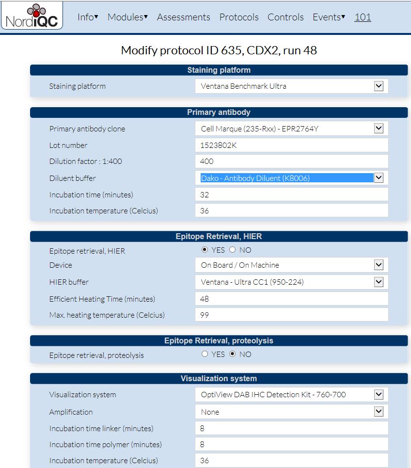
48 Lung ALK run 45, 176 labs 5A4 ALK1 ALCL Optimal Lung adenocarcinoma Poor

49 Lung ALK run 45, 176 labs


50 Lung ALK The immunoassay must fit for the purpose: Identify the antibody useful for the specific task The right external controls must be used: Tissue with high epitope expression to identify the right antibody Appendix Appendix - ALK Tissue with low epitope expression to assure the sensitivity: ALK-positive lung adenocarcinoma Tissue with no epitope expression to assure the specificity e.g., liver

51 Lung ALK run 45, 176 labs 5A4 ALK1 Appendix Optimal Poor Lung adenocarcinoma


52 External Quality Assurance PDL1 >50% <50% 52
53 Misleading data sheets Pan-Cytokeratin
54 Inappropriate retrieval (31%) AE1/AE3 + HIER TP Liver RCC FN AE1/AE3 + proteolysis TP 54 FN

55 IHC - NordiQC 2014 AE1/AE3 : Optimal results only obtained by HIER in NordiQC runs Dako: RTU HIER Leica: RTU Proteolysis Thermo: VMS: RTU - Proteolysis Conc: Proteolysis or HIER Conc: HIER Conc: HIER Quanto Proteolysis UltraVision Misleading data sheets + Wrong control material used 55

56 CEA run 47, 2016, 255 labs


57 CEA Optimal Urothelial carcinoma Poor: TF3H8-1 Optimal Liver Poor: TF3H8-1

58 CDX2 run 48


59 CDX2 Optimal Pancreas Poor: Colon adenocarcinoma

60 Calretinin - run 45 Concentrates Optimal Poor Adrenal gland


61 Calretinin 113 labs using RTUs Optimal Insufficient Malignant Mesothelioma

62 S-100 Mal. melanoma Optimal Insufficient
63 CD31 Angiosarcoma


64 Biotin based system giving false postives Synaptophysin Labelled Steptavidin-Biotin system No antibody Labelled Streptavidin-Biotin system: Neg. reagent control mandatory Synaptophysin Polymer based system 64

65 Platform dependent antibodies: PAX5 Hodgkin lymphoma NS clone SP34 RTU VMS/CM x200 clone 24 RTU VMS/CM x200

66 IHC Most common pitfalls
67 Tailored recommendations Replace less successful antibodies (conc./rtu) Calibrate the antibody concentration Use HIER (instead of proteolysis or no retrieval) Increase HIER time / temperature / buffer ph For 95% of epitopes ph 8-9 is preferable to ph 6 Use a non-biotin based viz. system Use FDA approved kits instead of home-brews..... Improve the internal QC: Identify the right controls 67 Select well defined normal low expressor cells/tissues
68 Results of NordiQC recommendations 419 advices for 11 markers No. Improved % Positive Negative
69 Conclusion External Quality Assurance (EQA) Provides objective evidence of lab performance Identifies methodological errors Provides directions for improvements & controls The results of the NordiQC work indicate that Improvement of IHC is strongly needed EQA schemes, industry and KOL must align - describing the requirements for optimal IHC performance. 69
70 Perspective Almost 1/3 of all IHC stains produced by NordiQC participants are still insufficient! New labs New antibodies, techniques, platforms Increasing demands How many IHC stains produced by labs not participating in an EQA scheme are insufficient? How many scientific publications are based on insufficient IHC stains? What are the consequences for the patients?
71 External Quality Assurance ER correct false negative Through the inquiry, the public learned that between 1997 and 2005 nearly 400 of about 1,000 breast cancer patients received incorrect test results of the ER status of their breast tumors. Control 71 Craig Allred

72 When you believe in automation and stop thinking
73 The impact of proficiency testing on lab immunoassays Mogens Vyberg Professor of Clinical Pathology Director of NordiQC Aalborg University Hospital, Aalborg, Denmark Thank you for your attention
NordiQC External Quality Assurance in Immunohistochemistry
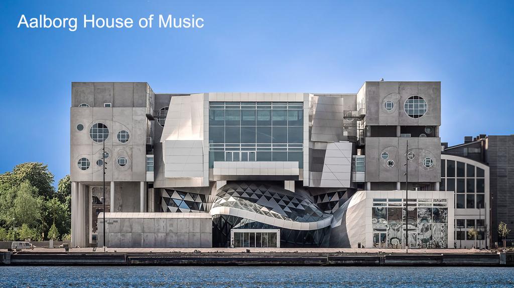
 NordiQC External Quality Assurance in Immunohistochemistry Mogens Vyberg Professor of Clinical Pathology Director of NordiQC Aalborg University Hospital, Aalborg, Denmark AALBORG (~ 200.000 inhabitants)
Quality Assurance in Immunohistochemistry: Experiences from NordiQC
 Nordic immunohistochemical Quality Control 2 Quality Assurance in Immunohistochemistry: Experiences from NordiQC Prof. Mogens Vyberg NordiQC Institute of Pathology Aalborg University Hospital Aalborg,
10 years of NordiQC Why are 30% of labs still getting it wrong?
 Mogens Vyberg & Søren Nielsen NordiQC Institute of Pathology Aalborg University Hospital Aalborg, Denmark May 29th 2015 10 years of NordiQC Why are 30% of labs still getting it wrong? Nothing to declare
Immunohistochemistry. Potential and challenges To be or not to be
 Immunohistochemistry Potential and challenges To be or not to be Søren Nielsen Scheme Manager NordiQC Aalborg University Hospital, Denmark Vårmöte 19.05.2016 Karlstad Overview IHC project coordinator at
Nordic Immunohistochemical Quality Control
 Nordic Immunohistochemical Quality Control Immunohistochemistry in the classifiation of neoplasias of the alimentary tract & External Quality Assurance of Immunohistochemistry for GI cancer markers Mogens
External Quality Assessment of Breast Marker Analysis. NordiQC data
 External Quality Assessment of Breast Marker Analysis NordiQC data Søren Nielsen Scheme Manager NordiQC Aalborg University Hospital, Denmark Aalborg 12.06 2015 Markers assessed in NordiQC Predictive markers
The unknown primary tumour: IHC classification part I, the primary panel - Antibody selection, protocol optimization, controls and EQA
 The unknown primary tumour: IHC classification part I, Mogens Vyberg Professor of Clinical Pathology Director of NordiQC Aalborg University Hospital, Aalborg, Denmark the primary panel - Antibody selection,
Optimization of antibodies, selection, protocols and controls Breast tumours
 Optimization of antibodies, selection, protocols and controls Breast tumours Søren Nielsen Project coordinator & Scheme Manager NordiQC Aalborg University Hospital, Denmark Breast panel: GCDFP-15 Mammaglobin
Breast cancer: Antibody selection, protocol optimzation controls and EQA
 Breast cancer: Antibody selection, protocol optimzation controls and EQA Workshop in Diagnostic Immunohistochemistry Oud St. Jan/ Old St. John Brugge (Bruges), Belgium June 13th 15nd 2018 Rasmus Røge,
Breast cancer: IHC classification. Mogens Vyberg Professor of Clinical Pathology Director of NordiQC Aalborg University Hospital, Aalborg, Denmark
 Breast cancer: IHC classification Mogens Vyberg Professor of Clinical Pathology Director of NordiQC Aalborg University Hospital, Aalborg, Denmark http://upload.wikimedia.org/wikipedia/commons/1/1a/breast.svg
The unkown primary tumour: IHC Classification, antibody selection, protocol optimization, controls and EQA (part I)
") The unkown primary tumour: IHC Classification, antibody selection, protocol optimization, Mogens Vyberg Professor of Clinical Pathology Director of NordiQC Aalborg University Hospital, Aalborg, Denmark
The unkown primary tumour: IHC Classification, antibody selection, protocol optimization, Mogens Vyberg Professor of Clinical Pathology Director of NordiQC Aalborg University Hospital, Aalborg, Denmark
NordiQC - update
 NordiQC - update 00-0 EQUALIS Uppsala 0 Tomas Seidal NordiQC participants NordiQC participants n:30 S DK N 6 F Ice Bel 54 NL 4 Ger 6 Aust USA 0 It 8 Argent 8.. 96% participation in S,DK & N ~ 60% in Finland
Carcinoembryonic antigen (CEA)
") Assessment Run 37 2013 Carcinoembryonic antigen (CEA) Material The slide to be stained for CEA comprised: 1. Appendix, 2. Liver, 3-4. Colon adenocarcinoma, 5. Urothelial carcinoma All tissues were fixed
Assessment Run 37 2013 Carcinoembryonic antigen (CEA) Material The slide to be stained for CEA comprised: 1. Appendix, 2. Liver, 3-4. Colon adenocarcinoma, 5. Urothelial carcinoma All tissues were fixed
Immunohistochemical classification of the unknown primary tumour (UPT) Part I. Prof. Mogens Vyberg NordiQC Institute of Pathology Aalborg, Denmark
 Part I. Prof. Mogens Vyberg NordiQC Institute of Pathology Aalborg, Denmark") Immunohistochemical classification of the unknown primary tumour (UPT) Part I Prof. Mogens Vyberg NordiQC Institute of Pathology Aalborg, Denmark Tumours of unknown origin: Histology Brain tumour - biopsy
Immunohistochemical classification of the unknown primary tumour (UPT) Part I Prof. Mogens Vyberg NordiQC Institute of Pathology Aalborg, Denmark Tumours of unknown origin: Histology Brain tumour - biopsy
Immunohistochemical classification of lung carcinomas and mesotheliomas. Prof. Mogens Vyberg NordiQC Institute of Pathology Aalborg, Denmark
 Immunohistochemical classification of lung carcinomas and mesotheliomas Prof. Mogens Vyberg NordiQC Institute of Pathology Aalborg, Denmark Endobronchial ultrasound guided transbronchial needle biopsy
SMH (Myosin, smooth muscle heavy chain)
") Material The slide to be stained for SMH comprised: Assessment Run 50 2017 SMH (Myosin, smooth muscle heavy chain) 1.Tonsil, 2. Esophagus, 3. Breast hyperplasia, 4. Breast ductal carcinoma in situ (DCIS),
Material The slide to be stained for SMH comprised: Assessment Run 50 2017 SMH (Myosin, smooth muscle heavy chain) 1.Tonsil, 2. Esophagus, 3. Breast hyperplasia, 4. Breast ductal carcinoma in situ (DCIS),
Assessment Run GATA3
 Assessment Run 44 2015 GATA3 Material The slide to be stained for GATA3 comprised: 1. Tonsil 2. Kidney, 3. Urothelial carcinoma, 4. Breast ductal carcinoma, 5. Colon adenocarcinoma All tissues were fixed
Schedule of Accreditation issued by United Kingdom Accreditation Service 2 Pine Trees, Chertsey Lane, Staines-upon-Thames, TW18 3HR, UK
 Schedule of ccreditation United Kingdom ccreditation Service 2 Pine Trees, Chertsey Lane, Staines-upon-Thames, TW18 3HR, UK External Quality ssessment Services for Cancer Diagnostics CIC Issue No: 005
Lung Anaplastic Lymphoma Kinase (lu-alk)
") Assessment Run 5 207 Lung Anaplastic Lymphoma Kinase (lu-alk) Material The slide to be stained for lu-alk comprised:. Appendix, 2. Tonsil, 3. Merkel cell carcinoma, 4. Anaplastic large cell lymphoma with
Assessment Run 5 207 Lung Anaplastic Lymphoma Kinase (lu-alk) Material The slide to be stained for lu-alk comprised:. Appendix, 2. Tonsil, 3. Merkel cell carcinoma, 4. Anaplastic large cell lymphoma with
Assessment Run C1 2017
 Assessment Run C1 2017 PD-L1 The first assessment in this new NordiQC Companion module C1 focused on the accuracy of the PD-L1 IHC assays performed by the participating laboratories to identify patients
Assessment Run NKX3.1 (NKX3.1)
") Assessment Run 49 2017 NKX3.1 (NKX3.1) Material The slide to be stained for NKX3.1 comprised: 1. Testis 2. Appendix 3-4. Prostate adenocarcinoma 5. Prostate hyperplasia All tissues were fixed in 10% neutral
Assessment Run 49 2017 NKX3.1 (NKX3.1) Material The slide to be stained for NKX3.1 comprised: 1. Testis 2. Appendix 3-4. Prostate adenocarcinoma 5. Prostate hyperplasia All tissues were fixed in 10% neutral
Diagnostic IHC in lung and pleura pathology
 Diagnostic IHC in lung and pleura pathology Mogens Vyberg Professor of Clinical Pathology Director of NordiQC Aalborg University Hospital, Aalborg, Denmark WHO 2004 and Web Malignant mesothelioma Epithelioid
Protocols for Zytomed Systems antibodies on fully automated IHC staining systems date of issue: September 20, 2012
 Protocols for Zytomed Systems antibodies on fully automated IHC staining systems date of issue: September 20, 2012 These protocols were provided by customers. Under no circumstances shall Zytomed Systems
Classification of the unknown primary tumour: the primary IHC panel
 CIQC/CAP-ACP SEMINAR 2013: DIAGNOSTIC IHC AND MOLECULAR PATHOLOGY Classification of the unknown primary tumour: the primary IHC panel Aalborg University Hospital Denmark Tumours of unknown origin: Histology
Immunohistochemical principles The technical test approach. Pre-analytical parametres
 Immunohistochemical principles The technical test approach Pre-analytical parametres Søren Nielsen Global Pathology Manager Agilent Technologies (Former Scheme Manager, NordiQC) 2 IHC project coordinator
Thyroid transcription factor-1 (TTF1) Assessment run
 Assessment run") Thyroid transcription factor- (TTF) Assessment run 39 203 The slide to be stained for TTF comprised:. Thyroid gland, 2. Liver, 3. Normal lung, 4. Lung adenocarcinoma 5. Colon adenocarcinoma, 6 & 7. Lung
Thyroid transcription factor- (TTF) Assessment run 39 203 The slide to be stained for TTF comprised:. Thyroid gland, 2. Liver, 3. Normal lung, 4. Lung adenocarcinoma 5. Colon adenocarcinoma, 6 & 7. Lung
Cytokeratin 19 (CK19)
") Assessment Run 34 202 Cytokeratin 9 (CK9) Material The slide to be stained for CK9 comprised:. Thyroid gland, 2. Appendix, 3. Esophagus, 4. Papillary thyroid carcinoma, 5 & 6. Pancreatic neuroendocrine
Assessment Run 34 202 Cytokeratin 9 (CK9) Material The slide to be stained for CK9 comprised:. Thyroid gland, 2. Appendix, 3. Esophagus, 4. Papillary thyroid carcinoma, 5 & 6. Pancreatic neuroendocrine
Estrogen receptor (ER)
") Material The slide to be stained for ER comprised: Assessment B25 208 Estrogen receptor (ER) No. Tissue ER-positivity* ER-intensity*. Uterine cervix 80-90% Moderate to strong 2. Tonsil < 2-5% Weak to strong
Material The slide to be stained for ER comprised: Assessment B25 208 Estrogen receptor (ER) No. Tissue ER-positivity* ER-intensity*. Uterine cervix 80-90% Moderate to strong 2. Tonsil < 2-5% Weak to strong
Assessment Run B HER2 IHC
 Assessment Run B24 2017 HER2 IHC Material The slide to be stained for HER2 comprised the following 5 materials: IHC: HER2 Score* (0, 1+, 2+, 3+) FISH: HER2 gene/chr 17 ratio** 1. Breast carcinoma, no.
Assessment Run C3 2018
 Assessment Run C3 2018 PD-L1 Amended version May 14 th 2018 The third assessment in NordiQC Companion module C3 focused on the accuracy of the PD-L1 IHC assays performed by the participating laboratories
Assessment Run
 Assessment Run 50 2017 S100 Material The slide to be stained for S100 comprised: 1. Appendix, 2. Tonsil, 3. Schwannoma, 4-5. Malignant melanoma, 6. Colon adenocarcinoma. All tissues were fixed in 10% neutral
Estrogen receptor (ER)
") Assessment Run B7 204 Estrogen receptor (ER) Material The slide to be stained for ER comprised: No. Tissue ER-positivity* ER-intensity*. Uterine cervix 80-90% Moderate to strong 2. Breast carcinoma 0%
Assessment Run B7 204 Estrogen receptor (ER) Material The slide to be stained for ER comprised: No. Tissue ER-positivity* ER-intensity*. Uterine cervix 80-90% Moderate to strong 2. Breast carcinoma 0%
Assessment Run B HER-2 IHC. HER-2/chr17 ratio**
 Assessment Run B2 20 HER-2 IHC Material The slide to be stained for HER-2 comprised the following 5 tissues: IHC HER-2 Score* (0, +, 2+,3+) FISH HER-2/chr7 ratio**. Breast ductal carcinoma 0..3 2. Breast
Estrogen receptor (ER)
") Material The slide to be stained for ER comprised: Assessment Run B26 2018 Estrogen receptor (ER) No. Tissue ER-positivity* ER-intensity* 1. Uterine cervix 80-90% Moderate to strong 2. Tonsil 1-5% Weak
Material The slide to be stained for ER comprised: Assessment Run B26 2018 Estrogen receptor (ER) No. Tissue ER-positivity* ER-intensity* 1. Uterine cervix 80-90% Moderate to strong 2. Tonsil 1-5% Weak
Single and Multiplex Immunohistochemistry
 Single and Multiplex Immunohistochemistry Steve Westra, BS Reagent Product Specialist Leica Biosystems IHC Theory Polyclonal vs Monoclonal Polyclonal reagents Detect a multitude of epitopes Batch to batch
External Quality Assessment of melanocytic marker analyses NordiQC experience
 External Quality Assessment of melanocytic marker analyses NordiQC experience Jan Klos MD, Department of Pathology Stavanger University Hospital Norway 1 Content 18 Runs = 2112 submissions between 2001-2014
Assessment Run B HER2 IHC
 Assessment Run B26 208 HER2 IHC Material The slide to be stained for HER2 comprised the following 5 materials: IHC: HER2 Score* (0, +, 2+, 3+) FISH: HER2 gene/chr 7 ratio**. Breast carcinoma, no. 2+..3
Epithelial cell-cell adhesion molecule (Ep-CAM)
") Assessment Run 3 011 Epithelial cell-cell adhesion molecule (Ep-CAM) Material The slide to be stained for Ep-CAM comprised: 1. Appendix,. Kidney, 3. Adrenal gland, 4. Lung carcinoid, 5 & 6. Renal clear
Assessment Run 3 011 Epithelial cell-cell adhesion molecule (Ep-CAM) Material The slide to be stained for Ep-CAM comprised: 1. Appendix,. Kidney, 3. Adrenal gland, 4. Lung carcinoid, 5 & 6. Renal clear
The unkown primary tumour: IHC Classification, antibody selection, protocol optimization, controls and EQA (part II)
") The unkown primary tumour: IHC Classification, antibody selection, protocol optimization, Mogens Vyberg Professor of Clinical Pathology Director of NordiQC Aalborg University Hospital, Aalborg, Denmark
The unkown primary tumour: IHC Classification, antibody selection, protocol optimization, Mogens Vyberg Professor of Clinical Pathology Director of NordiQC Aalborg University Hospital, Aalborg, Denmark
HER2 ISH (BRISH or FISH)
") Assessment Run H14 2018 HER2 ISH (BRISH or FISH) Material Table 1. Content of the multi-block used for the NordiQC HER2 ISH assessment, run H14 HER2 IHC* IHC score Dual - SISH** FISH*** FISH*** HER2/chr17
Assessment Run H14 2018 HER2 ISH (BRISH or FISH) Material Table 1. Content of the multi-block used for the NordiQC HER2 ISH assessment, run H14 HER2 IHC* IHC score Dual - SISH** FISH*** FISH*** HER2/chr17
Quality Control/Quality Assurance in Diagnostic Immunohistochemistry
 CIHRT Exhibit P- Page Quality Control/Quality Assurance in Diagnostic Immunohistochemistry Emina Torlakovic, MD, PhD College of Medicine University of Saskatchewan Emina Emilia Torlakovic, MD, PhD Associate
IHC Stainer platforms. Overview, pros and cons
 IHC Stainer platforms Overview, pros and cons Bart De Wiest Quality manager IHC OLV Hospital, Aalst, Belgium Donald Van Hecke Lab & Quality manager AZ St-Lucas, Brugge, Belgium Goal of this lecture: to
Product Introduction. Product Codes: HCL029, HCL030 and HCL031. Issue
 Product Introduction Product Codes: HCL029, HCL030 and HCL031 Issue 1. 180510 Contents Introduction to Estrogen Receptor 2 ER immunohistochemistry 3 Quality control 5 Cell lines as controls 6 Estrogen
Applications of IHC. Determination of the primary site in metastatic tumors of unknown origin
 Applications of IHC Determination of the primary site in metastatic tumors of unknown origin Classification of tumors that appear 'undifferentiated' by standard light microscopy Precise classification
Assessment Run CK19
 Assessment Run 29 200 CK9 The slide to be stained for CK9 comprised:. Appendix, 2. Thyroid gland, 3. Pancreas, 4. Ductal breast carcinoma, 5. Esophagus, 6. Papillary thyroid carcinoma. All tissues were
NEW IHC A n t i b o d i e s
 NEW IHC Antibodies TABLE OF CONTENTS NEW IHC ANTIBODIES from Cell Marque CITED1 (5H6).... 1 Claudin 7 (5D10F3).... 1 GATA1 (4F5).... 1 Transgelin (2A10C2).... 1 NEW IHC ANTIBODIES using RabMAb Technology
Schedule of Accreditation issued by United Kingdom Accreditation Service 2 Pine Trees, Chertsey Lane, Staines-upon-Thames, TW18 3HR, UK
 2 Pine Trees, Chertsey Lane, Staines-upon-Thames, TW18 3HR, UK UCL-Advanced Diagnostics 1st Floor, Rockefeller Building 21 University Street London WC1E 6JJ Contact: David Allen Tel: +44 (0)20 7679 6912
Assessment Run B HER-2
 Assessment Run B1 2006 HER-2 The slide to be stained for HER-2 comprised: 1. Cell line JIMT-1 (Amplified)* 2. Cell line MDA-453 (Amplified) 3. Cell line MCF-7 (Not amplified) 4. Cell line BT474 (Amplified)
Image analysis in IHC overview, considerations and applications
 Image analysis in IHC overview, considerations and applications Rasmus Røge, MD, Institute of Pathology, Aalborg University Hospital NordiQC workshop September 2016 Aalborg, Denmark Outline Theory Image
In Situ Hybridization Immunohistochemistry
 2013-2014 In Situ Hybridization Immunohistochemistry A History of Delivering Superior Products Genemed Biotechnologies, Inc. was founded in 1987 and is located in the center of the South San Francisco
Product Introduction
 Product Introduction Product Codes: HCL026, HCL027 and HCL028 Contents Introduction to HER2 2 HER2 immunohistochemistry 3 Cell lines as controls 5 HER2 Analyte Control DR IHC 7 HER2 Analyte Control DR
ADVANCED STAINING PRODUCT CATALOG. In Situ Hybridization Probes Immunohistochemistry Antibodies Detection Systems Ancillary Reagents
 2017 ADVANCED STAINING PRODUCT CATALOG In Situ Hybridization Probes Immunohistochemistry Antibodies Detection Systems Ancillary Reagents A History of Delivering Superior Products Genemed a wholly-owned
Sal-like protein 4 (SALL4)
") Assessment Run 43 205 Sal-like protein 4 (SALL4) The slide to be stained for SALL4 comprised:. Appendix, 2. Testis, 3. Renal clear cell carcinoma, 4. Seminoma, 5. Intratubular germ cell neoplasia (IGCN),
Assessment Run 43 205 Sal-like protein 4 (SALL4) The slide to be stained for SALL4 comprised:. Appendix, 2. Testis, 3. Renal clear cell carcinoma, 4. Seminoma, 5. Intratubular germ cell neoplasia (IGCN),
Thermo Scientific UltraVision Quanto for Immunohistochemistry The New Generation Micro-Polymer Detection System
 Thermo Scientific for Immunohistochemistry The New Generation Micro-Polymer Detection System highest sensitivity sharp crisp clear shorter incubation times UltraVision Quanto the new Micro-Polymer System
DOUBLE STAINS. Toll-Free: Direct:
 DOUBLE STAINS CD61 + CD71 DAB Brown: CD61 Alk. Phos. Red: CD71 Bone Marrow DAB Brown: Megakaryocytes Alk. Phos. Red: Erythroid Precursors 400x CD61 (2f2) 0.1 ml concentrate............. 161M-14 0.5 ml
Technology from Abcam
 CD2 (EP222) CD2 is one of the earliest T-cell lineage restricted antigens to appear during T-cell differentiation and only rare CD2+ cells can be found in the bone marrow. Anti-CD2 is a pan-t-cell antigen
C.L. Davis Foundation Descriptive Veterinary Pathology Course
 C.L. Davis Foundation 2015 Descriptive Veterinary Pathology Course IHC Resources IHC Identification Targets Antibodies Antibodies 1 Antibodies Specimens Antigen Retrieval Unmasks antigen epitopes Methods
Differential diagnosis of HCC
 Hepatocellular Carcinoma Quest for an Ideal Immunohistochemical Panel Sanjay Kakar, MD UCSF Differential diagnosis of HCC Hepatocellular lesions Adenoma, FNH, HG dysplasia Adenocarcinoma CholangioCA, metastasis
Assessment performed on Tuesday, July 29, 2014, at Lions Gate Hospital, North Vancouver
 Assessors report for ciqc Run 37: BRAF V600E (April 2014) Assessors: B Gilks, R Wolber, K Ung, P Tavassoli, J Garratt and J Won (recorder) Assessment performed on Tuesday, July 29, 2014, at Lions Gate
HistoCyte Laboratories Ltd
 HistoCyte Laboratories Ltd Progesterone Receptor: The neglected breast receptor! Dr Ian Milton & Colin Tristram November 2018 UKNEQAS Autumn meeting Introduction Progesterone is an important prognostic
The role of immunohistochemistry in surgical pathology of the uterine corpus and cervix
 The role of immunohistochemistry in surgical pathology of the uterine corpus and cervix Prof. Ben Davidson, MD PhD Department of Pathology, Norwegian Radium Hospital, Oslo University Hospital, Oslo, Norway
Review and Updates of Immunohistochemistry in Selected Salivary Gland and Head and Neck Tumors
 Review and Updates of Immunohistochemistry in Selected Salivary Gland and Head and Neck Tumors. Monophasic tumors : myoepithelioma, acinic cell carcinoma, and salivary duct carcinoma. Biphasic tumors includes
Tissue-based Immunohistochemical Biomarker Expression in Malignant Glandular Lesions of the Uterine Cervix: a Systematic Review
 Tissue-based Immunohistochemical Biomarker Expression in Malignant Glandular Lesions of the Uterine Cervix: a Systematic Review Sandra Lee MD, FRCPC 1 *, Vikrant V. Sahasrabuddhe, MBBS, DrPH 2 *, Diana
Immunotherapy in NSCLC Pathologist role
 Immunotherapy in NSCLC Pathologist role Pimpin Incharoen, M.D. Assistant Professor, Thoracic Pathology Department of Pathology, Ramathibodi Hospital Genetic alterations in NSCLC Khono et al, Trans Lung
Charles Halsey, DVM, PhD, DACVP Pfizer, Inc. IHC Resources
 Charles Halsey, DVM, PhD, DACVP Pfizer, Inc. IHC Resources 1 IHC Identification Targets Specimens Controls 2 Tissue controls Trouble Spots 3 The Key to Description IHC Description 4 Intermediate Filaments
Immunohistochemistry on Fluid Specimens: Technical Considerations
 Immunohistochemistry on Fluid Specimens: Technical Considerations Blake Gilks Dept of Pathology University of British Columbia, Vancouver, BC, Canada Disclosures None Learning Objectives At the end of
Breast cancer diagnostic solutions Deliver diagnostic confidence
 Breast cancer diagnostic solutions Deliver diagnostic confidence 2 Breast cancer diagnostic solutions Roche Tissue Diagnostics is committed to improving outcomes in breast cancer Breast cancer...the most
LUNG CANCER. pathology & molecular biology. Izidor Kern University Clinic Golnik, Slovenia
 LUNG CANCER pathology & molecular biology Izidor Kern University Clinic Golnik, Slovenia 1 Pathology and epidemiology Small biopsy & cytology SCLC 14% NSCC NOS 4% 70% 60% 50% 63% 62% 61% 62% 59% 54% 51%
Bihong Zhao, M.D, Ph.D Department of Pathology
 Bihong Zhao, M.D, Ph.D Department of Pathology 04-28-2009 Is tumor self or non-self? How are tumor antigens generated? What are they? How does immune system respond? Introduction Tumor Antigens/Categories
The Panel Approach to Diagnostics. Lauren Hopson International Product Specialist Cell Marque Corporation
 The Panel Approach to Diagnostics Lauren Hopson International Product Specialist Cell Marque Corporation Cell Marque Rocklin, California About Cell Marque: IVD primary antibody manufacturer Distributors
Instant Quality FISH. The name says it all.
 COMPANION DIAGNOSTICS Instant Quality FISH Instant Quality FISH. The name says it all. IQ: Instant Quality every time. Breast carcinoma stained with : Triple filter showing Blue DAPI colors nuclei, FITC
# Best Practices for IHC Detection and Interpretation of ER, PR, and HER2 Protein Overexpression in Breast Cancer
 #1034 - Best Practices for IHC Detection and Interpretation of ER, PR, and HER2 Protein Overexpression in Breast Cancer Richard W. Cartun, MS, PhD Andrew Ricci, Jr, MD Department of Pathology Hartford
The clinically challenging entity of liver metastasis from tumors of unknown primary
 The clinically challenging entity of liver metastasis from tumors of unknown primary Xuchen Zhang, MD, PhD Associate Professor of Pathology Department of Pathology Yale University School of Medicine Liver
Quality in Control. ROS1 Analyte Control. Product Codes: HCL022, HCL023 and HCL024
 Quality in Control ROS1 Analyte Control Product Codes: HCL022, HCL023 and HCL024 Contents What is ROS1? 2 The Role of ROS1 in Cancer 3 ROS1 Assessment 3 ROS1 Analyte Control Product Details 4 ROS1 Analyte
Quality in Control ALK-Lung Analyte Control (EML4-ALK) ALK-Lymphoma Analyte Control (NPM-ALK)
 ALK-Lymphoma Analyte Control (NPM-ALK)") Quality in Control ALK-Lung Analyte Control (EML4-ALK) ALK-Lymphoma Analyte Control (NPM-ALK) 10 Product Codes: HCL007, HCL008 and HCL009 HCL010, HCL011 and HCL012 Page 1 of Contents 1. What is ALK?...
Quality in Control ALK-Lung Analyte Control (EML4-ALK) ALK-Lymphoma Analyte Control (NPM-ALK) 10 Product Codes: HCL007, HCL008 and HCL009 HCL010, HCL011 and HCL012 Page 1 of Contents 1. What is ALK?...
Workflow. Connecting the Pieces For Total Patient Care
 Workflow Connecting the Pieces For Total Patient Care Biocare provides a full line of IHC and molecular pathology products for cancer and infectious disease diagnosis. From a full range of equipment: including
Welcome! HER2 TESTING DIAGNOSTIC ACCURACY 4/11/2016
 HER2 TESTING DIAGNOSTIC ACCURACY Can t We Finally Get It Right? Allen M. Gown, M.D. Medical Director and Chief Pathologist PhenoPath Laboratories Seattle, Washington Clinical Professor of Pathology University
PD-L1 Analyte Control DR
 Quality in Control PD-L1 Analyte Control DR PD-L1_PI_v2 Product Codes: HCL019, HCL020 and HCL021 Contents PD-L1 Analyte Control DR 2 What is PD-L1? 3 The Role of PD-L1 in Cancer 3 PD-L1 Assessment 4 PD-L1
INTERPRETATION OF IMMUNOHISTOCHEMICAL STAINS - DIFFICULTIES AND PITFALLS. Gabor Fischer Diagnostic Services Manitoba University of Manitoba
 INTERPRETATION OF IMMUNOHISTOCHEMICAL STAINS - DIFFICULTIES AND PITFALLS Gabor Fischer Diagnostic Services Manitoba University of Manitoba IHC INTERPRETATIONS LOCAL DATA Diagnostic Services Manitoba Number
Evolution of Pathology
 1 Traditional pathology Molecular pathology 2 Evolution of Pathology Gross Pathology Cellular Pathology Morphologic Pathology Molecular/Predictive Pathology Antonio Benivieni (1443-1502): First autopsy
Speaking to you. This statement by Dr. Rodney T. Miller, Director of
 Zyto_Facts 1-2013 News for pathology and immunohistochemistry +++Newsflash +++ Newsflash++ Speaking to you IHC algorithm poster now available in English. You can download the poster directly from our homepage
Cancers of unknown primary : Knowing the unknown. Prof. Ahmed Hossain Professor of Medicine SSMC
 Cancers of unknown primary : Knowing the unknown Prof. Ahmed Hossain Professor of Medicine SSMC Definition Cancers of unknown primary site (CUPs) Represent a heterogeneous group of metastatic tumours,
Role of the Pathologist in Guiding Immuno-oncological Therapies. Scott Rodig MD, PhD
 Role of the Pathologist in Guiding Immuno-oncological Therapies Scott Rodig MD, PhD Department of Pathology, Brigham & Women s Hospital Center for Immuno-Oncology, Dana-Farber Cancer Institute Associate
Interpretation Guide. Product Name: ALK Cell Line Analyte Control Product Codes: ALK2/CS and ALK2/CB. Page 1 of 9
 Interpretation Guide Product Name: ALK Cell Line Analyte Control Product Codes: ALK2/CS and ALK2/CB ALK2/CS/CB_IG_V_001 www.histocyte.com Page 1 of 9 Contents 1. What is ALK?... 2 2. Role of ALK in Cancer...
Schedule of Accreditation issued by United Kingdom Accreditation Service 2 Pine Trees, Chertsey Lane, Staines-upon-Thames, TW18 3HR, UK
 2 Pine Trees, Chertsey Lane, Staines-upon-Thames, TW18 3HR, UK The Princess Alexandra Hospital NHS Trust The Michael Letcher Department of Cellular Pathology The Princess Alexandra Hospital NHS Trust Hamstel
IHC Panels as an Aid in Diagnostic Decision Making
 IHC Antibody Test Selection Using a Panel Approach Steven Westra B.S. Reagent Product Specialist Leica Biosystems IHC Panels as an Aid in Diagnostic Decision Making Diagnostic Use of Tumors Using Algorithms
Breast - ductal carcinoma CK7 ER PR GATA3 Mammaglobin (50-70%) GCDFP-15 (50-70%) E-cadherin HMWCK CK20 PAX2 ER/PR/HER2 on all newly diagnosed cases
 GCDFP-15 (50-70%) E-cadherin HMWCK CK20 PAX2 ER/PR/HER2 on all newly diagnosed cases") Adrenal cortical carcinoma Inhibin Synap Melan-A Calretinin Vimentin Chromogr CK7 CK20 Breast - ductal carcinoma CK7 ER PR GATA3 Mammaglobin (50-70%) GCDFP-15 (50-70%) E-cadherin HMWCK CK20 PAX2 ER/PR/HER2
Adrenal cortical carcinoma Inhibin Synap Melan-A Calretinin Vimentin Chromogr CK7 CK20 Breast - ductal carcinoma CK7 ER PR GATA3 Mammaglobin (50-70%) GCDFP-15 (50-70%) E-cadherin HMWCK CK20 PAX2 ER/PR/HER2
Tumor Markers Yesterday, Today & Tomorrow. Steven E. Zimmerman M.D. Vice President & Chief Medical Director
 Tumor Markers Yesterday, Today & Tomorrow Steven E. Zimmerman M.D. Vice President & Chief Medical Director Tumor Marker - Definition Substances produced by cancer cells or other cells in response to cancer
COMPUTER-AIDED HER-2/neu EVALUATION IN EXTERNAL QUALITY ASSURANCE (EQA) OF BREAST CANCER SCREENING PROGRAMME
 OF BREAST CANCER SCREENING PROGRAMME") COMPUTER-AIDED HER-2/neu EVALUATION IN EXTERNAL QUALITY ASSURANCE (EQA) OF BREAST CANCER SCREENING PROGRAMME Maria Lunardi MD Anatomic Pathology Fracastoro Hospital San Bonifacio, Verona -Italy HER2-neu
COMPUTER-AIDED HER-2/neu EVALUATION IN EXTERNAL QUALITY ASSURANCE (EQA) OF BREAST CANCER SCREENING PROGRAMME Maria Lunardi MD Anatomic Pathology Fracastoro Hospital San Bonifacio, Verona -Italy HER2-neu
Quality assurance and quality control in pathology in breast disease centers
 Quality assurance and quality control in pathology in breast disease centers Judith Sandbank M.D. Pathology Assaf-Harofeh Medical Center ISRAEL jsandbank@asaf.health.gov.il 1 st IBDC, 28 th January, 2011
05/07/2018. Types of challenges. Challenging cases in uterine pathology. Case 1 ` 65 year old female Post menopausal bleeding Uterine Polyp
 Types of challenges Challenging cases in uterine pathology Nafisa Wilkinson Gynaecological Pathologist UCLH London Lack of complete history often, NO clinical history at all! Cases from other centres often
Simultaneous de-waxing and standardisation of antigen retrieval in immunohistochemistry using commercially available equipment
 Reprinted by permission of UK NEQAS Immunocytochemistry and David S. Gray Kind thanks to David S. Gray for allowing ThermoFisher Scientific, Lab Vision Products, to distribute this article. Immunocytochemistry
Vernieuwing en diagnostiek bij NSCLC: Immunotherapy: PD-L1 analyse: waar staan we
 9e avondsymposium: "Nieuwe ontwikkelingen in de behandeling van NSCLC" 9 november 2016, UMCG Vernieuwing en diagnostiek bij NSCLC: Immunotherapy: PD-L1 analyse: waar staan we Wim Timens Professor and Chair
Histopathological diagnosis of CUP
 Histopathological diagnosis of CUP Dr Karin Oien karin.oien@glasgow.ac.uk Disclosure slide Dr Karin Oien has no financial interests in any company mentioned in this presentation. Dr Karin Oien is conducting
Immunohistochemistry in Breast Pathology- Brief Overview of the Technique and Applications in Breast Pathology
 SMGr up Immunohistochemistry in Breast Pathology- Brief Overview of the Technique and Applications in Breast Pathology Bhanumathi K Rao 1 * 1 Department of Biochemistry, JSS Medical College, a constituent
Schedule of Accreditation issued by United Kingdom Accreditation Service 2 Pine Trees, Chertsey Lane, Staines-upon-Thames, TW18 3HR, UK
 2 Pine Trees, Chertsey Lane, Staines-upon-Thames, TW18 3HR, UK operating as North East Essex and Suffolk Pathology Service (NEESPS) Cellular Pathology Contact: Lynn Partridge Ipswich Hospital Tel: +44
I. Diagnosis of the cancer type in CUP
 Latest Research: USA I. Diagnosis of the cancer type in CUP II. Outcomes of site-specific therapy of the cancer type in CUP a. Prospective clinical trial b. Retrospective clinical trials 1 Latest Research:
Consultation interface between Pathologists and Forensic Science Experts for histopathology examination
 Consultation interface between Pathologists and Forensic Science Experts for histopathology examination V. Cirielli a,b, M. Brunelli c, F. Bortolotti a, Z. De Battisti a, G. Del Balzo a, A. De Salvia a,
HSL-Advanced Diagnostics 2018 / 19 Test & Service List
 HSL-Advanced Diagnostics 2018 / 19 Test & Service List 2018/19 TEST & SERVICE LIST Haematoxylin & Eosin H&E H&E per slide Routine Immunohistochemistry Immunohistochemical demonstration of an antigen in