Endoscopic monitoring of IBD patients for healing and dysplasia in 2018
|
|
|
- Samuel Welch
- 5 years ago
- Views:
Transcription


1 Endoscopic monitoring of IBD patients for healing and dysplasia in 2018 Marietta Iacucci, MD, PhD, FASGE Senior Associate Professor(Reader) University of Birmingham, UK Adjunct Associate Professor of Medicine University of Calgary, Canada
2 Advanced therapeutic endoscopy in IBD: A dawn of new era Mucosal Healing How deep is deep enough? Does it really help using the Optical diagnosis to assess and monitoring Inflammation and Mucosal Healing in UC? Dye Chromoendoscopy colonoscopy is the best endoscopic real time practice to detect colonic dysplastic lesions? Is it really true?
3 Advanced therapeutic endoscopy in the colon: A dawn of new era Do we need to characterize these colonic lesions? and How do we do it? Are they endoscopically resectable? When should I perform EMR and when ESD, when Colectomy? NEW ENDOSCOPES AT HORIZON!!!
4 How can we see more at endoscopy? Standard white light endoscopy Zoom endoscopy Dye spraying endoscopy High definition endoscopy (like HD TV) Electronic virtual chromoendoscopy Confocal laser endomicroscopy Endocyto scope

5 Optical Enhancement & Electronic (Virtual) Chromoendoscopy The New OE-iscan & NBI Near Focus & BLI Post Processing of emitted light i-scan 1 and 2 Optical Narrowing of light spectrum OE and NBI Effect: Surface analyis Vessels analysis Detection Pattern characterization In Vivo diagnosis Iacucci M et al Endoscopy 2017 Effect: Vessel analyis & Characterization Vessels Characterization

 = Vascular pattern Optical")
 + Digital Virtual")
6 New red flag techniques for detection and characterization Blue Laser Imaging 2 Laser sources 450nm (white-light) = White-light 410nm (blue laser) = Vascular pattern Optical Enhancement Optical Virtual Crhomoendoscopy (vascular pattern) + Digital Virtual Chromoendoscopy (surface pattern)


7 Narrow Banding Imaging
8 Mucosal Healing in Ulcerative Colitis--When Zero is Better. Boal Carvalho P, Dias de Castro F, Rosa B, Moreira MJ, Cotter J. J Crohns Colitis Jan;10(1):20-5
9 Evaluation of the Risk of Relapse in Ulcerative Colitis According to the Degree of Mucosal Healing (Mayo 0 vs 1): A Longitudinal Cohort StudyBarreiro-de Acosta M, Vallejo N, de la Iglesia D, Uribarri L, Bastón I, Ferreiro- Iglesias R, Lorenzo A, Domínguez-Muñoz JE. J Crohns Colitis Jan;10(1):13-9.

10


11 Endoscopic HD-UC score for Ulcerative Colitis Mayo UC Endoscopic Score =0 (Mucosal Healing) Mayo UC Endoscopic Score =1 (Mild disease: erythema, decrease vascular pattern,mild friability) Mayo UC Endoscopic score =2 Moderate disease: marked erythema, absent vascular pattern, friability, erosioms Mayo UC Endoscopic score=3 Severe Disease: spontaneous bleeding, ulcerations.
12 UCEIS Vascular pattern Normal (0) Patchy obliteration (1) Obliterated (2) Bleeding None (0) Mucosal (1) Luminal mild (2) Luminal moderate-severe (3) Erosions and ulcers None (0) Erosions (1) Superficial ulcer (2) Deep ulcer (3) Normal vascular pattern with arborisation of capillaries clearly defined, or with blurring or patchy loss of capillary margins Patchy obliteration of vascular pattern Complete obliteration of vascular pattern No visible blood Some spots or streaks of coagulated blood on the surface of the mucosa ahead of the scope, which can be washed away Some free liquid blood in the lumen Frank blood in the lumen ahead of endoscope or visible oozing from mucosa after washing intraluminal blood, or visible oozing from a haemorrhagic mucosa Normal mucosa, no visible erosions or ulcers Tiny ( 5mm) defects in the mucosa, of a white or yellow colour with a flat edge Larger (>5 mm) defects in the mucosa, which are discrete fibrin-covered ulcers in comparison with erosions, but remain superficial Deeper excavated defects in the mucosa, with a slightly raised edge Travis S et al. Gut 2012; 61:


:")


13 ULCERATIVE COLITIS ENDOSCOPIC INDEX OF SEVERITY (UCEIS): VASCULAR PATTERN Score 0 - Normal Score 1 - Patchy obliteration Score 2 - Obliterated BLEEDING Score 0 - None Score 1 - Mucosal Score 2 - Luminal mild Score 3 - Luminal moderate-severe ERORIONS AND ULCERS Score 0 - None Score 1 - Erosions Score 2 - Superficial ulcer Score 3 - Deep ulcer
14
![Current practice position 4.3 Patients in deep [clinical, biological, and endoscopic] remission probably have a lower risk of relapse after anti-tnf discontinuation.](/docs-images/94/122241934/images/15-0.jpg "Therefore, anti-tnf withdrawal should probably be considered only in patients in longstanding stable clinical, biological, and endoscopic remission Current practice position 4.")
15 Current practice position 4.3 Patients in deep [clinical, biological, and endoscopic] remission probably have a lower risk of relapse after anti-tnf discontinuation. Therefore, anti-tnf withdrawal should probably be considered only in patients in longstanding stable clinical, biological, and endoscopic remission Current practice position 4.7 A state of deep remission [clinical, biological, and endoscopic remission] probably decreases the risk of relapse after dose de-escalation Journal of Crohn's and Colitis, 2018, 17 31

16 Iacucci M et al GIE 2017

17 Picco M. GIE 2017
18 Virtual electronic chromoendoscopy (VEC) score in ulcerative colitis (UC) Mucosal architecture 0) No mucosal defect a) Continuous/regular crypts b) Crypts not visible (scar) c) Discontinuous and or dilated/elongated crypts I) Micro-erosions / crypt abscess 1) Discrete 2) Patchy 3) Diffuse II) Erosions size <5 mm 1 3) As above III) Ulcerations size >5 mm 1 3) As above Vascular architecture 0) Vessels; no dilatation a) Roundish following crypts b) Vessels not visible (scar) c) Sparse (deep) vessels I) Vessels; with dilatation a) Roundish b) Crowded / tortuous superficial vessels II) Intramucosal bleeding III) Luminal bleeding 1) Iacucci et al. Endoscopy ) Iacucci et al. Endoscopy )Iacucci et al. GIE 2017


19 PICaSSO mucosal architecture PICaSSO vascular architecture Microerosion /erosions Sparse vessels Intramucosal bleeding. Elongtaed crypts Ulcers Crowded vessels Intraluminal bleeding Roundish dilated vessels Scars
20 Diagnostic accuracy of PIcASSO score Iacucci et al GIE 2017
21 PICASSO MUCOSAL ARCHITECTURE

22 iscan Mucosal Healing Pattern in UC iscan-1 iscan-2 Waterimmersion+zoom Continuous/regular crypts


23 Mucosal Healing Crypts not visible (scar) Scars
24 PICASSO VASCULAR ARCHITECTURE
25 0-Vessels without dilatation C- Sparse (deep) vessels without dilatation iscan 1 iscan2 iscan3


26 I Vessels with dilatation I Micro-erosion 1. Discrete A- Roundish with dilatation Roundish with dilata7on

27 VIDEO 2
28 VIDEO 3 HD iscan 2 iscan 3 Mayo score: No erythema, intact vascular pattern Mayo 0 UCEIS: Vascular pattern: Normal Bleeding: None Erosions and ulcers: None UCEIS: 0 PICaSSO Mucosal: 0- No mucosal defect c) Elongated crypts Vascular: 0- Vessels without dilatation c) Sparse (deep) vessels without dilatation
29 The First Experience with OE-iscan in UC Beyond white light endoscopy : Optical enhancement in conjunction with magnification colonoscopy for the assessment of mucosal healing in Ulcerative Colitis Iacucci et al Endoscopy 2017
30

31 Sensitivity, Specificity,Accuracy,PPV & NPV of OE-iscan (relative to histology ECAP & RHI)
32
33 The Paddington International virtual ChromoendoScopy ScOre (PICaSSO) in ulcerative colitis exhibits very good inter-rater agreement after computerised module training: a multi-centre study across academic and community practice. Palak J. Trivedi, 1,2,3,4 Ralf Kiesslich, 5 James Hodson, 4 Neeraj Bhala, 3 Ralph A. Boulton, 3 Rachel Cooney, 3 Xianyong Gui, 6 Tariq Iqbal, 3 Ka-kit Li, 7 Saqib Mumtaz, 8 Shri Pathmakanthan, 3 Mohammed N. Quraishi, 3 Vandana M. Sagar, 1,2 Ashit Shah, 8 Naveen Sharma, 9 Keith Siau, 8 Samuel Smith, 3 Stephen Ward, 10 Monika M. Widlak, 11,12 Raf Bisschops, 13 Subrata Ghosh 3,4,14 and Marietta Iacucci. 3,4,14 GIE 2018 Epub

34
35 Summary: The approach to assessment of inflammation in IBD Novel optical virtual chromoendoscopy can better characterise mucosal and vascular pattern and aid the endoscopists to take targeted biopsies Develop and validate new and more precise endoscopic scores and relate to histological scores able to assess the full spectrum of inflammatory changes including subtle/ minimal inflammation Novel advanced electronic endoscopic techniques for real time assessment of inflammation, to assess response to treatment. Iacucci et al Gastroenterology 150(4):S129-S130 Iacucci M et al Endoscopy 2015;47:726

36 SURVEILLANCE IBD
37 The multistep endoscopy approach Detection Characterisation Treatment Follow-up
38 How do we detect the colonic lesions? Detection Characterization Treatment Follow-up
39 How do we detect the lesion? Statement 2: When performing surveillance with standard-definition colonoscopy, chromoendoscopy is recommended rather than white-light colonoscopy. (85% agreement; strong recommendation; moderatequality evidence) Statement 3: When performing surveillance with high-definition colonoscopy, chromoendoscopy is suggested rather than white-light colonoscopy. (84% agreement; conditional recommendation; lowquality evidence) SCENIC, Laine et al. Gastroenterology 2015; 148:
40 How do we do surveillance? ECCO statement 8H Colonoscopy surveillance is best performed when ulcerative colitis is in remission, because it is otherwise difficult to discriminate between dysplasia and inflammation on mucosal biopsies ECCO statement 8I Surveillance colonoscopy should take into account local expertise. Chromoendoscopy with targeted biopsies has been shown to increase dysplasia detection rate [EL2]. Alternatively, random biopsies (quadrantic biopsies every 10 cm) and targeted biopsies of any visible lesion should be performed if white light endoscopy is used [EL3]. High-definition endoscopy should be used if available ECCO guidelines Magro et al. J Crohns Colitis 2017; 11:
41 How do we do surveillance? Have optimal bowel preparation- the entire bowel mucosa should be without mucus, pus or stool. Surveillance colonoscopy should be performed in patients with minimal or no inflammation. Withdrawal time and antispasmodic agents have been clearly shown to be associated with improved adenoma detection in non-colitis patients (likely the same in IBD surveillance). Ghosh S, Iacucci M. Can J Gastroenterol. 2013; 29:236. Iacucci M et. al. Inflamm Bowel Dis. 2013; 19:

42 How do we do surveillance? Dye Chromoendoscopy (DCE) is gold standard for the surveillance. Both indigo carmine and methylene blue could be used SCENIC, Laine et al. Gastroenterology 2015; 148:


43 How do we do Dye Chromoendoscopy? Dye Chromoendoscopy Increase detection rate of intraepithelial neoplasia in IBD Characterize better the morphology and delineate margin of lesion
44 How do we do surveillance? Statement 4: When performing surveillance with standard-definition colonoscopy, narrow-band imaging (NBI) is not suggested in place of white-light colonoscopy. (84% agreement; conditional recommendation; lowquality evidence) Statement 5: When performing surveillance with high-definition colonoscopy, narrow-band imaging is not suggested in place of white-light colonoscopy. (80% agreement; conditional recommendation; moderate-quality evidence) SCENIC, Laine et al. Gastroenterology 2015; 148:

45 How do we do surveillance? NBI and CE do not differ significantly for detection of colitis associated dysplasia Given the shorter procedural time and easier applicability, NBI may replace CE in the future for surveillance of long-standing UC. Bisschops R, et al. Gut 2017;

46 How do we do surveillance? In this randomized trial VCE or HD-WLE is not inferior to dye spraying colonoscopy for detection of colonic neoplastic lesions during surveillance colonoscopy. HD-WLE alone was sufficient for detection of dysplasia, adenocarcinoma or all neoplastic lesions. Iacucci M et al Amer J Gastroenterol 2018; 113:
47 How do we characterise the colonic lesions? Detection Characterization Treatment Follow-up
48 How do we characterise the colonic lesions? Paris classification Kudo Pit pattern Characterization Margins Localization SCENIC, Laine et al. Gastroenterology 2015; 148:

49 How do we characterise the colonic lesions? Kaltenbach et al. Gastrointest Endosc 2017; 86:

50 How to characterize the colonic lesions? Kudo Pit pattern Non-Neoplastic Neoplastic Tanaka et al. Gastrointest Endosc 2006; 64:
51 How do we characterise the colonic lesions? Problems with Kudo Pit Pattern: Inflammatory activity may mimic neoplasia Regenerative hyplerplastic villous mucosa is difficult to distinguish from neoplastic pit patterns Serrated sessile adenomas (SSA) often have regular pit pattern-similar appearances as HP Iacucci M et al. Can J Gastroenterol 2014; 28: Sonwalkar S et al. Endoscopy 2006; 38:


52 HD i-scan 3 i-scan 2 Paris Classification Ulceration: Absent Kudo pit pattern: IIIL-IV Margins: Regular Histology: LGD


53 DCE NBI NBI Paris Classification Ulceration: Present Kudo pit pattern: IIO-IIIL-IV Margins: Regular Histology: LGD
54 How do we manage the lesions? Detection Characterization Treatment Follow-up
55 How do we manage the lesions? Endoscopically resectable : Margins of the lesion are identified The lesion appears to be enterely removed Histology confirms the completed removal Biopsies taken from adjagent mucosa to the removed lesion are free of dysplasia SCENIC, Laine et al. Gastroenterology 2015; 148:

56 How do we manage the lesions? Endoscopically resectable : Margins identified Enterely Removed En Block
57 How do we manage the colonic lesions? Endoscopically resectable Snare polypectomy or EMR: consider referral if necessary EMR or ESD; consider referral if necessary Resection should only be attempted by endoscopists skilled in EMR or ESD of flat/depressed lesions Biopsies takene of the flat endoscopically normal-appearing mucosa surrounding the resection site are placed in a separate container Tattoo site Review histology of resected lesion and biopsies of surrounding mucosa Yes No Close endoscopic surveillance at 1-6 and 12 months, with biopsies of resection site ASGE Guidelines, Gastrointestinal Endoscopy 2015; 81: Incomplete resection Repeat colonoscopy with attempted resection
58 How do we manage the colonic lesions? Suggested indications fro ESD in nonpolypoid colorectal dysplasia Patient selection Age >50 years; ESD in younger and healthy patients may not be suitable given the potential risk for metachronous lesions Colonoscopy shows remission to mild activity (Mayo 0 or 1) disease activity Patients with primary sclerosing cholangitis should be considered to have higher risk for colorectal cancer Number of lesions Macroscopic features Surface pattern of lesion not suitable for endoscopic resection Pathologic features Preferably with single lesion > 10 mm Lesion with clearly demarcated border Lesion without large depression Lesion with VN (invasive cancer) surface pattern Low-grade to high-grade dysplasia Sessile serrated adenoma/polyp with/out dysplasia Complete removal Well-differentiated carcinoma with invasion depth less than 1000 µm (in Japan) Not indicated: signet cell carcinoma and poorly differentiated carcinoma Follow-up Repeated colonoscopy within 6 to 12 months Biopsy scar site and surrounding tissue Team for endoscopipc resection of dysplasia in IBD Management of NP-CRD requires a team approach comprising not only an experienced endoscopy team but also a GI pathologist and an IBD surgeon Soetikno et al. Gastroenterol Endoscopy 2018; 87:
59 How do we manage the colonic lesions? Rates of metachronous dysplasia after ESD for non polypoid colorectal dysplasia Study Metachrounous dysplasia Follow-up, months (median) Pathologic features and number of lesions Iacopini et al 3/8 (38%) 24 LGD: 3 Suzuki et al 3/27 (11%) 33 Dysplasia: 3 Kinoshita et al 1/20 (5%) 21 HGD: 1 Summary 7/55 (135; 95% CI, 6%-25%) 27 Metachronous dysplasia is defined as further dysplasia occurring away from the ESD resection site. Soetikno et al. Gastroenterol Endoscopy 2018; 87:


60 HD DCE Paris Classification: IIa


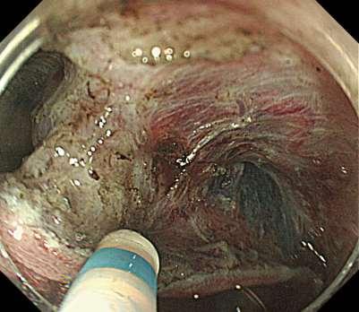


61 Severe Fibrosis


62
63 How do we follow-up the colonic lesions? Detection Characterization Treatment Follow-up
64 Summary: The approach to surveillance colonoscopy in IBD Dye Chromoendoscopy should be adopted in the daily practice as standard endoscopic technique for colonic lesions detection in IBD Selective virtual and dye chromoendoscopy with new optical enhancement diagnosis scopes may help to characterize the colonic lesions Optical enhancement diagnosis with or without magnification to assess margins and plan endoscopic therapeutic strategy. Local resection such as EMR or ESD in selected cases may be used to spare total colectomy. A distinct border of the lesion must be recognized for local resection Endoscopists must be dedicated to IBD patients for best diagnostic and therapeutic management. Laine L et al. Gastroenterology : Sugimoto et al GIE 2017;85:639

65 THE NEW FUTURE AT THE HORIZON


66 Endomicroscopy Mini probe Standard endoscope Field of view: 500x500µm Range: 0-250µm Lateral resolution: <1µm Kiesslich et al., Gastroenterology 2004


67 Normal colonic surface epithelium IMAGE CONTENT: Single crypt on the mucosal surface, with dark lumenal opening and mucus in the centre of each crypt. Goblet Cell Columnar Epithelial Cell Crypt Lumen


68 Confocal colon Normal crypt -daisy- Blood cells in lamina propria vessels
69 Atreya R & Neurath MF. Curr Opin Gastroenterol 2016 Molecular imaging aims at the identification and characterization of mucosal features because of their molecular composition rather than their morphological structure

70 Atreya R, et al. Nature Med 2014 Molecular imaging & response to Anti-TNFα ADALIMUMAB
71 Atreya R, et al. Nature Med 2014 Molecular imaging & response to Anti-TNFα ADALIMUMAB FITC FITC FITC FITC Fluorescein Isothiocyanate labeling
72 Atreya R, et al. Nature Med 2014 Molecular imaging & response to Anti-TNFα Ex vivo confocal imaging of mucosal specimens from patients with Crohn s disease incubated with FITC-adalimumab FITC FITC FITC FITC Fluorescein Isothiocyanate labelled Adalimumab


73 Adapted from Prof. Markus F Neurath Molecular imaging & response to Anti-TNFα In vivo colonic staining with FITC-adalimumab and confocal imaging in CD patients before anti-tnf therapy FITC FITC FITC FITC Fluorescein Isothiocyanate labelled Adalimumab


74 Rath T& Neurath MF. GIE 2017 Molecular imaging Endoscopic imaging Labelled antibody FITC-labelled antibody Antiα4/β7


75 Infrastructure

76

77 Maneuverability Normal Observation Magnifying Observation Endocyto Observation Similar maneuver as conventional zoom scopes Normal Observation Magnifying Observation Endocyto Observation /5/19

78 Principle of Endocyto observation Distal end of ECS scope Light guide Objectiv e lens Scattered light Tissue Dye stained tissue /5/19


79
80 The Future It s not about predicting histology It is about seeing histology

81 THANK YOU
How to characterize dysplastic lesions in IBD?
 How to characterize dysplastic lesions in IBD? Name: Institution: Helmut Neumann, MD, PhD, FASGE University Medical Center Mainz What do we know? Patients with IBD carry an increased risk of developing
Chromoendoscopy and Endomicroscopy for detecting colonic dysplasia
 Chromoendoscopy and Endomicroscopy for detecting colonic dysplasia Ralf Kiesslich I. Medical Department Johannes Gutenberg University Mainz, Germany Cumulative cancer risk in ulcerative colitis 0.5-1.0%
Dysplasia 4/19/2017. How do I practice Chromoendoscopy for Surveillance of Colitis? SCENIC: Polypoid Dysplasia in UC. Background
 SCENIC: Polypoid in UC Definition How do I practice for Surveillance of Colitis? Themos Dassopoulos, M.D. Director, BSW Center for IBD Themistocles.Dassopoulos@BSWHealth.org Tel: 469-800-7189 Cell: 314-686-2623
Chromoendoscopy - Should It Be Standard of Care in IBD?
 Chromoendoscopy - Should It Be Standard of Care in IBD? John F. Valentine, MD, FACG Professor of Medicine Division of Gastroenterology, Hepatology and Nutrition University of Utah What is the point of
Endoscopy in Inflammatory Bowel Disease DR. REENA KHANNA
 Endoscopy in Inflammatory Bowel Disease DR. REENA KHANNA ASSISTANT PROFESSOR, UNIVERSITY OF WESTERN ONTARIO Background Clinical trials in ulcerative colitis and Crohn s disease require validated instruments
Page 1. Is the Risk This High? Dysplasia in the IBD Patient. Dysplasia in the Non IBD Patient. Increased Risk of CRC in Ulcerative Colitis
 Screening for Colorectal Neoplasia in Inflammatory Bowel Disease Francis A. Farraye MD, MSc Clinical Director, Section of Gastroenterology Co-Director, Center for Digestive Disorders Boston Medical Center
Diagnostic and Therapeutic Approaches to Dysplasia in Inflammatory Bowel Diseases
 Diagnostic and Therapeutic Approaches to Dysplasia in Inflammatory Bowel Diseases Parakkal Deepak, M.B.B.S., M.S. Assistant Professor of Medicine Division of Gastroenterology John T. Milliken Department
Advances in Endoscopic Imaging
 Advances in Endoscopic Imaging SGNA meeting February 20, 2010 Amar R. Deshpande, MD Asst Professor of Medicine Division of Gastroenterology University of Miami Miller School of Medicine Objectives To recognize
CASE DISCUSSION: The Patient with Dysplasia: Surgery or Active Surveillance? Noa Krugliak Cleveland, MD David T. Rubin, MD
 CASE DISCUSSION: The Patient with Dysplasia: Surgery or Active Surveillance? Noa Krugliak Cleveland, MD David T. Rubin, MD Disclosure Statement NKC: No relevant conflicts to disclose. DTR: No relevant
Chromoendoscopy or Narrow Band Imaging with Targeted biopsies Should be the Cancer Surveillance Endoscopy Procedure of Choice in Ulcerative Colitis
 Chromoendoscopy or Narrow Band Imaging with Targeted biopsies Should be the Cancer Surveillance Endoscopy Procedure of Choice in Ulcerative Colitis Bret A. Lashner, M.D. Professor of Medicine Director,
University Mainz. Early Gastric Cancer. Ralf Kiesslich. Johannes Gutenberg University Mainz, Germany. Early Gastric Cancer 15.6.
 Ralf Kiesslich Johannes Gutenberg University Mainz, Germany DIAGNOSIS Unmask lesions - Chromoendoscopy -NBI Red flag technology - Autofluorescence Surface and detail analysis - Magnifying endoscopy - High
Paris classification (2003) 삼성의료원내과이준행
 삼성의료원내과이준행") Paris classification (2003) 삼성의료원내과이준행 JGCA classification - Japanese Gastric Cancer Association - Type 0 superficial polypoid, flat/depressed, or excavated tumors Type 1 polypoid carcinomas, usually attached
Paris classification (2003) 삼성의료원내과이준행 JGCA classification - Japanese Gastric Cancer Association - Type 0 superficial polypoid, flat/depressed, or excavated tumors Type 1 polypoid carcinomas, usually attached
ASGE and AGA Issue Consensus Statement on Surveillance and Management of Dysplasia in Patients With Inflammatory Bowel Disease
 ASGE and AGA Issue Consensus Statement on Surveillance and Management of Dysplasia in Patients With Inflammatory Bowel Disease DOWNERS GROVE, Ill., (March 5, 2015) The American Society for Gastrointestinal
Chromoendoscopy as an Adjunct to Colonoscopy
 Chromoendoscopy as an Adjunct to Colonoscopy Policy Number: 2.01.84 Last Review: 1/2018 Origination: 7/2017 Next Review: 7/2018 Policy Blue Cross and Blue Shield of Kansas City (Blue KC) will not provide
Endoscopic Corner CASE 1. Kimtrakool S Aniwan S Linlawan S Muangpaisarn P Sallapant S Rerknimitr R
 170 Endoscopic Corner Kimtrakool S Aniwan S Linlawan S Muangpaisarn P Sallapant S Rerknimitr R CASE 1 A 54-year-old woman underwent a colorectal cancer screening. Her fecal immunochemical test was positive.
Emerging Interventions in Endoscopy. Margaret Vance Nurse Consultant in Gastroenterology St Mark s Hospital
 Emerging Interventions in Endoscopy Margaret Vance Nurse Consultant in Gastroenterology St Mark s Hospital Colon Cancer Colon cancer is common. 1 in 20 people in the UK will develop the disease 19 000
BENEFIT APPLICATION BLUE CARD/NATIONAL ACCOUNT ISSUES
 Medical Policy BCBSA Ref. Policy: 2.01.84 Last Review: 11/15/2018 Effective Date: 11/15/2018 Section: Medicine Related Policies 2.01.87 Confocal Laser Endomicroscopy 6.01.32 Virtual Colonoscopy/Computed
An Atlas of the Nonpolypoid Colorectal Neoplasms in Inflammatory Bowel Disease
 An Atlas of the Nonpolypoid Colorectal Neoplasms in Inflammatory Bowel Disease Roy Soetikno, MD a, *, Silvia Sanduleanu, MD, PhD b, Tonya Kaltenbach, MD a KEYWORDS Inflammatory bowel disease Nonpolypoid
Where a licence is displayed above, please note the terms and conditions of the licence govern your use of this document.
 A Randomized Trial Comparing High Definition Colonoscopy Alone With High Definition Dye Spraying and Electronic Virtual Chromoendoscopy for Detection of Colonic Neoplastic Lesions During IBD Surveillance
COLON: Innovations 3 steps, 3 parts..
 COLON: Innovations 3 steps, 3 parts.. Detection: I see an abnormality (usually a polyp) Characterization: Is this abnormality neoplastic? (for example: an adenoma) Treatment: it is neoplastic. Can I treat
Diagnostic accuracy of pit pattern and vascular pattern in colorectal lesions
 Diagnostic accuracy of pit pattern and vascular pattern in colorectal lesions Digestive Disease Center, Showa University Northern Yokohama Hospital Department of Pathology Yoshiki Wada, Shin-ei Kudo, Hiroshi
Quality in Endoscopy: Can We Do Better?
 Quality in Endoscopy: Can We Do Better? Erik Rahimi, MD Assistant Professor Division of Gastroenterology, Hepatology, and Nutrition UT Health Science Center at Houston McGovern Medical School Ertan Digestive
Mucosal healing: does it really matter?
 Oxford Inflammatory Bowel Disease MasterClass Mucosal healing: does it really matter? Professor Jean-Frédéric Colombel, New York, USA Oxford Inflammatory Bowel Disease MasterClass Mucosal healing: does
Confocal Laser Endomicroscopy of the Colon
 clinical imaging Confocal Laser Endomicroscopy of the Colon Dan Ionut Gheonea, Adrian Saftoiu, Tudorel Ciurea, Carmen Popescu, Claudia Valentina Georgescu, Anca Malos Research Center of Gastroenterology
When and How to use Chromoendoscopy in IBD
 When and How to use Chromoendoscopy in IBD Samir A. Shah, MD, FACG, FASGE, AGAF Clinical Professor of Medicine, Brown University Chief of Gastroenterology, The Miriam Hospital Gastroenterology Associates,
Random biopsy is recommended in the US to detect
 Gastroenterology in Motion Ralf Kiesslich and Thomas D. Wang, Section Editors The Detection of Nonpolypoid (Flat and Depressed) Colorectal Neoplasms in Patients With Inflammatory Bowel Disease ROY SOETIKNO,
The Usefulness Of Narrow Band Imaging Endoscopy For The Real Time Characterization Of Colonic Lesions
 Acta Medica Marisiensis 2016;62(2):182-186 DOI: 10.1515/amma-2016-0004 RESEARCH ARTICLE The Usefulness Of Narrow Band Imaging Endoscopy For The Real Time Characterization Of Colonic Lesions Boeriu Alina
Predict, Resect and discard : Yes we can! (at least in some hands)
") Diminutive polyps : Real time endoscopic histology Predict, Resect and discard : Yes we can! (at least in some hands) Robert Benamouzig Hôpital Avicenne AP-HP & Paris 13 University France Why it is important?
Diminutive polyps : Real time endoscopic histology Predict, Resect and discard : Yes we can! (at least in some hands) Robert Benamouzig Hôpital Avicenne AP-HP & Paris 13 University France Why it is important?
Colon Polyps: Detection, Inspection and Characteristics
 Colon Polyps: Detection, Inspection and Characteristics Stephen Kim, M.D. Assistant Professor of Medicine Interventional Endoscopy Services UCLA Division of Digestive Diseases September 29, 2018 1 Disclosures
Narrow band imaging efficiency in evaluation of mucosal healing/ relapse of ulcerative colitis
 Original article Narrow band imaging efficiency in evaluation of mucosal healing/ relapse of ulcerative colitis Authors Seiko Sasanuma 1, Kazuo Ohtsuka 1, 2, Shin-ei Kudo 1, Noriyuki Ogata 1, Yasuharu
Description. Section: Medicine Effective Date: July 15, Subsection: Original Policy Date: September 13, 2012 Subject: Page: 1 of 17
 Page: 1 of 17 Last Review Status/Date: June 2016 Description Chromoendoscopy refers to the application of dyes or stains during endoscopy to enhance tissue differentiation or characterization. When used
Corporate Medical Policy
 Corporate Medical Policy Chromoendoscopy as an Adjunct to Colonoscopy File Name: Origination: Last CAP Review: Next CAP Review: Last Review: chromoendoscopy_as_an_adjunct_to_colonoscopy 7/2012 11/2017
Philip Chiu Associate Professor Department of Surgery, Prince of Wales Hospital The Chinese University of Hong Kong
 Application of Chromoendoscopy, NBI and AFI in Esophagus why, who, and how? Philip Chiu Associate Professor Department of Surgery, Prince of Wales Hospital The Chinese University of Hong Kong Cancer of
Management of pt1 polyps. Maria Pellise
 Management of pt1 polyps Maria Pellise Early colorectal cancer Malignant polyp Screening programmes SM Invasive adenocar cinoma Advances in diagnostic & therapeutic endoscopy pt1 polyps 0.75 5.6% of large-bowel
Difficult Polypectomy 2015 Tool of the Trade
 Difficult Polypectomy 2015 Tool of the Trade Jonathan Cohen, MD FACG FASGE Clinical Professor of Medicine NYU Langone School of Medicine Improving Therapeutics in the Colon Improved detection of polyp
Gastrointestinal Imaging
 Endoscopic Imaging of Gastroesophageal Reflux Disease Kerry B Dunbar, MD Assistant Professor of Medicine, Division of Gastroenterology and Hepatology, Johns Hopkins University School of Medicine Abstract
Chromoendoscopy is an image-enhanced endoscopic technique
 The Role of Chromoendoscopy in Evaluating Colorectal Dysplasia Anna M. Buchner, MD, PhD Dr Buchner is an assistant professor of medicine in the Division of Gastroenterology at the University of Pennsylvania
Advanced techniques for resection of large polyps. John G. Lee, MD February 2, 2018
 Advanced techniques for resection of large polyps John G. Lee, MD February 2, 2018 Background 1cm - large polyp on screening 2cm - large for polypectomy 3cm giant polyp 10-15% of polyps can t be removed
Diagnostic techniques for surveillance of dysplasia
 January 27th 2017, 8th Gastro Foundation Weekend for Fellows; Spier Hotel & Conference Centre, Stellenbosch Diagnostic techniques for surveillance of dysplasia Gerhard Rogler, Department of Gastroenterology
i-scan Mini-Atlas Case studies from clinical practice with HD + and i-scan.
 i-scan Mini-Atlas Case studies from clinical practice with and i-scan. Visible excellence. Gastrointestinal endoscopy with and i-scan. Index Introduction 3 and i-scan at a glance 4 5 PENTAX i-scan in characterization
Do any benign polyps require an operation?
 Do any benign polyps require an operation? Kevin Waschke MD.,CM., FRCPC, FASGE McGill University Health Center President Elect Canadian Association of Gastroenterology Colonoscopy Education Day - Tuesday
Learning Objectives:
 Crescent City GI Update 2018 Ochsner Clinic, NOLA Optimizing Endoscopic Evaluation of Barrett s Esophagus What Should I Do in My Practice? Gregory G. Ginsberg, M.D. Professor of Medicine University of
Management of Barrett s: From Imaging to Resection
 Management of Barrett s: From Imaging to Resection Michael Wallace, MD, MPH, FACG Professor of Medicine Mayo Clinic Florida Goals of Endoscopic Evaluation in Barrett s Detect Barrett s and dysplasia Reduce/eliminate
Alberta Colorectal Cancer Screening Program (ACRCSP) Post Polypectomy Surveillance Guidelines
 Post Polypectomy Surveillance Guidelines") Alberta Colorectal Cancer Screening Program (ACRCSP) Post Polypectomy Surveillance Guidelines June 2013 ACRCSP Post Polypectomy Surveillance Guidelines - 2 TABLE OF CONTENTS Background... 3 Terms, Definitions
Alberta Colorectal Cancer Screening Program (ACRCSP) Post Polypectomy Surveillance Guidelines June 2013 ACRCSP Post Polypectomy Surveillance Guidelines - 2 TABLE OF CONTENTS Background... 3 Terms, Definitions
Computer aided optical diagnosis of polyps. Dr Michael Byrne Vancouver General Hospital University of British Columbia
 Computer aided optical diagnosis of polyps Dr Michael Byrne Vancouver General Hospital University of British Columbia Conflict of Interest/disclosures Satis Operations Inc (partner in ai4gi venture)----ceo
Vital staining and Barrett s esophagus
 Marcia Irene Canto, MD, MHS Baltimore, Maryland Vital staining or chromoendoscopy refers to staining of endoscopic tissue or topical application of chemical stains or pigments to alter tissue appearances
Op#mizing)Management)in)IBD:) Mucosal)Healing)
Management)in)IBD:) Mucosal)Healing)") Op#mizing)Management)in)IBD:) Mucosal)Healing) Vipul&Jairath&MD&PhD& Associate&Professor&of&Medicine,&Epidemiology&and& Biosta=s=cs& Western&University&&& Division&of&Gastroenterology,&& London&Health&Sciences&Network&
Op#mizing)Management)in)IBD:) Mucosal)Healing) Vipul&Jairath&MD&PhD& Associate&Professor&of&Medicine,&Epidemiology&and& Biosta=s=cs& Western&University&&& Division&of&Gastroenterology,&& London&Health&Sciences&Network&
IBD. Crohn s. Outline. Ulcerative colitis versus Crohn s disease: is biopsy useful? UC vs. Crohn s? Is it easy? Biopsy settings 21/07/2017 IBD
 Outline Ulcerative colitis versus Crohn s disease: is biopsy useful? Roger Feakins Colorectal biopsies Ileal and upper GI biopsies Special situations New techniques Summary Inflammatory bowel disease (IBD)
Romanian Journal of Morphology and Embryology 2006, 47(3):
:") Romanian Journal of Morphology and Embryology 26, 7(3):239 23 ORIGINAL PAPER Predictive parameters for advanced neoplastic adenomas and colorectal cancer in patients with colonic polyps a study in a tertiary
Romanian Journal of Morphology and Embryology 26, 7(3):239 23 ORIGINAL PAPER Predictive parameters for advanced neoplastic adenomas and colorectal cancer in patients with colonic polyps a study in a tertiary
Endoscopy in IBD. F.Hartmann K.Kasper-Kliniken (St.Marienkrankenhaus) Frankfurt/M.
 Frankfurt/M.") F.Hartmann K.Kasper-Kliniken (St.Marienkrankenhaus) Frankfurt/M. F.Hartmann@em.uni-frankfurt.de Indications for endoscopy Diagnosis Management Surveillance Diagnosis Single most valuable tool: ileocolonoscopy
F.Hartmann K.Kasper-Kliniken (St.Marienkrankenhaus) Frankfurt/M. F.Hartmann@em.uni-frankfurt.de Indications for endoscopy Diagnosis Management Surveillance Diagnosis Single most valuable tool: ileocolonoscopy
PATIENTS WITH LONG-STANDING inflammatory
 Digestive Endoscopy 2016; 28: 266 273 doi: 10.1111/den.12634 Review Paradigm Shift in the Surveillance and Management of Dysplasia in Inflammatory Bowel Disease (West) Roy Soetikno, 1 Tonya Kaltenbach,
Novel endoscopic observation in Barrett s oesophagus using high resolution magnification endoscopy and narrow band imaging
 Alimentary Pharmacology & Therapeutics Novel endoscopic observation in Barrett s oesophagus using high resolution magnification endoscopy and narrow band imaging G. K. ANAGNOSTOPOULOS*, K. YAO*, P. KAYE,
Colonic Polyp. Najmeh Aletaha. MD
 Colonic Polyp Najmeh Aletaha. MD 1 Polyps & classification 2 Colorectal cancer risk factors 3 Pathogenesis 4 Surveillance polyp of the colon refers to a protuberance into the lumen above the surrounding
Confocal Laser Endomicroscopy
 Confocal Laser Endomicroscopy Policy Number: 2.01.87 Last Review: 3/2018 Origination: 3/2013 Next Review: 9/2018 Policy Blue Cross and Blue Shield of Kansas City (Blue KC) will not provide coverage for
Magnifying Endoscopy and Chromoendoscopy of the Upper Gastrointestinal Tract
 Magnifying Endoscopy and Chromoendoscopy of the Upper Gastrointestinal Tract Alina M.Boeriu 1, Daniela E.Dobru 1, Simona Mocan 2 1) Department of Gastroenterology, University of Medicine and Pharmacy;
malignant polyp Daily Challenges in Digestive Endoscopy for Endoscopists and Endoscopy Nurses BSGIE Annual Meeting 18/09/2014 Mechelen
 Plan Incidental finding of a malignant polyp 1. What is a polyp malignant? 2. Role of the pathologist and the endoscopist 3. Quantitative and qualitative risk assessment 4. How to decide what to do? Hubert
Endoscopic Submucosal Dissection ESD
 Endoscopic Submucosal Dissection ESD Peter Draganov MD Professor of Medicine Division of Gastroenterology, Hepatology and Nutrition University of Florida Gastrointestinal Cancer Lesion that Can be Treated
Role of random biopsies in surveillance of dysplasia in ulcerative colitis patients with high risk of colorectal cancer
 ORIGINAL ARTICLE pissn 1598-9100 eissn 2288-1956 http://dx.doi.org/10.5217/ir.2016.14.3.264 Intest Res 2016;14(3):264-269 Role of random biopsies in surveillance of dysplasia in ulcerative colitis patients
Adenoma to Carcinoma Pathway
 It is widely accepted that more than 95% of colorectal cancers arise from adenomatous polyps, which are generally defined as benign lesions with dysplastic epithelium that have variable potential for malignancy.
The Pathologist s Role in the Diagnosis and Management of Neoplasia in Barrett s Oesophagus Cian Muldoon, St. James s Hospital, Dublin
 The Pathologist s Role in the Diagnosis and Management of Neoplasia in Barrett s Oesophagus Cian Muldoon, St. James s Hospital, Dublin 24.06.15 Norman Barrett Smiles [A brief digression - Chair becoming
Supporting Information 2. ESGE QIC Lower GI Delphi voting process: Round 1 Working Group chair: Michal F. Kaminski, Poland
 Supporting Information 2. ESGE QIC Lower GI Delphi voting process: Round 1 Working chair: Michal F. Kaminski, Poland Population Interventions Comparator Outcome Additional evidence 1.1 Rate of adequate
Rectal EMR: Techniques and Tips
 Rectal EMR: Techniques and Tips Dr Paul Urquhart Epworth Eastern Eastern Health (Head of Endoscopy) The context of EMR Basic Technique Recurrence Perforation Bleeding Introduction 1 I don t treat rectal
Evaluation of the severity of ulcerative colitis using endoscopic dual red imaging targeting deep vessels
 Evaluation of the severity of ulcerative colitis using endoscopic dual red imaging targeting deep vessels Authors Makoto Naganuma 1, 2, Naohisa Yahagi 3,RiekoBessho 1, Keiko Ohno 1, Mari Arai 1, Makoto
Gastric Polyps. Bible class
 Gastric Polyps Bible class 29.08.2018 Starting my training in gastroenterology, some decades ago, my first chief always told me that colonoscopy may seem technically more challenging but gastroscopy has
Improving Your Adenoma Detection Rate
 Improving Your Adenoma Detection Rate JILL TINMOUTH, ASSOCIATE PROFESSOR, UNIVERSITY OF TORONTO JERRY MCGRATH, ASSOCIATE PROFESSOR, MEMORIAL UNIVERSITY OF NEWFOUNDLAND FEB. 11 2017 X CanMEDS Roles Covered
Polypectomy and Local Resections of the Colorectum Structured Pathology Reporting Proforma
 Polypectomy and Local Resections of the Colorectum Structured Pathology Reporting Proforma Mandatory questions (i.e. protocol standards) are in bold (e.g. S1.03). Family name Given name(s) Date of birth
When is a programmed follow-up meaningful and how should it be done? Professor Alastair Watson University of Liverpool
 When is a programmed follow-up meaningful and how should it be done? Professor Alastair Watson University of Liverpool Adenomas/Carcinoma Sequence Providing Time for Screening Normal 5-20 yrs 5-15 yrs
Oesophagus and Stomach update dysplasia and early cancer
 Oesophagus and Stomach update dysplasia and early cancer Dr Tim Bracey STR teaching 13/4/16 Please check pathkids.com for previous talks One of the biggest units in the country (100 major resections per
Review Article Confocal Endomicroscopy of Colorectal Polyps
 Gastroenterology Research and Practice Volume 2012, Article ID 545679, 6 pages doi:10.1155/2012/545679 Review Article Confocal Endomicroscopy of Colorectal Polyps Vivian M. Ussui and Michael B. Wallace
Experience and challenges of implementing optical diagnosis into clinical practice UK and European Perspective
 Experience and challenges of implementing optical diagnosis into clinical practice UK and European Perspective WEO Image Enhanced Endoscopy San Diego, USA Dr James East Consultant Gastroenterologist Honorary
Surveying the Colon; Polyps and Advances in Polypectomy
 Surveying the Colon; Polyps and Advances in Polypectomy Educational Objectives Identify classifications of polyps Describe several types of polyps Verbalize rationale for polypectomy Identify risk factors
FEP Medical Policy Manual
 FEP Medical Policy Manual Effective Date: April 15, 2018 Related Policies: 2.01.87 Confocal Laser Endomicroscopy 6.01.32 Virtual Colonoscopy/CT Colonography Chromoendoscopy as an Adjunct to Colonoscopy
Pitfalls in the Diagnosis of Inflammatory Bowel Disease
 Pitfalls in the Diagnosis of Inflammatory Bowel Disease Robert H Riddell MD Mt Sinai Hospital Toronto Prof of Lab. Medicine and Pathobiology University of Toronto Atypical gross / endoscopic distribution
Screening & Surveillance Guidelines
 Chapter 2 Screening & Surveillance Guidelines I. Eligibility Coloradans ages 50 and older (average risk) or under 50 at elevated risk for colon cancer (personal or family history) that meet the following
Index. Note: Page numbers of article titles are in boldface type.
 Note: Page numbers of article titles are in boldface type. A Aberrant crypt foci, chromocolonoscopy for, 539 540 Absorptive stains, for chromocolonoscopy, 522 524 Accessories, for colonoscopy, 680 684
Prevalence and Characteristics of Nonpolypoid Colorectal Neoplasm in an Asymptomatic and Average-Risk Chinese Population
 CLINICAL GASTROENTEROLOGY AND HEPATOLOGY 2009;7:463 470 Prevalence and Characteristics of Nonpolypoid Colorectal Neoplasm in an Asymptomatic and Average-Risk Chinese Population HAN MO CHIU,*,, JAW TOWN
EMR, ESD and Beyond. Peter Draganov MD. Professor of Medicine Division of Gastroenterology, Hepatology and Nutrition University of Florida
 EMR, ESD and Beyond Peter Draganov MD Professor of Medicine Division of Gastroenterology, Hepatology and Nutrition University of Florida Gastrointestinal Cancer Lesion that Can be Treated by Endoscopy
Clinical Study Clinical Study of the Relation between Mucosal Healing and Long-Term Outcomes in Ulcerative Colitis
 Hindawi Publishing Corporation Gastroenterology Research and Practice Volume 2013, Article ID 192794, 6 pages http://dx.doi.org/10.1155/2013/192794 Clinical Study Clinical Study of the Relation between
Volumetric laser endomicroscopy can target neoplasia not detected by conventional endoscopic measures in long segment Barrett s esophagus
 E318 Volumetric laser endomicroscopy can target neoplasia not detected by conventional endoscopic measures in long segment esophagus Authors Institution Arvind J. Trindade, Benley J. George, Joshua Berkowitz,
NATIONAL INSTITUTE FOR HEALTH AND CLINICAL EXCELLENCE SCOPE
 NATIONAL INSTITUTE FOR HEALTH AND CLINICAL EXCELLENCE 1 Guideline title SCOPE Colorectal cancer: colonoscopic surveillance for prevention of colorectal cancer in patients with ulcerative colitis, Crohn
The Paris classification of colonic lesions
 The Paris classification of colonic lesions Training to improve the interobserver agreement among international experts Sascha van Doorn, MD, PhD-student in CRC-reserach group of Evelien Dekker Introduction
Comparison of the Diagnostic Usefulness of Conventional Magnification and Near-focus Methods with Narrow-band Imaging for Gastric Epithelial Tumors
 ORIGINAL ARTICLE ISSN 1738-3331, http://dx.doi.org/10.7704/kjhugr.2015.15.1.39 The Korean Journal of Helicobacter and Upper Gastrointestinal Research, 2015;15(1):39-43 Comparison of the Diagnostic Usefulness
Pathology in Slovenian CRC screening programme:
 Pathology in Slovenian CRC screening programme: Findings, organisation and quality assurance Snježana Frković Grazio University Medical Center Ljubljana, Slovenia Slovenia s population: 2 million Incidence
Histopathology of Endoscopic Resection Specimens from Barrett's Esophagus
 Histopathology of Endoscopic Resection Specimens from Barrett's Esophagus Br J Surg 38 oct. 1950 Definition of Barrett's esophagus A change in the esophageal epithelium of any length that can be recognized
Latest Endoscopic Guidelines for FAP, HNPCC, IBD, and the General Population
 Latest Endoscopic Guidelines for FAP, HNPCC, IBD, and the General Population David T. Rubin, M.D. Assistant Professor of Medicine Inflammatory Bowel Disease Center MacLean Center for Clinical Medical Ethics
THE BIG, AWKWARD, FLAT POLYP THAT CAN T BE REMOVED WITH A (SINGLE) SNARE THE CASE FOR EMR AND ESD
 SNARE THE CASE FOR EMR AND ESD") THE BIG, AWKWARD, FLAT POLYP THAT CAN T BE REMOVED WITH A (SINGLE) SNARE THE CASE FOR EMR AND ESD Surgical Oncology Network meeting Dr. Eric Lam MD FRCPC October 14, 2017 DISCLOSURES None OBJECTIVES Appreciate
THE BIG, AWKWARD, FLAT POLYP THAT CAN T BE REMOVED WITH A (SINGLE) SNARE THE CASE FOR EMR AND ESD Surgical Oncology Network meeting Dr. Eric Lam MD FRCPC October 14, 2017 DISCLOSURES None OBJECTIVES Appreciate
Expert panel observations
 Expert panel observations Professor Neil A Shepherd Gloucester and Cheltenham, UK Gloucestershire Cellular Pathology Laboratory Three big issues in BCSP pathology serrated pathology & what do we do about
5/2/2018. Low Grade Dysplasia of GI Tract. High Grade Dysplasia of GI Tract. Dysplasia in Gastrointestinal Tract: Practical Pearls and Issues
 Dysplasia in Gastrointestinal Tract: Practical Pearls and Issues Arief Suriawinata, M.D. Professor of Pathology and Laboratory Medicine Geisel School of Medicine at Dartmouth Department of Pathology and
Title: Serrated polyposis syndrome associated with long-standing inflammatory bowel disease
 Title: Serrated polyposis syndrome associated with long-standing inflammatory bowel disease Authors: Jesús Castro, Miriam Cuatrecasas, Francesc Balaguer, Elena Ricart, María Pellisé DOI: 10.17235/reed.2017.5068/2017
Magnifying image-enhanced endoscopy for collagenous colitis
 Magnifying image-enhanced endoscopy for collagenous colitis Authors Masaaki Kobayashi 1, Takahiro Hoshi 1, Shin-ich Morita 1,TsutomuKanefuji 1, Takeshi Suda 1,GoHasegawa 2, Shuji Terai 3 Institutions 1
Diagnosis of colorectal lesions with a novel endocytoscopic classification a pilot study
 Original article 869 Diagnosis of colorectal lesions with a novel endocytoscopic classification a pilot study Authors S-E Kudo 1, K. Wakamura 1, N. Ikehara 1,Y.Mori 1, H. Inoue 1, S. Hamatani 2 Institutions
Introduction. Piecemeal EMR (EPMR) Symposium
 Symposium") Symposium Symposium II - Lower GI : Colonoscopy Issues in 2015 Resection of Large Polyps Using Techniques other than Endoscopic Submucosal Dissection: Piecemeal Resection, Underwater Endoscopic Mucosal
Symposium Symposium II - Lower GI : Colonoscopy Issues in 2015 Resection of Large Polyps Using Techniques other than Endoscopic Submucosal Dissection: Piecemeal Resection, Underwater Endoscopic Mucosal
Factors for Endoscopic Submucosal Dissection in Early Colorectal Neoplasms: A Single Center Clinical Experience in China
 ORIGINAL ARTICLE Clin Endosc 2015;48:405-410 http://dx.doi.org/10.5946/ce.2015.48.5.405 Print ISSN 2234-2400 On-line ISSN 2234-2443 Open Access Factors for Endoscopic Submucosal Dissection in Early Colorectal
Finding and Removing Difficult Polyps (safely)
") Finding and Removing Difficult Polyps (safely) David Lieberman MD Chief, Division of Gastroenterology and Hepatology Oregon Health and Science University Colonoscopy Clouds Interval Cancers Interval Cancer:
Finding and Removing Difficult Polyps (safely) David Lieberman MD Chief, Division of Gastroenterology and Hepatology Oregon Health and Science University Colonoscopy Clouds Interval Cancers Interval Cancer:
Magnification endoscopy, high resolution endoscopy, and chromoscopy; towards a better optical diagnosis
 iv7 PAPER Magnification endoscopy, high resolution endoscopy, and chromoscopy; towards a better optical diagnosis M J Bruno... In the past few years, optical magnification endoscopy and chromoscopy have
SCREENING COLONOSCOPY IS very important for
 Digestive Endoscopy 2015; 27: 232 238 doi: 10.1111/den.12395 Review Advanced colonoscopic imaging using endocytoscopy Helmut Neumann, 1,2 Shin-Ei Kudo, 3 Ralf Kiesslich 4 and Markus F. Neurath 1,2 1 Department
Hyperplastische Polyps Innocent bystanders?
 Hyperplastische Polyps Innocent bystanders?? K. Geboes P th l i h O tl dk d Pathologische Ontleedkunde, KULeuven Content Historical Classification Relation Hyperplastic polyps carcinoma The concept cept
Fujiya M, Saitoh Y, Watari J, Moriichi K, Kohgo Y.
 Digestive Endoscopy (2007) 19(s1):S145-S149. Auto-Fluorescence Imaging is useful to assess the activity of ulcerative colitis Fujiya M, Saitoh Y, Watari J, Moriichi K, Kohgo Y. Auto-Fluorescence Imaging
REPORT. McMahon Publishing Group. PENTAX Medical i-scan Technology for Improved Endoscopic Evaluations
 Brought to You by MAY 2014 PENTAX Medical i-scan Technology for Improved Endoscopic Evaluations Endoscopy plays a vital role in the diagnosis and clinical management of diseases of the gastrointestinal
How to remove BE cancer: EMR or ESD? Expected outcome
 How to remove BE cancer: EMR or ESD? Expected outcome Presented by Horst Neuhaus Institution Dpt. of Gastroenterology Evangelisches Krankenhaus Düsseldorf, Germany Indications for endoscopic resection