NEUROTROPHINS AND THEIR EFFECTS ON BREAST CANCER CELL PROLIFERATION AND MIGRATION
|
|
|
- Sarah Marsh
- 6 years ago
- Views:
Transcription

1 Purdue University Purdue e-pubs Open Access Theses Theses and Dissertations Spring 2014 NEUROTROPHINS AND THEIR EFFECTS ON BREAST CANCER CELL PROLIFERATION AND MIGRATION Kayla Elise Minser Purdue University Follow this and additional works at: Part of the Cell Biology Commons, and the Molecular Biology Commons Recommended Citation Minser, Kayla Elise, "NEUROTROPHINS AND THEIR EFFECTS ON BREAST CANCER CELL PROLIFERATION AND MIGRATION" (2014). Open Access Theses. Paper 222. This document has been made available through Purdue e-pubs, a service of the Purdue University Libraries. Please contact epubs@purdue.edu for additional information.
2 01 14 PURDUE UNIVERSITY GRADUATE SCHOOL Thesis/Dissertation Acceptance Kayla E. Minser NEUROTROPHINS AND THEIR EFFECTS ON BREAST CANCER CELL PROLIFERATION AND MIGRATION Master of Science Julia Kirshner Elizabeth J. Taparowsky Dorothy Teegarden Thesis/Dissertation Agreement. Publication Delay, and Certification/Disclaimer (Graduate School Form 32) adheres to the provisions of Julia Kirshner Richard J. Kuhn 04/15/2014 Department
3 NEUROTROPHINS AND THEIR EFFECTS ON BREAST CANCER CELL PROLIFERATION AND MIGRATION A Thesis Submitted to the Faculty of Purdue University by Kayla E. Minser In Partial Fulfillment of the Requirements for the Degree of Master Of Science May 2014 Purdue University West Lafayette, Indiana
4 To my parents: for your unconditional love and undying support. ii
5 iii ACKNOWLEDGEMENTS I would like to thank my mentor, Dr. Julia Kirshner, for her continuous support through this process. Your expertise and guidance are greatly appreciated. I would like to thank Dr. Elizabeth Taparowsky and Dr. Dorothy Teegarden for their help and insight on this project. I would also like to thank Mukti Parikh, who helped so much with this project and was always there to offer support and direction in times of need. She is a great scientist, but an even better friend. Thanks go to all members of the Kirshner lab who have always encouraged me and have been wonderful friends. I couldn t have picked a better group of people to have spent these last couple years with. And finally, I would like to acknowledge my parents, who have never wavered in their undying support for me. I am lucky to have two such wonderful people in my life and there is absolutely no way that I could have completed this without them.
6 iv TABLE OF CONTENTS Page LIST OF TABLES...vi LIST OF FIGURES...vii LIST OF ABBREVIATIONS...ix ABSTRACT... x CHAPTER 1. INTRODUCTION Breast Cancer Breast Cancer Incidence Metastasis Neurons Neuronal Morphology and Function Neuronal Development Neuronal Development and Neurotrophins Neurotrophins and Signal Transduction Neurotrophins and Cancer... 8 CHAPTER 2. MATERIALS AND METHODS Tissue Culture Cell Lines and Cell Culture D Proliferation Assay rmet Model Setup Matrix Preparation rmet Assembly rmet Proliferation Assay Dome Assay for Cell Migration Immunofluorescence Neurotrophin Receptor Staining in 2-D... 13
7 v Page Neurotrophin Receptor Staining in rmet Primary Tumor Site Cell Isolation and Neurotrophin Receptor Immunofluorescence Invasive Fraction Neurotrophin Receptor Immunofluorescence rbm Fraction Neurotrophin Receptor Immunofluorescence Molecular Biology RNA Extraction Reverse Transcritption cdna Synthesis Real-Time quantitative PCR (rt-qpcr) CHAPTER 3. NEUROTROPHIC EFFECTS ON THE PROLIFERATION AND MIGRATORY CAPABILITIES OF BREAST CANCER CELLS Neurotrophins and Breast Cancer Cell Proliferation Neurotrophin Receptor Identification D Cell Culture D Cell Culture rt-qpcr Confirmation of Neurotrophin Receptor Expression Neurotrophins and their effects on Breast Cancer Cell Proliferation D Cell Culture rmet Model Neurotrophins and Cancer Cell Migration Neurotrophins and MCF10CA1a Cell Migration: Dome Assay Neurotrophins and their receptors as potential treatment targets in breast cancer CHAPTER 4: NEUROTROPHINS AND THEIR POTENTIAL FOR THE TREATMENT OF BREAST CANCER AND ITS METASTASIS LIST OF REFERENCES... 37
8 vi LIST OF TABLES Table Page Table I. Neurotrophin Receptor Primers... 16
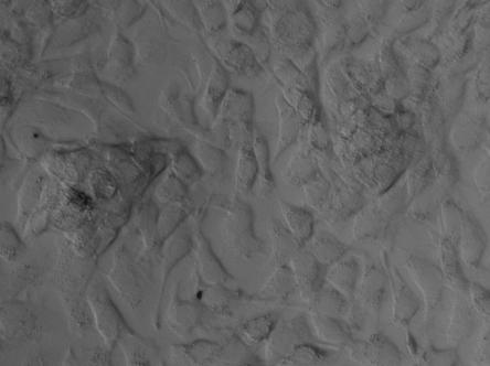
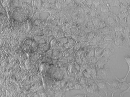
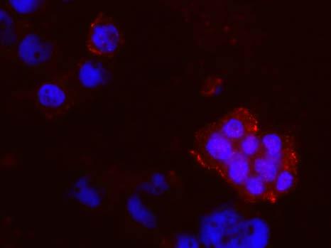
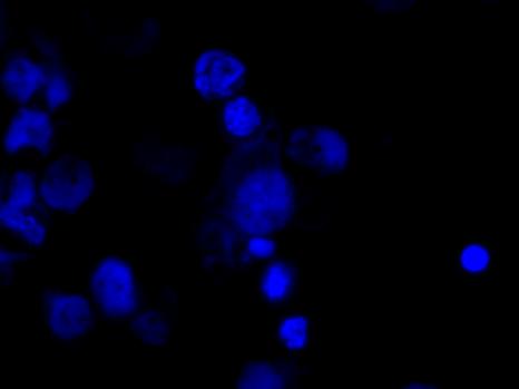
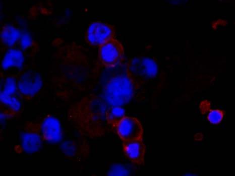
9 vii LIST OF FIGURES Figure Page Figure 1-1 Neurotrophin Binding and signaling pathways... 7 Figure 3-1The setup of the rmet model recapitulates the metastatic migration from a mammary environment, located in the top transwell, to a secondary site, the bone, located at the bottom (a). Cells are seeded in the top transwell [(b) left], and after 7-14 days of culture in the model, give rise to three distinct populations of cells [(b) right] Figure 3-2 A population of cells that colonize the rbm fraction have a neuronal-like morphology. The cells display multiple axon-like extensions, black arrowhead (a), and axon-like extensions that are uncharacteristically long for cancer cells, white arrowhead (b) Figure 3-3 Immunofluorescence of neurotrophin receptor expression in 2-D in MCF10CA1a cells. Cells were cultured in a chamber slide for 24 hours and then stained for the receptors. Scale bar 50 µm Figure 3-4 Immunofluorescence of neuotrophin receptor expression in the rmet model in MCF10CA1a cells. Cells were cultured in the model for 14 days and then isolated from each fraction to be stained for the receptors. Immunofluorescence of nuerotrophin receptor expression in the primary tumor fraction (a) is low and distinct to clusters of cells. The expression in the invasive layer (b) and rbm fraction (c) are expressed over a wider variety of cells, though still heterogeneous. Scale bar, 50 µm Figure 3-5 Fold change in each of the receptors for MCF10CA1a cells in 2D and from two fractions of the rmet model. RT-qPCR was completed using mrna from cells cultured in 2D, and cells isolated from the primary tumor site and the rbm fraction of the rmet model. The neurotrophin receptor genes are expressed in 2-D and are upregulated in the primary tumor fraction and further upregulated in the rbm colonizing fraction. The greatest upregulation is TrkA (b) in the rbm, followed by TrkB (c) in the rbm
10 viii Figure Page Figure 3-6 Effects of neurotrophins on proliferation of MCF10CA1a cells in 2D culture. Viable cells were counted using Trypan-blue and a hemacytometer. Graphs show the most effective concentrations for each neurotrophin. The most effective concentrations for NGF (a) and NT 4/5 (c) are 5 ng/ml, p<0.05. The most effective concentration for BDNF is 50 ng/ml, p< Figure 3-7 Effects of neurotrophins on proliferation of MCF10CA1a cells in the rmet model. In order to determine proliferation, cells were stained with Ki67, a marker of proliferation, and the total percentage of cells positive were determined. At 24 hours post-neurotrophin treatment, NGF causes the greatest increase in proliferation (pvalue=0.3677) (a). At 72 hours post-treatment, BDNF decreases the proliferation of the cells (p-value=0.179), with a small increase in NGF (b) Figure 3-8 Setup of the Dome Assay Figure 3-9 Dome Assay for Migration NT 4/5 and NGF induce a significant increase in cell migration towards the neurotrophin stimulus (**p-value <0.01; ***p-value<0.0001). 32
11 ix LIST OF ABBREVIATIONS rmet TrkA TrkB NGFR NGF BDNF Reconstructed Metastasis Model Receptor Tyrosine Kinase A Receptor Tyrosine Kinase B Nerve Growth Factor Receptor Nerve Growth Factor Brain-derived Neurotrophic Factor NT 4/5 Neurotrophic Factor 4/5 ECM EMT NSC MAPK PI3K PLC-γ NF-κB FBS rbm rend CNS Extracellular Matrix Epithelial to Mesenchymal Transition Neural Stem Cell Mitogen-Activated Protein Kinase Phosphatidylinositol 3-kinase Phospholipase C-γ Nuclear Factor Kappa-light-chain-enhancer of Activated B cells Fetal Bovine Serum Reconstructed Bone Marrow Matrix Reconstructed Endosteum Central Nervous System
12 x ABSTRACT Minser, Kayla E. M.S., Purdue University, May Neurotrophins and Their Effects on Breast Cancer Cell Proliferation and Migration. Major Professor: Julia Kirshner. Cancer is a large health issue in all parts of the world. In the United States alone, approximately 1 in 4 deaths are cancer related. Breast cancer is a particularly prevalent form, accounting for a little over 14 percent of all cancer incidence. The largest obstacle to overcome for breast cancer morbidity is metastasis. Over 90 percent of all breast cancer related deaths are due to metastasis. Because metastasis is a complex, multi-step process, it is difficult to treat. A recent observation in the Kirshner lab has revealed a type of phenotypic plasticity, where migratory cancer cells have a neuronallike morphology with multiple axon-like projections. This observation has led to an interest in the characteristics of these cells and how they relate to metastasis. One of the characteristics is receptivity to neurotrophins, which are neuronal growth factors. We have shown that MCF10CA1a cells, a malignant progression of the MCF10 series, do express three neuronal receptors, TrkA, TrkB, and p75 NTR in both 2D and 3D cell culture. Addition of neurotrophins, NGF, BDNF, and NT 4/5 increase proliferation between 24 and 72 hours of treatment in 2D cell culture. Further, addition of NGF and NT 4/5 act as a chemoattractant to these cells, increasing their migration towards the neurotrophin. From these results we conclude that neurotrophins do have an effect on the proliferation and migration of breast cancer cells. This could serve as a therapeutic target for future treatment of breast cancer and breast cancer metastasis.
13 1 CHAPTER 1. INTRODUCTION 1.1 Breast Cancer Breast Cancer Incidence Cancer is a large health obstacle in all parts of the world. In the United States alone, approximately 1 in 4 deaths are cancer related. Each type of cancer has its own complications, symptoms, and treatments, but the commonality in all of them is the diminished quality of life and an increased mortality rate. Breast cancer is a particularly prevalent form, accounting for a little over 14 percent of all cancer incidence 1. The American Cancer Society estimates that over 235,000 people in the United States will be diagnosed with breast cancer in 2014 alone and over 40,000 of them will succumb to the disease. It is vital to study and research breast cancer in order to reduce the mortality rate and improve quality of life for those diagnosed with the disease Metastasis The largest obstacle to overcome for breast cancer morbidity is metastasis. Over 90 percent of all breast cancer related deaths are due to metastasis 2. Over time the rates of breast cancer metastasis have increased. The American Medical Association has reported that metastatic breast cancer in women under 40 has increased 2 percent a year, ever year, from 1976 to Because metastasis is a complex and multi-step process, it is very difficult to treat and even more difficult to research the mechanisms involved in the process. Unfortunately, little is known about the populations of cells that are able to leave a primary site and colonize a secondary site in the body, nor is there much information on the signaling pathways that are involved in the process. The heterogeneous nature of metastasis makes it difficult to understand. In order to treat breast cancer and reduce the mortality of the disease, it is important to understand the mechanisms of metastasis so that they can be targeted as methods of treatment.
14 2 Metastasis is characterized by cells that are able to leave the primary tumor site and eventually colonize and proliferate in a secondary site in the body 4. The bone is one of the most common secondary sites of breast cancer metastasis. Other sites include brain, liver, lung, and lymph nodes 5. Metastasis was previously characterized as a latent, acquired phenotype of primary breast cancer tumorigenic cells 6. More recently, however, it was shown that the ability of cells to metastasize is an inherent feature of breast cancer 7. The process of metastasis is very complex and is referred to as the invasionmetastasis cascade. It is characterized by specific steps that allow epithelial cells to colonize distant sites in the body. In order for metastasis to occur, the first step that an epithelial cell must take is to invade the local tissue area. This includes breaking through the extracellular matrix (ECM) components and basement membrane, followed by invasion of the surrounding stroma 8. The basement membrane is a crucial part of the metastastic cascade because of the growth factors that it contains and the vital role that it plays in signal transduction pathways of the carcinoma cell 9. These pathways include changing of cell polarity, invasiveness, proliferation and survival 10. To further overcome invasion obstacles some cancer cells will undergo epithelial-to-mesenchymal transition(emt) 8. EMT includes the suppression of epithelial markers, such as E- cadherin and cytokeratin, that allow the cell to exhibit mesenchymal traits, most importantly, increased invasiveness The next step is intravasation, whereby the cancer cell must break through components of vessels such as the pericyte and endothelial cell barriers and enter into a blood or lymphatic vessel 14. This process may be facilitated by increased angiogenesis in the primary tumor area 15. Further, the new vessels that are formed near the tumor area can be leaky and poorly constructed aiding in an easier invasion of the vessel itself 16. After the cell completes intravasation it must then be able to survive in a non-adherent state as it travels through the vasculature. This is seemingly difficult for an epithelial cell because cell survival is generally dependent on integrin-dependent adhesion to ECM components 17. Cells unable to adhere usually undergo anoikis 18. Once the cell is in circulation, it will arrest in a capillary in a distant location. It is unknown whether the metastatic tropism arises from the large epithelial cell simply getting stuck in a microvessel, or if certain cancer cells have a preference for particular organs and their ECM components 19. After the cell is arrested in the microvessel it must break through the lumina of the vessel and enter into
15 3 the functional tissue of the organ, a process called extravasation 20. The cell must then survive in a new tissue microenvironment, which includes a new ECM makeup, growth factors and cytokines, and different types of stromal cells. The cell must survive long enough to form micrometastases and turn on proliferative programs that allow for neoplastic growth and full colonization of the distant site 21. This is particularly difficult for the cells to accomplish as they have limited supply to vessels and must adjust to a new microenvironment. In addition, the cells must have a high proliferation capacity in order to sustain a population of cells that successfully colonize a distant site. It is obvious that the metastatic cascade is extremely complex 22. There are many growth factors, cytokines, signaling pathways and genes that are involved, which is where difficulties arise in studying the process and further treating it in patients. In order to reduce the morbidity of breast cancer, it is important to understand more about these pathways and factors in the metastatic cascade. It is possible that some of the pathways involved in the metastatic cascade are not as obvious as it would seem. A recent study in the Kirshner lab has used a reconstructed metastasis model (rmet) as a way to study metastasis in vitro and has led to a new vein of research involving the connection of breast cancer cells and neurons. It has been observed in the rmet model that a population of breast cancer cells that have migrated and colonized the secondary environment display a neuronal-like morphology. This includes multiple axon-like extensions and even growth cone-like structures. This has been observed in multiple breast cancer cell lines and even other cancer cell lines such as lung and prostate cancers. This has led to the interest in the characteristics of these neuronal-like cells and how these characteristics contribute to metastasis. 1.2 Neurons Neuronal Morphology and Function Neurons are a unique class of cells that make up the nervous system. They are able to transmit information through chemical and electric signals. Though they contain similar internal structures to all other cells in the body, they have many morphological differences that make them unique. Structurally, they can be divided into three different parts: the cell body or soma, dendrites, and axons 23. The soma is the main body of the neuron and is where the nucleus and other cellular compartments are located. The soma can range from 6 to 80 µm in diameter 23. Dendrites and axons are both
16 4 extensions from the cell body. The axon can be classified into four distinct sections: the axon hillock, the initial segment, the axon proper, and the axon termination 23. The axon is responsible for sending chemical and electrical signals away from the cell body. They are composed of neurotubules and neurofilaments 24. Electric signals originate in the axolemma of the initial segment. The terminal end of the axon widens at the synaptic regions, where synapses will send chemical or electrical signals to other neurons 25. Dendrites are also arranged around the soma in a stellate manner and are responsible for afferent communications. They extend from the cell body in a single trunk but branch out towards the end to form a dendritic tree 26. Axon terminals have a common boundary with dendrites. The synaptic area at the axon terminal is where communication between neurons occurs. Synaptic messages can either be excitatory, inhibitory or modulatory Neuronal Development Developmentally, neurons arise from the ectoderm in the form of the neural tube 28. The neural tube is a single layer of epithelial cells and these epithelial cells are the progenitors of both neurons and glial cells 29. Neural stem cells (NSC) are extremely prolific at the beginning of development, but their numbers are quickly diluted with the presence of restricted progenitor cells and differentiated neural or glial cells. NSCs are derived from pluripotent embryonic stem cells 30. Neuroepithelial cells of the neural tube are self-renewing but do not always have unlimited divisions 31. They can be multipotent, giving rise to multiple, limited cell types, or possible even unipotent, giving rise to only one cell type. Additionally, their divisions might be symmetric, with both daughter cells identical to the mother, or asymmetric, giving rise to two functionally separate daughter cells, one identical to the mother cell and one that is not 32. Neuroepithelial cells can undergo symmetric, proliferative division, giving rise to two identical neuroepithelial cells. Further divisions, however, may be asymmetric and account for neurogenic division 32. This division will give rise to two functionally different nervous system cells, a glial cell and a neural progenitor that will give rise to two identical neurons. The glial cell can then undergo another asymmetric division, giving rise to an identical daughter cell and another neural progenitor that will give rise to two neurons 33. Neural progenitor cells themselves are non-functional cells and have a limited number of divisions 32. The radial cell can also undergo symmetric division with two neural progenitor daughter cells that will in total yield four neurons 34. Once the cells differentiate into neurons, they are
17 5 terminally differentiated and post-mitotic. The ability of a cell to undergo symmetric or asymmetric division is dependent upon the location of the cleavage plane during division. If the cleavage plane is positioned vertically, the division will be symmetric because equal apical and basal components will be transferred to both daughter cells. Conversely, if the plane is oriented horizontally, the division will be asymmetric because apical components will be given to one daughter cell and basal components will be given to the other daughter cell 32. After differentiation, cells migrate through the body to their specified area of the CNS Neuronal Development and Neurotrophins After migration, the neurons go through a maturation stage. In this stage their number of processes increases and they create a complex connection of neuronal cells that form the central nervous system. The manner in which these neuronal cells differentiate and form the complex network of the CNS is still not fully understood. There is however, evidence that neurotrophins play an important role in the development 35. Neurotrophins are dimeric, secretory proteins found primarily in the developing nervous system. They are extremely important for neuronal differentiation, proliferation, survival, and axon wiring, and exist in a family of four proteins 36. They are Nerve Growth Factor (NGF), Brain-Derived Neurotrophic Factor (BDNF), Neurotrophin-3 (NT-3) and Neurotrophin-4/5 (NT-4/5). The neurotrophin transcripts are all translated into precursor proteins called proneurotrophins. They are all relatively the same size and contain the mature neurotrophin within them The proneurotrophins are cleaved at a specific amino acid site. This cleavages yields the mature neurotrophin. The neurotrophins exert their effects through their receptors, p75 NTR, a low-affinity receptor, and three tyrosine kinase receptors (TrkA, TrkB, TrkC), all high-affinity receptors 38. It has been shown that all four neurotrophins are involved in both tropic and trophic axonal outgrowth. Chemotropic effects have been observed in growth cones of certain neuronal populations in vitro as well 40. The effectiveness of the neurotrophin on neurite outgrowth is not uniform; some neurotrophins such as BDNF, having a greater impact on neurite outgrowth and lamellepodial activity than others 41. Further, there is evidence that there must be a balance between the presence of the different neurotrophins because some neurotrophins have inhibitory effects on other members of the family 42. Not only do neurotrophins have effects on axonal outgrowth, they also can exert their effects on
18 6 dendritic growth. They can have effects on the connections that neurons make with each other and there is evidence that suggests that neurotrophins may even be involved in synapse formation and maturation 37. Not only can neurotrophins have an effect on neurons, but there are also a wide variety of cell types that show proliferation, differentiation or survival benefits when in the presence of neurotrophins Neurotrophins and Signal Transduction It is obvious that neurotrophins play a role in the growth, survival and proliferation of different cell types. In order to accomplish this, neurotrophins exert their effects through their receptors and the subsequent signaling pathways that are activated. As previously mentioned, there are four neurotrophin receptors. P75 NTR, is a low-affinity receptor. The other three, TrkA, TrkB, and TrkC, are all tyrosine kinase receptors that are high-affinity receptors. The Trk receptors are characterized by an intracellular tyrosine kinase domain, a transmembrane domain, and the extracellular domain containing an immunoglobulin-like domain and a leucine-rich region in between two cysteine-rich regions 38. All four of the neurotrophins bind with equal affinity to p75 NTR. The Trk receptors bind more specifically, but there is still overlap in the neurotrophins and the receptors 44. For example, NGF binds primarily to TrkA, but both BDNF and NT-4/5 bind to TrkB. NT-3 binds primarily to TrkC, but for the purpose of the research put forward, will not be covered in depth (Figure 1-1). When a neurotrophin binds to it s receptor, the trk receptors dimerize, which allows for autophosphorylation 44. The overall process can lead to the activation of a number of signaling pathways such as mitogen-activated protein kinase (MAPK), phosphatidylinositol 3-kinase (PI3K), and phospholipase C-γ (PLC-γ) 45, and nuclear factor kappa-light-chain-enhancer of activated B cells (NF-κΒ) 46. The particular pathway that is activated is dependent on the dimerization of the receptors and the neurotrophins that have bound to them. There are a variety of ways that the receptors can dimerize and certain receptors have higher affinity for each other than others 47. All neurotrophins can bind to p75 NTR 45. This receptor can dimerize with other receptors, but can also activate subsequent signaling pathways without dimerization. When a neurotrophin binds to p75 NTR and it does not dimerize, there is evidence that NF-κΒ is activated, which can lead to cell survival, or resistance to apoptosis 46. This pathway is also activated when NT-4/5 or BDNF bind to TrkB and TrkB dimerizes with p75 NTR. Though TrkB can dimerize with p75 NTR, there is no
19 7 BDNF NT 4/5 NGF TrkB p75 NTR TrkA? Resistance to Proliferation Apoptosis or Invasion Figure 1-1 Neurotrophin Binding and signaling pathways. Dashed lines show low affinity binding to p75 NTR. Solid lines show high affinity binding.
20 8 evidence that TrkA or TrkC have any affinity for binding to p75 NTR 47. When NT-4/5 or BDNF bind to TrkB and TrkB dimerizes with another copy of itself the pathway activated is still unknown but has been suggested that it can lead to increased invasion capabilities. Binding of NGF to TrkA, where TrkA dimerizes with another TrkA, leads to the activation of one of three pathways. They are the MAPK pathway, which leads to proliferation and possibly even an increase in invasion, PI3K pathway, which can increase cell survival, and the PLC-γ pathway, which in neurons aids in synaptic plasticity 45. The interaction between NGF and TrkA also leads to sensory neuron s ability to detect temperature and pain 48. It is obvious that the pathways and the binding of the receptors are not very straightforward. The ways in which neurotrophins can bind to their receptors and the way the receptors dimerize is complex and leads to difficulties in fully understanding the subsequent pathways that are activated. More work in the field will need to be completed to fully understand the way in which the neurotrophins exert their effects on not only neuronal cells during neuronal development, but also other cell types. 1.4 Neurotrophins and Cancer More recently there has been an increase in the research conducted on neurotrophins and their effects on cancer cells. Because there is growing evidence that neurotrophins can exert their effects on non-neuronal cells, it seems that there might be a connection between neurotrophins and cancer as well. There have been many groups that have identified the expression of neurotrophin receptors in cancers such as lung, breast, prostate, nueroblastoma and medulloblastoma We can reasonably infer that if the receptors are actively expressed in cancer cells that neurotrophins might have an effect on the proliferation and survival of those cells. Of particular interest are the Trk receptors. The Trk gene has been cloned as an oncogene and can be involved in human cancers as a proto-oncogene 53. When the Trk receptor is fused with the tropomyosin in the intracellular domain, the receptor becomes constitutively active 54. This leads to continuous cell proliferation. This fusion of the Trk receptor has been observed in acute myeloid leukemia, colon cancer and thyroid cancers 52,55. It has also been shown that alteration of the immunoglobulin-domain of the Trk receptors in the extracellular domain causes constitutive phosphorylation and dimerization of the TrkA receptor, which can lead to a malignant phenotype of the cells 56. Interestingly, these
21 9 characteristics were originally found in cancers that are not of neuronal origin. As a proto-oncogene, there is evidence that mutation or rearrangement of the DNA of the Trk gene can lead directly to tumorigenesis in cancers of neuronal origin 52. In neuroblastomas there is an increased expression of TrkB, which paired with it s neurotrophin binding partners can confer an increased invasion and metastatic capability to the cancer cells 57,58. It has been suggested that Trk intracellular signaling is deregulated in cancer cells leading to increased activity of proliferation pathways 52. In addition to this research there has been more interest in the expression of neurotrophin receptors and the neurotrophins in breast cancer. Similar to the work completed in neuroblastomas, there has been work concerning TrkA and NGF in breast cancer as well. It has been shown that when NGF binds to TrkA, there is stimulation on cell proliferation through the MAPK pathway in multiple breast cancer tumor-derived cell lines 59. It has also been shown that NGF binding to p75 NTR leads to an anti-apoptotic effect in breast cancer cell lines 60. The autocrine stimulation of these pathways has confirmed NGF as a growth factor that can contribute to the progression of breast cancer. TrkA has also been shown to work with HER2 to activate breast cancer cell proliferation 59. There is also evidence that NGF is overexpressed in many breast cancer tumors 61. Additionally, TrkB and its ligands BDNF and NT4/5 are expressed in breast cancer cell lines and this activity can lead to resistance to anoikis 62. The knowledge regarding the full activities and pathways that are activated in breast cancer are still lacking, however, which has led to the current vein of research. It is important to understand the growth factors and pathways that could be involved in the malignancy that is observed in breast cancer. If particular proliferation pathways or pathways that lead to aversion of apoptosis are evident in breast cancer, it is possible to develop treatments that can block these pathways. For example, tamoxifen has been shown to block the proliferative effects of NGF 63. However, relatively little is known about the neurotrophins and their effects on non-neuronal cells. Preliminary research has shown that this is a promising direction to take in cancer research. Metastasis is the cause of most breast cancer morbidity and the neurotrophins and their signaling pathways may be able to reduce the migratory and invasive capabilities of metastatic breast cancer cells. If we can treat metastasis more specifically, then we can significantly decrease the mortality rates of breast cancer and increase the quality of life of those who are diagnosed.
22 10 CHAPTER 2. MATERIALS AND METHODS 2.1 Tissue Culture Cell Lines and Cell Culture MCF10CA1a cells were obtained from Barbara Ann Karmanos Cancer Institute and were cultured in DMEM/F-12/HAM media (Sigma) supplemented with 5% horse serum (Sigma). MCF7 (ATCC) cells were cultured in RPMI-1640 media (Sigma) supplemented with 10% fetal bovine serum (FBS) and 1% penicillin-streptomycin (Sigma). Immortalized human fetal bone marrow mesenchymal stem cells (htert- MSC) were a kind gift from Dr. Carlotta Glackin (Beckman Research Institute, City of Hope National Medical Center) and were cultured in αmem (Sigma) supplemented with 10% FBS, 1% penicillin-streptomycin, and 1% L-Glutamine (Sigma). All cell lines were cultured at 37 C in 5% CO 2 tissue culture incubator. Bone marrow growth media (BMGM) consisted of RPMI-1640 supplemented with 6.2 x 10⁻⁴ M CaCl 2, 1x10⁻⁶ M sodium succinate, 1x10⁻⁶ M hydrocortisone, 20% FBS, and 1% penicillin-streptomycin. Bone marrow condition media (BMCM) was obtained by collecting BMGM that had been conditioned by cultures of htert-mscs D Proliferation Assay MCF10CA1a cells were grown to confluency in MCF10CA1a growth medium. The cells were then dissociated from the surface of the tissue culture vessel by 0.25% trypsin-edta (Sigma) digestion. Eighty thousand cells were seeded into each well of a 48-well tissue culture plate (BD Falcon) in 400 µl of growth medium. Twenty-four hours post-plating, the growth media was removed and cells were washed with 1X PBS (Sigma). Neurotrophins: BDNF, NT4/5, and NGF were diluted from an initial stock concentration of 0.5 mg/ml in DMEM/F-12/HAM supplemented with 1% horse serum. Working concentrations for each neurotrophin were 5, 50, 100, and 200 ng/ml[reference]. The vehicle control treatment consisted of DMEM/F-12/HAM
23 11 supplemented with 1% horse serum and 0.1% bovine serum albumin (Sigma). Bovine serum albumin was the vehicle control because it was used to make the stock concentrations of neurotrophins, NGF and NT 4/5. Cells were treated with neurotrophins for 24 or 48 hours, treatments were removed by aspiration, and cells were rinsed with 1X PBS. To determine the effect of each neurotrophin concentration on cell growth, viable cells were counted based Trypan Blue (Sigma) exclusion rmet Model Setup Matrix Preparation Rat-tail collagen type I (BD Biosciences) was first diluted to 2 mg/ml solution in neutralization buffer that contains 100 nm HEPES (Sigma) in 2X PBS with a ph of Reconstructed endosteum (rend) was made using the 2 mg/ml rat-tail collagen type I (BD Biosciences) and fibronectin from human plasma (Millipore) to a final concentration of 77 µl/ml and 29 µl/ml, respectively, in a 1X PBS (Sigma). rbm comprised of 3.7 parts Matrigel (BD Biosciences), 2.4 parts fibronectin (1 mg/ml) and 1 part collagen I (2 mg/ml) rmet Assembly To set up the distant metastatic site rend, rbm and BMCM were used in a tissue culture plate well. The wells of a 24 well plate (BD Falcon) were first coated with recontstructed endosteum (rend) for 1 hour at 37 C. The excess liquid was removed and 75 µl of reconstructed bone marrow (rbm) was added to each well and incubated for 1 hour and 37 C. This process forms a semi-solid layer of matrix in a tissue culture well. After this, 1 ml of warm BMCM was added to the well to complete the distant metastatic site setup. The primary tumor environment was set up in 24-well format cell culture inserts with membrane pores of 8 µm (BD Biosciences). MCF10CA1a cells grown to confluency were mixed with Matrigel at cells/5 µl 1X PBS per insert. The Matrigel/cell suspension was added to the insert and incubated for 30 minutes at 37 C. After the 30-minute incubation period, 0.5 ml of warm epithelial growth medium [EGM: RPMI-1640 supplemented with 1% horse serum and 1% penicillin-streptomycin (Sigma)] was added to the top of the Matrigel/cell mixture.
24 rmet Proliferation Assay Following the culture of MCF10CA1a cells in the rmet model for 14 days, cell culture inserts were removed from their wells and discarded. The wells were then rinsed with warm 1X PBS (Sigma). Metastatic cells growing in the bottom chamber of the rmet assay were treated with neurotrophins as follows: BDNF at 5 ng/ml, NT4/5 at 50 ng/ml, NGF at 50 ng/ml, all diluted in DMEM/F-12/HAM supplemented with 1% horse serum. These concentrations were chosen based on the data obtained from the 2-D proliferation assay. The vehicle control treatment consisted of 0.1% bovine serum albumin in DMEM/F-12/HAM supplemented with 1% horse serum. The BMCM medium from each well was removed and rinsed with PBS before new treatments were added. Each treatment was added to its respective well at a volume of 0.4 ml. Cells were treated for 24 or 72 hours, the treatments were removed and wells were washed with 1X PBS. Cells were then fixed in 10% neutral buffer formalin (1X PBS with 10% formalin) for 15 minutes. The cell membranes were then permeabilized with a solution of 0.1% Triton X- 100 (Sigma) in 1X PBS for 10 minutes. Non-specific binding sites of cells were blocked with a 1% bovine serum albumin in 1X PBS for 1 hour. Ki67 antibody (Rabbit mab, Alexa Fluor 647 Conjugate; Cell Signaling) was added at a dilution of 1:50 in 1% BSA in 1X PBS for a total volume of.15 ml per well, for 1 hour at room temperature. Nuclear Isolation and Staining Solution (NPE Systems) was added at 1:25 dilution and was incubated for 5 minutes. Each step was preceded by two 1X PBS washes to remove each solution. Cells were then imaged using Zeiss AxioObserver inverted microscope equipped with AxioVision software (Zeiss) Dome Assay for Cell Migration MCF10CA1a cells were grown to confluency in growth media. Cells were counted, centrifuged, and after centrifugation, supernatant was removed and cells were resuspended in 5 µl PBS per sample. Cell suspension was added at a volume of 5 µl to 20 µl matrigel. A circle in the center of the well, as well as a dotted grid were drawn on the bottom of a 24 well plate (BD Falcon). The 25 µl of Matrigel/cell mixture was carefully pipetted into the center circle that was drawn on the bottom of the plate, and the plate was incubated for 30 minutes at 37 C. For the treatment ring of the dome assay, neurotrophins were diluted to their final concentration in 75 µl matrigel (BDNF at 50 ng/ml, NT4/5 at 5 ng/ml, NGF at 5 ng/ml). The 75 µl matrigel/neurotrophin mixture
25 13 was added to the periphery of the well and incubated for 30 minutes at 37 C. Finally, 0.5 ml of warm MCF10CA1a growth media was carefully added to each well and plates were incubated for 24 hours at 37 C in a 5% CO 2 incubator. To visualize spreading cells, wells were rinsed with warm PBS and stained with calcein (Life Technologies) with a final concentration of 1 µm. Cells were imaged using the Zeiss AxioObserver inverted microscope equipped with AxioVision software (Zeiss). Counts were completed based on cell migration out of the dome, and into the treatment ring. 2.2 Immunofluorescence Neurotrophin Receptor Staining in 2-D MCF10CA1a cells were seeded at in 8 well chamber slides (Tetra-Tek) in MCF10CA1a growth media. Twenty-four hours post-seeding, cells were rinsed with 1X PBS and fixed for 15 minutes in NBF. Cells were washed with 1X PBS and the primary antibodies against NGFR, TrkA, and TrkB (R&D) were added to their respective wells at a concentration of 1:200 in 1% BSA in PBS, for a final volume of 150 µl per well, at room temperature for 1 hour. After primary antibody incubation cells were washed with 1X PBS. Secondary antibodies were added at a concentration of 1:200 in PBS for a final volume of 150 µl per well [Alexa Fluorofluor 555 donkey anti-mouse IgG (H+L) and Alexa Flurofluor 647 donkey anti-goat IgG (H+L); Life Technologies] at room temperature for one hour. After the incubation, Nuclear Isolation and Staining Solution (NPE Systems) was added for 5 minutes at a concentration of 1:25 in 1X PBS. Cells were immediately imaged on a Zeiss AxioObserver inverted microscope equipped with AxioVision software (Zeiss) Neurotrophin Receptor Staining in rmet MCF10CA1a cells were first seeded in the rmet model as described above. The cells were cultured in the model for 14 days. On day 14, the rmet model was deconstructed to isolate cells from the three separate levels of the model, the primary tumor site, the invasive fraction, and the secondary site at the bottom of the model.
26 Primary Tumor Site Cell Isolation and Neurotrophin Receptor Immunofluorescence The transwells were removed from the culture plate and rinsed twice with cold 1X PBS. Cell Recovery solution (BD Biosciences) was added to each transwell at a volume of 186 µl and vigorously pippetted. The Matrigel, cells, and recovery solution were then transferred to a 15 ml tube (BD Biosciences). An additional 279 µl of Cell Recovery Solution was added to each transwell and vigorously pippetted. The solution from each transwell was transferred to a second 15 ml tube. Samples incubated on ice for 2 hours. After incubation, tubes one and two were combined and 3 ml of MCF10CA1a growth media was added. The cell suspension was centrifuged for 8 minutes at 1800 rpm. The supernatant was removed and cells were resuspended in 120 µl of 1X PBS. Twenty µl of the suspension was pippetted and spread onto a glass slide (VWR Micro Slide). Slides were allowed to air dry, and then cells were fixed, stained and imaged using the same procedure and reagents described above Invasive Fraction Neurotrophin Receptor Immunofluorescence After the Matrigel/cell mixture was removed from the transwells, the membranes of the transwells were removed from the transwell using a razor blade. The membranes were then placed into their own individual wells in an 8 well chamber slide (Tetra-Tek) and were fixed, stained and as described above rbm Fraction Neurotrophin Receptor Immunofluorescence Media was first removed from the wells and the rbm/cell layer was rinsed twice with warm 1X PBS. Wells were then fixed for 15 minutes in NBF. Cells were washed with 1X PBS and the primary antibodies against NGFR, TrkA, and TrkB (R&D) were added to their respective wells at a concentration of 1:200 in 1% BSA in 1X PBS, for a final volume of 200 µl per well, at room temperature for 1 hour. After primary antibody incubation, cells were washed with 1X PBS. Secondary antibodies were added at a concentration of 1:200 in 1X PBS for a final volume of 200 µl per well[alexa Fluorofluor 555 donkey anti-mouse IgG (H+L) and Alexa Flurofluor 647 donkey anti-goat IgG (H+L); Life Technologies] at room temperature for 1 hour. After the incubation, Nuclear Isolation and Staining Solution (NPE Systems) was added for 5 minutes at a
27 15 concentration of 1:25 in 1X PBS. Cells were immediately imaged on a Zeiss AxioObserver inverted microscope equipped with AxioVision software (Zeiss). 2.3 Molecular Biology RNA Extraction RNA extraction was completed using a Direct-Zol RNA Miniprep kit (Zymo Research). Extraction was completed according to the manufacturer s instructions and RNA was stored at -80 C. RNA was extracted from MCF10CA1a cells from the 2-D culture, and from the primary tumor and rbm fractions of MCF10CA1a cells grown in the rmet model Reverse Transcritption cdna Synthesis cdna synthesis was completed using the Accuscript High Fidelity 1 st Strand cdna Synthesis kit from Agilent Technologies according to the manufacturer s instructions Real-Time quantitative PCR (rt-qpcr) rt-qpcr was completed using a DyNAmo HS Sybr Green qpcr kit (Thermo Scientific). Four reactions were completed for each cell sample, one for each of the neurotrophin receptors p75 NTR, TrkA, TrkB and GAPDH (Table I) 64. Cycling protocol was completed on a 7300 Real Time PCR System (Applied Biosciences) and followed: 95 C for 15 minutes; 30 cycles of 94 C for 10 seconds, 60 C for 30 seconds, 72 C for 30 seconds; followed by a melting curve analysis at 72 C -95 C for 20 minutes.
28 16 Table I. Neurotrophin Receptor Primers Receptor Forward Primer (5 3 ) Reverse Primer (5 3 ) p75 NTR TGGACAGCGTCACGTTCTCC GATCTCCTCGCATCGGCGT TrkA GCATCTGGAGCTCCGTGATC CTCTGCCCAGCACGTCAAGT TrkB AGACACTCAGGATTTGTACTGCC TCCGTGTGATTGGTAACATGTATT GAPDH CTGGGCTACACTGAGCACC AAGTGGTCGTTGAGGGCAATG
29 17 CHAPTER 3. NEUROTROPHIC EFFECTS ON THE PROLIFERATION AND MIGRATORY CAPABILITIES OF BREAST CANCER CELLS 3.1 Neurotrophins and Breast Cancer Cell Proliferation Neurotrophins are important growth factors for the growth and development of neuronal cells. Recent observations in the Kirshner lab have shown a phenotypic plasticity of certain metastatic cancer lines that results in a population of cells that resemble neurons. This observation has sparked an interest in neurotrophins and the effects that they could exert on the proliferation and metastatic spread of breast cancer cells. In order to study the metastatic migration of cancer cells, an in vitro model developed in the Kirshner lab was used 65. This reconstructed metastasis, the rmet model, is used to recapitulate multiple steps of the metastatic spread in vitro. This model is a 3-D method that recapitulates breast to bone metastasis, but is also applicable to other tumors and secondary sites with manipulation of ECM components. It is composed of a transwell containing extracellular matrix (ECM) components to mimic the mammary gland environment, or the primary tumor environment. The transwell is inserted into a cell culture plate where the ECM and media mimic a distant site of metastasis, in this case bone 66,67 (Fig 3-1). Breast cancer cells seeded in the top must migrate through the Matrigel, which is a mix of ECM components that is biologically active and mimics the basement membrane, travel through the membrane of the transwell, and then successfully attach and invade the reconstructed bone marrow matrix (rbm) after existing in a non-adherent state in the bone marrow media (Figure 3-1b). rbm contains collagen I, fibronectin, collagen IV and laminin, which are all the necessary ECM components to recapitulation bone marrow. The rmet model allows for observation and study of the populations of cells that are capable of movement to a secondary site. Further, it allows for manipulation of signaling pathways and growth factors to determine their respective roles in metastasis. The growing cells in rmet gives

 left], and after 7-14 days of culture in the")
30 18 (a) (b) Figure 3-1 The setup of the rmet model recapitulates the metastatic migration from a mammary environment, located in the top transwell, to a secondary site, the bone, located at the bottom (a). Cells are seeded in the top transwell [(b) left], and after 7-14 days of culture in the model, give rise to three distinct populations of cells [(b) right].
31 19 rise to three distinct populations: 1) the cells on the top, remaining within the transwell, which form tumor-like clusters, 2) the cells that have migrated through Matrigel to the transwell membrane, but have remained attached, deemed the invasive layer, and 3) the cells that have migrated and colonized the rbm. After approximately 5 days of culturing cells in the rmet model, interesting morphologies were observed in a population of cells that migrate to the rbm. These cells resemble neurons in that they have multiple, axon-like extensions and do not closely resemble mesenchymal or epithelial morphology that is characteristic of cancer cells (Figure 3-2). This observation has led to the current hypothesis that neuronal growth factors could play an important role in breast cancer metastasis. It is known that neurotrophins are important factors in neuronal growth and development, but it could be possible that they could exert their effects to non-neuronal cell types as well. It has been difficult to determine all of the pathways that are involved with neurotrophin signaling because the pathway activation depends on the neurotrophin, receptor, and subsequent receptor dimerization. It is known that when NGF binds to TrkA and it homodimerizes that the MAPK pathway is activated 45. This leads to cell proliferation. The signaling pathways involved with the other two neurotrophins, BDNF and NT4/5 and their receptors are less obvious and could lead to a variety of activated pathways. Thus, it is necessary to determine if the treatment with these neurotrophins can also lead to activation of a proliferative pathway. Cancer is characterized by a variety of characteristics, but particularly by the unchecked growth and proliferation of cancer cells. This proliferative quality of cancer is a good potential target for treatment. Current efforts to treat this have limited success, particularly in metastasis, which has led to new avenues of research to try and overcome this. In order to discover possible therapeutic targets, however, more must be understood about the signaling pathways that are involved. Due to an observation of phenotypic plasticity in metastatic cancer cells that results in cells that resemble neurons, a new path of research has led us to investigate whether neuronal growth factors such as neurotrophins could be involved in the proliferation of metastatic breast cancer cells. In order to determine this interaction it is important to first determine if the neurotrophin receptors, p75 NTR, TrkA, and TrkB are expressed in an aggressive metastatic cancer line, such as MCF10CA1a, using both 2-D and 3-D cell culture. After
, and axon-like extensions that are uncharacteristically long for")
32 Figure 3-2 A population of cells that colonize the rbm fraction have a neuronallike morphology. The cells display multiple axon-like extensions, black arrowhead (a), and axon-like extensions that are uncharacteristically long for cancer cells, white arrowhead (b). 20
33 21 the confirmation of neurotrophin receptor expression, the proliferative effects of neurotrophins can be tested. 3.2 Neurotrophin Receptor Identification D Cell Culture The first step to determine neurotrophin signaling in breast cancer metastasis, is to determine whether the neurotrophin receptors are expression by the metastatic breast cancer cells. The cell line of interest is MCF10CA1a, which is a malignant breast epithelial cell line, and was chosen because of its aggressive growth and migratory capabilites in the rmet model. Figure 3-3 shows the expression of the three selected neurotrophin receptors, NGFR(p75 NTR ), TrkA, and TrkB, in MCF10CA1a cells in 2-D culture. All three receptors are expressed in MCF10CA1a cells in 2-D. NGFR and TrkB have a heterogeneous expression in the cell line, while TrkA expression seems quite consistent with all cells (Figure 3-3). In addition, the expression of NGFR and TrkA is localized to the plasma membranes of cells, while the experession of TrkB is seems more cytoplasmic D Cell Culture It is important to also determine the expression of the neurotrophin receptors in 3-D culture. Due to the fact that 2-D and 3-D cell culture are quite different, there is a possibility that the expression of the receptors is also different. In order to accomplish this, the rmet model was used to set-up the tissue microenvironment in 3-D. Because the rmet model gives rise to three distinct populations of cells, it is important to establish receptor expression in all three layers: the primary tumor fraction, invasive fraction, and rbm colonizing fraction. After 14 days culture in the model, each population was recovered from the ECM and stained for the three neurotrophin receptors. Figure 3-4 shows the expression of the receptors at the different levels. At the top of the rmet, there is a small population of cells that are positive for NGFR, TrkA, and TrkB (Figure 3-4(a)). The cells express the receptors in a very heterogeneous manner and the expression is greatest in cells that are located in clusters. Progression down to the invasive (Figure3-4(b)) and rbm (Figure 3-4(c)) layers show higher number of cells positive for each the receptor, though the heterogeneity of receptor expression is retained in all fractions within the rmet. This shows that in a more physiologically


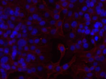

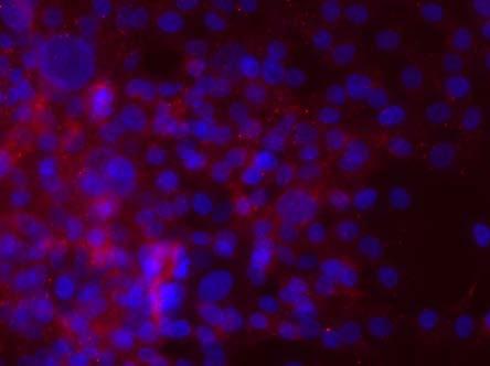
34 22 DIC DAPI Receptor Merged TrkB TrkA NGFR Figure 3-3 Immunofluorescence of neurotrophin receptor expression in 2-D in MCF10CA1a cells. Cells were cultured in a chamber slide for 24 hours and then stained for the receptors. Scale bar 50 µm.


35 23 (a) Receptor DAPI Merged (b) Receptor DAPI Merged TrkB TrkA NGFR TrkB TrkA NGFR


 and rbm fraction (c) are")
36 24 (c) Receptor DAPI Merged TrkB TrkA NGFR Figure 3-4 Immunofluorescence of neuotrophin receptor expression in the rmet model in MCF10CA1a cells. Cells were cultured in the model for 14 days and then isolated from each fraction to be stained for the receptors. Immunofluorescence of nuerotrophin receptor expression in the primary tumor fraction (a) is low and distinct to clusters of cells. The expression in the invasive layer (b) and rbm fraction (c) are expressed over a wider variety of cells, though still heterogeneous. Scale bar, 50 µm.
37 25 relevant setting, MCF10CA1a cells do express the three neurotrophin receptors with higher proportion expressed by the cells with migratory capacity rt-qpcr Confirmation of Neurotrophin Receptor Expression To confirm that the cells do in fact express the three neurotrophin receptors, RT- PCR was perform to quantify the mrna expression levels of each receptor. Primers were designed to the extracellular domains of each three receptor 64. RNA was extracted from MCF10CA1a cells from the flask (2-D), and from the top and rbm fractions of the rmet model (3-D). The results show that there is first is a low expression of all three receptors in 2-D culture. For all three receptors, there is an increase in the expression when the cells are cultured in the rmet model. Further, cells that are capable of migrating and surviving in the rbm fraction show a very large upregulation of all three neurotrophin receptors. TrkA (Figure 3-5b) has the highest fold change in the rbm, where the expression is upregulated approximately 240-fold as compared to the cells in the top. This similar pattern, is seen in both NGFR (Figure 3-5a) and TrkB (Figure3-5c) as well. 3.3 Neurotrophins and their effects on Breast Cancer Cell Proliferation D Cell Culture MCF10CA1a cells were treated with varying concentrations of neurotrophins: NGF, BDNF, and NT4/5 in 2-D cell culture to determine the proliferative effects that they have on the cells, and to determine the most effective concentration of each particular neurotrophin. Data was collected at 24- and 48-hours post neuorotrophin treatment for each concentration. For NGF (Figure 3-6a) and NT 4/5 (Figure 3-6c) the most effective concentration between 24 and 72 hours was 5 ng/ml. This is the concentration in which the proliferation of cells was significantly larger at 72 hours than it was at 24. For BDNF (Fig 3-6b) the most effective concentration was 50 ng/ml. It is important to choose the particular concentration that has a pro-proliferative effect on the cells because it has been shown that there are both pro- and anti-proliferative effects of neurotrophins depending on their concentration. These results show that there is a concentration dependent effect of neurotrophins on the proliferation of MCF10CA1a cells.
38 26 (a) (b) (c) Figure 3-5 Fold change in each of the receptors for MCF10CA1a cells in 2D and from two fractions of the rmet model. RT-qPCR was completed using mrna from cells cultured in 2D, and cells isolated from the primary tumor site and the rbm fraction of the rmet model. The neurotrophin receptor genes are expressed in 2-D and are upregulated in the primary tumor fraction and further upregulated in the rbm colonizing fraction. The greatest upregulation is TrkA (b) in the rbm, followed by TrkB (c) in the rbm.
39 27 (a) (b) (c) Figure 3-6 Effects of neurotrophins on proliferation of MCF10CA1a cells in 2D culture. Viable cells were counted using Trypan-blue and a hemacytometer. Graphs show the most effective concentrations for each neurotrophin. The most effective concentrations for NGF (a) and NT 4/5 (c) are 5 ng/ml, p<0.05. The most effective concentration for BDNF is 50 ng/ml, p<0.01.
40 rmet Model Based on 2-D proliferation experiments, the most effective concentrations for each neurotrophin were used in a proliferation experiments in the rmet model. The concentrations chosen were 50 ng/ml for BDNF, and 5 ng/ml for both NT4/5 and NGF. MCF10CA1a cells were cultured in the rmet model for 14 days. At that point, the transwell was removed from the rbm fraction and a treatment of neurotrophin in reduced serum media was added. This was done to specifically treat the population of migratory cells that are able to migrate and survive in the rbm fraction. In order to determine proliferation, cells were stained with Ki67, a marker for proliferation, at 24- and 72-hours post neurotrophin treatment. The longer time point in this experiment was chosen because original optimization of the experiment revealed little to no difference in the results between 24 and 48 hours. The results at 24 hours (Figure 3-7a) show that NGF stimulates the highest level of proliferation among all of the neurotrophins. This is consistent with other publications that have cited NGF as a mitogenic factor for MCF7 cells, but not for noncancerous cells 68. Seventy-two hours post neurotrophin treatment (Figure 3-7b) shows a slight increase in proliferation of cells treated with NGF, but more significantly, there is a large reduction in the proliferation of cells treated with BDNF. At 24 hours, BDNF does not show a significant increase in proliferation, suggesting that adding additional BDNF to cells in the rbm fraction could be detrimental to their proliferative capabilities. In order to determine the potential effects that neurotrophins can have on metastatic cancer cells, a series of experiments determining the role of neurotrophins and their effects on cancer cell proliferation were completed. There is some evidence that certain neurotrophins can induce proliferation of breast cancer cells, but do not have an effect on non-cancerous cells. Particularly, there is evidence that NGF has a mitogenic effect on MCF7 and MDA-MB-231 cells 68. The work completed on metastatic breast cancer lines, however, is lacking and needs further elucidation. Further, most research with neurotrophins and breast cancer cell lines has been completed in 2-D cell culture, where cells are grown on the surface of the tissue culture plastic. 2-D cell culture work is a good foundation on which to begin, but lacks physiological relevance due to a number of shortfalls, including lack of ECM components. It is important that in vitro cancer research mimics physiologically relevant conditions, which makes 3-D cell culture a vital avenue to take.

41 29 (a) (b) Figure 3-7 Effects of neurotrophins on proliferation of MCF10CA1a cells in the rmet model. In order to determine proliferation, cells were stained with Ki67, a marker of proliferation, and the total percentage of cells positive were determined. At 24 hours post-neurotrophin treatment, NGF causes the greatest increase in proliferation (p-value=0.3677) (a). At 72 hours posttreatment, BDNF decreases the proliferation of the cells (p-value=0.179), with a small increase in NGF (b).
42 Neurotrophins and Cancer Cell Migration Neurotrophins are vital players in the migration of neuronal cells during development. With evidence that neurotrophins can exert their effects on non-neuronal cells, and with the confirmation that MCF10CA1a cells express the three neurotrophin receptors, we wanted to determine whether the neurotrophins exert an effect on the migration and dissemination of breast cancer cells during metastasis. There have been a handful of studies that have studied the effects of neurotrophins and the effects that they have on the migration and invasiveness of cancer cells. Despite this, however, still little is known about the receptors involved and which particular signaling pathways are involved in this. For example, Lagadec et al. discovered an increase in TrkA expression in breast cancer tumors and discovered that the overexpression of TrkA in MDA-MB-231 cells increased their invasiveness 69. Further, it has been shown that neurotrophins NGF and NT 4/5 increase the migration and invasive properties of prostate cancer cell lines in a dose-dependent manner 70. This is consistent with other studies showing that addition of neurotrophins NGF and NT4/5 increase the invasiveness of pancreatic and ovarian cancer cells 71,72. Despite these studies, there is still gaping knowledge on how the increased invasiveness can aid in the treatment of metastasis. This leads to the current vein of research, to determine if similar migratory effects can be seen in aggressive breast cancer cell lines. The activation of signaling pathways leading to migration and invasion are not very clear in cancer cells, but further work should be able to elucidate the relationship between neurotrophins, the NTRs and their signaling pathways. It has been suggested that TrkA has the biggest influence on invasion of cancer cells, but this data needs more confirmation. Metastasis is a complex process that is extremely hard to target with current treatments. Current treatments and morbidity of breast cancer due to metastasis can be greatly improved if a signaling pathway and model of activation can be determined. 3.5 Neurotrophins and MCF10CA1a Cell Migration: Dome Assay In order to determine the role that neurotrophins play in the migration of breast cancer cell lines, a dome assay was created to determine the chemotactic potential of the neurotrophins on MCF10CA1a cells. In this assay, cells are mixed with Matrigel and are seeded in a dome in the center of a well in a tissue culture plate. In the surrounding area of the well, the treatment ring, Matrigel is added with the concentrations of
43 31 neurotrophins that were deemed most effective in the 2-D proliferation study (Figure 3-8). These concentrations are also fairly consistent with the concentrations used in other migration studies. After 24 hours, cells were imaged and counted based on their migration out of the dome and into the cell-free ring of Matrigel. The neurotrophin with the greatest stimulation of migration was NGF, with over 10% of cells migrating to the treatment ring (Figure 3-9). NT 4/5 had over a 3% increase in cell migration, which is a significant portion of cells compared to the vehicle control. BDNF had no significant effect on the migration of cells in the dome assay. 3.6 Neurotrophins and their receptors as potential treatment targets in breast cancer In this study, we can see that neurotrophins can play a role in the proliferation of breast cancer cells. In both 2-D and 3-D cell culture, MCF10CA1a cells express three neurotrophin receptors. Further, in 3-D, the migratory cells that colonize and proliferate in the rbm upregulate the receptors, which is evidence that highly migratory cells might be more receptive to neurotrophic effects. This is extremely useful and also congruent with the results that have shown that breast cancer tumors overexpress TrkA. If cells that have shown that they are capable of migrating and surviving in a secondary location, upregulate the neurotrophin receptors, they may be more receptive to neurotrophins than normal cells, which could greatly improve treatment options for breast cancer. Further, with the increase of the expression of the neurotrophin receptors, it could be expected that neurotrophins could possibly have increased effects in breast cancer cells. In 2-D cell culture we see an increase in cell proliferation at particular concentrations of neurotrophins. We also see that cells in the rbm have a slight increase in proliferation in the presence of NGF, which is consistent with the upregulation of TrkA in these cells because TrkA is the high-affinity receptor for NGF. Metastatic cells that are able to colonize at distant sites and form micrometastases must have increased proliferative properties in order to survive for an extended period of time at a distant site. It is possible that these cells are exploiting certain receptors and signaling pathways to increase their survival. If an effective inhibitor of TrkA could be developed, this could possibly have an effect on the unchecked proliferation that is characteristic of breast cancer cells.

44 32 Figure 3-8 Setup of the Dome Assay Figure 3-9 Dome Assay for Migration NT 4/5 and NGF induce a significant increase in cell migration towards the neurotrophin stimulus (**p-value <0.01; ***p-value<0.0001).
Protocol for A-549 VIM RFP (ATCC CCL-185EMT) TGFβ1 EMT Induction and Drug Screening
 TGFβ1 EMT Induction and Drug Screening") Protocol for A-549 VIM RFP (ATCC CCL-185EMT) TGFβ1 EMT Induction and Drug Screening Introduction: Vimentin (VIM) intermediate filament (IF) proteins are associated with EMT in lung cancer and its metastatic
Protocol for A-549 VIM RFP (ATCC CCL-185EMT) TGFβ1 EMT Induction and Drug Screening Introduction: Vimentin (VIM) intermediate filament (IF) proteins are associated with EMT in lung cancer and its metastatic
Animal Tissue Culture SQG 3242 Biology of Cultured Cells. Dr. Siti Pauliena Mohd Bohari
 Animal Tissue Culture SQG 3242 Biology of Cultured Cells Dr. Siti Pauliena Mohd Bohari The Culture Environment Changes of Cell s microenvironment needed that favor the spreading, migration, and proliferation
stem cell products Basement Membrane Matrix Products Rat Mesenchymal Stem Cell Growth and Differentiation Products
 stem cell products Basement Membrane Matrix Products Rat Mesenchymal Stem Cell Growth and Differentiation Products Stem Cell Qualified Extracellular Matrix Proteins Stem cell research requires the finest
Pluricyte Cardiomyocytes
 Pluricyte Cardiomyocytes Manual Version 2.1 / March 2018 Contents 1. Introduction 2 2. Equipment, Materials and Reagents 3 3. Methods 4 3.1 Coating of tissue culture plates 4 3.2 Thawing Pluricyte Cardiomyocytes
In vitro scratch assay: method for analysis of cell migration in vitro labeled fluorodeoxyglucose (FDG)
") In vitro scratch assay: method for analysis of cell migration in vitro labeled fluorodeoxyglucose (FDG) 1 Dr Saeb Aliwaini 13/11/2015 Migration in vivo Primary tumors are responsible for only about 10%
In vitro scratch assay: method for analysis of cell migration in vitro labeled fluorodeoxyglucose (FDG) 1 Dr Saeb Aliwaini 13/11/2015 Migration in vivo Primary tumors are responsible for only about 10%
Human ipsc-derived Ventricular Cardiomyocytes. Protocol version 3.1
 Human ipsc-derived Ventricular Cardiomyocytes Protocol version 3.1 Protocol version 3.1 Table of Contents Product Information 2 Recommendations 2 Preparing Cardiomyocyte Maintenance Medium 3 Cardiomyocyte
Immature organoids appear after hours.
 THE ESSENTIALS OF LIFE SCIENCE RESEARCH GLOBALLY DELIVERED Allison Ruchinskas, B.S., and James Clinton, Ph.D. ATCC Cell Systems, Gaithersburg, MD INTRODUCTION Figure 1. Mouse small intestinal organoid
THE ROLE OF VITAMIN D IN BREAST CANCER:
 THE ROLE OF VITAMIN D IN BREAST CANCER: Investigating Potential Inhibition Through Matrix Metalloproteinase 2 Student Author Hyesoo Chae is a professional student in the Purdue University College of Pharmacy.
Striatal Neuron Medium Kit
 Striatal Neuron Medium Kit Product Information What are included in the Striatal Neuron Medium Kit (ax0333): 2x 250 ml Striatal Neuron Basal Medium (Store at 4 o C upon receipt) 2x 7.5 ml Striatal Neuron
Enzyme-coupled Receptors. Cell-surface receptors 1. Ion-channel-coupled receptors 2. G-protein-coupled receptors 3. Enzyme-coupled receptors
 Enzyme-coupled Receptors Cell-surface receptors 1. Ion-channel-coupled receptors 2. G-protein-coupled receptors 3. Enzyme-coupled receptors Cell-surface receptors allow a flow of ions across the plasma
Cancer Biology Course. Invasion and Metastasis
 Cancer Biology Course Invasion and Metastasis 2016 Lu-Hai Wang NHRI Cancer metastasis Major problem: main reason for killing cancer patients, without it cancer can be cured or controlled. Challenging questions:
Product Use HPSC-CC are for research use only. It is not approved for human or animal use, or for application in in vitro diagnostic procedures.
 HPSC-derived Cardiomyocyte Cells (HPSC-CC) Catalog #6240 Cell Specification Human primary cardiomyocytes and cardiac tissue are superior modeling systems for heart disease studies, drug discovery and toxicity
Introduction: 年 Fas signal-mediated apoptosis. PI3K/Akt
 Fas-ligand (CD95-L; Fas-L) Fas (CD95) Fas (apoptosis) 年 了 不 度 Fas Fas-L 力 不 Fas/Fas-L T IL-10Fas/Fas-L 不 年 Fas signal-mediated apoptosis 度降 不 不 力 U-118, HeLa, A549, Huh-7 MCF-7, HepG2. PI3K/Akt FasPI3K/Akt
Phosphate buffered saline (PBS) for washing the cells TE buffer (nuclease-free) ph 7.5 for use with the PrimePCR Reverse Transcription Control Assay
 for washing the cells TE buffer (nuclease-free) ph 7.5 for use with the PrimePCR Reverse Transcription Control Assay") Catalog # Description 172-5080 SingleShot Cell Lysis Kit, 100 x 50 µl reactions 172-5081 SingleShot Cell Lysis Kit, 500 x 50 µl reactions For research purposes only. Introduction The SingleShot Cell Lysis
Catalog # Description 172-5080 SingleShot Cell Lysis Kit, 100 x 50 µl reactions 172-5081 SingleShot Cell Lysis Kit, 500 x 50 µl reactions For research purposes only. Introduction The SingleShot Cell Lysis
An Investigation into the Effects of the Addition of Synthetic Receptor on Chemokine Induced Jurkat T-Cell Migration
 An Investigation into the Effects of the Addition of Synthetic Receptor on Chemokine Induced Jurkat T-Cell Migration Jessica Jurado, Jianfang Hu, Avery August, PhD PSU Undergraduate Animal Bioscience July
Cell Migration and Invasion Assays INCUCYTE LIVE-CELL ANALYSIS SYSTEM. Real-time automated measurements of cell motility inside your incubator
 Cell Migration and Invasion Assays INCUCYTE LIVE-CELL ANALYSIS SYSTEM Real-time automated measurements of cell motility inside your incubator See the whole story Real-time cell motility visualization and
Introduction. Cancer Biology. Tumor-suppressor genes. Proto-oncogenes. DNA stability genes. Mechanisms of carcinogenesis.
 Cancer Biology Chapter 18 Eric J. Hall., Amato Giaccia, Radiobiology for the Radiologist Introduction Tissue homeostasis depends on the regulated cell division and self-elimination (programmed cell death)
CytoSelect Tumor- Endothelium Adhesion Assay
 Product Manual CytoSelect Tumor- Endothelium Adhesion Assay Catalog Number CBA- 215 100 assays FOR RESEARCH USE ONLY Not for use in diagnostic procedures Introduction Cancer metastasis comprises several
INSTRUCTIONS Pierce Primary Cardiomyocyte Isolation Kit
 INSTRUCTIONS Pierce Primary Cardiomyocyte Isolation Kit 88281 Number Description 88281 Pierce Primary Cardiomyocyte Isolation Kit, contains sufficient reagents to isolate cardiomyocytes from 50 neonatal
Extended Mammosphere Culture of Human Breast Cancer Cells
 Extended Mammosphere Culture of Human Breast Cancer Cells Application Note The PromoCell 3D Tumorsphere Medium XF The PromoCell 3D Tumorsphere Medium XF has been designed to meet your requirements for
Tumor microenvironment Interactions and Lung Cancer Invasiveness. Pulmonary Grand Rounds Philippe Montgrain, M.D.
 Tumor microenvironment Interactions and Lung Cancer Invasiveness Pulmonary Grand Rounds Philippe Montgrain, M.D. February 26, 2009 Objectives Review epithelial mesenchymal transition (EMT), and its implications
(A) PCR primers (arrows) designed to distinguish wild type (P1+P2), targeted (P1+P2) and excised (P1+P3)14-
 PCR primers (arrows) designed to distinguish wild type (P1+P2), targeted (P1+P2) and excised (P1+P3)14-") 1 Supplemental Figure Legends Figure S1. Mammary tumors of ErbB2 KI mice with 14-3-3σ ablation have elevated ErbB2 transcript levels and cell proliferation (A) PCR primers (arrows) designed to distinguish
1 Supplemental Figure Legends Figure S1. Mammary tumors of ErbB2 KI mice with 14-3-3σ ablation have elevated ErbB2 transcript levels and cell proliferation (A) PCR primers (arrows) designed to distinguish
VIII Curso Internacional del PIRRECV. Some molecular mechanisms of cancer
 VIII Curso Internacional del PIRRECV Some molecular mechanisms of cancer Laboratorio de Comunicaciones Celulares, Centro FONDAP Estudios Moleculares de la Celula (CEMC), ICBM, Facultad de Medicina, Universidad
Optimization of the Fuse-It-mRNA Protocol for L929 Cells in the µ-plate 24 Well
 Optimization of the Fuse-It-mRNA Protocol for L929 Cells in the µ-plate 24 Well 1. General Information... 1 2. Background... 1 3. Material and Equipment Required... 2 4. Experimental Procedure and Results...
CRIPTO-1 A POSSIBLE NEW BIOMARKER IN GLIOBLASTOMA MULTIFORME PIA OLESEN, MD, PHD STUDENT
 CRIPTO-1 A POSSIBLE NEW BIOMARKER IN GLIOBLASTOMA MULTIFORME PIA OLESEN, MD, PHD STUDENT Glioblastoma WHO Grade IV Glioma Heterogenic Undiffenrentiated phenotype 50% of all Gliomas Around 600 patients
Protocol for Thawing Cryopreserved Hepatocytes
 cell and tissue-based products Protocol for Thawing Cryopreserved Hepatocytes Product Instruction The following procedure may be carried out in a biosafety containment hood to reduce the risk of contamination
Corning BioCoat Matrigel Invasion Chamber
 Corning BioCoat Matrigel Invasion Chamber Catalog No. 354480, 354481 Guidelines for Use Discovery Labware, Inc., Two Oak Park, Bedford, MA 01730, Tel: 1.978.442.2200 (U.S.) CLSTechServ@Corning.com www.corning.com/lifesciences
Suppl Video: Tumor cells (green) and monocytes (white) are seeded on a confluent endothelial
 and monocytes (white) are seeded on a confluent endothelial") Supplementary Information Häuselmann et al. Monocyte induction of E-selectin-mediated endothelial activation releases VE-cadherin junctions to promote tumor cell extravasation in the metastasis cascade
Supplementary Information Häuselmann et al. Monocyte induction of E-selectin-mediated endothelial activation releases VE-cadherin junctions to promote tumor cell extravasation in the metastasis cascade
The Avatar System TM Yields Biologically Relevant Results
 Application Note The Avatar System TM Yields Biologically Relevant Results Liquid biopsies stand to revolutionize the cancer field, enabling early detection and noninvasive monitoring of tumors. In the
Supplementary Materials. for Garmy-Susini, et al, Integrin 4 1 signaling is required for lymphangiogenesis and tumor metastasis
 Supplementary Materials for Garmy-Susini, et al, Integrin 4 1 signaling is required for lymphangiogenesis and tumor metastasis 1 Supplementary Figure Legends Supplementary Figure 1: Integrin expression
Supplementary Figure (OH) 22 nanoparticles did not affect cell viability and apoposis. MDA-MB-231, MCF-7, MCF-10A and BT549 cells were
 22 nanoparticles did not affect cell viability and apoposis. MDA-MB-231, MCF-7, MCF-10A and BT549 cells were") Supplementary Figure 1. Gd@C 82 (OH) 22 nanoparticles did not affect cell viability and apoposis. MDA-MB-231, MCF-7, MCF-10A and BT549 cells were treated with PBS, Gd@C 82 (OH) 22, C 60 (OH) 22 or GdCl
Supplementary Figure 1. Gd@C 82 (OH) 22 nanoparticles did not affect cell viability and apoposis. MDA-MB-231, MCF-7, MCF-10A and BT549 cells were treated with PBS, Gd@C 82 (OH) 22, C 60 (OH) 22 or GdCl
Supplementary Figure 1
 Supplementary Figure 1 The average sigmoid parametric curves of capillary dilation time courses and average time to 50% peak capillary diameter dilation computed from individual capillary responses averaged
Overview of methodology, tools and reagents for evaluating cell proliferation and invasion using multicellular tumor spheroids.
 The Next Step in the Evolution of 3D Culture: Utilizing Extracellular Matrix to Enhance Multicellular Tumor Spheroid Models for Proliferation and Invasion Overview of methodology, tools and reagents for
Neoplasia part I. Dr. Mohsen Dashti. Clinical Medicine & Pathology nd Lecture
 Neoplasia part I By Dr. Mohsen Dashti Clinical Medicine & Pathology 316 2 nd Lecture Lecture outline Review of structure & function. Basic definitions. Classification of neoplasms. Morphologic features.
4/18/2011. Physiology 67 Lecture on Neural Development
 Physiology 67 Lecture on Neural Development 1 2 3 4 5 6 Neural cell categories After the ectodermal tissue has folded into the neural tube, another series of signaling interactions determine the type of
Brain Development III
 Brain Development III Neural Development In the developing nervous system there must be: 1. The formation of different regions of the brain. 2. The ability of a neuron to differentiate. 3. The ability
Genetics and Cancer Ch 20
 Genetics and Cancer Ch 20 Cancer is genetic Hereditary cancers Predisposition genes Ex. some forms of colon cancer Sporadic cancers ~90% of cancers Descendants of cancerous cells all cancerous (clonal)
- is a common disease - 1 person in 3 can expect to contract cancer at some stage in their life -1 person in 5 can expect to die from it
 MBB157 Dr D Mangnall The Molecular Basis of Disease CANCER Lecture 1 One of the simpler (and better) definitions of cancer comes from the American Cancer Society, who define cancer as; 'Cancer is a group
Instructions. Fuse-It-mRNA easy. Shipping and Storage. Overview. Kit Contents. Specifications. Note: Important Guidelines
 Membrane fusion is a highly efficient method for transfecting various molecules and particles into mammalian cells, even into sensitive and primary cells. The Fuse-It reagents are cargo-specific liposomal
Pluricyte Cardiomyocytes. using the Multiwell MEA System from Multi Channel Systems
 Pluricyte Cardiomyocytes using the Multiwell MEA System from Multi Channel Systems User Guide Version 3.1 / March 2018 Contents 1. Introduction 2 2. Workflow 3 3. Important Recommendations 4 4. Equipment,
Human Pluripotent Stem Cell Cardiomyocyte Differentiation Kit (PSCCDK) Introduction Kit Components Cat. # # of vials Reagent Quantity Storage
 Introduction Kit Components Cat. # # of vials Reagent Quantity Storage") Human Pluripotent Stem Cell Cardiomyocyte Differentiation Kit (PSCCDK) Catalog #5901 Introduction Human pluripotent stem cells (hpsc), including embryonic stem cells (ESC) and induced pluripotent stem
Human Pluripotent Stem Cell Cardiomyocyte Differentiation Kit (PSCCDK) Catalog #5901 Introduction Human pluripotent stem cells (hpsc), including embryonic stem cells (ESC) and induced pluripotent stem
PROTOCOL: OPTIMIZATION OF LENTIVIRAL TRANSDUCTION USING SPINFECTION
 Last Modified: April 2018 Last Review: October 2018 PROTOCOL: OPTIMIZATION OF LENTIVIRAL TRANSDUCTION USING SPINFECTION Table of Contents 1. Brief Description 1 2. Materials and Reagents.1 3. Optimization
Part II The Cell Cell Division, Chapter 2 Outline of class notes
 Part II The Cell Cell Division, Chapter 2 Outline of class notes 1 Cellular Division Overview Types of Cell Division Chromosomal Number The Cell Cycle Mitoses Cancer Cells In Vitro Fertilization Infertility
LQB383 Testbank. Week 8 Cell Communication and Signaling Mechanisms
 LQB383 Testbank Week 8 Cell Communication and Signaling Mechanisms Terms to learn match the terms to the definitions --------------------------------------------------------------------------------------------------------------------------
APPLICATION SPECIFIC PROTOCOL ANGIOGENESIS... 1 TABLE OF CONTENTS... 1 MONOLAYER FORMATION... 2 OPTION 1: APPLICATION OF ANGIOGENIC STIMULI...
 APPLICATION SPECIFIC PROTOCOL ANGIOGENESIS AIM 3D Cell Culture Chips offer a new perspective in studying angiogenesis by allowing the growth of new vascular sprouts in a 3D matrix from a pre-existing endothelial
(a) Significant biological processes (upper panel) and disease biomarkers (lower panel)
 Significant biological processes (upper panel) and disease biomarkers (lower panel)") Supplementary Figure 1. Functional enrichment analyses of secretomic proteins. (a) Significant biological processes (upper panel) and disease biomarkers (lower panel) 2 involved by hrab37-mediated secretory
Supplementary Figure 1. Functional enrichment analyses of secretomic proteins. (a) Significant biological processes (upper panel) and disease biomarkers (lower panel) 2 involved by hrab37-mediated secretory
The Development of Lymphocytes: B Cell Development in the Bone Marrow & Peripheral Lymphoid Tissue Deborah A. Lebman, Ph.D.
 The Development of Lymphocytes: B Cell Development in the Bone Marrow & Peripheral Lymphoid Tissue Deborah A. Lebman, Ph.D. OBJECTIVES 1. To understand how ordered Ig gene rearrangements lead to the development
MTC-TT and TPC-1 cell lines were cultured in RPMI medium (Gibco, Breda, The Netherlands)
") Supplemental data Materials and Methods Cell culture MTC-TT and TPC-1 cell lines were cultured in RPMI medium (Gibco, Breda, The Netherlands) supplemented with 15% or 10% (for TPC-1) fetal bovine serum
Supplemental data Materials and Methods Cell culture MTC-TT and TPC-1 cell lines were cultured in RPMI medium (Gibco, Breda, The Netherlands) supplemented with 15% or 10% (for TPC-1) fetal bovine serum
Chapter 11 Introduction to the Nervous System and Nervous Tissue Chapter Outline
 Chapter 11 Introduction to the Nervous System and Nervous Tissue Chapter Outline Module 11.1 Overview of the Nervous System (Figures 11.1-11.3) A. The nervous system controls our perception and experience
NINDS Repository Human Induced Pluripotent Stem Cell (ipsc) Handling Protocols (Matrigel and mtesr Media)
 Handling Protocols (Matrigel and mtesr Media)") General Guidelines for Handling Human ipsc cells ipsc are cryopreserved in plastic cryovials and shipped on dry ice. If storing the ipsc before thawing, store in liquid nitrogen vapor. Storage directly
General Guidelines for Handling Human ipsc cells ipsc are cryopreserved in plastic cryovials and shipped on dry ice. If storing the ipsc before thawing, store in liquid nitrogen vapor. Storage directly
Supplemental Information. Tissue Myeloid Progenitors Differentiate. into Pericytes through TGF-b Signaling. in Developing Skin Vasculature
 Cell Reports, Volume 18 Supplemental Information Tissue Myeloid Progenitors Differentiate into Pericytes through TGF-b Signaling in Developing Skin Vasculature Tomoko Yamazaki, Ani Nalbandian, Yutaka Uchida,
1.The metastatic cascade. 2.Pathologic features of metastasis. 3.Therapeutic ramifications
 Metastasis 1.The metastatic cascade 2.Pathologic features of metastasis 3.Therapeutic ramifications Sir James Paget (1814-1899) British Surgeon/ Pathologist Paget s disease of bone Paget s disease of the
G-Protein Signaling. Introduction to intracellular signaling. Dr. SARRAY Sameh, Ph.D
 G-Protein Signaling Introduction to intracellular signaling Dr. SARRAY Sameh, Ph.D Cell signaling Cells communicate via extracellular signaling molecules (Hormones, growth factors and neurotransmitters
STRUCTURAL ELEMENTS OF THE NERVOUS SYSTEM
 STRUCTURAL ELEMENTS OF THE NERVOUS SYSTEM STRUCTURE AND MAINTENANCE OF NEURONS (a) (b) Dendrites Cell body Initial segment collateral terminals (a) Diagrammatic representation of a neuron. The break in
Stem cells and Cancer. John Glod. December 2, 2009
 Stem cells and Cancer John Glod Lehigh University Lehigh University December 2, 2009 The Tumor Microenvironment Littlepage et al Cancer Cell 2005 Cancer Stem Cells A small group of cells within the larger
Instructions for Use. APO-AB Annexin V-Biotin Apoptosis Detection Kit 100 tests
 3URGXFW,QIRUPDWLRQ Sigma TACS Annexin V Apoptosis Detection Kits Instructions for Use APO-AB Annexin V-Biotin Apoptosis Detection Kit 100 tests For Research Use Only. Not for use in diagnostic procedures.
Mouse Hepatic Progenitor Organoid Culture: Supplementary Protocols
 TECHNICAL BULLETIN Mouse Hepatic Progenitor Organoid Culture: The following are supplementary protocols for the culture of hepatic organoids with HepatiCult Organoid Growth Medium (Mouse) (Catalog #06030).
Plasticity of Cerebral Cortex in Development
 Plasticity of Cerebral Cortex in Development Jessica R. Newton and Mriganka Sur Department of Brain & Cognitive Sciences Picower Center for Learning & Memory Massachusetts Institute of Technology Cambridge,
PRODUCT INFORMATION & MANUAL
 PRODUCT INFORMATION & MANUAL Nuclear Extraction Kit NBP2-29447 Research use only. Not for diagnostic or therapeutic procedures. www.novusbio.com - P: 888.506.6887 - technical@novusbio.com Novus kits are
Src-INACTIVE / Src-INACTIVE
 Biology 169 -- Exam 1 February 2003 Answer each question, noting carefully the instructions for each. Repeat- Read the instructions for each question before answering!!! Be as specific as possible in each
Human Induced Plutipotent Stem Cell (ipsc) Handling Protocols: Matrigel and mtesr/e8 Media
 Handling Protocols: Matrigel and mtesr/e8 Media") General Guidelines for Handling Human ipsc cells ipsc are cryopreserved in plastic cryovials and shipped on dry ice. If storing the ipsc before thawing, store in liquid nitrogen vapor. Storage directly
General Guidelines for Handling Human ipsc cells ipsc are cryopreserved in plastic cryovials and shipped on dry ice. If storing the ipsc before thawing, store in liquid nitrogen vapor. Storage directly
HOMEWORK RUBRICS MECHANOTRANSDUCTION UNIT: HOMEWORK #1 (20 pts towards your grade)
") HOMEWORK RUBRICS MECHANOTRANSDUCTION UNIT: HOMEWORK #1 (20 pts towards your grade) 1. Mesenchymal stem cells (MSC) cultured on extracellular matrices with different stiffness exhibit diverse lineage commitment
HOMEWORK RUBRICS MECHANOTRANSDUCTION UNIT: HOMEWORK #1 (20 pts towards your grade) 1. Mesenchymal stem cells (MSC) cultured on extracellular matrices with different stiffness exhibit diverse lineage commitment
Human Histology The Nervous System. Dr. Rawaa Salim Hameed
 Human Histology The Nervous System Dr. Rawaa Salim Hameed The organization of the nervous system Anatomically, the nervous system is divided into:- Neurohistology Structurally, nerve tissue consists of
PREPARED FOR: U.S. Army Medical Research and Materiel Command Fort Detrick, Maryland
 AD Award Number: W81XWH-12-1-0225 TITLE: Epstein Barr virus and blood brain barrier in Multiple Sclerosis PRINCIPAL INVESTIGATOR: Luwen Zhang CONTRACTING ORGANIZATION: University of Nebraska Lincoln, NE
Human Anatomy and Physiology - Problem Drill 11: Neural Tissue & The Nervous System
 Human Anatomy and Physiology - Problem Drill 11: Neural Tissue & The Nervous System Question No. 1 of 10 The human body contains different types of tissue. The tissue is formed into organs and organ systems.
Bio 111 Study Guide Chapter 11 Cell Communication
 Bio 111 Study Guide Chapter 11 Cell Communication BEFORE CLASS: Reading: Read the introduction on p. 210, and for Concept 11.1, read from the first full paragraph on p. 212. Read all of Concept 11.2. Pay
Single Cell Quantitative Polymer Chain Reaction (sc-qpcr)
") Single Cell Quantitative Polymer Chain Reaction (sc-qpcr) Analyzing gene expression profiles from a bulk population of cells provides an average profile which may obscure important biological differences
Single Cell Quantitative Polymer Chain Reaction (sc-qpcr) Analyzing gene expression profiles from a bulk population of cells provides an average profile which may obscure important biological differences
Aberrant cell Growth. Younas Masih New Life College of Nursing Karachi. 3/4/2016 Younas Masih ( NLCON)
") Aberrant cell Growth Younas Masih New Life College of Nursing Karachi 1 Objectives By the end of this session the learners will be able to, Define the characteristics of the normal cell Describe the characteristics
Aberrant cell Growth Younas Masih New Life College of Nursing Karachi 1 Objectives By the end of this session the learners will be able to, Define the characteristics of the normal cell Describe the characteristics
Assessment of pro-arrhythmic effects using Pluricyte Cardiomyocytes. on the ACEA xcelligence RTCA CardioECR
 Assessment of pro-arrhythmic effects using Pluricyte Cardiomyocytes on the ACEA xcelligence RTCA CardioECR User Guide Version 3.0 / March 2018 Contents 1. Introduction 2 2. Workflow 3 3. Important recommendations
10.00 PBS OVA OVA+isotype antibody 8.00 OVA+anti-HMGB1. PBS Methatroline (mg/ml)
") RESEARCH ARTICLE Penh (100% of PBS) 1 PBS 8.00 +anti-hmgb1 6.00 4.00 p=0.054 Cellular & Molecular Immunology advance online publication, PBS 3.12 6.25 Methatroline (mg/ml) Neutrophil isolation and culture
RESEARCH ARTICLE Penh (100% of PBS) 1 PBS 8.00 +anti-hmgb1 6.00 4.00 p=0.054 Cellular & Molecular Immunology advance online publication, PBS 3.12 6.25 Methatroline (mg/ml) Neutrophil isolation and culture
Cancer. The fundamental defect is. unregulated cell division. Properties of Cancerous Cells. Causes of Cancer. Altered growth and proliferation
 Cancer The fundamental defect is unregulated cell division. Properties of Cancerous Cells Altered growth and proliferation Loss of growth factor dependence Loss of contact inhibition Immortalization Alterated
CytoSelect 24- Well Cell Invasion Assay (Basement Membrane, Fluorometric Format)
") Product Manual CytoSelect 24- Well Cell Invasion Assay (Basement Membrane, Fluorometric Format) Catalog Number CBA- 111 12 assays FOR RESEARCH USE ONLY Not for use in diagnostic procedures Introduction
Product Manual CytoSelect 24- Well Cell Invasion Assay (Basement Membrane, Fluorometric Format) Catalog Number CBA- 111 12 assays FOR RESEARCH USE ONLY Not for use in diagnostic procedures Introduction
Signaling Vascular Morphogenesis and Maintenance
 Signaling Vascular Morphogenesis and Maintenance Douglas Hanahan Science 277: 48-50, in Perspectives (1997) Blood vessels are constructed by two processes: vasculogenesis, whereby a primitive vascular
1. The metastatic cascade. 3. Pathologic features of metastasis. 4. Therapeutic ramifications. Which malignant cells will metastasize?
 1. The metastatic cascade 3. Pathologic features of metastasis 4. Therapeutic ramifications Sir James Paget (1814-1899) British Surgeon/ Pathologist Paget s disease of Paget s disease of the nipple (intraductal
T-cell activation T cells migrate to secondary lymphoid tissues where they interact with antigen, antigen-presenting cells, and other lymphocytes:
 Interactions between innate immunity & adaptive immunity What happens to T cells after they leave the thymus? Naïve T cells exit the thymus and enter the bloodstream. If they remain in the bloodstream,
SIBLINGs, cancer's multifunctional weapons
 SIBLINGs, cancer's multifunctional weapons 6/18/08 Akeila Bellahcène and Vincent Castronovo of the Metastasis Research laboratory of the University of Liège are among the first researchers to have discovered
Evaluation of directed and random motility in microslides Assessment of leukocyte adhesion in flow chambers
 Evaluation of directed and random motility in microslides Motility experiments in IBIDI microslides, image acquisition and processing were performed as described. PMN, which ended up in an angle < 180
(A) Dose response curves of HMLE_shGFP (blue circle), HMLE_shEcad (red square),
 Dose response curves of HMLE_shGFP (blue circle), HMLE_shEcad (red square),") Supplementary Figures and Tables Figure S1. Validation of EMT-selective small molecules (A) Dose response curves of HMLE_shGFP (blue circle), HMLE_shEcad (red square), and HMLE_Twist (black diamond) cells
Supplementary Figures and Tables Figure S1. Validation of EMT-selective small molecules (A) Dose response curves of HMLE_shGFP (blue circle), HMLE_shEcad (red square), and HMLE_Twist (black diamond) cells
Supplementary Figure 1. SA-β-Gal positive senescent cells in various cancer tissues. Representative frozen sections of breast, thyroid, colon and
 Supplementary Figure 1. SA-β-Gal positive senescent cells in various cancer tissues. Representative frozen sections of breast, thyroid, colon and stomach cancer were stained with SA-β-Gal and nuclear fast
Supplementary Figure 1. HOPX is hypermethylated in NPC. (a) Methylation levels of HOPX in Normal (n = 24) and NPC (n = 24) tissues from the
 Methylation levels of HOPX in Normal (n = 24) and NPC (n = 24) tissues from the") Supplementary Figure 1. HOPX is hypermethylated in NPC. (a) Methylation levels of HOPX in Normal (n = 24) and NPC (n = 24) tissues from the genome-wide methylation microarray data. Mean ± s.d.; Student
Supplementary Figure 1. HOPX is hypermethylated in NPC. (a) Methylation levels of HOPX in Normal (n = 24) and NPC (n = 24) tissues from the genome-wide methylation microarray data. Mean ± s.d.; Student
Supplemental Data. TGF-β-mediated mir-181a expression promotes breast cancer metastasis by targeting Bim.
 Supplemental Data TGF-β-mediated mir-181a expression promotes breast cancer metastasis by targeting Bim. Molly A. Taylor 1, Khalid Sossey-Alaoui 2, Cheryl L. Thompson 3, David Danielpour 4, and William
Supplementary data Supplementary Figure 1 Supplementary Figure 2
 Supplementary data Supplementary Figure 1 SPHK1 sirna increases RANKL-induced osteoclastogenesis in RAW264.7 cell culture. (A) RAW264.7 cells were transfected with oligocassettes containing SPHK1 sirna
Erzsebet Kokovay, Susan Goderie, Yue Wang, Steve Lotz, Gang Lin, Yu Sun, Badrinath Roysam, Qin Shen,
 Cell Stem Cell, Volume 7 Supplemental Information Adult SVZ Lineage Cells Home to and Leave the Vascular Niche via Differential Responses to SDF1/CXCR4 Signaling Erzsebet Kokovay, Susan Goderie, Yue Wang,
PBS Class #2 Introduction to the Immune System part II Suggested reading: Abbas, pgs , 27-30
 PBS 803 - Class #2 Introduction to the Immune System part II Suggested reading: Abbas, pgs. 15-25, 27-30 Learning Objectives Compare and contrast the maturation of B and T lymphocytes Compare and contrast
Human IL-2. Pre-Coated ELISA Kit
 Human IL-2 (Interleukin 2) Pre-Coated ELISA Kit Catalog No: 90-2083 1 96 well Format (96 tests) Detection Range: 31.2 2000 pg/ml Sensitivity: < 18.75 pg/ml This immunoassay kit allows for the in vitro
Notch Signaling Pathway Notch CSL Reporter HEK293 Cell line Catalog #: 60652
 Notch Signaling Pathway Notch CSL Reporter HEK293 Cell line Catalog #: 60652 Background The Notch signaling pathway controls cell fate decisions in vertebrate and invertebrate tissues. Notch signaling
Unit Six The Nervous System
 Unit Six The Nervous System I. Introduction A. Definition a coordinating system of the body, composed of highly specialized cells that conduct nerve impulses to a center so responses can be made. The nervous
Supplemental Information. Otic Mesenchyme Cells Regulate. Spiral Ganglion Axon Fasciculation. through a Pou3f4/EphA4 Signaling Pathway
 Neuron, Volume 73 Supplemental Information Otic Mesenchyme Cells Regulate Spiral Ganglion Axon Fasciculation through a Pou3f4/EphA4 Signaling Pathway Thomas M. Coate, Steven Raft, Xiumei Zhao, Aimee K.
Neoplasia 18 lecture 8. Dr Heyam Awad MD, FRCPath
 Neoplasia 18 lecture 8 Dr Heyam Awad MD, FRCPath ILOS 1. understand the angiogenic switch in tumors and factors that stimulate and inhibit angiogenesis. 2. list the steps important for tumor metastasis
SUPPLEMENTARY INFORMATION
 Supplementary Figures Supplementary Figure S1. Binding of full-length OGT and deletion mutants to PIP strips (Echelon Biosciences). Supplementary Figure S2. Binding of the OGT (919-1036) fragments with
Olfactory ensheathing glia
 Olfactory ensheathing glia From Wikipedia, the free encyclopedia Neuroglia of the brain shown by Golgi's method. Olfactory ensheathing glia (OEG), also known as olfactory ensheathing cells (OECs) or olfactory
Nervous System. Master controlling and communicating system of the body. Secrete chemicals called neurotransmitters
 Nervous System Master controlling and communicating system of the body Interacts with the endocrine system to control and coordinate the body s responses to changes in its environment, as well as growth,
Influenza B Hemagglutinin / HA ELISA Pair Set
 Influenza B Hemagglutinin / HA ELISA Pair Set Catalog Number : SEK11053 To achieve the best assay results, this manual must be read carefully before using this product and the assay is run as summarized
Major Structures of the Nervous System. Brain, cranial nerves, spinal cord, spinal nerves, ganglia, enteric plexuses and sensory receptors
 Major Structures of the Nervous System Brain, cranial nerves, spinal cord, spinal nerves, ganglia, enteric plexuses and sensory receptors Nervous System Divisions Central Nervous System (CNS) consists
Islet Oxygen Consumption Assay using the XF24 Islet Capture Microplate Shirihai Lab and Seahorse Bioscience Aug
 1 Islet Oxygen Consumption Assay using the XF24 Islet Capture Microplate Shirihai Lab and Seahorse Bioscience Aug 6 2009 XF Analyzers are most commonly used with an adherent monolayer of cells attached
Biochemistry of Carcinogenesis. Lecture # 35 Alexander N. Koval
 Biochemistry of Carcinogenesis Lecture # 35 Alexander N. Koval What is Cancer? The term "cancer" refers to a group of diseases in which cells grow and spread unrestrained throughout the body. It is difficult
Cancer and Oncogenes Bioscience in the 21 st Century. Linda Lowe-Krentz
 Cancer and Oncogenes Bioscience in the 21 st Century Linda Lowe-Krentz December 1, 2010 Just a Few Numbers Becoming Cancer Genetic Defects Drugs Our friends and family 25 More mutations as 20 you get older
axion Protocol Cell Culture on Microelectrode Arrays Cell Type: QBM Cell Science - E14,15 Embryonic C57 Mouse Cortical Neurons BioSystems v. 1.
 axion BioSystems Cell Culture on Microelectrode Arrays Cell Type: QBM Cell Science - E14,15 Embryonic C57 Mouse Cortical Neurons Protocol v. 1.1 Trademarks Axion BioSystems, Inc. and the logo are trademarks
Development of the Nervous System 1 st month
 Development of the Nervous System 1 st month day 1 - fertilization of egg day 6 - uterine implantation day 18 - trilaminar (3-layered) disc (blastoderm, embryo) ectoderm (dorsal) - nervous system and skin