The Muir-Torre syndrome
|
|
|
- Spencer Merritt
- 6 years ago
- Views:
Transcription
1 Lesions and syndromes: Cutaneous markers of systemic problems Philip E. LeBoit, M.D. Depts. of Pathology and Dermatology, University of California, San Francisco In medicine and psychology, a syndrome is the association of several clinically recognizable features, signs (observed by a physician), symptoms (reported by the patient), phenomena or characteristics that often occur together, so that the presence of one feature alerts the physician to the presence of the others. In recent decades, the term has been used outside medicine to refer to a combination of phenomena seen in association. The term syndrome derives from its Greek roots (σύνδρομος) and means literally "run together", as the features do. It is most often used to refer to the set of detectable characteristics when the reason that they occur together (the pathophysiology of the syndrome) has not yet been discovered. A familiar syndrome name often continues to be used even after an underlying cause has been found, or when there are a number of different primary causes that all give rise to the same combination of symptoms and signs. Many syndromes are named after the physicians credited with first reporting the association; these are "eponymous" syndromes (see also the list of eponymous diseases, many of which are called "syndromes"). Otherwise, disease features or presumed causes, as well as references to geography, history or poetry, can lend their names to syndromes. -Wikipedia, Syndrome, The Muir-Torre syndrome 1


2 5/24/2014 2

 Microsatellite instability can silence")
3 Muir-Torre syndrome Autosomal dominant condition Variant form of the hereditary non-polyposis colorectal carcinoma or Lynch syndrome Produced by germline defects in several genes coding for DNA mismatch repair proteins. Two most commonly encountered mis-match repair proteins that are defective are hmsh2 and hmlh1 A defect in a mismatch repair protein results in socalled microsatellite instability Microsatellites are repeated DNA sequences (often comprised of repeats of the bases C and A Muir-Torre syndrome (con t) Microsatellite instability can silence tumor suppressor genes Most common carcinomas are colon, bladder, female genital tract, renal Colonic carcinomas behave less agressively 3


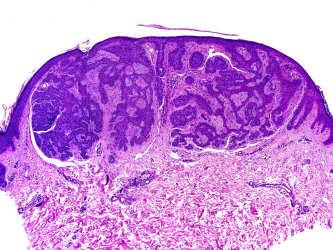
4 Sebaceous hyperplasia 4


5 5


6 Sebaceoma 6


7 7


8 Cystic sebaceous neoplasm 8
:405, October 1999.")


9 TABLE 1. Cystic Sebaceous Tumors as Marker Lesions for the Muir-Torre Syndrome: A Histopathologic and Molecular Genetic Study. Rutten, Arno; Burgdorf, Walter; Hugel, Heino; Kutzner, Heinz; Hosseiny-Malayeri, Hamid; Friedl, Waltraut; Propping, Peter; Kruse, Roland American Journal of Dermatopathology. 21(5):405, October TABLE 1. Clinical findings in eight patients with MTS with CST*Current number in this study; in parentheses running number in genetic study (see ref. 1).+Current age. In Patient 4, metastatic colorectal cancer caused death at age 69.Internal tumors and skin tumors: in parentheses number of tumors. mx, multiple tumors Lippincott Williams & Wilkins, Inc. Published by Lippincott Williams & Wilkins, Inc. 2 TABLE 2. TABLE 3. Cystic Sebaceous Tumors as Marker Lesions for the Muir-Torre Syndrome: A Histopathologic and Molecular Genetic Study. Rutten, Arno; Burgdorf, Walter; Hugel, Heino; Kutzner, Heinz; Hosseiny-Malayeri, Hamid; Friedl, Waltraut; Propping, Peter; Kruse, Roland American Journal of Dermatopathology. 21(5):405, October Cystic Sebaceous Tumors as Marker Lesions for the Muir-Torre Syndrome: A Histopathologic and Molecular Genetic Study. Rutten, Arno; Burgdorf, Walter; Hugel, Heino; Kutzner, Heinz; Hosseiny-Malayeri, Hamid; Friedl, Waltraut; Propping, Peter; Kruse, Roland American Journal of Dermatopathology. 21(5):405, October TABLE 2. CST in eight patients with MTS: histopathologic and molecular genetic datacst, cystic sebaceous tumors; MSI, microsatellite instability (instable markers/all examined markers); NE, not examined; ND, not detected. TABLE 3. Summary of the spectrum of histopathologic findings in CST 1999 Lippincott Williams & Wilkins, Inc. Published by Lippincott Williams & Wilkins, Inc Lippincott Williams & Wilkins, Inc. Published by Lippincott Williams & Wilkins, Inc. 4 9
:405, October 1999.")
. 1999 Lippincott Williams & Wilkins, Inc. Published by Lippincott Williams & Wilkins, Inc. 5 1999 Lippincott Williams & Wilkins, Inc.")
.")

10 FIG. 1. FIG. 2. Cystic Sebaceous Tumors as Marker Lesions for the Muir-Torre Syndrome: A Histopathologic and Molecular Genetic Study. Rutten, Arno; Burgdorf, Walter; Hugel, Heino; Kutzner, Heinz; Hosseiny-Malayeri, Hamid; Friedl, Waltraut; Propping, Peter; Kruse, Roland American Journal of Dermatopathology. 21(5):405, October Cystic Sebaceous Tumors as Marker Lesions for the Muir-Torre Syndrome: A Histopathologic and Molecular Genetic Study. Rutten, Arno; Burgdorf, Walter; Hugel, Heino; Kutzner, Heinz; Hosseiny-Malayeri, Hamid; Friedl, Waltraut; Propping, Peter; Kruse, Roland American Journal of Dermatopathology. 21(5):405, October FIG. 1. Multiple sebaceous tumors of the nasal region (Patient 4). FIG. 2. Cystic sebaceous adenoma (Patient 1) Lippincott Williams & Wilkins, Inc. Published by Lippincott Williams & Wilkins, Inc Lippincott Williams & Wilkins, Inc. Published by Lippincott Williams & Wilkins, Inc. 6 FIG. 4. Cystic Sebaceous Tumors as Marker Lesions for the Muir-Torre Syndrome: A Histopathologic and Molecular Genetic Study. Rutten, Arno; Burgdorf, Walter; Hugel, Heino; Kutzner, Heinz; Hosseiny-Malayeri, Hamid; Friedl, Waltraut; Propping, Peter; Kruse, Roland American Journal of Dermatopathology. 21(5):405, October Figure 4 Cystic Sebaceous Tumors as Marker Lesions for the Muir-Torre Syndrome: A Histopathologic and Molecular Genetic Study. Rutten, Arno; Burgdorf, Walter; Hugel, Heino; Kutzner, Heinz; Hosseiny-Malayeri, Hamid; Friedl, Waltraut; Propping, Peter; Kruse, Roland American Journal of Dermatopathology. 21(5):405, October FIG. 4. A: Cystic sebaceous adenoma with a central opening to the skin surface (Patient 6). B: Cyst wall with a small rim of nonvacuolated immature sebocytes at the periphery and a broad zone of vacuolated fully differentiated sebocytes toward the center. Figure 4. Continued 1999 Lippincott Williams & Wilkins, Inc. Published by Lippincott Williams & Wilkins, Inc Lippincott Williams & Wilkins, Inc. Published by Lippincott Williams & Wilkins, Inc. 8 10
:405, October 1999.")
 with a folded cyst wall and a higher proportion of immature sebocytes.")

11 FIG. 3. Fig. 5. Cystic Sebaceous Tumors as Marker Lesions for the Muir-Torre Syndrome: A Histopathologic and Molecular Genetic Study. Rutten, Arno; Burgdorf, Walter; Hugel, Heino; Kutzner, Heinz; Hosseiny-Malayeri, Hamid; Friedl, Waltraut; Propping, Peter; Kruse, Roland American Journal of Dermatopathology. 21(5):405, October Cystic Sebaceous Tumors as Marker Lesions for the Muir-Torre Syndrome: A Histopathologic and Molecular Genetic Study. Rutten, Arno; Burgdorf, Walter; Hugel, Heino; Kutzner, Heinz; Hosseiny-Malayeri, Hamid; Friedl, Waltraut; Propping, Peter; Kruse, Roland American Journal of Dermatopathology. 21(5):405, October FIG. 3. Cystic sebaceous adenoma (Patient 2). Fig. 5. A: Proliferating cystic sebaceous tumor (Patient 3) with a folded cyst wall and a higher proportion of immature sebocytes. B: Part of the infolded cyst wall with an irregular proliferation of immature sebocytes Lippincott Williams & Wilkins, Inc. Published by Lippincott Williams & Wilkins, Inc Lippincott Williams & Wilkins, Inc. Published by Lippincott Williams & Wilkins, Inc. 10 Figure 5 Cystic Sebaceous Tumors as Marker Lesions for the Muir-Torre Syndrome: A Histopathologic and Molecular Genetic Study. Rutten, Arno; Burgdorf, Walter; Hugel, Heino; Kutzner, Heinz; Hosseiny-Malayeri, Hamid; Friedl, Waltraut; Propping, Peter; Kruse, Roland American Journal of Dermatopathology. 21(5):405, October Figure 5. Continued Cowden s syndrome 1999 Lippincott Williams & Wilkins, Inc. Published by Lippincott Williams & Wilkins, Inc

 Extracutaneous manifestations more often in women Fibroadenomas of the breast, breast adenocarinomas, thyroid adenomas and carcinomas of several types, ovarian,")
12 Cowden s syndrome Also called multiple hamartoma syndrome Multiple cutaneous lesions, usually trichilemmomas (in 85% of patients), and visceral cancers Named after a patient, Rachel Cowden Trichilemmomas, also gingival, labial and lingual papillomatosis giving gingival mucosa with a cobblestone appearance, scrotal tongue, wart-like acral keratoses. Cowden fibroma= sclerotic fibroma Cutaneous lesions often appear in early 20s. Cowden s syndrome (cont d) Extracutaneous manifestations more often in women Fibroadenomas of the breast, breast adenocarinomas, thyroid adenomas and carcinomas of several types, ovarian, endometrial, urinary tract and gastrointestinal cancer. Skeletal abnormalities including macrocephaly, kyphosis, an arched palate, mental retardation and seizure disorders can all be part of the syndrome. Germline mutations in the PTEN (phosphatase and TENsin homologue) gene (chr. 10q22 23). About 15%) of cases of Cowden s syndrome have mutations in succinate dehydrogenase. Other syndromes associated with PTEN mutation include the Bannayan-Riley-Ruvalcaba syndrome (BRRS) and Proteus syndrome. 12


13 Infundibulum Isthmus Stem Lower segment Bulb 13


14 5/24/2014 Infundibulum Tricholemmal differentiation 1.- At the isthmus: Isthmus Stem Lower segment A peripheral layer of keratinocytes with palisading arrangement on a thick basement membrane, several layers of polygonal keratinocytes with large eosinophilic cytoplasm and abrupt keratinization without granular layer, resulting in compact, orthokeratotic and eosinophilic keratin. 2.- At the lower segment: One or several layers of clear cells, with peripheral palisading over a thick basement membrane. Bulb 14
: Tricholemmoma, inverted follicular keratosis Tricholemmal cyst Proliferating tricholemmal tumor Tumor")
15 CLASSIFICATION OF FOLLICULAR PROLIFERATIONS ACCORDING TO THEIR TYPE OF DIFFERENTIATION Differentiation toward the infundibulum: Nevus comedonicus Dilated pore Infundibular cyst Folliculo-sebaceous cystic hamartoma Trichoadenoma Differentiation toward the isthmus (tricholemmal differentiation): Tricholemmoma, inverted follicular keratosis Tricholemmal cyst Proliferating tricholemmal tumor Tumor of follicular infundibulum Pilar sheath acanthoma Differentiation toward the mantle: Fibrofolliculoma Trichodiscoma Differentiation toward matrical cells: Pilomatricoma BCC with matrical differentiation Pilomatrixcarcinoma Differentiation toward germinative cells: Trichoblastoma BCC with follicular differentiation Differentiation toward the entire follicle: Basaloid follicular hamartoma Trichofolliculoma Fibrous papule Panfolliculoma Tricholemmoma Clinical features Small solitary papule with keratotic surface, usually on the face. Multiple tricholemmomas may be a cutaneous marker of Cowden s syndrome. Ackerman considered tricholemmomas as old warts with tricholemmal differentiation. MULTIPLE TRICHOLEMMOMAS IN COWDEN SYNDROME 15


16 Tricholemmoma Histopathologic features Sharply circumscribed lesions composed of one or more lobules of epithelium with tricholemmal differentiation: peripheral layer of columnar cells with nuclear palisading over a thick basement membrane, resembling the outer root sheath. Lobules of clear or pale cells containing abundant glycogen within their cytoplasm. Areas of pseudocarcinomatous hyperplasia in desmoplastic tricholemmoma. 16


17 17


18 5/24/


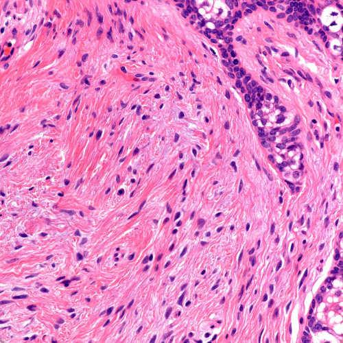
19 Sclerotic fibroma Well circumscribed, dermal or superifial subcutaneous mass Pauci-cellular Plywood sclerosis CD34+ 19


20 20


21 21
:588-590, December 2007.")


22 FIGURE 1 FIGURE 2 Congenital Hand Lesion. Calonje, Eduardo; Fletcher, Christopher; Mentzel, Thomas American Journal of Dermatopathology. 29(6): , December DOI: /DAD.0b013e d Congenital Hand Lesion. Calonje, Eduardo; Fletcher, Christopher; Mentzel, Thomas American Journal of Dermatopathology. 29(6): , December DOI: /DAD.0b013e d FIGURE 1. Ceribriform lesion on the left palm. FIGURE 2. Low-power view showing an exophytic lesion Lippincott Williams & Wilkins, Inc. Published by Lippincott Williams & Wilkins, Inc Lippincott Williams & Wilkins, Inc. Published by Lippincott Williams & Wilkins, Inc. 3 FIGURE 3 Congenital Hand Lesion. Calonje, Eduardo; Fletcher, Christopher; Mentzel, Thomas American Journal of Dermatopathology. 29(6): , December DOI: /DAD.0b013e d FIGURE 4 Congenital Hand Lesion. Calonje, Eduardo; Fletcher, Christopher; Mentzel, Thomas American Journal of Dermatopathology. 29(6): , December DOI: /DAD.0b013e d FIGURE 3. Spindle cell proliferation with a storiform growth pattern. FIGURE 4. The spindle cells show uniform appearance with tapering nuclei and indistinct cytoplasm Lippincott Williams & Wilkins, Inc. Published by Lippincott Williams & Wilkins, Inc Lippincott Williams & Wilkins, Inc. Published by Lippincott Williams & Wilkins, Inc. 5 22
 located on chr. 5q21-22 APC Encodes a protein that inhibits the Wnt signalling pathway by binding to and then down-regulting betacatenin.")
23 Gardner s syndrome Gardner s syndrome Multiple and often unusual cutaneous cysts Gastrointestinal polyposis, osteomas of the mandible and skull A phenotypic variant of Familial Adenomatous Polyposis. Adenomatous Polyposis Coli APC) located on chr. 5q21-22 APC Encodes a protein that inhibits the Wnt signalling pathway by binding to and then down-regulting betacatenin. APC mutations produce stop codons in the gene, leading to truncation of the protein. With the loss of APC function, beta-catenin accumulates in the cytoplasm, leading to problems in cell migration and differentitation. Affected patients often have over 100 colonic polyps, and an increased risk of small bowel and colon cancer. Sigmoidoscopy starting at age 12 and procto-colectomy and terminal ileostomy at young adult age have been advocated as preventive measures. Figure 5.24 The Biology of Cancer ( Garland Science 2007) 23


24 Cutaneous lesions in Gardner s syndrome Infunbular-matrical hybrid cysts Pilomatricomas 24


25 Matrical differentiation Round, monomorphous epithelial cells with vesicular nuclei, scant cytoplasm, prominent nucleoli, many mitotic figures and scattered dendritic melanocytes Shadow cells: Abortive attempts of hair shaft formation. Pilomatricoma Clinical features Frequent in children on the head, neck and upper extremities. Nodular, firm well-circumscribed lesion Multiple pilomatricomas may be associated with myotonic dystrophy, Turner s and Gardner s syndrome. Rare variants: Giant, anetodermic, perforating, etc. PILOMATRICOMA 25


26 5/24/2014 Pilomatricoma Histopathologic features Cystic neoplasm with three types of neoplastic cells: matrical, transitional and shadow cells. Many mitotic figures in matrical cells ( proliferating pilomatricoma ). Frequent calcification and ossification of shadow cells. Foreign body granuloma around shadow cells. Melanin in 25% of the cases. Rare findings: Extramedullary hematopoiesis Transepidermal elimination: Perforating pilomatricoma Anetodermic changes Matricoma: Multiple discrete neoplastic aggregations of matrical and shadow cells Melanocytic matricoma: Pilomatricoma with abundant dendritic melanocytes scattered within the aggregations of matrical cells Pilomatricoma-like changes in the cyst wall of infundibular cysts in patients with Gardner s syndrome Pilomatrixcarcinoma Histopathologic features General architecture of malignant neoplasm: Ulceration of the epidermis, necrosis en masse, etc. Most of the neoplastic aggregations are composed of basophilic matrical cells, with pleomorphic vesicular nuclei and numerous mitotic figures. Focally, matrical differentiation in the form of shadow cells. 26


27 5/24/2014 PILOMATRICOMA PILOMATRIXCARCINOMA 27


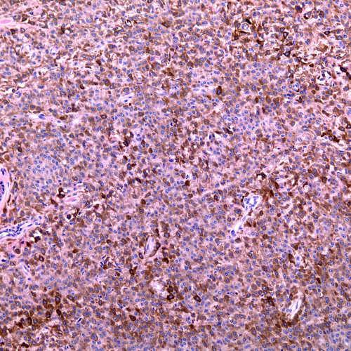
28 28
29 Birt-Hogg Dubee syndrome Birt-Hogg-Dubee syndrome Several types of cutaneous lesions (usually stated as trichodiscomas,fibrofolliculomas and acrochordons) appear on facial skin, usually in patients in their twenties The lesions are tiny papules, sometimes umbillicated, clinically resembling closed comedones. Occasionally, they can affect the trunk. Birt-Hogg-Dubé syndrome Clinical features Autosomal dominant inheritance. Mutation in chromosoma 17p11.2, locus at the folliculin gene. Fibrofolliculomas and trichodiscomas on the face: Asymptomatic skin-colored papules on the face. Most cases develop in the third decade of the life. Renal tumors, often bilateral. Oncocytoma, chromophobous carcinoma. Spontaneous pneumothorax, pulmonary cyst, emphysema. Birt-Hogg-Dubee syndrome, cont d Mutations of tumor suppressor gene called BHD/FLCN, chromosome 17p11.2. Encodes folliculin, expressed in the skin and in several other tissues. Folliculin down-regulates mtor activity Extracutaneous manifestations: Spontaneous pneumothorax Emphysema Pulmonary cysts. Renal tumors include oncocytomas, chromophobe renal cell carcinomas and combinations of both cell types, or oncocytic hybrid tumors. 29


30 5/24/2014 BIRT-HOGG-DUBÉ SYNDROME BIRT-HOGG-DUBÉ SYNDROME Fibrofolliculoma-Trichodiscoma Histopathologic features Fibrofolliculomas and trichodiscomas seem to be the two ends of the spectrum of a single lesion. Fibrofolliculoma: Cords and strands of epithelial basaloid cells radiating from a follicular structure and surrounded by loose connective tissue. Trichodiscoma: Loose and finely fibrillary connective tissue surrounded by folliculosebaceous collarettes at both lateral margins. 30


31 5/24/


32 32


33 Fibrofoliculoma Tricodiscoma Ackerman, Reddy, Soyer


34 34


35 Angiofibroma-like lesions in Birt- Hogg-Dubee syndrome 35


36 36


37 37
 Papillary renal carcinoma (15%) Renal")
 Renal")
 Clear-cell renal")

38 RENAL TUMORES GENERAL PATIENTS Clear-cell renal carcinoma (75%) Papillary renal carcinoma (15%) Renal oncocytoma (5%) Chromophobe carcinoma (5%) PATIENTS WITH BHD SYNDROME Chromophobe carcinoma (34%) Renal oncocytoma (5%) Hybrid tumors between chromophobe carcinoma and renal oncocytoma (50%) Clear-cell renal carcinoma (9%) Papillary renal carcinoma (2%) Pavlovich CP et al. Am J Surg Pathol 2002;26: BIRT-HOGG-DUBÉ SYNDROME 38

 Enzymatic testing in skin fibroblasts Molecular")
39 Hereditary leiomyomas, renal cell carcinoma and fumarate hydratase deficiency Hereditary leiomyomatosis and renal cell carcinoma syndrome (HLRCC) Cutaneous and uterine leiomyomas, and renal cell carcinoma Cutaneous lesions appear in young adults, increase in number with age Segmental leiomyomas are a marker for HLRCC Uterine leiomyomas appear in affected women, from their twenties to middle age The renal cell carcinomas include papillary, tubulo-papillary and collecting duct neoplasms, and often behave aggressively Hereditary leiomyomatosis and renal cell carcinoma syndrome (HLRCC) Enzymatic testing in skin fibroblasts Molecular genetic testing 39


40 40

41 Carney s complex Carney s complex Autosomal dominant condition Angiomyxomas, epithelioid blue nevi, and mucosal lentigines Endocrine abnormalities, including neoplasms CNC1 gene located on chromosome 17q22-24 and the CNC2 gene mapped to 2p16 are considered responsible CNC1 gene, also known as the PRKAR1A gene, is a tumor suppressor gene that the type 1A regulatory subunit of c-amp-dependent protein kinase A. Carney s complex, cont d Superficial angiomyxoma Unlike banal cutaneous myxomas, those of Carney s complex often feature the induction of hair follicular structures from the overlying epidermis, in the form of rudimentary follicular bulbs and papillae. Especially in external ear canal Atrial myxoma 41


42 42


43 43


44 44


45 45


46 Pigmented epithelioid melanocytoma Is it distinct from epithelioid blue nevus of Carney complex and from animal type melanoma? Protein kinase regulatory subunit R1α loss a possible marker for sporadic PEM Many cases with sentinel nodal involvement, few with distant metastases (Zembowicz, USCAP 2009) 46


47 47


48 48


49 49
 and thus slowing down GLI1 translocation to the cell")

50 Basal cell nevus syndrome Basal cell nevus syndrome The basal cell nevus syndrome (BCNS) is due to mutations in the patched or PTCH1 gene. This gene encodes a transmembrane protein that is part of the sonic hedgehog pathway, normally inhibiting smoothened (SMO) and thus slowing down GLI1 translocation to the cell nucleus. Sporadic BCCs can have mutations in PTCH1 that render it ineffective, or mutations in SMO; either can result in increased GLI1 translocation. GLI1 translocation, in turn causes proliferation and inhibits differentiation. PTCH1 is also mutated in trichoepitheliomas, and PTCH1 knockout mice develop trichoblastomas- hence, mutation is necessary but not sufficient for the formation of BCC in many cases. 50
 Basal cell nevus syndrome (BCNS, Gorlin-Goltz")

51 Figure 5.23 The Biology of Cancer ( Garland Science 2007) Basal cell nevus syndrome (BCNS, Gorlin-Goltz Syndrome) Mutation in patched gene (ptchd) Basal cell carcinomas at early age Palmar pits Odontogenic keratocysts Infundibulocystic BCCs in clinically normal skin Anti-ptchd therapy in clinical trials (e.g. GDC-0449) Figure 16.40a The Biology of Cancer ( Garland Science 2007) 51


52 Infundibular differentiation Cystic lesions lined by epithelium similar to follicular infundibulum Basal layer, several layers of keratinocytes, a granular layer, and a cornified layer composed of laminated or basket-weave corneocytes 52


53 53


54 54


55 55


56 56


57 57


58 58


59 59


60 60


61 61


62 Brooke s syndrome Brooke s syndrome Aka Brooke-Spiegler syndrome, hereditary cylindromatosis, multiple familial trichoepithelioma Susceptibility to trichoepitheliomas, cylindromas, spiradenomas, pure or in combination Most lesions on head/neck Brooke s syndrome, cont d Prediliction for face, scalp Entire scalp can be involved in cylindromatosis Rarely, cylindrocarcinomas, spiradenocarcinomas, trichoblastic carcinomas 62


63 Brooke s syndrome, cont d Familial cylindromatosis mapped to 16q12-16q13 CYLD, a tumor suppressor gene identified in this locus, and then found in sporadic cylindromas CYLD interacts with NF-kB signalling pathway 63


64 64


65 65


66 66


67 67


68 68


69 69


70 70


71 71


72 72


73 73


74 Basaloid follicular hamartoma Clinical features Clinical variants: Systematized, multiple, localized and linear. Generalized forms are familiar with autosomal dominant inheritance. Generalized variants may be associated with hypotrichosis and myasthenia gravis. In all clinical variants: Small papular lesions centered in hair follicles. GENERALIZED BASALOID FOLLICULAR HAMARTOMA LOCALIZED BASALOID FOLLICULAR HAMARTOMA LOCALIZED BASALOID FOLLICULAR HAMARTOMA 74


75 Basaloid follicular hamartoma Histopathologic features Individual hair follicles are replaced by strands and branching cords of undifferentiated basaloid cells. Anastomosing basaloid cords Scant fibrous stroma Histopathologic differential diagnosis: Infundibulocystic BCC, trichoepithelioma and Pinkus fibroepithelioma: BFH has less stroma and interfollicular dermis is not involved. 75


76 76

77 77
Samer Ghosn, MD Associate professor, Derpartment of Dermatology American University of Beirut Medical Center. Follicular lesions
 Samer Ghosn, MD Associate professor, Derpartment of Dermatology American University of Beirut Medical Center Follicular lesions Introduction Follicular lesions are important to recognize: For proper management
Appendageal skin tumors
 Appendageal skin tumors Ibrahim Khalifeh, M.D. Associate Professor Department of Pathology American University of Beirut Medical Center Beirut, Lebanon Appendageal tumors Neoplasms whose differentiation
Keratinocyte tumors. Actinic Keratosis. Squamous cell carcinoma in situ. Squamous Cell Carcinoma. (aka Bowen s disease)
") Actinic Keratosis Keratinocyte tumors Prepared by Kurt Schaberg Precancerous, risk of malignancy ~8-20% per year (progresses to SCC); Due to chronic sun exposure Rough scaly plaque; typically due to sun
Actinic Keratosis Keratinocyte tumors Prepared by Kurt Schaberg Precancerous, risk of malignancy ~8-20% per year (progresses to SCC); Due to chronic sun exposure Rough scaly plaque; typically due to sun
Cowden Syndrome PTEN Hamartoma Tumor Syndrome. ACCME/Disclosure. 1. Background. Outline
 MASSACHUSETTS GENERAL HOSPITAL HARVARD MEDICAL SCHOOL PATHOLOGY Cowden Syndrome PTEN Hamartoma Tumor Syndrome ACCME/Disclosure Vania Nosé, MD, PhD Professor of Pathology Director of Anatomic Pathology
SEBACEOUS NEOPLASMS. Dr. Prachi Saraogi Clinical Fellow in Dermatology
 SEBACEOUS NEOPLASMS Dr. Prachi Saraogi Clinical Fellow in Dermatology Sebaceous neoplasms Sebaceous adenoma (Benign) Sebaceous carcinoma (Malignant) SEBACEOUS ADENOMA Benign tumours composed of incompletely
What is new on adnexal neoplasms. Omar P. Sangueza, MD. Professor and Director of Dermatopathology. Wake Forest University School of Medicine
 What is new on adnexal neoplasms Omar P. Sangueza, MD Professor and Director of Dermatopathology Wake Forest University School of Medicine Winston Salem, North Carolina Carney complex is an autosomal dominant
10/2/17. A 45-year-old female presents with a lesion on the cheek, rule out basal cell carcinoma. Overview
 10/2/17 Overview Desmoplastic trichoepithelioma vs. morpheaform BCC vs. microcystic adnexal carcinoma Trichilemmomas & Cowden s syndrome Cutaneous Adnexal Tumours with Clinical Impact Rajiv M. Patel, M.D.
Cutaneous Adnexal Tumors
 Cutaneous Adnexal Tumors Lesions with Predominant Follicular Differentiation Special Emphasis on Basal Cell Carcinoma 2014-04-01 Prof. Dr. med. Katharina Glatz Pathologie Cutaneous Adnexal Tumors Hair
USCAP Neuropathology night panel CASE 2
 USCAP Neuropathology night panel CASE 2 B.K. Kleinschmidt-DeMasters MD University of Colorado at Denver and Health Sciences Center Denver, Colorado The Chinese Wall, Flat Tops Wilderness, Colorado Clinical
Basal cell carcinoma 5/28/2011
 Goal of this Presentation A practical approach to the diagnosis of cutaneous carcinomas and their mimics Thaddeus Mully, MD University of California San Francisco To review common non-melanoma skin cancers
- Selected Tumors of the Skin Appendages - Primary vs. Metastasis
 - Selected Tumors of the Skin Appendages - Primary vs. Metastasis Napa Valley 2018 Victor G. Prieto, MD, PhD Chair of Pathology UT MD Anderson Cancer Center vprieto@mdanderson.org Napa Valley in May Introduction
Benign and malignant epithelial lesions: Seborrheic keratosis: A common benign pigmented epidermal tumor occur in middle-aged or older persons more
 Benign and malignant epithelial lesions: Seborrheic keratosis: A common benign pigmented epidermal tumor occur in middle-aged or older persons more common on the trunk; but extremities, head and neck are
Disclosure. Relevant Financial Relationship(s) None. Off Label Usage None MFMER slide-1
 None. Off Label Usage None MFMER slide-1") Disclosure Relevant Financial Relationship(s) None Off Label Usage None 2013 MFMER slide-1 Case Presentation A 43 year old male, with partial nephrectomy for a right kidney mass 2013 MFMER slide-2 2013
Disclosure Relevant Financial Relationship(s) None Off Label Usage None 2013 MFMER slide-1 Case Presentation A 43 year old male, with partial nephrectomy for a right kidney mass 2013 MFMER slide-2 2013
Simplified approach to cutaneous adnexal tumors
 Simplified approach to cutaneous adnexal tumors Chandra Smart, MD Associate Clinical Professor UCLA Department of Pathology Introduction: Cutaneous adnexal neoplasms (CANs) are a diverse group of tumors
Pathology of the skin. 2nd Department of Pathology, Semmelweis University
 Pathology of the skin 2nd Department of Pathology, Semmelweis University Histology of the skin Epidermis: Stratum corneum Stratum granulosum Stratum spinosum Stratum basale Dermis: papillary and reticular
Apocrine and eccrine adnexal tumors
 Apocrine and eccrine adnexal tumors Timothy McCalmont, MD University of California San Francisco, CA ECCRINE TUMORS Generally uncommon, because the sweat apparatus has little proliferative potential (hyperproliferation
Desmoplastic Melanoma R/O BCC. Clinical Information. 74 y.o. man with lesion on left side of neck r/o BCC
 R/O BCC Sabine Kohler, M.D. Professor of Pathology and Dermatology Dermatopathology Service Stanford University School of Medicine Clinical Information 74 y.o. man with lesion on left side of neck r/o
Apocrine and eccrine adnexal tumors
 Apocrine and eccrine adnexal tumors Timothy McCalmont, MD University of California San Francisco, CA Past misguided notions Syringoma is eccrine Eccrine poroma rather than eccrine or apocrine poroma Spiradenoma
Pathology Slides. [Pathology]
![Pathology Slides. [Pathology]](/thumbs/94/120604575.jpg "Pathology Slides. [Pathology]") Pathology Slides MedicoNotes provides real laboratory pathological slides to aid you to differentiate between different pathological structures under microscope. www.mediconotes.com Histology slides example
Pathology Slides MedicoNotes provides real laboratory pathological slides to aid you to differentiate between different pathological structures under microscope. www.mediconotes.com Histology slides example
Diseases of the breast (1 of 2)
") Diseases of the breast (1 of 2) Introduction A histology introduction Normal ducts and lobules of the breast are lined by two layers of cells a layer of luminal cells overlying a second layer of myoepithelial
Diseases of the breast (1 of 2) Introduction A histology introduction Normal ducts and lobules of the breast are lined by two layers of cells a layer of luminal cells overlying a second layer of myoepithelial
Skin Adnexal Tumors - A Histopathological Spectrum at a Tertiary Care Hospital
 Original Article GCSMC J Med Sci Vol (VI) No (I) January-June 2017 Skin Adnexal Tumors - A Histopathological Spectrum at a Tertiary Care Hospital Neeraja Barve*, Hansa Goswami**, Urvi Parikh *** Abstract
Corporate Medical Policy
 Corporate Medical Policy Genetic Testing for PTEN Hamartoma Tumor Syndrome File Name: Origination: Last CAP Review: Next CAP Review: Last Review: genetic_testing_for_pten_hamartoma_tumor_syndrome 4/2013
colorectal cancer Colorectal cancer hereditary sporadic Familial 1/12/2018
 colorectal cancer Adenocarcinoma of the colon and rectum is the third most common site of new cancer cases and deaths in men (following prostate and lung or bronchus cancer) and women (following breast
Genodermatoses with predisposition toward internal malignancy
 Genodermatoses with predisposition toward internal malignancy Associate Professor of Dermatology and Child Health Department of Dermatology University of Missouri Columbia Columbia, Missouri DISCLOSURE
Malignant Peripheral Nerve Sheath Tumor
 C H A P T E R 120 Malignant Peripheral Nerve Sheath Tumor Currently, malignant peripheral nerve sheath tumor (MPNST) is the most commonly used generic name for the neoplasms known in the past as neurosarcoma,
Prior Authorization. Additional Information:
 Genetic Testing for Cowden Syndrome - PTEN Gene MP9488 Covered Service: Prior Authorization Required: Additional Information: Yes when meets criteria below Yes as shown below Pre and post-test genetic
Development of Carcinoma Pathways
 The Construction of Genetic Pathway to Colorectal Cancer Moriah Wright, MD Clinical Fellow in Colorectal Surgery Creighton University School of Medicine Management of Colon and Diseases February 23, 2019
Financial disclosures
 Mesenchymal Neoplasms with Melanocytic Differentiation By Konstantinos Linos MD, FCAP, FASDP Bone, Soft Tissue and Dermatopathology Assistant Professor of Pathology Dartmouth-Hitchcock Medical Center Geisel
Malignant tumors of melanocytes: Part 1. Deba P Sarma, MD., Omaha
 Malignant tumors of melanocytes: Part 1 Deba P Sarma, MD., Omaha The melanocytic tumor is one of the most difficult and confusing areas in Dematopathology. It is true that most (95%) of such lesions are
MECHANISMS OF HUMAN DISEASE: LABORATORY SESSION PATHOLOGY OF THE SKIN LAB. Friday, February 13, :30 am 11:00 am
 MECHANISMS OF HUMAN DISEASE: LABORATORY SESSION PATHOLOGY OF THE SKIN LAB Friday, February 13, 2009 9:30 am 11:00 am FACULTY COPY GOALS: Describe the basic clinical and morphologic features of various
Renal Cell Carcinoma: Genetics & Imaging Srinivasa R Prasad University of Texas San Antonio
 Renal Cell Carcinoma: Genetics & Imaging Srinivasa R Prasad University of Texas HSC @ San Antonio No financial disclosures Acknowledgements Dr. Peter Choyke, NIH My Gurus @ MIR, MGH 2004 WHO Taxonomy of
International Journal of Health Sciences and Research ISSN:
 International Journal of Health Sciences and Research www.ijhsr.org ISSN: 2249-9571 Original Research Article A Clinicopathological Study of Adnexal Tumors of Skin in a Tertiary Care Research Hospital
Familial and Hereditary Colon Cancer
 Familial and Hereditary Colon Cancer Aasma Shaukat, MD, MPH, FACG, FASGE, FACP GI Section Chief, Minneapolis VAMC Associate Professor, Division of Gastroenterology, Department of Medicine, University of
3. Histopathology. 1. Introduction. 2. Case History. Volume 6 Issue 4, April Licensed Under Creative Commons Attribution CC BY
 Spiradenocylindroma with Trichoepithelioma A Collision Tumor with Multiple Differentiation R. Lavanya 1, S. K. Sridevi 2, P. Viswanathan 3, P. V. S.Prasad 4 1 II nd Year Post Graduate, Department of Pathology,
Diagnostic Cytology of Cancer Cases
 Diagnostic Cytology of Cancer Cases Somporn Techangamsuwan Companion Animal Cancer Research Unit (CAC-RU) Department of Pathology, Faculty of Veterinary Science, Chulalongkorn University 1 Tumor or Non-tumor
American College of Mohs Surgery. Diagnostic Quality Control Program (Review of Answers)
") American College of Mohs Surgery Diagnostic Quality Control Program 2010 (Review of Answers) Question 1 A flesh-colored plaque on the right ala of an otherwise healthy 57 year-old male is referred for
American College of Mohs Surgery Diagnostic Quality Control Program 2010 (Review of Answers) Question 1 A flesh-colored plaque on the right ala of an otherwise healthy 57 year-old male is referred for
Journal of International Academy of Forensic Science & Pathology (JIAFP)
") Journal of International Academy of Forensic Science & Pathology (JIAFP) ISSN 2395-0722 MICROCYSTIC ADNEXAL CARCINOMA-A CASE REPORT WITH REVIEW OF LITERATURE Case Report Sulakshana M S 1,Natarajan M 2
Journal of International Academy of Forensic Science & Pathology (JIAFP) ISSN 2395-0722 MICROCYSTIC ADNEXAL CARCINOMA-A CASE REPORT WITH REVIEW OF LITERATURE Case Report Sulakshana M S 1,Natarajan M 2
A 5 Year Histopathological Study of Skin Adnexal Tumors at a Tertiary Care Hospital
 IOSR Journal of Dental and Medical Sciences (IOSR-JDMS) e-issn: 2279-0853, p-issn: 2279-0861.Volume 14, Issue 4 Ver. VII (Apr. 2015), PP 01-05 www.iosrjournals.org A 5 Year Histopathological Study of Skin
Dermatopathology. Dr. Rafael Botella Estrada. Hospital La Fe de Valencia
 Dermatopathology Dr. Rafael Botella Estrada. Hospital La Fe de Valencia Melanoma and mimics Dr. Martin Mihm Malignant lesions result from the accumulation of mutations Class I lesions (benign) Class II
Basaloid follicular hamartoma (BFH) is a rare, benign,
 is a rare, benign,") Resident Short Reviews Basaloid Follicular Hamartoma N Basaloid follicular hamartoma is a benign lesion of important consideration because it can be mistaken both clinically and histologically for basal
Resident Short Reviews Basaloid Follicular Hamartoma N Basaloid follicular hamartoma is a benign lesion of important consideration because it can be mistaken both clinically and histologically for basal
Trichofolliculoma of the Guinea Pig 1,2
 Trichofolliculoma of the Guinea Pig 1,2 Raymond D. Ediger, Garrett S. Dill, Jr., and Robert M. Kovatch, Aerobiology and Evaluation Laboratories and Medical Sciences Laboratories, Fort Detrick, Frederick,
Familial and Hereditary Colon Cancer
 Familial and Hereditary Colon Cancer Aasma Shaukat, MD, MPH, FACG, FASGE, FACP GI Section Chief, Minneapolis VAMC Associate Professor, Division of Gastroenterology, Department of Medicine, University of
A rare association of parotid oncocytoma, lipoma, multinodular goitre and uterine adenomysosis
 ISPUB.COM The Internet Journal of Otorhinolaryngology Volume 7 Number 2 A rare association of parotid oncocytoma, lipoma, multinodular goitre and uterine adenomysosis P Sinha, S Sharma, S Agarwal, R Ray
Diploma Examination. Dermatopathology: First paper. Tuesday 20 March Candidates must answer FOUR questions. Time allowed: 3 hours
 Dermatopathology: First paper Tuesday 20 March 2018 Candidates must answer FOUR questions Time allowed: 3 hours 1. Give an account of the genetic aberrations encountered in Spitzoid neoplasms and how these
Papillary Lesions of the breast
 Papillary Lesions of the breast Emad Rakha Professor of Breast Pathology The University of Nottingham Papillary lesions of the breast are a heterogeneous group of disease, which are characterised by neoplastic
Various hereditary, acquired and neoplastic conditions can lead to cyst formation in the kidney.
 Dr. Fatima AlAl-Hashimi Hashimi,, MD, FRCPath Salmaniya Medical Complex, Bahrain Various hereditary, acquired and neoplastic conditions can lead to cyst formation in the kidney. The most frequently encountered
A clinicopathologic study of skin appendageal tumors
 Net Study A clinicopathologic study of skin appendageal tumors Pradeep S. Nair Department of Dermatology and Venereology, Medical College Hospital, Trivandrum- 695 011, Kerala, India Address for correspondence:
Pathology perspective of colonic polyposis syndromes
 Pathology perspective of colonic polyposis syndromes When are too many polyps too many? David Schaeffer Head and Consultant Pathologist, Department of Pathology and Laboratory Medicine, Vancouver General
21/07/2017. Hobnail endothelial cells are not the same as epithelioid endothelial cells
 UPDATE IN CUTANEOUS VASCULAR S DERMATOPATHOLOGY SESSION BELFAST PATHOLOGY JUNE 21/2017 Dr E Calonje St John s Institute of Dermatology, London, United Kingdom THE FAMILY OF VASCULAR S WITH EPITHELIOID
A neoplasm is defined as "an abnormal tissue proliferation, which exceeds that of adjacent normal tissue. This proliferation continues even after
 NEOPLASIA Neoplasia is a very important topic in pathology because neoplasms are both common and serious diseases. A neoplasm literally means a new growth, and this term is used interchangeably with a
MECHANISMS OF HUMAN DISEASE: LABORATORY SESSION PATHOLOGY OF THE SKIN LAB. Friday, February 12, :30 am 11:00 am
 MECHANISMS OF HUMAN DISEASE: LABORATORY SESSION PATHOLOGY OF THE SKIN LAB Friday, February 12, 2012 9:30 am 11:00 am FACULTY COPY GOALS: Describe the basic clinical and morphologic features of various
Simulators of melanoma
 Simulators of melanoma Philip E. LeBoit, M.D. Depts. of Pathology and Dermatology University of California, San Francisco Simulators of melanoma Simulators of melanoma in situ Melanocytic Non-melanocytic
Case Report A Rare Cutaneous Adnexal Tumor: Malignant Proliferating Trichilemmal Tumor
 Case Reports in Medicine Volume 2015, Article ID 742920, 4 pages http://dx.doi.org/10.1155/2015/742920 Case Report A Rare Cutaneous Adnexal Tumor: Malignant Proliferating Trichilemmal Tumor Omer Alici,
Cutaneous metastases. Thaddeus Mully. University of California, San Francisco Professor, Departments of Pathology and Dermatology
 Cutaneous metastases Thaddeus Mully University of California, San Francisco Professor, Departments of Pathology and Dermatology DISCLOSURE OF RELATIONSHIPS WITH INDUSTRY Thaddeus Mully Course C005 Essential
Case Report Nevus Lipomatosus Superficialis with a Folliculosebaceous Component: Report of 2 Cases
 SAGE-Hindawi Access to Research Pathology Research International Volume 2011, Article ID 105973, 4 pages doi:10.4061/2011/105973 Case Report Nevus Lipomatosus Superficialis with a Folliculosebaceous Component:
Epithelial tumors. Dr. F.F. Khuzin, PhD Dr. M.O. Mavlikeev
 Epithelial tumors Dr. F.F. Khuzin, PhD Dr. M.O. Mavlikeev Epithelial tumors Tumors from the epithelium are the most frequent among tumors. There are 2 group features of these tumors: The presence in most
Paul K. Shitabata, M.D. Dermatopathology Institute
 Paul K. Shitabata, M.D. Dermatopathology Institute Key Points Subtle dermatologic signs may suggest significant gastrointestinal and systemic disease Check family history Several dermatologic disorders
Colonic polyps and colon cancer. Andrew Macpherson Director of Gastroentology University of Bern
 Colonic polyps and colon cancer Andrew Macpherson Director of Gastroentology University of Bern Improtance of the problem of colon cancers - Epidemiology Lifetime risk 5% Incidence/10 5 /annum (US Detroit
Myxo-inflammatory Fibroblastic sarcoma
 AKA Myxo-inflammatory Fibroblastic sarcoma Acral Myxoinflammatory fibroblastic sarcomaam.j.surg.path1998; 22; 911-924 Inflammatory myxoid tumour of soft parts with bizarre giant cells [Pathol.Res.Pract.
BASAL CELL CARCINOMA WITH ECCRINE DIFFERENTIATION: A RARE ENTITY Divvya B 1, Rehana Tippoo 2, P. Viswanathan 3, B. Krishnaswamy 4, A.
 BASAL CELL CARCINOMA WITH ECCRINE DIFFERENTIATION: A RARE ENTITY Divvya B 1, Rehana Tippoo 2, P. Viswanathan 3, B. Krishnaswamy 4, A. Anvar Ali 5 HOW TO CITE THIS ARTICLE: Divvya B, Rehana Tippoo, P. Viswanathan,
Disorders of Cell Growth & Neoplasia. Histopathology Lab
 Disorders of Cell Growth & Neoplasia Histopathology Lab Paul Hanna April 2010 Case #84 Clinical History: 5 yr-old, West Highland White terrier. skin mass from axillary region. has been present for the
Table of Contents. Preface xi. Acknowledgments xiii. Part I Overview of the Diagnostic Process 1. 1 Overview of Grading and Staging 3
 Table of Contents Preface xi Acknowledgments xiii Part I Overview of the Diagnostic Process 1 1 Overview of Grading and Staging 3 Identification of the process 3 Identification of tumor types 5 Grading
Clinical History. 29 yo woman with polyhydramnios Cardiac mass at fetal ultrasound At 35 weeks, newborn died 30 minutes after delivery
 CASE 1 a Clinical History 29 yo woman with polyhydramnios Cardiac mass at fetal ultrasound At 35 weeks, newborn died 30 minutes after delivery Interface between tumor and normal myocardium Smaller well-demarcated
Whitney A. High, MD, JD, MEng
 ADS Dermatopathology Meeting 2014 Selected Adnexal Tumors Whitney A. High, MD, JD, MEng Associate Professor, Dermatology & Pathology Director of Dermatopathology (Dermatology) University of Colorado School
First described by Muir et al 1 in 1967 and Torre 2 in 1968, Muir-Torre Syndrome. Resident Short Review
 Resident Short Review Muir-Torre syndrome (MTS) is a rare autosomal-dominant genodermatosis characterized by sebaceous neoplasms and one or more visceral malignancies. Sebaceous tumors include sebaceous
CASE REPORT SOLITARY SEBACEOUS NEVUS OF JADASSOHN COMPLICATED BY SQUAMOUS CELL CARCINOMA AND BASAL CELL CARCINOMA
 CASE REPORT Dennis H. Kraus, MD, Section Editor SOLITARY SEBACEOUS NEVUS OF JADASSOHN COMPLICATED BY SQUAMOUS CELL CARCINOMA AND BASAL CELL CARCINOMA Ahmad Ridzwan Arshad, FRCS, 1 Wan S. Azman, MS, 1 Ayadurai
Histopathology: skin pathology
 Histopathology: skin pathology These presentations are to help you identify, and to test yourself on identifying, basic histopathological features. They do not contain the additional factual information
27
 26 27 28 29 30 31 32 33 34 35 Diagnosis:? Diagnosis: Juvenile Polyposis with BMPR1A Mutation 36 Juvenile Polyposis Syndrome Rare Autosomal Dominant Disorder with Multiple Juvenile Polyps in GI Tract Juvenile
Familial Adenomatous Polyposis
 Familial Adenomatous Polyposis 1 in 10,000 incidence 100 s to 1000 s of colonic adenomas by teens Cancer risk: colon, gastric, duodenum (periampulla), small bowel, pancreas, papillary thyroid, childhood
Gross appearance of nodular hyperplasia in material obtained from suprapubic prostatectomy. Note the multinodular appearance and the admixture of
 Tiền liệt tuyến Tiền liệt tuyến Gross appearance of nodular hyperplasia in material obtained from suprapubic prostatectomy. Note the multinodular appearance and the admixture of solid and microcystic areas.
PATHOLOGY OF THE SKIN 2. Tumours of the skin
 PATHOLOGY OF THE SKIN 2. Tumours of the skin Máirín E. McMenamin MB MRCPI FRCPath Dip (Dermatopathol) RCPath St. James s Hospital and University of Dublin, Trinity College Tumour (Neoplasia) Benign or
IN THE NAME OF GOD Dr. Kheirandish Oral and maxillofacial pathology
 IN THE NAME OF GOD Dr. Kheirandish Oral and maxillofacial pathology ORAL FOCAL MUCINOSIS Uncommon Tumorlike Cutaneous myxoid cyst Overproduction of hyaluronic acid by firoblasts Young adults Female Gingiva
Eruptive Tumors of the Follicular Infundibulum: An Unexpected Diagnosis of Hypopigmented Macules
 Dermatol Ther (Heidelb) (2015) 5:207 211 DOI 10.1007/s13555-015-0079-0 CASE REPORT Eruptive Tumors of the Follicular Infundibulum: An Unexpected Diagnosis of Hypopigmented Macules Poonkiat Suchonwanit.
Salivary Glands 3/7/2017
 Salivary Glands 3/7/2017 Goals and objectives Focus on the entities unique to H&N Common board type facts Information for your future practice Salivary Glands Salivary Glands Major gland. Paratid. Submandibular.
Lesions Mimicking Adenoid Cystic Carcinoma. Diagnostic Problems in Salivary Gland Pathology An Update 5/29/2009
 Diagnostic Problems in Salivary Gland Pathology An Update Lesions Mimicking Adenoid Cystic Carcinoma Stacey E. Mills, M.D. W.S. Royster Professor of Pathology Director of Surgical and Cytopathology University
Dermatopathology: The tumor is composed of keratinocytes which show atypia, increase mitoses and abnormal mitoses.
 Squamous cell carcinoma (SCC): A common malignant tumor of keratinocytes arising in the epidermis, usually from a precancerous condition: 1- UV induced actinic keratosis, usually of low grade malignancy.
Populations Interventions Comparators Outcomes Individuals: With clinical signs of a PTEN hamartoma tumor syndrome. are:
 Protocol Genetic Testing for PTEN Hamartoma Tumor Syndrome (20488) Medical Benefit Effective Date: 07/01/14 Next Review Date: 05/18 Preauthorization Yes Review Dates: 05/13, 05/14, 05/15, 05/16, 05/17
DISCLOSURE OF RELEVANT RELATIONSHIPS WITH INDUSTRY
 DISCLOSURE OF RELEVANT RELATIONSHIPS WITH INDUSTRY David R. Carr, MD I HAVE NO RELEVENT RELATIONSHIPS WITH ANY COMPANIES The most likely diagnosis is A. Actinic lentigo B. Eschar C. Blue nevus D. Melanoma
ARIZONA SOCIETY OF PATHOLOGISTS 13 TH APRIL 2013 HEAD AND NECK CYTOPATHOLOGY. F ZAHRA ALY, MD, PhD
 ARIZONA SOCIETY OF PATHOLOGISTS 13 TH APRIL 2013 HEAD AND NECK CYTOPATHOLOGY F ZAHRA ALY, MD, PhD The main areas sites amenable for cytopathology include lymph nodes, thyroid, major salivary glands especially
المركب النموذج--- سبيتز وحمة = Type Spitz's Nevus, Compound SPITZ NEVUS 1 / 7
 SPITZ NEVUS 1 / 7 Epidemiology An annual incidence rate of 1.4 cases of Spitz nevus per 100,000 individuals has been estimated in Australia, compared with 25.4 per 100,000 individuals for cutaneous melanoma
Clinicopathological Study of 1016 Consecutive Adnexal Skin Tumors
 ORIGINAL REPORT Clinicopathological Study of 1016 Consecutive Adnexal Skin Tumors Kambiz Kamyab-Hesari 1, Kamran Balighi 2, Nasim Afshar 2, Nessa Aghazadeh 2, Ziba Rahbar 2, Maryam Seraj 2, and Maede Rayati
أملس عضلي غرن = Leiomyosarcoma. Leiomyosarcoma 1 / 5
 Leiomyosarcoma 1 / 5 EPIDEMIOLOGY Exact incidence is unknown, but older studies suggest that leiomyosarcomas comprise approximately 3 percent of soft-tissue sarcomas. Superficial leiomyosarcoma occurs
Case year female. Routine Pap smear
 Case 1 57 year female Routine Pap smear Diagnosis? 1. Atypical glandular cells of unknown significance (AGUS) 2. Endocervical AIS 3. Endocervical adenocarcinoma 4. Endometrial adenocarcinoma 5. Adenocarcinoma
A Clinicopathological study of skin and adnexal neoplasms at a rural based tertiary teaching hospital
 A Clinicopathological study of skin and adnexal neoplasms at a rural based tertiary teaching hospital V. V. Narhire 1*, S.Y. Swami 2, B. D. Baste 1,S. A. Khadase 3,G. F. D costa 4 1 Assistant professor,
Risk of Colorectal Cancer (CRC) Hereditary Syndromes in GI Cancer GENETIC MALPRACTICE
 Hereditary Syndromes in GI Cancer GENETIC MALPRACTICE") Identifying the Patient at Risk for an Inherited Syndrome Sapna Syngal, MD, MPH, FACG Director, Gastroenterology Director, Familial GI Program Dana-Farber/Brigham and Women s Cancer Center Associate Professor
Identifying the Patient at Risk for an Inherited Syndrome Sapna Syngal, MD, MPH, FACG Director, Gastroenterology Director, Familial GI Program Dana-Farber/Brigham and Women s Cancer Center Associate Professor
Problem diagnoses. Current issues in Anatomic pathology. Problem Diagnoses in Tumors of the Oral Cavity 5/29/2009
 Current issues in Anatomic pathology Problem Diagnoses in Tumors of the Oral Cavity Richard Jordan DDS PhD FRCPath Professor of Oral Pathology & Pathology Director, UCSF Oral Pathology Diagnostic Laboratory
What s New in Pathology of Genitourinary Tumors. Jiaoti Huang, MD, PhD Department of Pathology Duke University School of Medicine
 What s New in Pathology of Genitourinary Tumors Jiaoti Huang, MD, PhD Department of Pathology Duke University School of Medicine Kidney Tumors Multilocular cystic renal neoplasm of low malignant potential
Multistep nature of cancer development. Cancer genes
 Multistep nature of cancer development Phenotypic progression loss of control over cell growth/death (neoplasm) invasiveness (carcinoma) distal spread (metastatic tumor) Genetic progression multiple genetic
IT S FUNDAMENTAL MY DEAR WATSON! A SHERLOCKIAN APPROACH TO DERMATOLOGY
 IT S FUNDAMENTAL MY DEAR WATSON! A SHERLOCKIAN APPROACH TO DERMATOLOGY Skin, Bones, and other Private Parts Symposium Dermatology Lectures by Debra Shelby, PhD, DNP, FNP-BC, FADNP, FAANP Debra Shelby,
Benign versus Cancerous Lesions How to tell the difference FMF 2014 Christie Freeman MD, CCFP, DipPDerm, MSc
 1 Benign versus Cancerous Lesions How to tell the difference FMF 2014 Christie Freeman MD, CCFP, DipPDerm, MSc Benign lesions Seborrheic Keratoses: Warty, stuck-on Genetics and birthdays Can start in late
Appendix : Dermoscopy
 Go Back to the Top To Order, Visit the Purchasing Page for Details APP Appendix : Dermoscopy Dermoscopy, also known as dermatoscopy, epiluminoscopy and epiluminescent microscopy, is an effective non-invasive
Skin (Integumentary System) Wheater, Chap. 9
 Wheater, Chap. 9") Skin (Integumentary System) Wheater, Chap. 9 Skin (Integument) Consists of skin and associated derivatives Largest organ of body (21 ft 2 ; 9 lbs.; has 11 miles of blood vessels) Functions: Protection
Skin (Integumentary System) Wheater, Chap. 9 Skin (Integument) Consists of skin and associated derivatives Largest organ of body (21 ft 2 ; 9 lbs.; has 11 miles of blood vessels) Functions: Protection
ABCD rule. apocrine glands. arrector pili. ceruminous glands. contact dermatitis
 ABCD rule assessing moles: asymmetric, broder irregularity, color, diameter (larger than 6mm) apocrine glands arrector pili sweat glands in the pubic and underarm areas that secrete thicker sweat, that
BSD 2015 Case 19. Female 21. Nodule on forehead. The best diagnosis is:
 BSD 2015 Case 19 Female 21. Nodule on forehead. The best diagnosis is: A. mixed tumour of skin B. porocarcinoma C. nodular hidradenoma D. metastatic adenocarcinoma BSD 2015 Case 19 Female 21 Nodule on
Pathology reports, related operative reports and consult letters must be provided with a request for assessment.
 Page 1 of 6 Polyposis Syndromes Inherited risk for colorectal cancer is associated with a number of polyposis syndromes (genes), some of which are well-defined and others are less common. Identification
Four Different Tumors Arising in a Nevus Sebaceous
 Published online: April 20, 2016 2016 The Author(s) Published by S. Karger AG, Basel 1662 6567/16/0081 0075$39.50/0 This article is licensed under the Creative Commons Attribution-NonCommercial 4.0 International
Case Based Urology Learning Program
 Case Based Urology Learning Program Resident s Corner: UROLOGY Case Number 18 CBULP 2011 041 Case Based Urology Learning Program Editor: Associate Editors: Manager: Case Contributors: Steven C. Campbell,
Spectrum of Preneoplastic and Neoplastic Cystic Lesions of the Kidney in Adult. by dr. Banan Burhan Mohammed Lecturer in Pathology Department
 Spectrum of Preneoplastic and Neoplastic Cystic Lesions of the Kidney in Adult by dr. Banan Burhan Mohammed Lecturer in Pathology Department Various hereditary, acquired, and neoplastic conditions can
Hereditary Leiomyomatosis and Renal Cell Carcinoma Variant of Reed s Syndrome - A Rare Case Report
 American Research Journal of Urology Volume 1, Issue 1, pp:26-30 Case Hereditary Leiomyomatosis and Renal Cell Carcinoma Variant of Reed s Syndrome - A Rare Case Manas Babu, Devesh Bansal, Sony Mehta,