Neoplasia. Fatima Obeidat, MD Assistant Professor of Neuropathology
|
|
|
- Cleopatra Bennett
- 6 years ago
- Views:
Transcription
1 Neoplasia Fatima Obeidat, MD Assistant Professor of Neuropathology 1
2 - Neoplasia literally means "new growth - Neoplastic cells are transformed because they continue to replicate, independent of normal regulatory influences. - Neoplasms enjoy a certain degree of autonomy and tend to increase in size; Their autonomy is not complete, some neoplasms require endocrine support, and depend on the host for their nutrition and blood supply
3 - a neoplasm is referred to as a tumor and study of tumors is called oncology ( oncos, "tumor," and logos, "study of"). - The neoplasms are divided into benign and malignant 3
4 I. Benign tumors a. its microscopic and gross characteristics are considered to be relatively innocent, b. it will remain localized and amenable to local surgical removal; the patient generally survives 4
5 II. Malignant tumors are collectively referred to as cancers a. Implies that the lesion can invade adjacent structures b. Spread to distant sites (metastasize) to cause death Note: Not all cancers pursue a deadly course, the most aggressive may be curable but the term constitutes a red flag 5
6 The tumor has two basic components: (1) Parenchyma, made up of transformed or neoplastic cells - It determines its biologic behavior, and - Is this component from which the tumor derives its name (2) the supporting, host-derived, non-neoplastic stroma, - Made up of connective tissue, blood vessels, and, is crucial to the growth of the neoplasm, since it carries the blood supply and provides support for the growth of parenchymal cells
7 Nomenclature 1.Benign Tumors of mesenchymal tissues - In general, are designated by attaching the suffix -oma to the cell type from which the tumor arises. a. A benign tumor of fibrous tissue is a fibroma; b. A benign cartilaginous tumor is a chondroma c. A benign tumor of bone is called osteoma
8 2. Benign epithelial tumors are classified either: a. on the basis of their microscopic pattern b. Or on the basis of their macroscopic pattern. c. Others are classified by their cells of origin 8
9 I. Adenoma : - Is applied to benign epithelial neoplasm producing gland patterns and to neoplasms derived from glands but not necessarily forming glands a. A benign epithelial neoplasm arising from renal tubule, cells and growing in glandlike patterns called adenoma b. A mass of benign epithelial cells that produces no glandular patterns but has its origin in the adrenal cortex
10 Colonic polyp
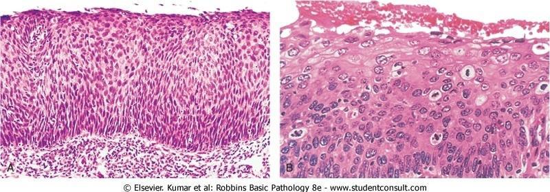
11 II. Papillomas: - Are benign epithelial neoplasms, growing on any surface, that produce microscopic or macroscopic finger-like fronds III. A polyp: - A mass that projects above a mucosal surface, as in the gut, to form a macroscopically visible structure and although this term commonly is used for benign tumors, some malignant tumors may grow as polyps, IV. Cystadenomas - Are hollow cystic masses that typically arise in the ovary 11

12 Colonic polyp

13 Colonic polyp 13
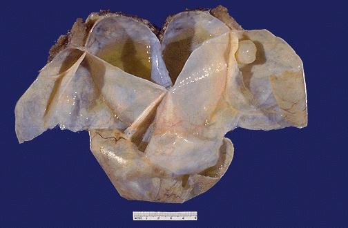
14 Ovarian cystadenoma 14
15 The nomenclature of malignant tumors I. Malignant neoplasms arising in "solid" mesenchymal tissues or its derivatives are called sarcomas, a. A cancer of fibrous tissue origin is a fibrosarcoma, b. Cancer of chondrocytes is a chondrosarcoma II. Whereas those arising from the mesenchymal cells of the blood are called leukemias or lymphomas.
16 III. While the epithelia are derived from all three germ cell layers, malignant neoplasms of epithelial cells are called carcinomas regardless of the tissue of origin. a. Thus, a malignant neoplasm arising in the renal tubular epithelium (mesoderm) is a carcinoma, b. As are the cancers arising in the skin (ectoderm) c. and lining epithelium of the gut (endoderm). 16
17 - Carcinomas are subdivided further. a. Carcinomas that grow in glands are adenocarcinomas b. Those that produce squamous cells are called squamous cell carcinomas. - Sometimes the tissue or organ of origin can be identified, as in the designation of renal cell adenocarcinoma.
18 Adenocarcinoma 18

19 Well differentiated squamous cell carcinoma 19
20 -The transformed cells in a neoplasm, resemble each other, as though all had been derived from a single progenitor, consistent with the monoclonal origin of tumors. - In some instances, however, the tumor cells undergo divergent differentiation, creating so-called mixed tumors.
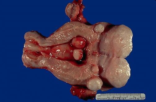
21 1.Mixed tumor of salivary gland: - ( pleomorphic adenoma: ) - It has epithelial, components dispersed throughout a fibromyxoid stroma with islands of cartilage or bone 2. Fibroadenoma of the female breast: - This benign tumor contains a mixture of ductal elements (adenoma) embedded in a loose fibrous tissue (fibroma). 21

22 Benign mixed tumor of the parotid

23 Fibroadenoma of the breast
24 Teratoma:- - Is a special type of mixed tumor that contains recognizable mature or immature tissues representative of more than one germ cell layer. - originate from totipotential germ cells such as those normally present in the ovary and testis - Teratomas they may give rise to neoplasms that have bone, epithelium, muscle, nerve, and other tissues 24

25 Teratoma 25
26 Some glaring inconsistencies may be noted. a. Lymphoma: malignant tumor of lymphocytes b. Mesothelioma : malignant tumor of mesothelium c. Melanoma :malignant tumor of melanocytes d. Seminoma : malignant tumor of germ cells in testis. e. Astrocytoma: malignant tumor in brain.
27 Hamartoma : Is a mass of disorganized tissue indigenous to the particular site and examples include:. - Lung hamartoma is a mass composed of disorganized islands of cartilage, bronchi, and blood vessels - Hamartomas have traditionally been considered congenital but genetic studies suggesta neoplastic origin 27

28 Hamartoma 28
29 Choristoma - Called hetrotopia - Means presence of normal tissue in another tissue and is a congenital anomaly. - For example, a small nodule of normally organized pancreatic tissue may be found in the submucosa of the stomach, duodenum, or small intestine 29
30 CHARACTERISTICS OF BENIGN AND MALIGNANT NEOPLASMS 30
31 - In general, benign tumors are genetically "simple," harboring fewer mutations than cancers, and genetically stable, changing little in genotype over time. - In practice, the determination of benign versus malignant is made with remarkable accuracy using long-established clinical and anatomic criteria, but some neoplasms defy easy characterization but certain features may indicate innocence, and others may indicate malignancy.
32 I. Differentiation and anaplasia - Differentiations is seen only in the parenchymal cells and refers to the extent to which tumor cells resemble their normal forebears morphologically and functionally A. Benign neoplasms : Are composed of welldifferentiated cells that closely resemble their normal counterparts - A chondroma is made up of mature cartilage cells that synthesize their usual cartilaginous matrix-evidence of morphologic and functional differentiation - In benign tumors, mitoses are rare and are of normal configuration
33 B. Malignant neoplasms : can be well differentiated tor completely undifferentiated. - For example, well-differentiated carcinomas of the thyroid may contain normal-appearing follicles, such tumors may be difficult to distinguish from benign proliferations. Note:- The stroma carrying the blood supply is crucial to the growth of tumors but does not aid in the separation of benign from malignant ones and the amount of stroma does determine, however, the consistency of a neoplasm Desmoplasia : Production of abundant amount of stroma by some cancers and these tumors called scirrhous tumors

34 Invasive ductal carcinoma of breast showing desmoplasia

35 Adenocarcinoma with desmoplasia 35
36 Anaplastic tumors: Are malignant neoplasms that are composed of un-differentiated cells. - Lack of differentiation, or anaplasia, literally means backward formation"-implying dedifferentiation, or loss of the structural and functional differentiation of normal cells. - But some cancers arise from stem cells ; therefore, failure of differentiation, rather than dedifferentiation of specialized cells, accounts for their undifferentiatiation - In some cases, dedifferentiation of apparently mature cells does occur during carcinogenesis 36
37 - Anaplastic cells display a. Marked pleomorphism (i.e., variation in size and shape) b. The nuclei are extremely hyperchromatic (dark-staining c. An increased nuclear-to-cytoplasmic ratio that may approach 1:1 instead of the normal 1:4 or 1:6 d. Prominent nucleoli and giant cells. e. Mitoses often are numerous and distinctly atypical; tripolar or quadripolar mitotic figures f. Fail to develop recognizable patterns of orientation to one another ( they lose normal polarity), they may grow in

38 Anaplastic malignant tumor

39 Anaplastic tumor cells with abnormal mitoses
40 sheets, with total loss of glandular or squamous architecture - The more differentiated the tumor cell, the more it retains the functional capabilities of its normal counterparts. a. Benign neoplasms and even well-differentiated cancers of endocrine glands frequently elaborate the hormones characteristic of their origin. b. Well-differentiated squamous cell carcinomas produce keratin
41 Note: In other instances, unanticipated functions emerge. - Some cancers may elaborate fetal proteins not produced by comparable cells in the adult - Cancers of nonendocrine origin may produce so-called ectopic hormones For example, certain lung carcinomas may produce adrenocorticotropic hormone (ACTH), parathyroid hormone-like hormone,. - Despite exceptions, the more rapidly growing and the more anaplastic a tumor, the less likely it is to have specialized functional activity.
42 Dysplasia,: disorderly but non-neoplastic proliferation. - Dysplasia is encountered principally in epithelial lesions. - It is a loss in the uniformity of individual cells and in their architectural orientation Dysplastic cells exhibit a. considerable pleomorphism with hyperchromatic nuclei b. Mitotic figures are more abundant than usual and appear in abnormal locations within the epithelium, for example In dysplastic stratified. squamous epithelium, mitoses may be seen at all levels and even in surface cells. 42
43 Carcinoma insitu-cervix
44 c. There is considerable architectural anarchy, For example, the usual progressive maturation of tall cells in the basal layer to flattened squames on the surface may be lost and replaced by a disordered of dark basal-appearing cells - When dysplastic changes involve the entire thickness of the epithelium, the lesion is referred to as carcinoma in situ, a preinvasive stage of cancer. - The term dysplasia is not synonymous with cancer; - Mild to moderate dysplasias sometimes regress completely, particularly if inciting causes are removed
45 II. Rate of Growth : - Most benign tumors grow slowly, and most cancers grow much faster but there are exceptions - Some benign tumors grow more rapidly than some cancers, for example, the rate of growth of leiomyomas (benign smooth muscle tumors) of the uterus is influenced by estrogens levels, they may increase rapidly in size during pregnancy and then cease growing after menopause. - Adequacy of blood supply may affect the growth rate of benign tumors, for example, pituitary adenoma locked in the sella turcica may shrink suddenly because they
46 undergo a wave of necrosis as progressive enlargement compresses their blood supply. - Despite these caveats, it is true that most benign tumors increase in size slowly over of months to years. - The rate of growth of malignant tumors usually correlates inversely with their level of differentiation.,in other words, poorly differentiated tumors tend to grow more rapidly than do well-differentiated tumors - However, there is wide variation in the rate of growth, Some grow slowly for years and then enter a phase of rapid growth signifying the emergence
47 Uterus-leiomyomas
48 , of an aggressive subclone of transformed cells. - Others grow relatively slowly and steadily; - Despite these rarities, most cancers progressively enlarge over time, some slowly, others rapidly, - Rapidly growing malignant tumors often contain central areas of ischemic necrosis, because the tumor blood supply, derived from the host, fails to keep pace with the oxygen needs of the expanding mass of cells.
49 III. INVASION - A benign neoplasm remains localized at its site of origin, It does not have the capacity to infiltrate or invade - For example, as adenomas slowly expand, most develop an enclosing fibrous capsule separating them from the host tissue and this capsule probably is derived from the stroma of the host tissue and the stroma of the tumor Note: Not all benign neoplasms are encapsulated. - For example, the leiomyoma of the uterus is discretely demarcated from the surrounding smooth muscle by a zone of compressed myometrium, but there is no well-
50 developed capsule but has a well-defined cleavage plane - Some benign vascular neoplasms of the dermis are neither encapsulated nor discretely defined; therfore, the lack of a capsule does not mean that a tumor is malignant Cancers : - Grow by progressive infiltration, invasion, destruction, and penetration of the surrounding tissue and do not develop well-defined capsules. - There are occasional instances in which a slowly growing malignant tumor deceptively appears to be encapsulated
51 but microscopic examination usually reveals tiny crablike feet penetrate the margin and infiltrate adjacent structures. - The infiltrative growth pattern makes it necessary to remove a wide margin of surrounding normal tissue when surgical excision of a malignant tumor is attempted and pathologists carefully examine the margins of resected tumors to ensure that they are devoid of cancer cells (clean margins) - Next to the development of metastases, local invasiveness is the most reliable feature that distinguishes malignant from benign tumors.
52 IV. Metastasis - Are secondary implants of a tumor that are discontinuous with the primary tumor and located in remote tissues - More than any other attribute, the property of metastasis identifies a neoplasm as malignant. - Not all cancers have equivalent ability to metastasize, a. Basal cell carcinomas of the skin and most primary central nervous system,rarely metastasize. b. At the other extreme are osteosarcomas which usually have have metastasized to the lungs at the time of initial discovery
53 .- Approximately 30% of patients with newly diagnosed solid tumors (excluding skin cancers other than melanomas) present with clinically evident metastases - In general, the more anaplastic and the larger the tumor the more likely is metastatic spread, but as with most rules, there are exceptions; small cancers have been known to metastasize,conversely, some large may not - Dissemination may preclude, the possibility of curing the disease, so obviously, short of prevention of cancer, no achievement would confer greater benefit on patients than the prevention of metastases

54 Metastatic adenocarcinoma in lymph node
55 Malignant neoplasms disseminate by one of three pathways 1. Spread by seeding: Occurs when neoplasms invade a natural body cavity and it is characteristic of a. Ovarian cancers which often cover the peritoneal surfaces b. Medulloblastoma of the cerebellum may be carried by the cerebrospinal fluid to reimplant on the meninges 2. Lymphatic spead is more typical of carcinomas, whereas hematogenous spread is favored by sarcomas - There are numerous interconnections, between the
56 lymphatic and vascular systems, so all forms of cancer may disseminate through either or both systems Note: - The pattern of lymph node involvement depends principally on the site of the primary neoplasm and the natural pathways of local lymphatic drainage. --. Carcinoma of the breast usually arises in the upper outer quadrant and first spreads to the axillary nodes - A "sentinel lymph node" is the first regional lymph node that receives lymph flow from a primary tumor and can be identified by injection of blue dyes near the primary tumor
57 - Biopsy of sentinel lymph nodes allows determination of the extent of tumor spread and can be used to plan treatment Note, - Although enlargement of nodes near a primary tumor should arouse concern for metastatic spread, it does not always imply cancerous involvement :the necrotic products of the tumor evoke immunologic responses in the nodes, such as proliferation of sinus histiocytosis) ; Thus, histopathologic verification of tumor within an enlarged lymph node is required.
58 3- Hematogenous spread is the favored pathway for sarcomas, but carcinomas use it as well - arteries are penetrated less readily than are veins. a- With venous invasion, malignant cells follow the venous flow draining the site of the neoplasm - Since all portal area drainage flows to the liver, and all caval blood flows to the lungs, the liver and lungs are the most frequently involved sites in hematogenous spread b- Cancers arising near the vertebral column such as prostate carcinoma often embolize through the. paravertebral plexus
59 - Some carcinomas may grow within veins,renal cell carcinoma may invade the renal vein to grow In a snake-like fashion up the inferior vena cava, and may reach the right side of the heart and such intravenous growth may not be accompanied by widespread dissemination - Many observations suggest that the anatomic localization of a neoplasm and its venous drainage cannot wholly explain the systemic distributions of metastases, For example: Lung carcinomas tend to involve the adrenals - Note: skeletal muscles, although rich in capillaries, are rarely the site of secondary deposits
60 EPIDEMIOLOGY :- - Contribute to knowledge about the origin of cancer. - The concept that cigarette smoking is associated with lung cancer arose primarily from epidemiologic studies. - A comparison of the incidence rates for colon cancer and dietary patterns in the Western world and in Africa led to the recognition that dietary fat and fiber content may figure importantly in the causation of this cancer. - Causes of cancer can be known from epidemiologic studies that relate particular environmental, racial and cultural influences to the occurrence of specific neoplasms.
61 Cancer Incidence -- Over several decades, the death rates for many forms of cancer have changed, There was significant increase in the overall cancer death rate among men that was attributable largely to lung cancer, but this has finally begun to drop - By contrast, the overall death rate among women has fallen slightly, mostly as a result of the decline in death rates for cancers of the cervix, stomach, and large bowel.
62 - The declining death rate from cervical cancer is related to widespread use of cytologic smear studies for early detection of this tumor and its precursor lesions. - The development of the human papillomavirus (HPV) vaccine may eliminate this cancer in the coming years. - The causes of decline in death rates for cancers of the stomach are obscure; but might be due to decreasing exposure to dietary carcinogens - There is striking climb in the rate of lung cancer in women, which was uncommon form of neoplasia in this sex
63 -Geographic and Environmental Variables : - Many advances in understanding the molecular pathogenesis of cancer have been made by analyzing hereditary cancers, it is fair to state that environmental factors are the predominant cause of the most common sporadic cancers - Death rates from breast cancer are about five times higher in the United States than in Japan and conversely, the death rate for stomach carcinoma is about seven times higher in Japan than in the US - Liver cell carcinoma is relatively infrequent in the US but
64 is the most lethal cancer among many African populations - Nearly all the evidence indicates that these geographic differences are environmental rather than genetic in origin. - The carcinogens lurk in environment, in food, and in personal practices and the most distressing environmental influences, in terms of prevention are those incurred in personal practices, such as cigarette smoking and chronic alcohol consumption. -The risk of cervical cancer is linked to age at first intercourse and the number of sex partners (pointing to a causal role for transmission of the oncogenic virus HPV).
65 Age: In general, the frequency of cancer increases with age and Most cancer deaths occur between ages 55 and 75; - The rising incidence with age may be explained a. by the accumulation of somatic mutations associated with the emergence of malignant tumors b. The decline in immune competence with ageing. - Cancer causes slightly more than 10% of all deaths among children younger than 15 years and the major lethal cancers in children are leukemias, tumors of the central nervous system, lymphomas, and soft tissue and bone sarcomas.
66 Heredity :-. Hereditary forms of cancer are divided into three categories I. Autosomal Dominant Cancer Syndromes: a. Are cancers in which inheritance of a single mutant gene greatly increases the risk of developing a tumor b. The predisposition to these tumors shows an autosomal.. dominant pattern of inheritance Example: Retinoblastoma in which 40% are familial and. inherited mutations in a tumor suppressor gene (RB) are responsible for the development of this tumor in families
67 - Carriers of this gene have a 10,000-fold increased risk of developing retinoblastoma c. The Tumors in this category may be bilateral : patients with familial retinoblastoma develop bilateral tumors, - And they also have a greatly increased risk of developing a second cancer, particularly osteosarcoma d. Tumors are associated with a specific marker phenotype., 1. In Familial polyposis coli syndrome, there may be multiple colonic polyps (benign tumors) in the colon 2. Many endocrine tumors in multiple endocrine neoplasia syndromes
68 II.Autosomal Recessive Syndromes of Defective DNA Repair - A group of rare autosomal recessive disorders collectively characterized by chromosomal or DNA, instability and high rates of certain cancers. --One of the best-studied is xeroderma pigmentosum, in which DNA repair is defective.
69 3. Familial Cancers of Uncertain Inheritance - All the common types of sporadic cancers were reported to occur in familial forms where the inheritance pattern is unclear:examples are carcinomas of colon, breast, ovary. - Features that characterize familial cancers include a. Early age at onset, and might be multiple or bilateral b. tumors arising in two or more close relatives of the case c. Are not associated with specific marker phenotypes, - in contrast with the familial polyposis coli, familial colonic cancers do not arise in preexisting benign polyps.
70 d. In general, siblings have a relative risk between 2 and NOTE: no more than 5% to 10% of all human cancers fall into one of the three aforementioned categories - What can be said about the influence of heredity in the large preponderance of malignant tumors? - There is emerging evidence that the influence of hereditary factors is subtle and sometimes indirect: The genotype may influence the likelihood of developing environmentally induced cancers; For example, polymorphisms in drug-metabolizing enzymes confer genetic predisposition to lung cancer in people who smoke cigarettes.
71 Acquired Preneoplastic Lesions : - Are referred to as preneoplastic lesions or "precancers - These designations are unfortunate because they imply inevitability, but although such lesions increase the likelihood of malignancy but, most do not progress to cancer. - In many instances, precursor lesions arise in the setting of chronic injury or inflammation, which may increase the likelihood of malignancy by stimulating continuing regeneration or by exposing cells to byproducts of inflammation, both of which can lead to somatic mutations
72 - Many precursor lesions possess some of the genetic lesions found in their associated cancers. - Clinically, these precursor lesions are important to recognize, because their removal or reversal may prevent the development of a cancer and Examples include: 1. Squamous dysplasia of the bronchial mucosa, seen in habitual smokers-a risk factor for lung cancer 2. Endometrial hyperplasia and dysplasia, seen in women risk factor for endometrial carcinoma 3. Villous adenomas of the colon, associated with a high risk of colon adenocarcinoma
73 "What is the risk of malignant change in a benign neoplasm?"-or, stated differently, "Are benign tumors precancerous?" - In general the answer is no, but there are exceptions, and perhaps it is better to say that each type of benign tumor is associated with a particular level of risk, ranging from high to virtually nonexistent. - For example, villous adenomas of the colon as they enlarge can undergo malignant transformation in 50% of cases;and malignant change is extremely rare in leiomyomas of the uterus.
74 THE MOLECULAR BASIS OF CANCER - Nonlethal genetic damage lies at the heart of carcinogenesis and such genetic damage (or mutation) : a. may be acquired by environmental agents, such as chemicals, radiation, or viruses, b. or it may be inherited in the germ line. - The genetic hypothesis implies that a tumor results from the clonal expansion of a single progenitor cell that has incurred genetic damage (i.e., tumors are monoclonal).
75 Four classes of normal regulatory genes- are the principal targets of genetic damage a.growth-promoting proto-oncogenes b. Growth-inhibiting tumor suppressor genes c. Genes that regulate (i.e., apoptosis), d. Genes involved in DNA repair - Oncogenes: Are genes that induce a transformed phenotype when expressed in cells and most oncogenes are mutated or over expressed versions of normal cellular genes called proto-oncogenes
76 - They are considered dominant because mutation of a single allele can lead to cellular transformation Tumor suppressor genes : - Are genes that normally prevent uncontrolled growth and, when mutated allow the transformed phenotype to develop. a. Usually both normal alleles of tumor suppressor genes must be damaged for transformation to occur b. Tumor suppressor genes are usefully placed into two general groups, "governors" and "guardians 76
77 1-"Governors Such as RB, where it smutation leads to transformation by removing an important brake on cellular proliferation. 2- Guardian" genes are responsible for sensing genomic damage and some of these genes initiate "damage control response and this response leads to the cessation of proliferation or, if the damage is too great to be repaired, the induction of apoptosis a. TP53, the so-called "guardian of the genome, b. Genes are involved in repairing specific kinds of DNA damage 77
78 - Mutation of TP53 does not directly transform cells, and loss of guardian function has no direct effect on cellular proliferation or apoptosis. - Instead, loss of the guardian genes permits the acquisition of mutations in oncogenes and tumor suppressor genes that can lead to cancer development and this increase in mutation rate is often referred to as a mutator phenotype Note: Genes that regulate apoptosis and DNA repair may act like proto-oncogenes (loss of one copy is sufficient) or tumor suppressor genes (loss of both copies).
79 CARCINOGENESIS: A MULTISTEP PROCESS - Carcinogenesis is a multistep process resulting from the accumulation of multiple genetic alterations that collectively give rise to the transformed phenotype. - Many cancers arise from non-neoplastic precursor lesions, which already possess some of the mutations needed to establish a full-blown cancer. - Malignant neoplasms have several phenotypes such as excessive growth, local invasiveness, and metastasis - And over a period of time, many tumors become more aggressive and acquire greater malignant potential
80 - This phenomenon is referred to as tumor progression and is not represented simply by an increase in tumor size. - Increasing malignancy is acquired in incremental fashion - At the molecular level, tumor progression are most likely to result from multiple mutations that accumulate in different cells, generating subclones with different characteristics such as ability to invade,, metastatic ability, hormonal responsiveness, and susceptibility to antineoplastic drugs. - Some of the mutations may be lethal; others may spur cell growth by affecting proto-oncogenes or suppressor genes
81 . - Thus even though most malignant tumors are monoclonal in origin, by the time they become clinically evident their constituent cells may be extremely heterogeneous. - During progression, tumor cells are subjected to immune and nonimmune selection pressures.,for example, cells that are highly antigenic are destroyed by host defenses, whereas those with reduced growth factor requirements are positively selected.
82 - When tumors recur after chemotherapy, the recurrent tumor is always resistant to the drug regimen if it is given again and this acquired resistance, is a manifestation of selection, as subclones that by chance bear mutations imparting drug resistance survive - Thus, genetic evolution and selection can explain two of the most pernicious properties of cancers: the tendency for cancers to become (1) more aggressive and (2) less responsive to therapy over time
Neoplasia literally means "new growth.
 NEOPLASIA Neoplasia literally means "new growth. A neoplasm, defined as "an abnormal mass of tissue the growth of which exceeds and is uncoordinated with that of the normal tissues and persists in the
ONCOLOGY. Csaba Bödör. Department of Pathology and Experimental Cancer Research november 19., ÁOK, III.
 ONCOLOGY Csaba Bödör Department of Pathology and Experimental Cancer Research 2018. november 19., ÁOK, III. bodor.csaba1@med.semmelweis-univ.hu ONCOLOGY Characteristics of Benign and Malignant Neoplasms
number Done by Corrected by Doctor Maha Shomaf
 number 16 Done by Waseem Abo-Obeida Corrected by Zeina Assaf Doctor Maha Shomaf MALIGNANT NEOPLASMS The four fundamental features by which benign and malignant tumors can be distinguished are: 1- differentiation
NEOPLASIA-I CANCER. Nam Deuk Kim, Ph.D.
 NEOPLASIA-I CANCER Nam Deuk Kim, Ph.D. 1 2 Tumor in the hieroglyphics of the Edwin Smith papyrus (1,600 B.C., Breasted s translation 1930) 3 War on Cancer (National Cancer Act, 1971) 4 Cancer Acts in Korea
Neoplasia 2018 Lecture 2. Dr Heyam Awad MD, FRCPath
 Neoplasia 2018 Lecture 2 Dr Heyam Awad MD, FRCPath ILOS 1. List the differences between benign and malignant tumors. 2. Recognize the histological features of malignancy. 3. Define dysplasia and understand
A neoplasm is defined as "an abnormal tissue proliferation, which exceeds that of adjacent normal tissue. This proliferation continues even after
 NEOPLASIA Neoplasia is a very important topic in pathology because neoplasms are both common and serious diseases. A neoplasm literally means a new growth, and this term is used interchangeably with a
Neoplasia part I. Dr. Mohsen Dashti. Clinical Medicine & Pathology nd Lecture
 Neoplasia part I By Dr. Mohsen Dashti Clinical Medicine & Pathology 316 2 nd Lecture Lecture outline Review of structure & function. Basic definitions. Classification of neoplasms. Morphologic features.
Abdulrahman Alhanbali. Bahaa Najjar. Maha shomaf
 14 Abdulrahman Alhanbali Bahaa Najjar Maha shomaf 1 Neoplasia In this lecture we will talk about neoplasia, its features and the nomenclature of different types of tumors. Neoplasia (neo: new and plasia:
Lecture 2. [Pathophysiology]
![Lecture 2. [Pathophysiology]](/thumbs/83/88432253.jpg "Lecture 2. [Pathophysiology]") II. Rate of Growth Most benign tumors grow slowly, and most cancers (malignant tumors) grow much faster. However, there are some exceptions to this generalization e.g., the rate of growth of leiomyomas
II. Rate of Growth Most benign tumors grow slowly, and most cancers (malignant tumors) grow much faster. However, there are some exceptions to this generalization e.g., the rate of growth of leiomyomas
NEOPLASIA. 3. Which of the following tumour is benign a. Chondrosarcoma b. Osteochondroma c. Chondroblastoma d. Ewing s tumour e.
 NEOPLASIA 1. malignant neoplasms a. are independent of hormonal influence b. are always composed of homogenous cell lines c. arise from differentiated cells by a process of anaplasia d. display abnormal
Dr Rodney Itaki Lecturer Anatomical Pathology Discipline. University of Papua New Guinea School of Medicine & Health Sciences Division of Pathology
 Neoplasia Dr Rodney Itaki Lecturer Anatomical Pathology Discipline University of Papua New Guinea School of Medicine & Health Sciences Division of Pathology General Considerations Overview: Neoplasia uncontrolled,
NEOPLASIA! Terminology and Classification of Neoplastic cells! Objectives: Asst. Prof. Prasit Suwannalert, Ph.D. Leading Questions
 NEOPLASIA! Asst. Prof. Prasit Suwannalert, Ph.D. (Email: prasit.suw@mahidol.ac.th)! Department of Pathobiology Faculty of Science, Mahidol University! Objectives: After learning, students should be able
Epithelial tumors. Dr. F.F. Khuzin, PhD Dr. M.O. Mavlikeev
 Epithelial tumors Dr. F.F. Khuzin, PhD Dr. M.O. Mavlikeev Epithelial tumors Tumors from the epithelium are the most frequent among tumors. There are 2 group features of these tumors: The presence in most
NEOPLASIA! Terminology and Classification of Neoplastic cells! Asst. Prof. Prasit Suwannalert, Ph.D. Objectives:
 NEOPLASIA! Asst. Prof. Prasit Suwannalert, Ph.D. (SCPA 202: Feb 20, 2018) (Email: prasit.suw@mahidol.ac.th)! Department of Pathobiology Faculty of Science, Mahidol University! 1! Topic: Neoplasia Lecturer
SESSION 1: GENERAL (BASIC) PATHOLOGY CONCEPTS Thursday, October 16, :30am - 11:30am FACULTY COPY
 PATHOLOGY CONCEPTS Thursday, October 16, :30am - 11:30am FACULTY COPY") SESSION 1: GENERAL (BASIC) PATHOLOGY CONCEPTS Thursday, October 16, 2008 9:30am - 11:30am FACULTY COPY GOAL: Describe the basic morphologic (structural) changes which occur in various pathologic conditions.
SESSION 1: GENERAL (BASIC) PATHOLOGY CONCEPTS Thursday, October 16, 2008 9:30am - 11:30am FACULTY COPY GOAL: Describe the basic morphologic (structural) changes which occur in various pathologic conditions.
Tumour Structure and Nomenclature. Paul Edwards. Department of Pathology and Cancer Research UK Cambridge Institute, University of Cambridge
 Tumour Structure and Nomenclature Paul Edwards Department of Pathology and Cancer Research UK Cambridge Institute, University of Cambridge Malignant Metastasis Core idea of cancer Normal Cell Slightly
DUSTURBANCES OF GROWTH. MLS Basic histological diagnosis MLS HIST 422 Semester 8- batch 7 L8 Uz: Musa
 DUSTURBANCES OF GROWTH MLS Basic histological diagnosis MLS HIST 422 Semester 8- batch 7 L8 Uz: Musa Agnesia: means complete absence of an organ (Kidney). Aplasia: s defined in general as "defective development
Test Bank for Robbins and Cotran Pathologic Basis of Disease 9th Edition by Kumar
 Link full download:https://getbooksolutions.com/download/test-bank-for-robbinsand-cotran-pathologic-basis-of-disease-9th-edition-by-kumar Test Bank for Robbins and Cotran Pathologic Basis of Disease 9th
number Done by Corrected by Doctor مها شوماف
 number 15 Done by Ali Yaghi Corrected by Waseem Alhaj Doctor مها شوماف 1 P a g e Epidemiology Epidemiology is the study of the incidence of a disease. It can give us information about the possible causes
Note: The cause of testicular neoplasms remains unknown
 - In the 15- to 34-year-old age group, they are the most common tumors of men. - Tumors of the testis are a heterogeneous group of neoplasms that include: I. Germ cell tumors : 95%; all are malignant.
CODING TUMOUR MORPHOLOGY. Otto Visser
 CODING TUMOUR MORPHOLOGY Otto Visser INTRODUCTION The morphology describes the tissue of the tumour closest to normal tissue Well differentiated tumours are closest to normal Undifferentiated tumours show
BY Mrs. K.SHAILAJA., M. PHARM., LECTURER DEPT OF PHARMACY PRACTICE, SRM COLLEGE OF PHARMACY
 BY Mrs. K.SHAILAJA., M. PHARM., LECTURER DEPT OF PHARMACY PRACTICE, SRM COLLEGE OF PHARMACY Cancer is a group of more than 100 different diseases that are characterized by uncontrolled cellular growth,
Aberrant cell Growth. Younas Masih New Life College of Nursing Karachi. 3/4/2016 Younas Masih ( NLCON)
") Aberrant cell Growth Younas Masih New Life College of Nursing Karachi 1 Objectives By the end of this session the learners will be able to, Define the characteristics of the normal cell Describe the characteristics
Aberrant cell Growth Younas Masih New Life College of Nursing Karachi 1 Objectives By the end of this session the learners will be able to, Define the characteristics of the normal cell Describe the characteristics
Neoplasia 2018 Lecture 1. Dr Heyam Awad MD, FRCPath
 Neoplasia 2018 Lecture 1 Dr Heyam Awad MD, FRCPath Dear All Welcome to this part of your course ( introduction to pathology) where we will study neoplasia in detail. Please note that each lecture builds
TUMOR,NEOPLASM. Pathology Department, Zhejiang University School of Medicine,
 TUMOR,NEOPLASM Pathology Department, Zhejiang University School of Medicine, 马丽琴,maliqin198@zju.edu.cn The points in this chapter What is a neoplasm (conception) Morphology of neoplasm Macroscopy of Neoplasm
number Done by Corrected by Doctor Maha Shomaf
 number 19 Done by Waseem Abo-Obeida Corrected by Abdullah Zreiqat Doctor Maha Shomaf Carcinogenesis: the molecular basis of cancer. Non-lethal genetic damage lies at the heart of carcinogenesis and leads
Maram Abdaljaleel, MD Dermatopathologist and Neuropathologist University of Jordan, School of Medicine
 Maram Abdaljaleel, MD Dermatopathologist and Neuropathologist University of Jordan, School of Medicine The most common non-skin malignancy of women 2 nd most common cause of cancer deaths in women, following
number Done by Corrected by Doctor Maha shomaf
 number 17 Done by Ahmad rawajbeh Corrected by أسامة الخضر Doctor Maha shomaf 0 P a g e In this lecture, we are going to: complete the differentiation between benign and malignant tumors. - -start to study
Test Bank for Robbins and Cotran Pathologic Basis of Disease 9th Edition by Kumar
 Link full download: http://testbankair.com/download/test-bank-for-robbins-cotran-pathologic-basis-of-disease-9th-edition-bykumar-abbas-and-aster Test Bank for Robbins and Cotran Pathologic Basis of Disease
Disorders of Cell Growth & Neoplasia. Histopathology Lab
 Disorders of Cell Growth & Neoplasia Histopathology Lab Paul Hanna April 2010 Case #84 Clinical History: 5 yr-old, West Highland White terrier. skin mass from axillary region. has been present for the
Chapter 9, Part 1: Biology of Cancer and Tumor Spread
 PATHOPHYSIOLOGY Name Chapter 9, Part 1: Biology of Cancer and Tumor Spread I. Cancer Characteristics and Terminology Neoplasm new growth, involves the overgrowth of tissue to form a neoplastic mass (tumor).
Part II The Cell Cell Division, Chapter 2 Outline of class notes
 Part II The Cell Cell Division, Chapter 2 Outline of class notes 1 Cellular Division Overview Types of Cell Division Chromosomal Number The Cell Cycle Mitoses Cancer Cells In Vitro Fertilization Infertility
-The cause of testicular neoplasms remains unknown
 - In the 15- to 34-year-old age group, they are the most common tumors of men. - include: I. Germ cell tumors : (95%); all are malignant. II. Sex cord-stromal tumors: from Sertoli or Leydig cells; usually
Chapter 3. Neoplasms. Copyright 2015 Cengage Learning.
 Chapter 3 Neoplasms Terminology Related to Neoplasms and Tumors Neoplasm New growth Tumor Swelling or neoplasm Leukemia Malignant disease of bone marrow Hematoma Bruise or contusion Classification of Neoplasms
Female Reproduc.ve System. Kris.ne Kra7s, M.D.
 Female Reproduc.ve System Kris.ne Kra7s, M.D. Female Reproduc.ve System Outline Cervix Uterus Ovaries Breast Female Reproduc.ve System Outline Cervix Cervical carcinoma Cervical Carcinoma Once the most
colorectal cancer Colorectal cancer hereditary sporadic Familial 1/12/2018
 colorectal cancer Adenocarcinoma of the colon and rectum is the third most common site of new cancer cases and deaths in men (following prostate and lung or bronchus cancer) and women (following breast
B. Environmental Factors. a. The major risk factor to papillary thyroid cancer is exposure to ionizing radiation, during the first 2 decades of life.
 B. Environmental Factors. a. The major risk factor to papillary thyroid cancer is exposure to ionizing radiation, during the first 2 decades of life. b. Deficiency of dietary iodine: - Is linked with a
Gynaecological Malignancies
 Gynaecological Malignancies Dr Rodney Itaki Lecturer Anatomical Pathology Discipline University of Papua New Guinea Division of Pathology School of Medicine & Health Sciences Overview Genital tract tumors
MVST BOD & NST PART IB Thurs. 2 nd & Fri. 3 rd March 2017 Pathology Practical Class 23
 MVST BOD & NST PART IB Thurs. 2 nd & Fri. 3 rd March 2017 Pathology Practical Class 23 Neoplasia I Neoplasia I: Benign and malignant neoplasms in glandular epithelium and mesenchyme 1.0. Aims 1. To understand
- A cancer is an uncontrolled, independent proliferation of robust, healthy cells.
 1 Cancer A. What is it? - A cancer is an uncontrolled, independent proliferation of robust, healthy cells. * In some the rate is fast; in others, slow; but in all cancers the cells never stop dividing.
BREAST PATHOLOGY. Fibrocystic Changes
 BREAST PATHOLOGY Lesions of the breast are very common, and they present as palpable, sometimes painful, nodules or masses. Most of these lesions are benign. Breast cancer is the 2 nd most common cause
BIT 120. Copy of Cancer/HIV Lecture
 BIT 120 Copy of Cancer/HIV Lecture Cancer DEFINITION Any abnormal growth of cells that has malignant potential i.e.. Leukemia Uncontrolled mitosis in WBC Genetic disease caused by an accumulation of mutations
Cell Death and Cancer. SNC 2D Ms. Papaiconomou
 Cell Death and Cancer SNC 2D Ms. Papaiconomou How do cells die? Necrosis Death due to unexpected and accidental cell damage. This is an unregulated cell death. Causes: toxins, radiation, trauma, lack of
Cancer Fundamentals. Julie Randolph-Habecker, Ph.D. Director, Experimental Histopathology Shared Resource
 Cancer Fundamentals Julie Randolph-Habecker, Ph.D. Director, Experimental Histopathology Shared Resource Cancer Overview Leading cause of death in US 1.2 million diagnosed each year More common after age
Acute: Symptoms that start and worsen quickly but do not last over a long period of time.
 Cancer Glossary Acute: Symptoms that start and worsen quickly but do not last over a long period of time. Adjuvant therapy: Treatment given after the main treatment. It usually refers to chemotherapy,
Diseases of the breast (2 of 2) Breast cancer
 Breast cancer") Diseases of the breast (2 of 2) Breast cancer Epidemiology & etiology The most common type of cancer & the 2 nd most common cause of cancer death in women 1 of 8 women in USA Affects 7% of women Peak at
Diseases of the breast (2 of 2) Breast cancer Epidemiology & etiology The most common type of cancer & the 2 nd most common cause of cancer death in women 1 of 8 women in USA Affects 7% of women Peak at
Cancer arises from the mutation of a normal gene. A factor which brings about a mutation is called a mutagen.
 Cancer Single cells divide by mitosis to form many cells. This cells undergo physical and chemical changes in order to perform specific functions. (we say the cells have Differentiated) in this way we
Diseases of the breast (1 of 2)
") Diseases of the breast (1 of 2) Introduction A histology introduction Normal ducts and lobules of the breast are lined by two layers of cells a layer of luminal cells overlying a second layer of myoepithelial
Diseases of the breast (1 of 2) Introduction A histology introduction Normal ducts and lobules of the breast are lined by two layers of cells a layer of luminal cells overlying a second layer of myoepithelial
Normal thyroid tissue
 Thyroid Pathology Overview Normal thyroid tissue Normal thyroid tissue with follicles filled with colloid. Thyroid cells form follicles, spheres of epithelial cells (always single layered in health, usually
Clinically Microscopically Pathogenesis: autoimmune not lifetime
 Vulvar Diseases: Can be divided to non-neoplastic and neoplastic diseases. The neoplastic diseases are much less common. Of those, squamous cell carcinoma is the most common. most common in postmenopausal
Mody. AIS vs. Invasive Adenocarcinoma of the Cervix
 Common Problems in Gynecologic Pathology Michael T. Deavers, M.D. Houston Methodist Hospital, Houston, Texas Common Problems in Gynecologic Pathology Adenocarcinoma in-situ (AIS) of the Cervix vs. Invasive
- is a common disease - 1 person in 3 can expect to contract cancer at some stage in their life -1 person in 5 can expect to die from it
 MBB157 Dr D Mangnall The Molecular Basis of Disease CANCER Lecture 1 One of the simpler (and better) definitions of cancer comes from the American Cancer Society, who define cancer as; 'Cancer is a group
Information for You and Your Family
 Information for You and Your Family What is Prevention? Cancer prevention is action taken to lower the chance of getting cancer. In 2017, more than 1.6 million people will be diagnosed with cancer in the
Multistep nature of cancer development. Cancer genes
 Multistep nature of cancer development Phenotypic progression loss of control over cell growth/death (neoplasm) invasiveness (carcinoma) distal spread (metastatic tumor) Genetic progression multiple genetic
CNS TUMORS. D r. Ali Eltayb ( U. of Omdurman. I ). M. Path (U. of Alexandria)
. M. Path (U. of Alexandria)") CNS TUMORS D r. Ali Eltayb ( U. of Omdurman. I ). M. Path (U. of Alexandria) CNS TUMORS The annual incidence of intracranial tumors of the CNS ISmore than intraspinal tumors May be Primary or Secondary
CNS TUMORS D r. Ali Eltayb ( U. of Omdurman. I ). M. Path (U. of Alexandria) CNS TUMORS The annual incidence of intracranial tumors of the CNS ISmore than intraspinal tumors May be Primary or Secondary
PATHOBIOLOGY OF NEOPLASIA
 PATHOBIOLOGY OF NEOPLASIA Department of Pathology Gadjah Mada University School of Medicine dr. Harijadi Blok Biomedis, 6 Maret 2009 [12] 3/17/2009 1 The pathobiology of neoplasia Normal cells Malignant
performed to help sway the clinician in what the appropriate diagnosis is, which can substantially alter the treatment of management.
 Hello, I am Maura Polansky at the University of Texas MD Anderson Cancer Center. I am a Physician Assistant in the Department of Gastrointestinal Medical Oncology and the Program Director for Physician
3 cell types in the normal ovary
 Ovarian tumors 3 cell types in the normal ovary Surface (coelomic epithelium) the origin of the great majority of ovarian tumors (neoplasms) 90% of malignant ovarian tumors Totipotent germ cells Sex cord-stromal
Lung tumors & pleural lesions
 Lung tumors & pleural lesions A brief introduction 95% of lung tumors are carcinomas Among the remaining 5%, we will discuss: -Hamartoma the most common benign lung tumor spherical, coin lesion on x-rays
University Journal of Pre and Para Clinical Sciences
 ISSN 2455 2879 Volume 2 Issue 1 2016 Metaplastic carcinoma breast a rare case report Abstract : Metaplastic carcinoma of the breast is a rare malignancy with two distinct cell lines described as a breast
Oncology 101. Cancer Basics
 Oncology 101 Cancer Basics What Will You Learn? What is Cancer and How Does It Develop? Cancer Diagnosis and Staging Cancer Treatment What is Cancer? Cancer is a group of more than 100 different diseases
Salivary Glands 3/7/2017
 Salivary Glands 3/7/2017 Goals and objectives Focus on the entities unique to H&N Common board type facts Information for your future practice Salivary Glands Salivary Glands Major gland. Paratid. Submandibular.
Female Reproduc.ve System. Kris.ne Kra7s, M.D.
 Female Reproduc.ve System Kris.ne Kra7s, M.D. Female Reproduc.ve System Outline Cervix Uterus Ovaries Breast Cervical Carcinoma Once the most common cancer in women now not even in top 10. Decrease due
AllinaHealthSystems 1
 Overview Biology and Introduction to the Genetics of Cancer Denise Jones, MS, CGC Certified Genetic Counselor Virginia Piper Cancer Service Line I. Our understanding of cancer the historical perspective
Introduction to Basic Oncology
 Introduction to Basic Oncology Cancer Cell AHS 102 Med Term Dr. Susie Turner 1/3/13 General Oncology Study of Tumors Neoplasms/Tumors Abnormal growth of new tissue Are either; Benign or Malignant Onc/o
CANCER = Malignant Tumor = Malignant Neoplasm
 CANCER = Malignant Tumor = Malignant Neoplasm A tissue growth: Not necessary for body s development or repair Invading healthy tissues Spreading to other sites of the body (metastasizing) Lethal because
1.Acute and Chronic Cervicitis - At the onset of menarche, the production of estrogens by the ovary stimulates maturation of the cervical and vaginal
 Diseases of cervix I. Inflammations 1.Acute and Chronic Cervicitis - At the onset of menarche, the production of estrogens by the ovary stimulates maturation of the cervical and vaginal squamous mucosa
Biochemistry of Cancer and Tumor Markers
 Biochemistry of Cancer and Tumor Markers The term cancer applies to a group of diseases in which cells grow abnormally and form a malignant tumor. It is a long term multistage genetic process. The first
PLEOMORPHIC ADENOMA ( BENIGN MIXED TUMOR )
") ( BENIGN MIXED TUMOR ) Grossly, the tumor is freely movable, solid, sometimes lobulated and occasionally cystic. If recurrent, multinodular masses are common. Histologically, within a fibrous capsule,
( BENIGN MIXED TUMOR ) Grossly, the tumor is freely movable, solid, sometimes lobulated and occasionally cystic. If recurrent, multinodular masses are common. Histologically, within a fibrous capsule,
Cancer. Chapter 31 Lesson 2
 Cancer Chapter 31 Lesson 2 Tumors All cancers are tumors- masses of tissue. Not all tumors are cancers. Some tumors are benign- noncancerous. These tumors are surrounded by membranes that prevent them
Primary bone tumors > metastases from other sites Primary bone tumors widely range -from benign to malignant. Classified according to the normal cell
 Primary bone tumors > metastases from other sites Primary bone tumors widely range -from benign to malignant. Classified according to the normal cell counterpart and line of differentiation. Among the
Major tips: The importance of early detection: Hx, risk factors
 Cancer Chapter 3 Introduction Cancer ranks 2nd to cardiovascular disease as the leading cause of death in the Gaza Strip. Death rate increased from 10.3% in 2007 to 13.6 in 2012. Some related factors :
Review of the AP Part II Practical Examination. Dr David Clift Co Chief Examiner
 Review of the AP Part II Practical Examination Dr David Clift Co Chief Examiner General Remarks The part II practical examination involved 15 cases which were presented with sufficient clinical data to
Mousa. Israa Ayed. Abdullah AlZibdeh. 0 P a g e
 1 Mousa Israa Ayed Abdullah AlZibdeh 0 P a g e Breast pathology The basic histological units of the breast are called lobules, which are composed of glandular epithelial cells (luminal cells) resting on
Biochemistry of Carcinogenesis. Lecture # 35 Alexander N. Koval
 Biochemistry of Carcinogenesis Lecture # 35 Alexander N. Koval What is Cancer? The term "cancer" refers to a group of diseases in which cells grow and spread unrestrained throughout the body. It is difficult
Chapter 10-3 Regulating the Cell Cycle
 Chapter 10-3 Regulating the Cell Cycle Vocabulary: Cyclin Cancer Key Concepts: How is the cell cycle regulated? How are cancer cells different from other cells? I. Introduction A. An Interesting Fact About
Diseases of the vulva
 Diseases of the vulva 1. Bartholin Cyst - Infection of the Bartholin gland produces an acute inflammation within the gland (adenitis) and may result in an abscess. Bartholin duct cysts - Are relatively
DISORDERS OF THE SALIVARY GLANDS Neoplasms Dr.M.Baskaran Selvapathy S IV
 DISORDERS OF THE SALIVARY GLANDS Neoplasms Dr.M.Baskaran Selvapathy S IV NEOPLASMS A) Epithelial I. Benign Pleomorphic adenoma( Mixed tumour) Adenolymphoma (Warthin s tumour) Oxyphil adenoma (Oncocytoma)
CERVIX. MLS Basic histological diagnosis MLS HIST 422 Semester 8- batch 7 L12 : Dr. Ali Eltayb.
 CERVIX MLS Basic histological diagnosis MLS HIST 422 Semester 8- batch 7 L12 : Dr. Ali Eltayb. CERVIX Most cervical lesions are: Most are Cervicitis. cancers ( common in women worldwide). CERVICITIS Extremely
Tumors of kidney and urinary bladder
 Tumors of kidney and urinary bladder Overview of kidney tumors Benign and malignant Of the benign: papillary adenoma -cortical -small (0.5cm) -in 40% of population -clinically insignificant The most common
Cerebral Parenchymal Lesions: I. Metastatic Neoplasms
 Chapter 4 Cerebral Parenchymal Lesions: I. Metastatic Neoplasms After one has reasonably ruled out the possibility of a nonneoplastic diagnosis (see Chap. 3), one is left with considering a diagnosis of
Colonic Polyp. Najmeh Aletaha. MD
 Colonic Polyp Najmeh Aletaha. MD 1 Polyps & classification 2 Colorectal cancer risk factors 3 Pathogenesis 4 Surveillance polyp of the colon refers to a protuberance into the lumen above the surrounding
Neoplasia 18 lecture 6. Dr Heyam Awad MD, FRCPath
 Neoplasia 18 lecture 6 Dr Heyam Awad MD, FRCPath ILOS 1. understand the role of TGF beta, contact inhibition and APC in tumorigenesis. 2. implement the above knowledge in understanding histopathology reports.
Kidney Case 1 SURGICAL PATHOLOGY REPORT
 Kidney Case 1 Surgical Pathology Report February 9, 2007 Clinical History: This 45 year old woman was found to have a left renal mass. CT urography with reconstruction revealed a 2 cm medial mass which
Tumor suppressor genes D R. S H O S S E I N I - A S L
 Tumor suppressor genes 1 D R. S H O S S E I N I - A S L What is a Tumor Suppressor Gene? 2 A tumor suppressor gene is a type of cancer gene that is created by loss-of function mutations. In contrast to
CPC 4 Breast Cancer. Rochelle Harwood, a 35 year old sales assistant, presents to her GP because she has noticed a painless lump in her left breast.
 CPC 4 Breast Cancer Rochelle Harwood, a 35 year old sales assistant, presents to her GP because she has noticed a painless lump in her left breast. 1. What are the most likely diagnoses of this lump? Fibroadenoma
Bone, soft tissue and skin tumors. By: Shefaa qa qa
 Bone, soft tissue and skin tumors By: Shefaa qa qa Bone tumors Most bone neoplasms develop during the first several decades of life and have a propensity for the long bones of the extremities. The occurrence
The Future of Cancer. Lawrence Tsui Global Risk Products Actuary Swiss Reinsurance Company Hong Kong. Session Number: WBR8
 Lawrence Tsui Global Risk Products Actuary Swiss Reinsurance Company Hong Kong Session Number: WBR8 Agenda Cancer the basics Cancer past and present Cancer the future CANCER THE BASICS Cancer the basics
CELL BIOLOGY - CLUTCH CH CANCER.
 !! www.clutchprep.com CONCEPT: OVERVIEW OF CANCER Cancer is a disease which is primarily caused from misregulated cell division, which form There are two types of tumors - Benign tumors remain confined
Gross appearance of nodular hyperplasia in material obtained from suprapubic prostatectomy. Note the multinodular appearance and the admixture of
 Tiền liệt tuyến Tiền liệt tuyến Gross appearance of nodular hyperplasia in material obtained from suprapubic prostatectomy. Note the multinodular appearance and the admixture of solid and microcystic areas.
Basement membrane in lobule.
 Bahram Memar, MD Basement membrane in lobule. Normal lobule-luteal phase Normal lobule-follicular phase Lactating breast Greater than 95% are adenocarcinomas in situ carcinomas and invasive carcinomas.
Case year female. Routine Pap smear
 Case 1 57 year female Routine Pap smear Diagnosis? 1. Atypical glandular cells of unknown significance (AGUS) 2. Endocervical AIS 3. Endocervical adenocarcinoma 4. Endometrial adenocarcinoma 5. Adenocarcinoma
Hyperplastische Polyps Innocent bystanders?
 Hyperplastische Polyps Innocent bystanders?? K. Geboes P th l i h O tl dk d Pathologische Ontleedkunde, KULeuven Content Historical Classification Relation Hyperplastic polyps carcinoma The concept cept
CELL AND TISSUE INJURY COURSE-II PATHOLOGY LABORATORY. PATHOLOGY of MASS LESIONS and TISSUE DEFECTS -MACROSCOPY Assoc. Professor Rengin Ahıskalı
 CELL AND TISSUE INJURY COURSE-II PATHOLOGY LABORATORY PATHOLOGY of MASS LESIONS and TISSUE DEFECTS -MACROSCOPY Assoc. Professor Rengin Ahıskalı M1 - RENAL TUBERCULOSIS cavitary areas caseous necrosis fibrous
FINALIZED SEER SINQ QUESTIONS
 0076 Source 1: WHO Class CNS Tumors pgs: 33 MP/H Rules/Histology--Brain and CNS: What is the histology code for a tumor originating in the cerebellum and extending into the fourth ventricle described as
AGGRESSIVE VARIANTS OF PAPILLARY THYROID CARCINOMA DIAGNOSIS AND PROGNOSIS
 AGGRESSIVE VARIANTS OF PAPILLARY THYROID CARCINOMA DIAGNOSIS AND PROGNOSIS PAPILLARY THYROID CARCINOMA Clinical Any age Microscopic to large Female: Male= 2-4:1 Radiation history Lymph nodes Prognosis
Basal cell carcinoma 5/28/2011
 Goal of this Presentation A practical approach to the diagnosis of cutaneous carcinomas and their mimics Thaddeus Mully, MD University of California San Francisco To review common non-melanoma skin cancers
Lecture 1: Carcinogenesis
 Lecture 1: Carcinogenesis Anti-cancer (oncology agents): These are perhaps the most dangerous of drugs, other than the narcotic analgesics. This is due to their toxicities. Killing or inhibiting cancer
2 to 3% of All New Visceral Cancers Peak Incidence is 6th Decade M:F = 2:1 Grossly is a Bright Yellow, Necrotic Mass with a Pseudocapsule
 GENITOURINARY PATHOLOGY Kathleen M. O Toole, M.D. Renal Cell Carcinoma 2 to 3% of All New Visceral Cancers Peak Incidence is 6th Decade M:F = 2:1 Grossly is a Bright Yellow Necrotic Mass Grossly is a Bright