Search for the cause of cancer
|
|
|
- Brendan Sherman
- 5 years ago
- Views:
Transcription
1 ONCOGENES I and II Terminology Viruses and Cancer RNA tumour viruses DNA tumour viruses Genetics and Cancer Activation of proto-oncogenes Function of proteins encoded by oncogenes 1

2 Hanahan and Weinberg
3 Search for the cause of cancer Before 1800 few cancers (people died of infectious diseases, malnutrition, accidents, etc.) 1761 snuff linked to nasal tumours Early 1900 s X-Rays linked to leukemia and skin cancer, discovery of a transmissible agent by Rous By 1950 s, cigarette smokers were showing times the lung cancer rate of nonsmokers 1953 after discovery of the DNA double helix, it was predicted that mutations could occur in genes 1971 v-src gene discovered (first oncogene) 1976 cellular src discovered 1982 discovery of first human oncogene (ras) 1987 cloning of the first tumour suppressor gene (RB1) 3
4 TERMINOLOGY Oncogene = cancer causing gene Gene product is present and altered in structure and/or amount in such a way as to induce one or more aspects of the cancer phenotype Acts in a dominant manner Proto-oncogene = normal gene with potential to become an oncogene An oncogene is an altered form of a proto-oncogene Sarcoma derived from mesodermal cells (muscle, bone, blood vessels, fibroblasts) Carcinoma derived from epithelial cells of endodermal (bladder, pancreas, liver, lung, colon) or ectodermal (skin, neuronal cells, glial cells) origin Leukemia and lymphoma derived from blood and lymph systems Genome genetic material of a cell/organism Karyotype set of chromosomes in a cell 4
5 TERMINOLOGY- cont d Transformation changes in cell morphology and growth regulation (in vitro properties) Neoplasm/tumour abnormal growth of cells, benign or malignant Cancer abnormal growth of cells, malignant (invasive, metastatic) Metastatic tumour spreads to other sites in body, property of malignant cells 5
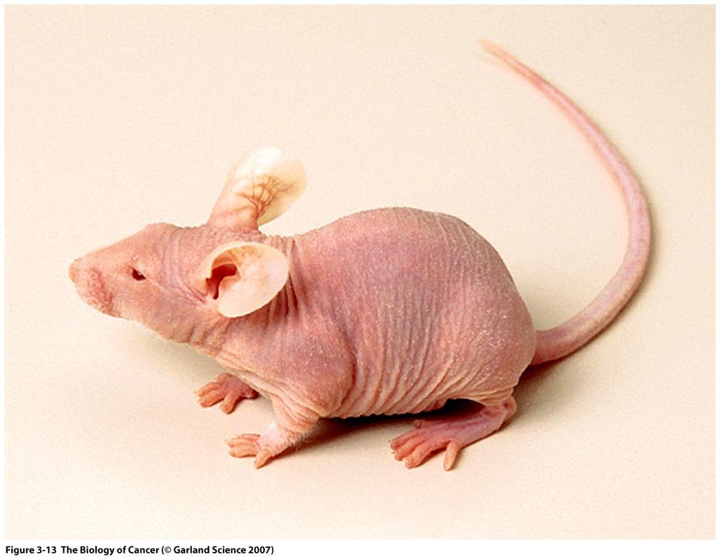
6 GROWTH OF NORMAL AND NEOPLASTIC FIBROBLASTS IN CULTURE TRANSFORMATION Growth Characteristic Normal Cells Tumour Cells *Density-dependent inhibition of growth Present Absent Growth factor requirement High Low *Anchorage dependence Present Absent *Proliferative life span Finite Immortal Contact inhibition of movement Present Absent Adhesiveness High Low Morphology Flat Rounded TUMORIGENICITY Growth Characteristic Normal Cells Tumour Cells Tumour formation in nude or SCID mice No Yes 6
7 7
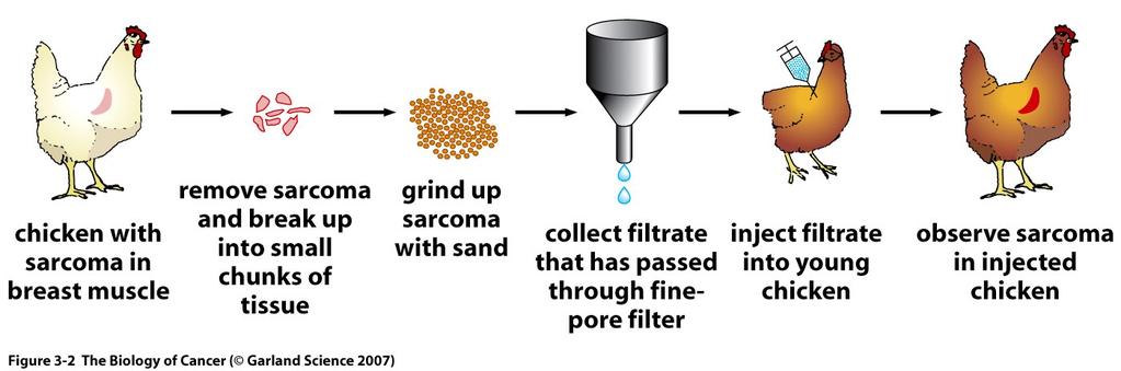
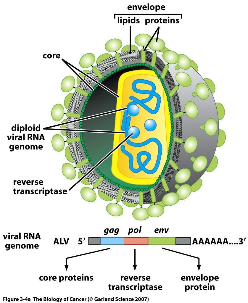
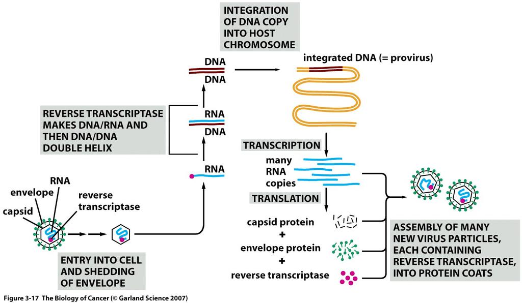
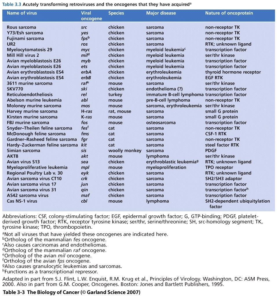
8 VIRUSES AND CANCER First oncogenic virus described in 1911 by Rous. Cellfree filtrate prepared from a chicken sarcoma tumour produced sarcomas in chicken. Rous sarcoma virus (RSV) - RNA tumour virus (retrovirus) 1950 s - Cultures of chicken fibroblasts + RSV = transformation Two classes of retroviruses (i) acute transforming viruses (e.g. RSV) which rapidly induce tumours and efficiently transform cells in culture and (ii) chronic tumour viruses (e.g. ALV avian leukosis virus) which induce tumours after long latent periods (several months) and don t transform cells in culture RSV has an extra gene oncogene src not required for viral growth. ALV has no oncogene, induces tumours by inserting themselves next to host genes and altering the expression of these genes. 8
9 9
10 10
11 11
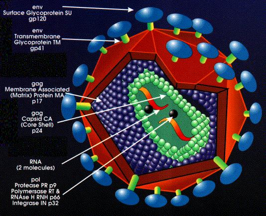
12 Basic retrovirus genome GAG: internal proteins ENV: envelope glycoproteins POL: enzymes, includes reverse transcriptase and integrase 12
13 13
14 14
15 Retrovirus 15
16 16
17 Where do oncogenes come from? Virus or cell? (1) RNA was isolated from RSV and used to probe DNA from various tissues of the chicken. Found sequences homologous to src in the DNA of all tissues of virus-free chickens. (2) Furthermore, homologous sequences to src are found in yeast, mice, rats, Drosophila, as well as humans. (3) Eukaryotic genes are composed of coding regions (exons) and non-coding regions (introns). When mrna is made, the introns are spliced out. Cellular oncogenes have exons and introns, while viral oncogenes are mrna-like. Unlikely that the viruses infected the cells first and then developed introns and exons. Furthermore, size and number of introns and exons in different species are conserved. 17
18 Where do oncogenes come from cont d (4) Localization and linkage of proto-oncogenes is conserved in different species. If viral oncogene introduced into cells after speciation, unlikely to find conserved gene structure and linkage. (5) Animals infected with a RNA tumour virus that doesn t contain an oncogene occasionally develop tumours that produce viruses that now carry an oncogene. Conclusion oncogenes are of cellular origin 18
19 Proto-oncogenes oncogenes >30 viral oncogenes have been identified each of which has a normal cellular counterpart. In normal cells, these proto-oncogenes are expressed and don t produce tumours. Proto-oncogenes normal cellular genes that can be altered to become oncogenes 19
20 20
21 Four human retroviruses HTLV-1 associated with cancer HTLV-2 may be associated with cancer HIV-1 human immunodeficiency virus - causes AIDS (acquired immunodeficiency syndrome) HIV-2 causes AIDS 21
22 Human RNA tumour viruses HTLV-1 human T cell-leukemia virus type 1 Associated with adult T-cell leukemia, a rare cancer found in Southern Japan, Caribbeans Latency a few years to >40 years Does not carry a host derived oncogene Viral genome Rex protein is essential for viral replication enhances expression of incompletely spliced viral transcripts that encode the viral structural proteins Tax protein is the transforming component of HTLV-1 22
23 HTLV-1 cont d Cellular pathways or genes responsive to Tax include: (i) NF-κB pathway which is activated by Tax resulting in induction of cytokines and genes associated with apoptosis and the cell cycle, and (ii) DNA polymerase ß, p53 and p16 genes which are repressed by Tax resulting in defects in DNA repair and the cell cycle 23
24 DNA tumour viruses RNA tumour viruses oncogenes are derived from host; oncoproteins do not elicit immune response; oncogenes are not required for viral replication DNA tumour viruses oncogenes are virally encoded; oncoproteins tend to elicit an immune response; often associated with immunocompromised hosts; oncogenes (early genes) are required for viral replication and/or promote cell cycle progression 24
25 DNA tumour viruses Four families of DNA viruses have oncogenic potential: hepadna-, papova-, adeno-, and herpes-virus. The transforming proteins of DNA tumour viruses large T antigen in SV40, E6 and E7 in papillomavirus, E1A and E1B in adenovirus have no cellular counterparts. Virus Family Approximate Genome Size (kb) Hepadna Hepatitis B viruses 3 Polyomaviridae Papillomaviridae SV40 and Polyomavirus Papillomaviruses 5 8 Adeno Adenoviruses 35 Herpes Herpesviruses
26 26

27 Hepatitis B virus: Hepatitis B virus: - strongly associated with the development of human hepatocellular carcinoma. Frequency of this cancer is increased 100X in individuals chronically infected with HBV. HBV DNA is commonly integrated in the genome of hepatocellular carcinoma cells. Adjacent cellular genes may be activated, increased cell proliferation. 27
28 Polyomaviridae SV40, JC, BK and polyoma virus (not directly linked to human cancer): Genomes encode 6 to 9 proteins. Contain both early (before viral DNA synthesis) and late (after viral DNA synthesis) genes. Rely on host DNA synthesis machinery to replicate viral DNA. Early proteins trigger cells to enter S phase and are responsible for immortalization and transformation. Polyomaviridae interact with susceptible cells in two different ways: (i) in permissive cells, viral DNA is replicated, coat protein made and progeny virion assembled. Virus particles are then released resulting in cell lysis and cell death. (ii) in non-permissive cells, viral DNA integrates randomly into the host chromosomes. A small proportion of cells with integrated viral DNA will become transformed and tumorigenic. 28
29 Simian Virus 40 (SV40) and polyomavirus - extensively characterized DNA tumour viruses : SV40 found to be a contaminant of polio virus vaccine injected into millions of people - SV40 (permissive in monkey cells) causes tumours in newborn and immunodeficient rodents but has not been directly associated with human cancers - early proteins are called T antigens (large, middle and small in polyomavirus; large and small in SV40). T antigens are multifunctional proteins that regulate viral DNA replication and transcription - transgenic mice carrying SV40 large T antigen under the control of tissue-specific promoters develop tumours in the appropriate tissues. - SV40 large T antigen is used to immortalize rodent cells to establish permanent lines. Although much less efficient, human cells can also be immortalized with SV40 large T antigen. - large T antigen binds to tumour suppressors p53 and prb. 29
30 Papillomaviruses -associated with warts and cervical carcinoma (found in 90% of cervical carcinomas). In benign warts, virus is maintained as an episome. In cervical carcinomas, HPV DNA is integrated in the host genome. - the early genes E6 and E7 encode proteins that have transforming potential. E7 binds to prb while E6 binds to p53. - >100 types of papillomaviruses; HPV16 and HPV18 (high-risk HPV) can extend lifespan of human fibroblasts and keratinocytes in culture. - infection with HPV is not sufficient to cause cancer. - HPV vaccine made with late proteins (empty capsids) which generate a strong antibody response 30
31 Adenovirus - permissive in humans; most m people have antibodies to these viruses - adenoviruses are not known to cause human cancer - rodent cells transformed with adenoviruses contain an incomplete viral genome that always includes E1A and E1B. E1A binds RB protein while E1B binds p53. 31
32 Herpesvirus - >60 proteins encoded by viral genome - EBV (Epstein-Barr virus) infects a large percentage of the population (90%) - EBV-infected human B lymphocytes become immortalized - genes required for immortalization: EBNA-2, EBNA-3 and the membrane protein LMP-1. - in immunocompromised individuals (e.g. AIDS patients, organ transplant recipients), EBV may induce lymphomas. - EBV has been isolated from several types of cancers: 97% of Burkitt lymphoma (BL) in tropical forest region; 20% of sporadic BL; 30% of AIDSassociated BL; common in nasopharyngeal carcinoma; 50% of Hodgkin s lymphoma. - HHV-8 is involved in a number of malignancies including Kaposi s sarcoma which is a frequent complication of AIDS 32
33 Oncolytic viruses - oncolytic viruses have intrinsic or engineered tumour selectivity - oncolytic viruses destroy tumour cells (e.g. by replicating selectively in cancer cells) - clinical trials have been or are being carried out with oncolytic DNA viruses (including adenovirus and Herpes simplex virus - HSV) and RNA viruses (including reovirus Calgary) - HSV has the following advantages: can insert a considerable amount of foreign DNA, doesn t integrate in the genome. Disadvantage: hard to work with because of its large genome - reoviruses are non-pathogenic to humans (cause mild infections of the respiratory or gastrointestinal tracts). Reoviruses replicate selectively in cells with activated Ras pathway (30% of human cancers have activated Ras pathway) - approx. 300 clinical trials with oncolytic viruses; little toxicity reported; encouraging results in some clinical trials (data not conclusive) 33
34 GENETICS AND CANCER Evidence that cancer is a genetic disease (associated with genetic alterations/mutations) before the mid 1970s: (1) Chromosomal abnormalities in tumour cells (translocations, double minute chromosomes) (2) Association of tumour development with DNAdamaging agents (such as chemical carcinogens, e.g., 2 napthyl amine) (3) Increased incidence of cancer in hereditary diseases such as Ataxia telangiectasia (AT), Xeroderma pigmentosa (XP), retinoblastoma (RB), familial polyposis coli, and other forms of hereditary cancers. (4) Genetic material from RNA and DNA tumour viruses carry information that allow rapid induction of malignant transformation in tissue culture and in animals. 34

35 Assay for cell transformation DNA transfection DNA of RSV-transformed sarcoma cells normal chicken embryo fibroblasts ~2 weeks foci of transformed cells 35
36 Can normal cells be transformed with DNA from non-virally-infected cancer cells? - use normal mouse fibroblasts (NIH3T3) (normal = immortalized, growth inhibited by contact, anchorage dependent, don t form tumours in nude mice). - transfect NIH3T3 cells with genomic DNA from: (a) chemically transformed cells and (b) from a bladder carcinoma cell line (late 1970 s/early 1980 s). - presence of transformed foci (cells piling up) indicates that the NIH3T3 cells have been transformed by introduced genomic DNA. - tests for anchorage independence and tumour formation in nude mice provide additional evidence that the foci are transformed and tumorigenic. If isolate DNA from the transformed cells and use this DNA to transfect NIH3T3 still works (i.e. obtain transformed foci) Is the transformed phenotype caused by an oncogene? Normal cells have proto-oncogenes Do cancer cells have oncogenes? 36
37 Events leading to the identification of cellular oncogenes In 1980 identification of Alu repetitive elements (~600,000 repeats = 1/6000 bp). By probing with Alu repetitive DNA, one can detect the human DNA retained in mouse NIH3T3 cells that have been transfected with human tumour DNA. If there is one Alu sequence/6000 bp of DNA, each intact gene should have multiple Alu sequences. By carrying out secondary and tertiary transfections and selecting for the transformed phenotype, the amount of human material present is reduced but the putative human oncogene is retained. A human oncogene was obtained by: (1) cloning the entire genome, (2) selecting the clones that had human DNA and (3) determining whether this DNA was capable of transforming NIH3T3. By probing the cloned DNA with viral oncogenes to see if the isolated gene might represent the cellular counterpart of a viral oncogene, the human oncogene in bladder carcinoma cell line was identified as H-ras (1982). 37
38 38
39 39
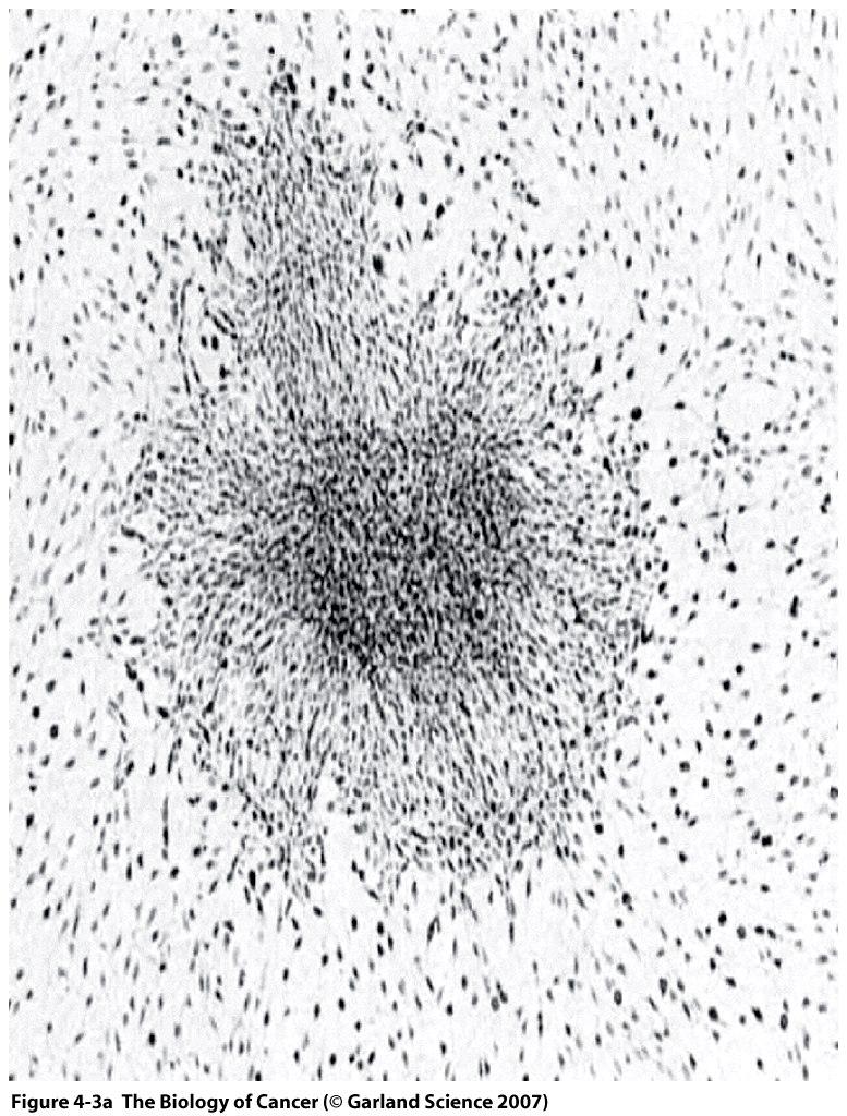
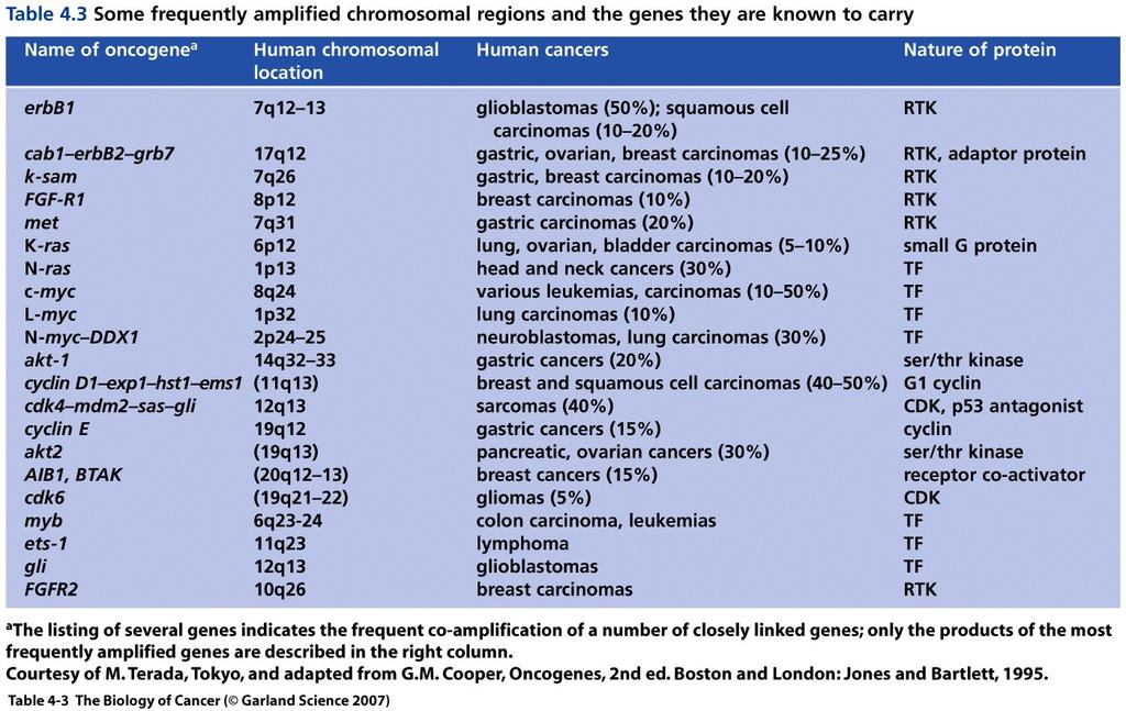
40 40
41 Activation of the H-ras protooncogene (point mutation) The NIH3T3 DNA transfection assay to identify activated cellular oncogenes was extremely popular in the early 1980 s. DNAs from a great variety of tumours were tested. 41
42 Assay most efficient at detecting ras oncogenes. 42
43 43
44 Activation of proto-oncogenes by chromosomal translocation Burkitt s lymphoma (8:14 translocation) Chronic myelogenous leukemia (CML) Philadelphia Chromosome (9:22 translocation) 44
45 45
46 46
47 ACTIVATION OF PROTO-ONCOGENES ONCOGENES BY GENE AMPLIFICATION Homogeneously staining regions (HSRs) and double minutes (DMs) HSRs and DMs found in a large number of tumours The first gene identified in HSRs/DMs was c-myc (MYC) (in HL60 a promyelocytic leukemia) Neuroblastoma tumours have N-myc (MYCN) amplified in HSRs or DMs. Patients with multiple copies of N- myc (amplification of the N-myc gene) have a poor clinical prognosis. 47
48 48


49 Gene amplification Double minutes HSR HSR MYCN amplification in neuroblastoma 49
50 50
 Estimates based on age at which cancer appears and incidence of cancer indicate that 3 to 8 mutations (2 mutations in the")
51 Is cellular proto-oncogene activation sufficient to cause cancer? Evidence suggesting that multiple hits are required for cancer formation: (1) Estimates based on age at which cancer appears and incidence of cancer indicate that 3 to 8 mutations (2 mutations in the case of retinoblastoma) are required for cancer formation (2) Some cancers go through several stages (3) Multiple oncogenes are required to transform cells in culture 51
52 (4) In some cases, infection of cells with a retrovirus carrying an oncogene suggests that additional events are required. For example, infection of bone marrow cells of mice with a retrovirus carrying the bcr/abl fusion gene found in CML results in CML in some cases only. Furthermore, it takes a long time for CML to develop, suggesting that additional genetic alterations are required (5) Multiple mutated genes are found in a single cancer cell. E.g., HL60 has both amplified myc gene and amplified N-ras gene. In colon carcinoma, at least 6 different genetic alterations have been identified (Kinsler and Vogelstein Cell 87: ). 52

53 CIS = carcinoma in situ CIN = cervical intraepithelial neoplasia DCIS = ductal carcinoma in situ PIN = prostatic intraepithelial neoplasia 53
54 Cellular proto-oncogenes, viral oncogenes and cellular oncogenes e.g. src Cellular proto-oncogene picture Viral oncogene multiple mutations, expressed at elevated levels picture Cellular oncogene single base pair substitutions, translocations resulting in fusion proteins or overexpression, gene amplification resulting in overexpression e.g. point mutation 54

55 Table 39.3 NB RSV 1-2 weeks sarcoma other RNA tumour viruses take longer RNA tumour viruses do not infect all cell types 55

56 Sites of action of oncogenes 56
57 Roles of proteins encoded by oncogenes 1. Growth factors -sis 2. Growth factor receptors -erbb - erbb2 (Her-2/neu, HER2) 3. Nonreceptor tyrosine kinases - src 4. GTP-binding proteins - ras 5. Serine/threonine kinase - raf 6. Nuclear transcription factors (DNA binding proteins) - myc - fos/jun 57
58 The Basic Science of Oncology, pg126 58
59 Functions of proteins encoded by oncogenes Normal cells balance between growth-promoting and growth-restraining/inhibiting properties. Cancer cells balance is altered so that growthpromoting properties are increased (oncogenes) or growth-inhibiting properties are decreased (tumour suppressors). 59
60 1. Oncogenes and growth factors (sis/pdgf) Cell growth and differentiation are triggered by extracellular signals at cell surface. When growth factor binds to cell surface receptor, a cascade of biochemical reactions is initiated that alters protein expression in the cell. Growth factors are short polypeptides that induce proliferation in appropriate target cells. Normal cell growth and differentiation depends on presence of growth factors. However, if growth factor is always expressed, this can result in continuous stimulation of cell growth (a step towards transformation and cancer). 60
 are responsive to sis.")
61 Growth factors cont d The sis (simian sarcoma virus) oncogene was discovered In 1983 identified as the B-subunit of the platelet-derived growth factor (PDGF). PDGF is a major growth factor used by fibroblasts and smooth muscle cells. Only cells that are normally responsive to PDGF (i.e. cells that have PDGF receptors) are responsive to sis. 61
62 62
63 Kaposi s sarcoma (autocrine signaling) PDGF TGF-β IGF-1 Ang2 CC18/14 CXCL11 Endothelin Plus all receptors for these ligands Plus HHV-8 produces two more ligands whose receptors are expressed in Kaposi sarcoma cells 63
64 2. Oncogenes and growth factor receptors (receptor protein tyrosine kinases) - a common way of regulating protein function is by phosphorylation. Many oncogenes encode growth factor receptors. Growth factor receptors are often protein tyrosine kinases that can phosphorylate themselves and other proteins at tyrosine residues resulting in the activation of a signaling cascade. Example 1: EGFR (ErbB) - the epidermal growth factor (EGF) receptor binds growth factor EGF, resulting in a conformational change that facilitates dimerization of growth factor receptors by exposing a dimerization loop. Receptor dimerization results in intermolecular phosphorylation of tyrosines which in turn allows access to ATP and protein substrates. - ErbB (is a truncated version of the EGF receptor (contains the PK domain and the transmembrane domain but lacks the extracellular ligand binding domain so no longer need EGF for stimulation). 64
65 The Basic Science of Oncology p145 65
66 Example 2: Her2/neu (ErbB2) - second family of oncogenes identified using the NIH3T3 assay to analyse rat tumours. Neu protein found in rat tumours is almost identical to c-erbb2 (one amino acid substitution). This amino acid substitution in the transmembrane domain causes ligand-independent dimerization of neu protein. - in humans, Her2/neu is amplified in many cancers including breast, ovarian, lung, colon, pancreatic. Her2/neu is amplified and over-expressed in ~30% of breast cancers and is associated with metastasis and a poor clinical outcome. As receptor concentration increases, there is a shift from monomeric to dimeric (active) form. 66
67 (ErbB2, Her2) 67
68 Breast cancer heterogeneous disease 15 distinct forms recognized by the American Joint Committee on Cancer Four distinct molecular subtypes of locally advanced breast cancer have been identified based on gene expression profiling: Luminal A (Estrogen receptor (ER) positive lower percentage of proliferating cells): good prognosis Luminal B (ER+ve higher percentage of proliferating cells): intermediate prognosis Basal-like or triple-negative (ER, PR and HER2 negative): poor prognosis HER2 positive: poor prognosis 68
69 Her2/neu has been targeted for the treatment of Her2/neuoverexpressing metastatic breast cancer - Trastuzumab (marketed as Herceptin): humanized monoclonal antibody that recognizes Her2/neu protein and inhibits proliferation of Her2/neuoverexpressing breast cancer cells. Trastuzumab has been recommended for the treatment of Her2/neu-overexpressing breast cancer patients. Problem; resistance to trastuzumab treatment. Need combination therapy. - other possibilities drugs that block tyrosine kinases, antisense oligonucleotides. Need specific delivery systems. 69
70 70
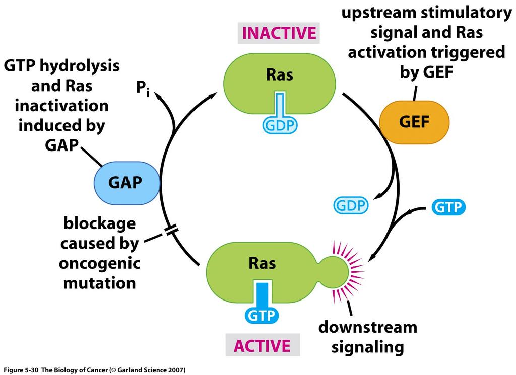
71 - targets of Her2/neu kinase: phosphatidylinositol-3- kinase (PI3-kinase/Akt), mitogen-activated protein kinase (MAPK), camp/protein kinase A. These pathways are involved in cell proliferation and cell survival; e.g. Akt phosphorylates p21 and disrupts its growth-inhibitory activity. - reduction in Her2/neu receptors on cell surface (after Herceptin treatment) may result in a reduction in PI3-kinase and Akt that protect cells from apoptosis, thus making these cells more sensitive to radiation treatment and chemotherapy. 71
72 Junttila et al. Cancer Cell 15, May 5, 2009 ErbB2 does not have a ligand and functions as a co-receptor with other ErbB receptors. ErbB3 is best suited to activate the PI3K/Akt signaling pathway as it has multiple binding sites for the regulatory subunits of PI3K. ErbB3 binds heregulins but has impaired kinase activity, and only signals as a complex with another ErbB, preferably ErbB2 Ligand-independent ErbB2/ErbB3/PI3K complexes function as a oncogenic unit in ErbB2-over-expressing breast cancer cells Trastuzumab destabilizes ligand-independent ErbB2/ErbB3/PI3K complexes, uncoupling ErbB3 from ErbB2 and blocking downstream PI3K signaling. However, PI3K mutations are common in breast cancer. Mutant PI3K remains at cell membrane and activates Akt in the presence of transtuzumab. Target PI3K pathway in trastuzumab-resistant ErbB2 overexpressing breast cancers. 72
73 3. Oncogenes and nonreceptor protein tyrosine kinases Src was the first oncogene shown to have protein kinase activity. Phosphorylates tyrosine residues. Cytoplasmic (intracellular) protein. A family of 9 closely related tyrosine kinases constitute the Src family. Implicated in signalling through growth factor receptor tyrosine kinases such as receptor for PDGF. 73
74 Tyrosine 416 is autophosphorylated by Src increases kinase activity and transforming potential Tyrosine 527 is phosphorylated by another proteintyrosine kinase (e.g. Csk or Chk) - downregulates kinase activity (tyrosine 527 is not present in viral src protein) Src homology domains SH4 Myristoylation site Targets Src to cytoplasmic membrane Binds proline-rich proteins Binds phosphotyrosine-containing proteins [e.g. PDGF receptor, FAK (focal adhesion kinase)] Many of Src substrates are part of signaling cascades 74
75 Src cont d BBA 1602: (2002) 75
 phosphorylation] BBA 1602: 114-130")
76 Src cont d Src is overexpressed in some epithelial cancers (colon, breast) Src is mutated in some colon cancers [affects Tyr527(530) phosphorylation] BBA 1602: (2002) 76
77 Src therapeutic target for cancer? - Activation of Src is common in solid tumors, whether or not its negative regulator Csk is present - Activated Src moves to cell membrane - Lipid rafts are cholesterol-enriched domains believed to be important for protein signaling in cells 77
78 Oneyama et al. Molecular Cell 30: (2008) - Csk is a cytoplasmic protein whereas activated Src is anchored to the cytoplasmic membrane - Csk uses adaptor proteins to access Src - Cbp is a Csk adaptor protein found in lipid rafts - In Csk-deficient fibroblasts, over-expression of wild-type Src can induce transformation - Cbp expression is dramatically downregulated in these transformed fibroblasts - Re-expression of Cbp suppresses Src transformation (in the absence of Csk); phosphorylated Cbp specifically binds to activated Src and sequesters it in lipid rafts Conclusion: lipid raft components such as Cbp may inactivate Src and suppress tumour formation 78
79 79
80 4. Oncogenes and guanine nucleotide binding proteins (G proteins) ras Cytoplasmic signaling. Regulate activity of target proteins in response to a variety of signals. Links growth factor receptors to cytoplasmic activation pathways. G proteins can bind GDP and GTP and have GTPase activity. Ras proto-oncogene responds to signals such as PDGF (in fibroblasts) or antigen stimulation (in T cells). In the presence of these growth factors, Ras is converted to active form (Ras-GTP). GTPase activity gradually converts GTP to GDP thereby returning Ras to its inactive form (Ras-GDP). Mutations of Ras at amino acids 12, 13, 61 decrease GTPase activity, thereby increasing the level of active Ras-GTP. 80
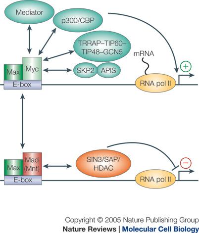
81 81
82 Proteins that regulate Ras GEFs (guanine nucleotide exchange factors) activation of Ras GAPs (GTPase activating proteins) inactivation of Ras increase GTPase activity of Ras Mutations affecting Ras-GEFs and Ras-GAPs can also contribute to cell transformation p120 GAP increases GTPase activity of normal Ras but not mutant Ras A second protein with GAP activity is the tumour suppressor NF1 neurofibromatosis gene. Lack of activity of this gene results in high levels of normal Ras being in the active GTP bound form GEFs function as positive regulators of Ras by stimulating the exchange of bound GDP for free GTP. An example of a GEF is Sos (son of sevenless) which can induce transformation of mammalian cells in culture Sos plays a critical role in activating Ras proteins in response to growth factor stimulation of protein-tyrosine kinases. Sos associates with an adaptor protein called Grb2 which has SH2 and SH3 domains. Grb2 binds Sos through its SH3 domain and phosphotyrosine-containing growth factor receptors through its SH2 domains 82

83 83
84 Best-studied downstream targets (effectors) of Ras: (i) Raf kinase Active Ras binds the protein kinase Raf and activates it. Raf phosphorylates MEK (MAP kinase kinases) which in turn phosphorylate MAP kinases (a.k.a. Erk1 and Erk2). MAP kinase is believed to change the activity of nuclear transcription factors such as Fos, Jun, and Myc. This pathway has been strongly implicated in the transformation of mouse cells. (ii) PI3-kinase Ras activates PI3 kinase which results in activation of Akt and Rho-GEF. Activated Akt inactivates Bad (thus suppressing apoptosis), inactivates GSK-3β (thus stimulating cell proliferation) and may activate mtor (stimulating proliferation). Activated PI3K alone cannot cause transformation of NIH3T3 cells but cooperates with activated Raf to transform cells. (iii) RaI (protein related to Ras) Ras communicates with Ral through RaI-GEF. Co-expression of activated Ral-GEF and activated Raf induces foci formation in NIH3T3. RaI-GEF (and not Raf) appears to play a key role in transformation of human cells by Ras. 84
85 85
86 5. Oncogenes and serine threonine kinases (Raf) - direct binding has been observed between the Ras and Raf proteins - interaction between Raf and Ras is required to recruit Raf to cell membrane - once activated, Raf can directly phosphorylate a family of serine/threonine kinases called the mitogen-activated protein kinase kinases (MAPKK or MEK) - activated MAPKKs activate MAPKs 86
87 The Biological Basis of Cancer (1998) 87

88 6. Oncogenes and transcription factors A number of proteins encoded by oncogenes are located in the nucleus and function as transcription factors. Transcription of genes in the nucleus involves interaction of transcription factors with regulatory elements. Regulatory elements (promoters, enhancers) are usually located upstream of genes. 88
89 Myc proteins c-myc (Myc), N-myc, L-myc Myc proteins are involved in many cancers (gene amplification, gene translocation). Activation of myc (proto-oncogene to oncogene) is through overexpression of myc protein. Myc proteins are induced rapidly following growth factor stimulation. Myc has a DNA binding domain and functions as a transcription factor. 89
90 Table 1 Myc overexpression in various cancers Cancerous tissue Frequency of Myc overexpression Solid tumours Glioblastoma 57 78% Breast 45% Colon 67% Medulloblastoma 31% Ovarian 66% Pancreatic 43% Lymphoma Burkitt s >90% Diffuse large B cell 10 25% Transformed follicular 70% Mantle cell Up to 100% Brooks, TA and Hurley, LH. Nature Reviews Cancer 9: ,
91 Myc cont d Myc interacts with Max. Like Myc, Max has a DNA binding domain. The Myc-Max complex binds to a specific DNA sequence called the E-box (CACGTG) and activates transcription which results in mitogenic stimulation. Further regulation is provided by Mnt and Mad which dimerize with Max but not Myc. Max can dimerize with Myc, Mad and Mnt with equal affinity. Max-Mnt and Max-Mad heterodimers repress transcription. Transcription repression by Mnt-Max and Mad-Max requires interaction with Sin3 which recruits histone deacetylases (HDACs). Max and Sin3 are constitutively expressed. The dimerization state of Max is determined by the relative levels of Myc and Mnt or Mad. Target genes for Myc include cyclin D2 and CDK4 involved in cell proliferation and VEGF involved in angiogenesis. Myc also regulates genes involved in glucose metabolism, nucleotide metabolism, and induces micrornas that affect E2F1 expression. 91
92 92
93 Myc can also bind to a second transcription factor called Miz-1. When bound to Miz-1, Myc functions as a transcriptional repressor (represses the expression of p21 and p15, cyclin-dependent kinase inhibitors which inhibit CDK4/6 and CDK2 involved in phosphorylating prb) 93

94 Myc pathways Jonas A Nilsson and John L Cleveland 94
95 Is Myc a potential target for cancer treatment? - Myc has a short half-life (20-30 minutes) - ~30,000 potential Myc binding sites in the human genome % of genes are bound by Myc - Transcription of the Myc gene induces negative supercoiling of DNA - Might Myc transcription be controlled through small molecules that target structural elements in the Myc promoter? 95

96 nature medicine volume 17 number 11 november
97 Second example of oncogenes that function as transcription factors Fos (murine osteogenic sarcoma virus) and Jun (avian sarcoma virus) Members of the Fos and Jun family form dimeric AP-1 transcription factors which bind to TGACTCA The levels and/or activity of members of the Fos and Jun family are elevated in the presence of activated Ras and other oncoproteins such as adenovirus E1A 97
98 Numerous AP-1 target genes have been identified. Different AP-1 heterodimers may regulate different target genes. Cell Cycle 6: 2633,
99 99
RAS Genes. The ras superfamily of genes encodes small GTP binding proteins that are responsible for the regulation of many cellular processes.
 ۱ RAS Genes The ras superfamily of genes encodes small GTP binding proteins that are responsible for the regulation of many cellular processes. Oncogenic ras genes in human cells include H ras, N ras,
Deregulation of signal transduction and cell cycle in Cancer
 Deregulation of signal transduction and cell cycle in Cancer Tuangporn Suthiphongchai, Ph.D. Department of Biochemistry Faculty of Science, Mahidol University Email: tuangporn.sut@mahidol.ac.th Room Pr324
VIRUSES AND CANCER Michael Lea
 VIRUSES AND CANCER 2010 Michael Lea VIRAL ONCOLOGY - LECTURE OUTLINE 1. Historical Review 2. Viruses Associated with Cancer 3. RNA Tumor Viruses 4. DNA Tumor Viruses HISTORICAL REVIEW Historical Review
Chapter 4 Cellular Oncogenes ~ 4.6 -
 Chapter 4 Cellular Oncogenes - 4.2 ~ 4.6 - Many retroviruses carrying oncogenes have been found in chickens and mice However, attempts undertaken during the 1970s to isolate viruses from most types of
Karyotype analysis reveals transloction of chromosome 22 to 9 in CML chronic myelogenous leukemia has fusion protein Bcr-Abl
 Chapt. 18 Cancer Molecular Biology of Cancer Student Learning Outcomes: Describe cancer diseases in which cells no longer respond Describe how cancers come from genomic mutations (inherited or somatic)
What causes cancer? Physical factors (radiation, ionization) Chemical factors (carcinogens) Biological factors (virus, bacteria, parasite)
 Chemical factors (carcinogens) Biological factors (virus, bacteria, parasite)") Oncogenes What causes cancer? Chemical factors (carcinogens) Physical factors (radiation, ionization) Biological factors (virus, bacteria, parasite) DNA Mutation or damage Oncogenes Tumor suppressor genes
Oncogenes What causes cancer? Chemical factors (carcinogens) Physical factors (radiation, ionization) Biological factors (virus, bacteria, parasite) DNA Mutation or damage Oncogenes Tumor suppressor genes
CANCER. Inherited Cancer Syndromes. Affects 25% of US population. Kills 19% of US population (2nd largest killer after heart disease)
") CANCER Affects 25% of US population Kills 19% of US population (2nd largest killer after heart disease) NOT one disease but 200-300 different defects Etiologic Factors In Cancer: Relative contributions
CANCER Affects 25% of US population Kills 19% of US population (2nd largest killer after heart disease) NOT one disease but 200-300 different defects Etiologic Factors In Cancer: Relative contributions
Cancer. The fundamental defect is. unregulated cell division. Properties of Cancerous Cells. Causes of Cancer. Altered growth and proliferation
 Cancer The fundamental defect is unregulated cell division. Properties of Cancerous Cells Altered growth and proliferation Loss of growth factor dependence Loss of contact inhibition Immortalization Alterated
Cell cycle, signaling to cell cycle, and molecular basis of oncogenesis
 Cell cycle, signaling to cell cycle, and molecular basis of oncogenesis MUDr. Jiří Vachtenheim, CSc. CELL CYCLE - SUMMARY Basic terminology: Cyclins conserved proteins with homologous regions; their cellular
Cancer. The fundamental defect is. unregulated cell division. Properties of Cancerous Cells. Causes of Cancer. Altered growth and proliferation
 Cancer The fundamental defect is unregulated cell division. Properties of Cancerous Cells Altered growth and proliferation Loss of growth factor dependence Loss of contact inhibition Immortalization Alterated
1. Basic principles 2. 6 hallmark features 3. Abnormal cell proliferation: mechanisms 4. Carcinogens: examples. Major Principles:
 Carcinogenesis 1. Basic principles 2. 6 hallmark features 3. Abnormal cell proliferation: mechanisms 4. Carcinogens: examples Carcinogenesis Major Principles: 1. Nonlethal genetic damage is central to
number Done by Corrected by Doctor Maha Shomaf
 number 19 Done by Waseem Abo-Obeida Corrected by Abdullah Zreiqat Doctor Maha Shomaf Carcinogenesis: the molecular basis of cancer. Non-lethal genetic damage lies at the heart of carcinogenesis and leads
CELL CYCLE MOLECULAR BASIS OF ONCOGENESIS
 CELL CYCLE MOLECULAR BASIS OF ONCOGENESIS Summary of the regulation of cyclin/cdk complexes during celll cycle Cell cycle phase Cyclin-cdk complex inhibitor activation Substrate(s) G1 Cyclin D/cdk 4,6
Determination Differentiation. determinated precursor specialized cell
 Biology of Cancer -Developmental Biology: Determination and Differentiation -Cell Cycle Regulation -Tumor genes: Proto-Oncogenes, Tumor supressor genes -Tumor-Progression -Example for Tumor-Progression:
ONCOGENES. Michael Lea
 ONCOGENES 2011 Michael Lea ONCOGENES - Lecture Outline I. Introduction 2. Identification of oncogenic genes in retroviruses 3. Homologous sequences in transformed and untransformed cells 4. Methods of
Carcinogenesis. Carcinogenesis. 1. Basic principles 2. 6 hallmark features 3. Abnormal cell proliferation: mechanisms 4. Carcinogens: examples
 Carcinogenesis 1. Basic principles 2. 6 hallmark features 3. Abnormal cell proliferation: mechanisms 4. Carcinogens: examples Major Principles (cont d) 4. Principle targets of genetic damage: 4 classes
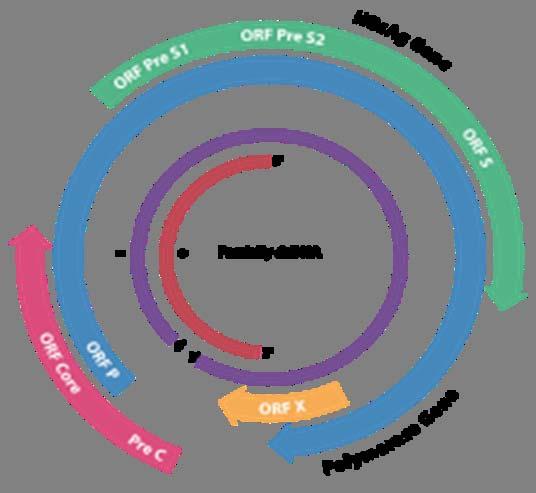
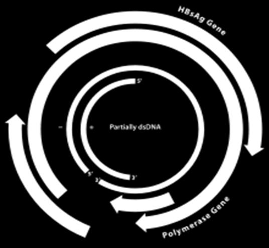
Genome of Hepatitis B Virus. VIRAL ONCOGENE Dr. Yahwardiah Siregar, PhD Dr. Sry Suryani Widjaja, Mkes Biochemistry Department
 Genome of Hepatitis B Virus VIRAL ONCOGENE Dr. Yahwardiah Siregar, PhD Dr. Sry Suryani Widjaja, Mkes Biochemistry Department Proto Oncogen and Oncogen Oncogen Proteins that possess the ability to cause
Molecular Cell Biology. Prof. D. Karunagaran. Department of Biotechnology. Indian Institute of Technology Madras
 Molecular Cell Biology Prof. D. Karunagaran Department of Biotechnology Indian Institute of Technology Madras Module 9 Molecular Basis of Cancer, Oncogenes and Tumor Suppressor Genes Lecture 2 Genes Associated
oncogenes-and- tumour-suppressor-genes)
") Special topics in tumor biochemistry oncogenes-and- tumour-suppressor-genes) Speaker: Prof. Jiunn-Jye Chuu E-Mail: jjchuu@mail.stust.edu.tw Genetic Basis of Cancer Cancer-causing mutations Disease of aging
Special topics in tumor biochemistry oncogenes-and- tumour-suppressor-genes) Speaker: Prof. Jiunn-Jye Chuu E-Mail: jjchuu@mail.stust.edu.tw Genetic Basis of Cancer Cancer-causing mutations Disease of aging
7.012 Problem Set 6 Solutions
 Name Section 7.012 Problem Set 6 Solutions Question 1 The viral family Orthomyxoviridae contains the influenza A, B and C viruses. These viruses have a (-)ss RNA genome surrounded by a capsid composed
Oncogenes and Tumor Suppressors MCB 5068 November 12, 2013 Jason Weber
 Oncogenes and Tumor Suppressors MCB 5068 November 12, 2013 Jason Weber jweber@dom.wustl.edu Oncogenes & Cancer DNA Tumor Viruses Simian Virus 40 p300 prb p53 Large T Antigen Human Adenovirus p300 E1A
Origin of oncogenes? Oncogenes and Proto-oncogenes. Jekyll and Hyde. Oncogene hypothesis. Retroviral oncogenes and cell proto-oncogenes
 Oncogenes and Proto-oncogenes Jekyll and Hyde A double edged sword Origin of oncogenes? Oncogene hypothesis Retroviral oncogenes and cell proto-oncogenes (v-onc) (c-onc) The role of c-onc in cancer How
Cancer and Gene Alterations - 1
 Cancer and Gene Alterations - 1 Cancer and Gene Alteration As we know, cancer is a disease of unregulated cell growth. Although we looked at some of the features of cancer when we discussed mitosis checkpoints,
Introduction. Cancer Biology. Tumor-suppressor genes. Proto-oncogenes. DNA stability genes. Mechanisms of carcinogenesis.
 Cancer Biology Chapter 18 Eric J. Hall., Amato Giaccia, Radiobiology for the Radiologist Introduction Tissue homeostasis depends on the regulated cell division and self-elimination (programmed cell death)
VIII Curso Internacional del PIRRECV. Some molecular mechanisms of cancer
 VIII Curso Internacional del PIRRECV Some molecular mechanisms of cancer Laboratorio de Comunicaciones Celulares, Centro FONDAP Estudios Moleculares de la Celula (CEMC), ICBM, Facultad de Medicina, Universidad
Signaling. Dr. Sujata Persad Katz Group Centre for Pharmacy & Health research
 Signaling Dr. Sujata Persad 3-020 Katz Group Centre for Pharmacy & Health research E-mail:sujata.persad@ualberta.ca 1 Growth Factor Receptors and Other Signaling Pathways What we will cover today: How
CELL BIOLOGY - CLUTCH CH CANCER.
 !! www.clutchprep.com CONCEPT: OVERVIEW OF CANCER Cancer is a disease which is primarily caused from misregulated cell division, which form There are two types of tumors - Benign tumors remain confined
Enzyme-coupled Receptors. Cell-surface receptors 1. Ion-channel-coupled receptors 2. G-protein-coupled receptors 3. Enzyme-coupled receptors
 Enzyme-coupled Receptors Cell-surface receptors 1. Ion-channel-coupled receptors 2. G-protein-coupled receptors 3. Enzyme-coupled receptors Cell-surface receptors allow a flow of ions across the plasma
Cell Signaling part 2
 15 Cell Signaling part 2 Functions of Cell Surface Receptors Other cell surface receptors are directly linked to intracellular enzymes. The largest family of these is the receptor protein tyrosine kinases,
BioSci 145A Lecture 15 - Oncogenes and Cancer
 BioSci 145A Lecture 15 - Oncogenes and Cancer Topics we will cover today Introduction to normal and cancer cells Characteristics of cells in culture Cancerous changes in cells Viruses can harbor transforming
Cancer. Questions about cancer. What is cancer? What causes unregulated cell growth? What regulates cell growth? What causes DNA damage?
 Questions about cancer What is cancer? Cancer Gil McVean, Department of Statistics, Oxford What causes unregulated cell growth? What regulates cell growth? What causes DNA damage? What are the steps in
Cancer genetics
 Cancer genetics General information about tumorogenesis. Cancer induced by viruses. The role of somatic mutations in cancer production. Oncogenes and Tumor Suppressor Genes (TSG). Hereditary cancer. 1
Name Section Problem Set 6
 Name Section 7.012 Problem Set 6 Question 1 The viral family Orthomyxoviridae contains the influenza A, B and C viruses. These viruses have a (-)ss RNA genome surrounded by a capsid composed of lipids
Lecture 8 Neoplasia II. Dr. Nabila Hamdi MD, PhD
 Lecture 8 Neoplasia II Dr. Nabila Hamdi MD, PhD ILOs Understand the definition of neoplasia. List the classification of neoplasia. Describe the general characters of benign tumors. Understand the nomenclature
Transcription and RNA processing
 Transcription and RNA processing Lecture 7 Biology 3310/4310 Virology Spring 2018 It is possible that Nature invented DNA for the purpose of achieving regulation at the transcriptional rather than at the
Biol403 MAP kinase signalling
 Biol403 MAP kinase signalling The mitogen activated protein kinase (MAPK) pathway is a signalling cascade activated by a diverse range of effectors. The cascade regulates many cellular activities including
Receptor mediated Signal Transduction
 Receptor mediated Signal Transduction G-protein-linked receptors adenylyl cyclase camp PKA Organization of receptor protein-tyrosine kinases From G.M. Cooper, The Cell. A molecular approach, 2004, third
G-Protein Signaling. Introduction to intracellular signaling. Dr. SARRAY Sameh, Ph.D
 G-Protein Signaling Introduction to intracellular signaling Dr. SARRAY Sameh, Ph.D Cell signaling Cells communicate via extracellular signaling molecules (Hormones, growth factors and neurotransmitters
Chapter 18- Oncogenes, tumor suppressors & Cancer
 Chapter 18- Oncogenes, tumor suppressors & Cancer - Previously we have talked about cancer which is an uncontrolled cell proliferation and we have discussed about the definition of benign, malignant, metastasis
Fayth K. Yoshimura, Ph.D. September 7, of 7 RETROVIRUSES. 2. HTLV-II causes hairy T-cell leukemia
 1 of 7 I. Diseases Caused by Retroviruses RETROVIRUSES A. Human retroviruses that cause cancers 1. HTLV-I causes adult T-cell leukemia and tropical spastic paraparesis 2. HTLV-II causes hairy T-cell leukemia
BIT 120. Copy of Cancer/HIV Lecture
 BIT 120 Copy of Cancer/HIV Lecture Cancer DEFINITION Any abnormal growth of cells that has malignant potential i.e.. Leukemia Uncontrolled mitosis in WBC Genetic disease caused by an accumulation of mutations
KEY CONCEPT QUESTIONS IN SIGNAL TRANSDUCTION
 Signal Transduction - Part 2 Key Concepts - Receptor tyrosine kinases control cell metabolism and proliferation Growth factor signaling through Ras Mutated cell signaling genes in cancer cells are called
Src-INACTIVE / Src-INACTIVE
 Biology 169 -- Exam 1 February 2003 Answer each question, noting carefully the instructions for each. Repeat- Read the instructions for each question before answering!!! Be as specific as possible in each
Oncogenes and tumour suppressor genes
 Cancer mutations disrupt cellular homeostasis Oncogenes and tumour suppressor genes Oncogenes: Gain of function mutations Proto-oncogene Tumour suppressor genes: loss of function mutations Normal cell
BCHM3972 Human Molecular Cell Biology (Advanced) 2013 Course University of Sydney
 2013 Course University of Sydney") BCHM3972 Human Molecular Cell Biology (Advanced) 2013 Course University of Sydney Page 2: Immune Mechanisms & Molecular Biology of Host Defence (Prof Campbell) Page 45: Infection and Implications for Cell
BCHM3972 Human Molecular Cell Biology (Advanced) 2013 Course University of Sydney Page 2: Immune Mechanisms & Molecular Biology of Host Defence (Prof Campbell) Page 45: Infection and Implications for Cell
Polyomaviridae. Spring
 Polyomaviridae Spring 2002 331 Antibody Prevalence for BK & JC Viruses Spring 2002 332 Polyoma Viruses General characteristics Papovaviridae: PA - papilloma; PO - polyoma; VA - vacuolating agent a. 45nm
Activation of cellular proto-oncogenes to oncogenes. How was active Ras identified?
 Dominant Acting Oncogenes Eugene E. Marcantonio, M.D. Ph.D. Oncogenes are altered forms of normal cellular genes called proto-oncogenes that are involved in pathways regulating cell growth, differentiation,
Genetics and Cancer Ch 20
 Genetics and Cancer Ch 20 Cancer is genetic Hereditary cancers Predisposition genes Ex. some forms of colon cancer Sporadic cancers ~90% of cancers Descendants of cancerous cells all cancerous (clonal)
Problem Set 5 KEY
 2006 7.012 Problem Set 5 KEY ** Due before 5 PM on THURSDAY, November 9, 2006. ** Turn answers in to the box outside of 68-120. PLEASE WRITE YOUR ANSWERS ON THIS PRINTOUT. 1. You are studying the development
Cellular Signaling Pathways. Signaling Overview
 Cellular Signaling Pathways Signaling Overview Signaling steps Synthesis and release of signaling molecules (ligands) by the signaling cell. Transport of the signal to the target cell Detection of the
Multistep nature of cancer development. Cancer genes
 Multistep nature of cancer development Phenotypic progression loss of control over cell growth/death (neoplasm) invasiveness (carcinoma) distal spread (metastatic tumor) Genetic progression multiple genetic
Herpesviruses. Virion. Genome. Genes and proteins. Viruses and hosts. Diseases. Distinctive characteristics
 Herpesviruses Virion Genome Genes and proteins Viruses and hosts Diseases Distinctive characteristics Virion Enveloped icosahedral capsid (T=16), diameter 125 nm Diameter of enveloped virion 200 nm Capsid
Chapter 15: Signal transduction
 Chapter 15: Signal transduction Know the terminology: Enzyme-linked receptor, G-protein linked receptor, nuclear hormone receptor, G-protein, adaptor protein, scaffolding protein, SH2 domain, MAPK, Ras,
Oncogenes and Tumor. supressors
 Oncogenes and Tumor supressors From history to therapeutics Serge ROCHE Neoplastic transformation TUMOR SURESSOR ONCOGENE ONCOGENES History 1911 1960 1980 2001 Transforming retrovirus RSV v-src is an oncogene
Introduction to Cancer Biology
 Introduction to Cancer Biology Robin Hesketh Multiple choice questions (choose the one correct answer from the five choices) Which ONE of the following is a tumour suppressor? a. AKT b. APC c. BCL2 d.
THE HALLMARKS OF CANCER
 THE HALLMARKS OF CANCER ONCOGENES - Most of the oncogenes were first identified in retroviruses: EGFR (ErbB), Src, Ras, Myc, PI3K and others (slightly more than 30) - Mutated cellular genes incorporated
MCB*4010 Midterm Exam / Winter 2008
 MCB*4010 Midterm Exam / Winter 2008 Name: ID: Instructions: Answer all 4 questions. The number of marks for each question indicates how many points you need to provide. Write your answers in point form,
Transformation of Normal HMECs (Human Mammary Epithelial Cells) into Metastatic Breast Cancer Cells: Introduction - The Broad Picture:
 into Metastatic Breast Cancer Cells: Introduction - The Broad Picture:") Transformation of Normal HMECs (Human Mammary Epithelial Cells) into Metastatic Breast Cancer Cells: Introduction - The Broad Picture: Spandana Baruah December, 2016 Cancer is defined as: «A disease caused
Transformation of Normal HMECs (Human Mammary Epithelial Cells) into Metastatic Breast Cancer Cells: Introduction - The Broad Picture: Spandana Baruah December, 2016 Cancer is defined as: «A disease caused
Chapter 9, Part 1: Biology of Cancer and Tumor Spread
 PATHOPHYSIOLOGY Name Chapter 9, Part 1: Biology of Cancer and Tumor Spread I. Cancer Characteristics and Terminology Neoplasm new growth, involves the overgrowth of tissue to form a neoplastic mass (tumor).
EBV infection B cells and lymphomagenesis. Sridhar Chaganti
 EBV infection B cells and lymphomagenesis Sridhar Chaganti How EBV infects B-cells How viral genes influence the infected B cell Differences and similarities between in vitro and in vivo infection How
Genetics of Cancer Lecture 32 Cancer II. Prof. Bevin Engelward, MIT Biological Engineering Department
 Genetics of Cancer Lecture 32 Cancer II rof. Bevin Engelward, MIT Biological Engineering Department Why Cancer Matters New Cancer Cases in 1997 Cancer Deaths in 1997 Genetics of Cancer: Today: What types
Oncogenes. Introduction. Viral-induced Tumours. Secondary article. Retroviruses. Amanda R Perry, Institute of Cancer Research, Sutton, Surrey, UK
 Amanda R Perry, Institute of Cancer Research, Sutton, Surrey, UK Oncogenes are the activated forms of normal cellular genes whose protein products are involved in key signalling pathways governing cell
Oncolytic virus strategy
 Oncolytic viruses Oncolytic virus strategy normal tumor NO replication replication survival lysis Oncolytic virus strategy Mechanisms of tumor selectivity of several, some of them naturally, oncolytic
CYTOKINE RECEPTORS AND SIGNAL TRANSDUCTION
 CYTOKINE RECEPTORS AND SIGNAL TRANSDUCTION What is Cytokine? Secreted popypeptide (protein) involved in cell-to-cell signaling. Acts in paracrine or autocrine fashion through specific cellular receptors.
The mutations that drive cancer. Paul Edwards. Department of Pathology and Cancer Research UK Cambridge Institute, University of Cambridge
 The mutations that drive cancer Paul Edwards Department of Pathology and Cancer Research UK Cambridge Institute, University of Cambridge Previously on Cancer... hereditary predisposition Normal Cell Slightly
Chapter13 Characterizing and Classifying Viruses, Viroids, and Prions
 Chapter13 Characterizing and Classifying Viruses, Viroids, and Prions 11/20/2017 MDufilho 1 Characteristics of Viruses Viruses Minuscule, acellular, infectious agent having either DNA or RNA Cause infections
Transcription and RNA processing
 Transcription and RNA processing Lecture 7 Biology W3310/4310 Virology Spring 2016 It is possible that Nature invented DNA for the purpose of achieving regulation at the transcriptional rather than at
Computational Systems Biology: Biology X
 Bud Mishra Room 1002, 715 Broadway, Courant Institute, NYU, New York, USA L#5:(October-18-2010) Cancer and Signals Outline 1 2 Outline 1 2 Cancer is a disease of malfunctioning cells. Cell Lineage: Adult
The elements of G protein-coupled receptor systems
 The elements of G protein-coupled receptor systems Prostaglandines Sphingosine 1-phosphate a receptor that contains 7 membrane-spanning domains a coupled trimeric G protein which functions as a switch
Negative Regulation of c-myc Oncogenic Activity Through the Tumor Suppressor PP2A-B56α
 Negative Regulation of c-myc Oncogenic Activity Through the Tumor Suppressor PP2A-B56α Mahnaz Janghorban, PhD Dr. Rosalie Sears lab 2/8/2015 Zanjan University Content 1. Background (keywords: c-myc, PP2A,
Neoplasia 2018 lecture 4. Dr Heyam Awad MD, FRCPath
 Neoplasia 2018 lecture 4 Dr Heyam Awad MD, FRCPath ILOS To understand the concept of the hallmarks of cancer and that they are phenotypic changes needed in all cancer cells. To list the tumor enablers
7.012 Quiz 3 Answers
 MIT Biology Department 7.012: Introductory Biology - Fall 2004 Instructors: Professor Eric Lander, Professor Robert A. Weinberg, Dr. Claudette Gardel Friday 11/12/04 7.012 Quiz 3 Answers A > 85 B 72-84
Cancer Genetics. What is Cancer? Cancer Classification. Medical Genetics. Uncontrolled growth of cells. Not all tumors are cancerous
 Session8 Medical Genetics Cancer Genetics J avad Jamshidi F a s a U n i v e r s i t y o f M e d i c a l S c i e n c e s, N o v e m b e r 2 0 1 7 What is Cancer? Uncontrolled growth of cells Not all tumors
Phospho-AKT Sampler Kit
 Phospho-AKT Sampler Kit E 0 5 1 0 0 3 Kits Includes Cat. Quantity Application Reactivity Source Akt (Ab-473) Antibody E021054-1 50μg/50μl IHC, WB Human, Mouse, Rat Rabbit Akt (Phospho-Ser473) Antibody
Chapt 15: Molecular Genetics of Cell Cycle and Cancer
 Chapt 15: Molecular Genetics of Cell Cycle and Cancer Student Learning Outcomes: Describe the cell cycle: steps taken by a cell to duplicate itself = cell division; Interphase (G1, S and G2), Mitosis.
BL 424 Test pts name Multiple choice have one choice each and are worth 3 points.
 BL 424 Test 3 2010 150 pts name Multiple choice have one choice each and are worth 3 points. 1. The plasma membrane functions as a a. selective barrier to the passage of molecules. b. sensor through which
Effects of Second Messengers
 Effects of Second Messengers Inositol trisphosphate Diacylglycerol Opens Calcium Channels Binding to IP 3 -gated Channel Cooperative binding Activates Protein Kinase C is required Phosphorylation of many
Howard Temin. Predicted RSV converted its genome into DNA to become part of host chromosome; later discovered reverse transciptase.
 Howard Temin Predicted RSV converted its genome into DNA to become part of host chromosome; later discovered reverse transciptase Nobel prize 1975 Figure 3.6 The Biology of Cancer ( Garland Science 2007)
COURSE: Medical Microbiology, PAMB 650/720 - Fall 2008 Lecture 16
 COURSE: Medical Microbiology, PAMB 650/720 - Fall 2008 Lecture 16 Tumor Immunology M. Nagarkatti Teaching Objectives: Introduction to Cancer Immunology Know the antigens expressed by cancer cells Understand
Emerging" hallmarks of cancer, a. Reprogramming of energy metabolism b. Evasion of the immune system, Enabling characteristics, a.
 HALLMARKS OF CANCER - Together dictate the malignant phenotype. 1. Self-sufficiency in growth signals 2. Insensitivity to growth inhibitory signals 3. Evasion of cell death 4. Limitless replicative potential
Early Embryonic Development
 Early Embryonic Development Maternal effect gene products set the stage by controlling the expression of the first embryonic genes. 1. Transcription factors 2. Receptors 3. Regulatory proteins Maternal
19/06/2013. Viruses are not organisms (do not belong to any kingdom). Viruses are not made of cells, have no cytoplasm, and no membranes.
. Viruses are not made of cells, have no cytoplasm, and no membranes.") VIRUSES Many diseases of plants and animals are caused by bacteria or viruses that invade the body. Bacteria and viruses are NOT similar kinds of micro-organisms. Bacteria are classified as living organisms,
VIRUSES Many diseases of plants and animals are caused by bacteria or viruses that invade the body. Bacteria and viruses are NOT similar kinds of micro-organisms. Bacteria are classified as living organisms,
Regulation of Cell Division. AP Biology
 Regulation of Cell Division 2006-2007 Coordination of cell division A multicellular organism needs to coordinate cell division across different tissues & organs critical for normal growth, development
Lecture 7: Signaling Through Lymphocyte Receptors
 Lecture 7: Signaling Through Lymphocyte Receptors Questions to Consider After recognition of its cognate MHC:peptide, how does the T cell receptor activate immune response genes? What are the structural
Viruses. Rotavirus (causes stomach flu) HIV virus
 HIV virus") Viruses Rotavirus (causes stomach flu) HIV virus What is a virus? A virus is a microscopic, infectious agent that may infect any type of living cell. Viruses must infect living cells in order to make more
Viruses Rotavirus (causes stomach flu) HIV virus What is a virus? A virus is a microscopic, infectious agent that may infect any type of living cell. Viruses must infect living cells in order to make more
Principles of Genetics and Molecular Biology
 Cell signaling Dr. Diala Abu-Hassan, DDS, PhD School of Medicine Dr.abuhassand@gmail.com Principles of Genetics and Molecular Biology www.cs.montana.edu Modes of cell signaling Direct interaction of a
Prof. R. V. Skibbens. Cell Cycle, Cell Division and Cancer (Part 2)
") Prof. R. V. Skibbens November 22, 2010 BIOS 10: BioScience in the 21 st Century Cell Cycle, Cell Division and Cancer (Part 2) Directionality - clocks go in only one direction G1 doesn t have replication-inducing
Prof. R. V. Skibbens November 22, 2010 BIOS 10: BioScience in the 21 st Century Cell Cycle, Cell Division and Cancer (Part 2) Directionality - clocks go in only one direction G1 doesn t have replication-inducing
Chapter 12. Regulation of Cell Division. AP Biology
 Chapter 12. Regulation of Cell Division Coordination of cell division! Multicellular organism " need to coordinate across different parts of organism! timing of cell division! rates of cell division "
Viruses and cancer: Should we be more afraid?
 Viruses and cancer: Should we be more afraid? Viruses and cancer: Should we be more afraid? During the past 30 years it has become exceedingly clear that several viruses play significant roles in the development
Section D. Genes whose Mutation can lead to Initiation
 This work is licensed under a Creative Commons Attribution-NonCommercial-ShareAlike License. Your use of this material constitutes acceptance of that license and the conditions of use of materials on this
mirna Dr. S Hosseini-Asl
 mirna Dr. S Hosseini-Asl 1 2 MicroRNAs (mirnas) are small noncoding RNAs which enhance the cleavage or translational repression of specific mrna with recognition site(s) in the 3 - untranslated region
Section D: The Molecular Biology of Cancer
 CHAPTER 19 THE ORGANIZATION AND CONTROL OF EUKARYOTIC GENOMES Section D: The Molecular Biology of Cancer 1. Cancer results from genetic changes that affect the cell cycle 2. Oncogene proteins and faulty
Grassroots Academy New Delhi NET JRF TEST 10.
 Grassroots Academy New Delhi NET JRF TEST 10 www.grassrootsacademy.in TEST-10 Cancer: Genetic rearrangements in progenitor cells, oncogenes, tumor suppressor genes, Cancer and the cell cycle, virus-induced
Concise Reference. HER2 Testing in Breast Cancer. Mary Falzon, Angelica Fasolo, Michael Gandy, Luca Gianni & Stefania Zambelli
 Concise Reference Testing in Breast Cancer Mary Falzon, Angelica Fasolo, Michael Gandy, Luca Gianni & Stefania Zambelli Extracted from Handbook of -Targeted Agents in Breast Cancer ublished by Springer
Molecular biology :- Cancer genetics lecture 11
 Molecular biology :- Cancer genetics lecture 11 -We have talked about 2 group of genes that is involved in cellular transformation : proto-oncogenes and tumour suppressor genes, and it isn t enough to
Prof. R. V. Skibbens
 Prof. R. V. Skibbens December 2, 2011 BIOS 10: BioScience in the 21 st Century Cell Cycle, Cell Division and Cancer (Part 2) Directionality The Cell Cycle clock goes in only one direction S-phase cells
Transformation and Oncogenesis
 Transformation and Oncogenesis Lecture 18 Biology 3310/4310 Virology Spring 2018 Cause and effect, means and ends, seed and fruit, cannot be severed; for the effect already blooms in the cause, the end
Biochemistry of Carcinogenesis. Lecture # 35 Alexander N. Koval
 Biochemistry of Carcinogenesis Lecture # 35 Alexander N. Koval What is Cancer? The term "cancer" refers to a group of diseases in which cells grow and spread unrestrained throughout the body. It is difficult
Signal Transduction Cascades
 Signal Transduction Cascades Contents of this page: Kinases & phosphatases Protein Kinase A (camp-dependent protein kinase) G-protein signal cascade Structure of G-proteins Small GTP-binding proteins,
Viruses Tomasz Kordula, Ph.D.
 Viruses Tomasz Kordula, Ph.D. Resources: Alberts et al., Molecular Biology of the Cell, pp. 295, 1330, 1431 1433; Lehninger CD Movie A0002201. Learning Objectives: 1. Understand parasitic life cycle of