Metastatic Disease: Interactions Between Tumor Cells and Host Environment During Cancer Cell Spread
|
|
|
- Valerie Cross
- 5 years ago
- Views:
Transcription

1 Western University Electronic Thesis and Dissertation Repository August 2011 Metastatic Disease: Interactions Between Tumor Cells and Host Environment During Cancer Cell Spread Jennifer M. MacLean University of Western Ontario Supervisor Drs Ann F Chambers The University of Western Ontario Joint Supervisor Ian C MacDonald The University of Western Ontario Graduate Program in Medical Biophysics A thesis submitted in partial fulfillment of the requirements for the degree in Doctor of Philosophy Jennifer M. MacLean 2011 Follow this and additional works at: Part of the Biological Phenomena, Cell Phenomena, and Immunity Commons, Cancer Biology Commons, Enzymes and Coenzymes Commons, Medical Biophysics Commons, Oncology Commons, and the Pharmaceutical Preparations Commons Recommended Citation MacLean, Jennifer M., "Metastatic Disease: Interactions Between Tumor Cells and Host Environment During Cancer Cell Spread" (2011). Electronic Thesis and Dissertation Repository This Dissertation/Thesis is brought to you for free and open access by Scholarship@Western. It has been accepted for inclusion in Electronic Thesis and Dissertation Repository by an authorized administrator of Scholarship@Western. For more information, please contact tadam@uwo.ca.
2 METASTATIC DISEASE: INTERACTIONS BETWEEN TUMOR CELLS AND HOST ENVIRONMENT DURING CANCER CELL SPREAD (Spine title: Tumor cell host interactions during metastasis) Thesis format: Integrated Article by Jennifer M MacLean Graduate Program in Medical Biophysics A thesis submitted in partial fulfillment of the requirements for the degree of Doctor of Philosophy The School of Graduate and Postdoctoral Studies The University of Western Ontario London, Ontario, Canada J MacLean 2011
3 THE UNIVERSITY OF WESTERN ONTARIO School of Graduate and Postdoctoral Studies CERTIFICATE OF EXAMINATION Supervisor Dr. Ann F. Chambers Dr. Ian C. MacDonald Supervisory Committee Dr. Alan B. Tuck Examiners Dr. Janusz Rak Dr. Vincent Morris Dr. Paula Foster Dr. John Lewis Dr. Chris G. Ellis Dr. Frank Beier The thesis by Jennifer Margaret MacLean entitled: Metastatic disease: Interactions between tumor cells and host environment during cancer cell spread is accepted in partial fulfillment of the requirements for the degree of Doctor of Philosophy Date Chair of the Thesis Examination Board ii
4 Abstract Tumor and metastasis formation are not cell autonomous phenomena, but rather an evolution of disease within and responding to the host environment. Metastatic spread from a primary tumor occurs as a result of a complex interplay between tumor cells and the host, wherein tumor cells must escape the primary tumor, enter the host vasculature, travel to and arrest in a distant tissue and survive and grow in that new organ. It is known that cells that progress through these stages must both escape and exploit host systems, yet the mechanisms used are not fully understood. Therefore, the goal of this work was to investigate the interactions between tumor cells and the host and to determine the role that host systems play in supporting tumor development and progression. Specifically, the interaction between hemostatic factors and metastatic cells was evaluated, and the effect of a primary tumor in influencing this interaction was investigated using a murine melanoma cell line. It was determined that tumor cells are capable of exploiting host hemostatic factors to increase lung metastasis, and stabilization of this interaction increased metastasis. Intriguingly, the presence of a primary tumor depleted circulating platelets leading to impaired hemostasis at the time of metastatic cell introduction. This led to reduced interaction between tumor cells and host hemostatic factors and decreased metastasis. To determine if this effect of a primary tumor was unique to melanoma, a human breast adenocarcinoma cell line was also used. In agreement with murine melanoma data, it was found that the presence of a breast primary tumor also reduced the development of lung metastases. The ability of metastatic cells to exploit host hemostatic factors and the identification of global modulation of this interaction by a primary tumor indicates that the interaction between primary tumor, host, and distant metastatic cells is complex and multi-faceted. Full understanding of the interplay between a primary tumor, the host and metastases will be essential to the development of strategies to inhibit metastatic progression, either before or after the surgical resection of the primary tumor. iii
5 Keywords Pre-clinical, Metastasis, Hemostasis, Melanoma, Breast Cancer, Concomitant tumor resistance iv
6 Co-Authorship Statement The following thesis contains material from previously a published manuscript from Clinical and Experimental Metastasis, a submitted manuscript, and a text chapter written for inclusion in Experimental and Clinical Metastasis: A Comprehensive Review. Copyright permissions as required for thesis publication are provided in Appendix A. Chapter 1 contains sections from a chapter included in Experimental and Clinical Metastasis: A Comprehensive Review - In Press (Springer), entitled Interactions of Normal Tissues and Systems with Metastatic Cells: Impact on Location, Survival and Growth, written with the assistance of AF Chambers. Chapter 2 had been published as Effect of anti-fibrinolytic therapy on experimental melanoma metastasis by JM Kirstein, KC Graham, LT MacKenzie, DE Johnston, LJ Martin, AB Tuck, IC MacDonald, and AF Chambers in Clinical and Experimental Metastasis 2009;26(2): I designed and carried out the experiments described in this publication. The B16F10- LacZ cell line was generated by KC Graham, LT MacKenzie provided assistance with animal injections, DE Johnston provided histological sectioning and staining, and LJ Martin performed confocal quantification as part of an undergraduate research project. IC MacDonald and AF Chambers provided supervision and assistance editing the manuscript. Chapter 3 has been submitted for review to the International Journal of Cancer as Primary melanoma tumor decreases metastasis through alterations in systemic hemostasis by JM Kirstein, MN Hague, PM McGowan, AB Tuck, and AF Chambers. I designed and carried out the experiments described in this publication. MN Hague, PM McGowan and I worked as a co-ordinated team during the extensive animal experiments. MN Hague provided expert assistance with animal injections, PM McGowan performed the platelet isolation and I prepared tumor cells for injection. MN Hague also performed confocal analysis. Assistance with interpretation of murine spleen data, was provided by v
7 I Welch at Animal Care and Veterinary Services at the University of Western Ontario. AB Tuck assisted with histological analysis and he and AF Chambers provided supervision and assistance editing the manuscript. Chapter 4 outlines ongoing work within our laboratory that will be included with Chapter 3 for submission. I designed and carried out the experiments described here and received assistance from MN Hague with animal injections. Confocal images were taken by CD MacMillan, flow cytometry was performed in collaboration between C Simedrea and DW Dales and BD Hedley, and histological staining was performed by CO Postenka. AB Tuck assisted with histological analysis and he and AF Chambers provided supervision and assistance editing the chapter. Throughout the thesis, figure formatting and image processing assistance was provided by KA MacLean. vi
8 Acknowledgments I would like to express gratitude to my thesis supervisors Drs Ann Chambers and Ian MacDonald for their support and guidance throughout my tenure in the lab. I am thoroughly indebted to Ann for her support of my tangential progress during my thesis work. Her guidance and support have allowed me to pursue many avenues of career development and allowed me the joy of raising a family. Thank you to Ian for showing me that it doesn t have to be pretty for it to work and thank you to Dr Alan Groom for his early guidance I will always hear your voice when preparing a presentation! I would also like to thank Dr Alan Tuck for his guidance that went well beyond an advisory committee member, as well as Drs Chris Ellis and Frank Beier for their valuable advice and support as members of my advisory committee. The work described in this thesis would not have been possible without the assistance of several key people. Lisa MacKenzie taught me everything I know about animal work and was always a joy to work with; Kevin Graham was an exceptional sounding board and brainstorming partner for the beginning of my thesis work; Nicole Hague and Patricia McGowan worked tirelessly as my team during 20-hour experiment days, and remain two of the best friends I have had. I would also like to thank those that assisted me with the logistics of writing a thesis, working part-time and parenting simultaneously, especially David Martin and Joseph Stellpflug for the solitude of the farm. Thank you to my parents for instilling just the right amount of stubbornness to take on a challenge and see it through to the end, while being able to ask for help when needed. To my husband Kyle, thank you for carrying a heavy load to help me through this thesis I promise (as you did) it will be my last. Lachlan, you have been an inspiration and a joy during the toughest days and have illuminated what is truly important in life. vii
9 Table of Contents CERTIFICATE OF EXAMINATION... ii Abstract... iii Co-Authorship Statement... v Acknowledgments... vii Table of Contents... viii List of Figures... xiii List of Appendices... xv Abbreviation List... xvi Chapter 1... xvii 1 Introduction Cancer is a disease of cell and host Hemostasis Vascular endothelium in hemostasis Platelets Coagulation Cascade Fibrinolysis Coagulation and Cancer The role of host systems during metastasis Tumor cell invasion and intravasation Survival and arrest in the vasculature Extravasation and growth initiation in secondary tissue Angiogenesis and sustained growth Host-tumor interactions as therapeutic targets viii
10 1.3 Pharmacologic modulation of hemostasis Heparin and anti-coagulant therapies Anti-coagulants and cancer Aprotinin and anti-fibrinolytic therapies Anti-fibrinolytics and cancer Summary: modulation of hemostasis in cancer patients Host-mediated inhibition of metastasis Molecular interactions limiting tumor metastasis Concomitant tumor resistance Conclusions and Rationale Objectives References Chapter Effect of anti-fibrinolytic therapy on experimental melanoma metastasis Synopsis Introduction Materials and Methods Cell culture and transfections In vitro growth assay Experimental metastasis model Quantification of lung metastases Drug treatment protocols Analysis of coagulation using confocal microscopy Histology ix
11 2.4 Results Aprotinin treatment increased experimental lung metastasis B16F10-LacZ cell growth is not directly affected by aprotinin Aprotinin treatment increases persistence of cell foci at early time points Histological analysis of thrombi associated with B16F10-LacZ cells Analysis of coagulation in lung capillaries using confocal microscopy Plasmin-specific inhibition increases lung metastasis Discussion References Chapter Primary melanoma tumor inhibits metastasis through alterations in systemic hemostasis Synopsis Introduction Materials and Methods Cell Culture Experimental Metastasis Model Quantification of lung metastases Assessment of circulating platelet numbers Platelet isolation and preparation Analysis of coagulation using confocal microscopy Histology Statistical analysis x
12 3.4 Results Primary tumor presence decreases metastasis Primary tumor decreases thrombus formation following metastatic cell arrest Primary tumor causes systemic changes including decreased platelet number and splenomegaly Re-establishment of platelet number in primary tumor-bearing mice restores thrombus formation Normal platelet count required for B16F10 metastasis Discussion References Chapter Primary tumor presence restricts metastatic establishment in a murine model of human breast cancer Synopsis Introduction Materials and Methods Cell culture and transfections Primary tumor and metastasis assay Quantification of lung metastasis Histology and Immunohistochemistry Western blot analysis of conditioned media Flow cytometry xi
13 4.4 Results Primary tumor presence decreases lung metastasis BMDC recruitment to the lung OPN expression by 231LNscr cells Tissue Factor expression Discussion References Chapter General Discussion Thesis summary Experimental Implications Hemostasis in cancer progression Concomitant tumor resistance and murine models of metastasis Clinical implications Future directions Appendices Curriculum Vitae xii
14 List of Figures Figure 1.1: The coagulation cascade... 4 Figure 1.2: Host-tumor cell interactions during metastasis Figure 2.1 In vivo aprotinin treatment significantly increased the number of lung surface metastases Figure 2.2 Aprotinin treatment increases the number of B16F10-LacZ cell foci at early time points Figure 2.3 Aprotinin treatment increased association between thrombi and B16F10-LacZ cells Figure 2.4 Analysis of the dynamic association between B16F10-LacZ cells and thrombi using confocal microscopy Figure 2.5 Aprotinin treatment increased association between B16F10-LacZ cells and fluorescent thrombi Figure 2.6 Plasmin-specific inhibition gives an equivalent increase in lung surface metastases as aprotinin treatment Figure 3.1 The presence of an intradermal B16F10 primary tumor significantly reduced lung metastasis from a secondary i.v. injection of B16F10-LacZ cells Figure 3.2 The presence of an intradermal B16F10 primary tumor reduced the association between thrombi and B16F10-LacZ cells in mouse lung microvasculature Figure 3.3 Interaction between a B16F10 primary tumor and platelet turnover Figure 3.4 Histological analysis of splenic tissue isolated from tumor-naïve (A, B) and tumor-bearing (C, D) animals xiii
15 Figure 3.5 Injection of isolated murine platelets re-established circulating platelet number and restores thrombotic tumor emboli.... Error! Bookmark not defined.79 Figure 3.6 Reconstitution of platelet number in tumor-bearing animals re-established lung metastasis formation Figure 4.1 Timeline of primary tumor and metastasis injection model Figure 4.2 Primary tumor presence significantly reduced pulmonary metastasis Figure 4.3 Histological analysis of lung tissue isolated from primary tumor-bearing and tumor naïve animals Figure 4.4 Western blot analysis of conditioned media from 231LN and 231LN-scr cells Figure 4.5 Flow cytometry analysis of TF expression xiv
16 List of Appendices Appendix A: Copyright Agreements and Approvals for Previously Published Work Appendix B: Animal Experimentation Ethics Approval xv
17 Abbreviation List 231LN metastatic variant of the MDA-MB-231 cell line 3D 3-dimensional AT antithrombin BMDC bone marrow derived cells BSGC buffered saline glucose citrate CAF carcinoma associated fibroblast CAM chorioallantoic membrane CMFDA Cell tracker Green (5-chloromethylfluorescein diacetate) CSF-1 colony stimulating Factor -1 EACA ε-amino caprioic acid ECM extracellular matrix EDTA ethylenediaminetetraacetic acid EGF epidermal growth factor EMT epithelial-mesenchymal transition FBS fetal bovine serum FN fibronectin H&E hematoxylin and eosin HBSS Hanks balanced salt solution HPC hematapoietic progenitor cells KIU kallikrein inhibiting units LLC lewis lung carcinoma LMWH low molecular weight heparin MFP mammary fat pad MMP matrix metalloprotease MV microvesicles NF- B - nuclear factor kappa-light-chain-enhancer of activated B cells NK natural killer NO nitric oxide OPN osteopontin xvi
18 PAF platelet-activating factor PAR protease activated receptor PAI plasminogen activator inhibitor PDGF platelet derived growth factor PL phospholipid PlGF placental growth factor PR platelet rich PVDF polyvinylidene fluoride SCC squamous cell carcinoma SDF-1 stromal derived factor-1 TAM tumor associated macrophage TBS-T tris-buffered saline with 0.1% Tween TF tissue factor TFPI-1 tissue factor pathway inhibitor -1 TGFβ tumor growth factor- β TNF tumor necrosis factor tpa tissue type plasminogen activator TSP thrombospondin TXA tranexamic acid UFH unfractionated heparin upa urokinase type plasminogen activator upar urokinase type plasminogen activator receptor VEGF vascular endothelial growth factor VEGFR vascular endothelial growth factor receptor VTE venous thromboembolism VWF von Willebrand factor xvii
19 1 Chapter 1 1 Introduction Cancer was responsible for 7.9 million deaths in 2007, accounting for approximately 13% of all deaths worldwide. This number is expected to climb to over 11 million deaths by Importantly, primary tumor formation is not generally responsible for this high level of mortality. Rather, the spread of tumor cells throughout the body in a process known as metastasis leads to the majority of cancer-related deaths 2. In this thesis I will focus on how tumor cells are capable of exploiting host cells, growth factors, pathways and systems during each of the key steps in metastasis. 1.1 Cancer is a disease of cell and host Tumor formation is not a cell autonomous phenomenon, but rather an evolution of disease within and responding to the host environment. In particular, metastatic spread from a primary tumor occurs as a result of a complex interplay between tumor cells and the host. In order to form successful metastases, tumor cells must escape the primary tumor, enter the host vasculature, travel to and arrest in a distant tissue and survive and grow in that new organ 2. Cells that progress through these stages must both escape and exploit host systems. As tumor cells acquire a metastatic phenotype, they do so through interacting with and manipulating host responses 3-5. The tissue microenvironment is significantly altered by the presence of a primary tumor, with changes in stromal cell composition and the presence of infiltrating immune cells. The individual components are specific to tumor type, but the net result is a cycle of mutual stimulation of host and tumor tissue, leading to increased tumor growth and aggressive behavior. Many components and host systems have been identified to play a role in tumor progression and metastasis, but the interaction between tumor cells and components of the host hemostatic system plays a particularly important and pervasive role in metastasis.
20 Hemostasis The human circulatory system has evolved to react to vascular injury in an explosive manner to prevent excessive blood loss. This rapid response exists in a delicate balance with tight controls and regulation; initiation of coagulation is followed closely by activation of fibrinolysis which enables the hemostatic system to stem excessive blood loss, without giving rise to thrombosis 6. An imbalance or defect in any component of the hemostatic system can lead to a clinical disorder, such as hemophilia upon the loss of Factor VIII 7. The major components and tightly regulated pathways and the interplay between coagulation, fibrinolysis, and tumor progression will be discussed here Vascular endothelium in hemostasis The vasculature is not just a passive conduit for blood circulation; rather the endothelial cells lining blood vessels are active participants in hemostasis 6. For example, endothelial cells secrete basement membrane and extracellular matrix (ECM) components such as collagen, fibronectin (FN), laminin, vitronectin and von Willebrand Factor (VWF), all of which are pro-thrombotic if allowed to contact blood components 6. To protect these proteins from spontaneous contact with the blood, endothelial cells secrete the antithrombotic molecules thrombomodulin and heparin sulphate on their surface. Upon stimulation by enzymes like thrombin or under hypoxic or shear stress, leads to tissue factor (TF) and VWF presentation on their cell surface and alters integrin expression. This facilitates platelet adhesion and increased FN, collagen and laminin binding. Thus, the endothelium potentiates a pro-coagulant response. Additionally, damage to the vessel wall causes blood exposure to sub-endothelial proteins which stimulate formation of platelet aggregates and thrombi Platelets Platelets are small (2.5 x 0.5 um) anuclear cytoplasmic bodies which are formed by fragmentation of megakaryocytes in the bone marrow 8. As a major component of the hemostatic system, x10 9 platelets per litre of blood normally circulate within the human vasculature, but do not adhere to the vascular wall unless stimulated to do so 8, 9. In response to vascular injury, platelets adhere to the exposed sub-endothelium and
21 3 become activated. This activation causes platelets to change from a flat discoid shape to a more spherical form with extensive pseudopodia and causes the release of proteins from α-granules 8. Activation also causes extensive changes on the platelet membrane with increased presentation of activated GPIIb/IIIa (α IIb β 3 ), which allows for binding to fibrinogen, VWF, FN and vitronectin. Release of fibrinogen from α-granules contributes to stabilization of the fibrin clot, as α IIb β 3 on the platelet surface mediates binding to fibrinogen as well as FN and VWF Coagulation Cascade Two separate proteolytic cascades are responsible for initial activation of coagulation the intrinsic and extrinsic pathways 6. The intrinsic pathway is activated by tissue damage when blood comes into contact with sub-endothelial tissues. It is responsible for the initial reaction to tissue damage, but is slower than the extrinsic pathway to activate the key protease, thrombin. The extrinsic pathway provides a rapid response to coagulation stimuli and functions mainly to augment the activity of the intrinsic pathway. Both the intrinsic and extrinsic pathways lead to the common pathway, which results in thrombin formation after activation of Factor X. The central component to cascade progression involves binding of the co-factor TF to phospholipid (PL, intrinsic) or Factor VIIa (extrinsic) to activate Factor IX and Factor X respectively. 6 All of the serine proteases involved in the coagulation cascade circulate as inactive zymogens which prevents spontaneous clot formation, while still enabling a rapid response to vascular injury. Figure 1.1 illustrates the progressive nature of the clotting cascade, and outlines the individual protein-cofactor interactions required. In general, exposure of the subendothelium triggers the intrinsic cascade leading to pro-thrombin conversion to activated thrombin 6. Thrombin cleaves fibrinogen to the self-polymerizing protein fibrin to initiate the second phase of clot formation, and also contributes to clot expansion through increasing activation of Factor VIII, X and XI. Formation of a stable fibrin clot occurs through thrombin-mediated cleavage of fibrinogen to fibrin, and the subsequent crosslinking by Factor XIIIa. This fibrin mesh binds platelets together and increases attachment of the thrombus to the damaged vessel wall and binds to the platelet receptor IIb β 3. Molecular bridges between fibrin and the plasma proteins FN and
22 4 Figure 1.1: The coagulation cascade Vessel wall damage initiates the intrinsic coagulation cascade where each activated protease is responsible for activating the next. The extrinsic pathway supports and augments the activity of the intrinsic pathway and leads to increased thrombin activation. The co-factor TF is essential to the cascade as it is required for activation of thrombin, the key protease responsible for thrombus formation.
23 5 thrombospondin (TSP), as well as bridging between proteins and platelets, and between platelets and the vessel wall lead to an increasingly stable clot that is resistant to dissolution Fibrinolysis During coagulation, release of tissue type-plasminogen activator (tpa) from the endothelium converts plasminogen to plasmin and facilitates the initiation of clot breakdown 6. The activity of plasmin is mitigated by its interaction with -antiplasmin which is directly crosslinked within the fibrin network and therefore influences the degree and location of fibrinolysis. The tightly regulated nature of hemostasis is due to the balance that exists between pro-coagulant and platelet aggregation cues, restingstate inhibition of coagulation, as well as pro-fibrinolytic and anti-fibrinolytic reactions. Following the rapid and efficient response to injury the clot is gradually reduced to allow for wound healing and tissue repair Coagulation and Cancer First recognized by Trusseau in , it has long been recognized that tumors instigate changes in the hemostatic system that function to support tumor progression 4, Tumor growth is associated with a global hypercoagulable state, platelet abnormalities and thromboembolism, often leading to patient mortality (reviewed in 15 ). A definitive role for components of coagulation in primary tumor growth has not been identified, as tumor growth is equivalent in fibrinogen-deficient and wild-type animals 16, 17. During metastasis however, coagulation factors play an important role in sustained cell arrest, tumor cell survival and extravasation The role of host systems during metastasis Tumor cell invasion and intravasation Excessive proliferation of neoplastic cells in a developing cancer leads to hypoxia and necrosis in the tumor microenvironment. Tumor and stromal cells react by secreting growth factors and cytokines such as colony stimulating factor (CSF)-1 and tumor growth factor-β (TGF-β), which are chemoattractants for immune cells 24. Further host reaction
24 6 to the developing neoplasm leads to recruitment of mesenchymal stem cells, activated fibroblasts, endothelial precursors, dendritic cells, macrophages, monocytes, lymphocytes, leukocytes and mast cells 25, 26. Initially, it is likely that this recruitment is a host defense mechanism, but the tumor is able to capitalize on the pro-growth factors and counteract the growth-inhibitory capabilities of the recruited cells 26. It would be expected that an abundance of immune cells would be beneficial for the host, yet it often correlates with poor clinical prognosis 27, 28. A major effect of the inflammatory response to tumor development is an increase in tumor invasiveness. Breast cancer cells cultured in macrophage-conditioned media, or co-cultured with macrophages, show a significant increase in invasive behavior in vitro 29, 30. The resulting increase in matrix metalloprotease (MMP) activity was found to aid tumor cell invasion 30. These results indicate that tumor cells can capitalize on the host immune response leading to increased invasiveness and subsequent metastasis. Tumor-associated macrophages (TAMs) are often the most common immune cell in the tumor microenvironment and play an essential role in tumor metastasis. Using an in vivo model of mammary carcinoma, it was found that TAMs are most likely to be found at the margin of a primary tumor, with decreasing numbers upon imaging deeper into the tumor 31. The few TAMs that were found in the tumor core were associated with blood vessels and were essential for tumor cell intravasation (Figure 1.2 a). Analysis of murine and clinical samples found that TAMs may guide breast cancer cells toward blood vessels through epidermal growth factor (EGF)-CSF-1 signaling, as cancer cells were often found in contact with perivascular macrophages. The density of these interactions in clinical samples correlated with the histological grade of the tumor and positively associated with the risk of distant metastasis formation 32. It has also been noted that macrophages are often present at the site of basement membrane breach and tumor cell dissemination 33. Neutrophils, lymphocytes and TAMs all express and secrete MMPs, which collectively can degrade every ECM protein. The association of these immune cells with the invasive border of a tumor leads to a degradation of the physical barrier that prevents tumor cell
25 7 dissemination. This degradation releases and activates many growth factors (TGFβ, tumor necrosis factor (TNF ), Fas Ligand, heparin bound-epidermal growth factor and others) that are normally sequestered in the ECM 34, 35. It is understood that a tumor is not a uniformly organized mass each tumor cell will have differential access to nutrients, oxygen and tumor stromal components depending on its individual location 36. Direct imaging of murine mammary tumor growth using a mammary window was able to visualize individual cells longitudinally and evaluate differences in their behavior depending on their initial location. It was found that those cells in close proximity to blood vessels showed increased migration and invasion and were more likely to spread from the primary tumor to the lung than those cells that did not have immediate access to the vasculature 36. Immune cells are a key component of tumor stroma, but the most abundant stromal cell is the carcinoma associated fibroblast (CAF) 37 (Figure 1.2 a), which is also associated with an increase in tumor cell invasion. These fibroblasts have been recruited as normal fibroblasts and are activated to become myofibroblasts, or have been recruited as bone marrow derived cells (BMDCs) and differentiate into fibroblasts at the tumor site 38. Using a 3-dimensional (3D) in vitro model of the epidermal/dermal microenvironment, it was found that invasion of squamous cell carcinoma (SCC) cells always followed a leading CAF 39. This leading fibroblast was able to create a track in the Matrigel matrix through both protease- and force-mediated remodeling that the SCC cells would follow. The track was found to be necessary and sufficient for SCC cell invasion as removal of the fibroblasts after track formation still allowed SCC cells to invade. These SCC cells have not undergone an epithelial-mesenchymal transition (EMT) and are non-invasive. It had been questioned how tumors that maintained an epithelial phenotype were able to intravasate; this work illustrates that those tumor cells that are not inherently invasive are able to co-opt host cells in order to metastasize 39. Components of the host coagulation system are also involved in regulating tumor cell invasiveness. TF is consistently upregulated in many human malignancies and is found to contribute to many facets of tumor aggressiveness 40, 41. TF is expressed by tumor cells, often at high levels, but also by many host cells such as endothelial cells, TAMs and CAFs. The main function of TF
26 8 is to activate thrombin which potentiates clot formation, but thrombin is also essential for activating protease activated receptor (PAR)-1 and -2. Activation of PAR-1 expressed by tumor cells leads to increased tumor invasion and metastasis through induction of proteases and cell adhesion molecules Survival and arrest in the vasculature The host coagulation system is known to play a significant role in tumor cell arrest and survival in the vasculature. Tumor cells activate or produce many components of the coagulation cascade such as thrombin, PAR-1, TF, fibrinogen, VWF, and plateletactivating factor (PAF), leading to a platelet mimicry phenotype 43. The hypoxic tumor environment increases TF expression by endothelial cells, TAMs and CAFs leading to thrombin production within the primary tumor. This pre-treatment with thrombin increases tumor cell adhesion to platelets and the vascular endothelium following tumor cell intravasation 44. Through expression of TF, tumor cells are able to exploit the host coagulation system to increase metastatic efficiency. Within five minutes of metastatic cell arrest in the lung there is evidence of tumor cell association with platelets and fibrin. 23 In an elegant series of papers, Palumbo et al , 45 evaluated the interplay between metastatic cells and the individual components of coagulation. They found that loss of host fibrinogen significantly decreased lung metastasis formation, yet had no impact on the number of cells that originally arrested in the lung following experimental metastasis cell injection. Fibrinogen was essential for sustained adherence of tumor cells in the lung vasculature 17. The role for fibrinogen in cancer progression appears restricted to metastasis however, as fibrinogen knock-out animals had reduced lung metastasis despite equivalent primary tumor formation in fibrinogen-null and wild type animals 16. Evaluation of metastasis in animals with activation-resistant platelets (platelets present in normal number, but not able to be activated by thrombin, adenosine diphosphate, or other coagulation stimuli) showed a significant decrease in experimental and spontaneous metastasis, again due to reduced survival or retention in the lung vasculature 18. Depletion of circulating natural killer (NK) immune cells prior to metastatic cell introduction resulted in equivalent
 A primary tumor is infiltrated with host-derived macrophages and fibroblasts that aid in tumor cell invasion and intravasation.")
27 9 Figure 1.2: Host-tumor cell interactions during metastasis. Interaction between metastatic tumor cells and the host environment in early stages of metastasis. (a) A primary tumor is infiltrated with host-derived macrophages and fibroblasts that aid in tumor cell invasion and intravasation. Upon arrest in a secondary site, tumor cells often stimulate formation of a thrombus (b), which provides adhesion contacts and protection from the host immune system. These arrested cells may undergo apoptosis due to release of nitric oxide from the vascular endothelium (c) or may extravasate, often with assistance from a host macrophage (d). Not all metastatic cells extravasate prior to initiating growth in a secondary organ, and intravascular micrometastases are found (e), especially in the lung. Extravascular micrometastatic growths (f) are also common, and often found to be associated with host macrophages. The site of metastatic growth is dependent on many factors, but formation of a premetastatic niche (g) is thought to direct and aid initial growth and survival of metastatic cells. (Reprinted from Kirstein JM and Chambers AF. Interactions of Normal Tissues and Systems with Metastatic Cells: Impact on Location, Survival and Growth. In: Experimental and Clinical Metastasis: A Comprehensive Review - In Press (Springer))
28 10 metastasis number in platelet mutant, fibrinogen knock-out and wild type animals, indicating that platelet- and fibrinogen-mediated thrombus formation protects tumor cells from NK cell surveillance in the lung vasculature 18. The role of NK-mediated cell killing was strengthened through work on Factor XIII, which stabilizes fibrin and other ECM components through catalysis of crosslinkages 6. Factor XIII was found to be essential in preventing NK cell immunosurveillance of tumor cells 45. Crosslinking between tumor cells and platelets also contributes to firm arrest in the vasculature, as normal platelet bridging to the vasculature (in response to vasculature damage) assists to tether tumor cells within the vessel 14. Thus, thrombi provide a physical barrier between tumor cells and circulating immune cells, and may actually lead to suppression of the immune response as TGFβ from platelets has been found to decrease the immunostimulatory factor interferon γ 46. The formation of a thrombus at the surface of an arrested tumor cell has also been linked to increased metastasis through maintenance of cell adherence in the pulmonary vasculature (Figure 1.2 b) 4, 23, 47. Metastatic cells protected in a fibrin clot were able to change from a rounded morphology and spread along the inside of a vessel. Those cells that showed stable adherence to the lung vasculature were able to form significantly more lung metastases than those prevented from spreading through treatment with anticoagulant agents 23. In accordance with this, prevention of thrombus formation with heparin 48, 49 or hiruden 50 is linked with reduced pulmonary metastasis due to decreased cell retention in the lung. Stable adherence of tumor cells to the vasculature upon arrest appears to be a major determinant of metastatic efficiency. Comparison of metastatic and non-metastatic cells injected into the circulation showed no difference in the original number of cells that arrested in the lung, however only those cell lines that had a metastatic phenotype were able to resist apoptosis and form micrometastases in the lung 51. Tumor cell arrest is also influenced by host expression of P-selectin. Platelets isolated from P-selectin knock-out mice were unable to bind to tumor cells in vitro, and experimental metastasis assays found that there was a decrease in the initial seeding of the lung tissue in P-selectin-null animals 47. Additionally, P-selectin was found to facilitate tumor cell tethering and rolling
29 11 along the pulmonary vasculature, but further binding by IIb β 3 was required to stabilize tumor cell adhesion 52. Integrin 3 β 1 is also involved in tumor cell adhesion to the vascular endothelium through sections of exposed basement membrane. Adhesion and migration of tumor cells was also stimulated by binding of TF on tumor cells to tissue factor pathway inhibitor -1 (TFPI-1) on tumor associated vessels 53. Tumor cellassociated thrombus formation may also increase metastatic cell survival in the vasculature, as activation of PAR-1 by thrombin leads to transmission of survival signals and prevention of apoptosis 54. Additionally, many growth and survival factors are released from platelets upon activation and are therefore present within thrombi 55. Tumor cells are able to bind to the provisional matrix provided by a fibrin clot thereby increasing metastasis (Figure 1.2 b) 16, 56, 57. Further, plasmin-mediated clot dissolution may aid tumor cells with the next step in metastasis extravasation from the host vasculature Extravasation and growth initiation in secondary tissue Compared with the other steps in metastasis, relatively little is known about tumor cell extravasation at a secondary site. Using cell accounting techniques Luzzi, et al. 58 found that the majority of B16F1 murine melanoma cells had extravasated from the liver vasculature within 3 days of cell injection 58. Importantly, very few of these cells went on to form micrometastases (2%) and even fewer were able to form macrometastases (0.02%). Two weeks following tumor cell injection, over one-third of injected cells remained in the liver as solitary, extravasated cells and 95% of those identifiable cells were not apoptotic or proliferating (as determined by histological staining for TUNEL and Ki67). The low rate of metastasis yet high level of extravasation in this model indicates that in the liver, extravasation may not be an essential part of metastatic inefficiency. Additionally, using the chick chorioallantoic membrane (CAM), Koop, S et al. 59 found that nearly all B16F1 cells were able to survive and extravasate following arrest. Tissue inhibitor of metalloproteinases-1-overexpressing B16F1 cells were poorly metastatic, and yet they were still able to successfully extravasate in the chick CAM model 59. Using ras-transformed and control fibroblasts, it was also found that extravasation was independent of metastatic ability 60. Nearly all ras-transformed
30 12 fibroblasts and control fibroblasts (89 and 96%, respectively) had extravasated from the chick CAM within 24 hours of initial injection. Additionally, migration of both cell types within the mesenchymal layer was equivalent, despite having differential invasion capabilities in vitro 60. Direct visualization of tumor cell extravasation was performed recently in a murine model of brain metastasis 61. Using a cranial window, single cancer cells were visualized throughout arrest and extravasation. MDA-MD-435 cells were found to arrest in microvessel branch points and extravasate as single cells. These cells began to proliferate only after successful extravasation and only when extravasated cells maintained contact with an abluminal endothelial cell of a brain capillary 61. Study of metastasis to the lung vasculature shows a distinct difference from that seen in the liver and chick CAM, however. Using the 4T1 murine mammary carcinoma cell line it was found that these cells arrest in the lung as individuals attached to the vascular endothelium. The cells were able to form small colonies within three weeks, some entirely maintained within the vasculature. The colonies were then able to extravasate as micro or macrometastases 62. Further to this, fewer than 2% of HT1080 cells had extravasated from the lung vasculature within 24 hours of tumor cell injection, and were found to form colonies within the lung vasculature within three days. These colonies showed tumor cells that projected outwards from the central focus as strings following within the capillaries (Figure 1.2 e) 63. Analysis of experimental metastasis of B16F10 melanoma cells in the mouse lung found that the majority of injected cells had extravasated, with no identifiable clusters or single cells within the pulmonary vasculature within 4 days of injection 64. Using an orthotopic prostate cancer model, however, the majority of metastatic tumor cells and tumor cell clusters were found within the vasculature of both the liver and the lung 65. Taken together, these data indicate that the role of extravasation in successful metastasis formation may be specific to the model, cell type and secondary organ of study. It is known that arrest of tumor cells is associated with the formation of a fibrin clot at the arrested cell site. These clots do not persist indefinitely clot dissolution is mediated by
31 13 the powerful protease plasmin 56. This clot breakdown may aid tumor cell extravasation through activation of MMPs and other proteases. Additionally, activated platelets are able to increase vascular permeability; platelet secretions and clot dissolution can both result in retraction of the endothelium to assist immune cell colonization at inflammatory sites, but during metastasis may enable tumor cell extravasation 21. Tumor cells that express high amounts of urokinase type plasminogen activator (upa) tend to be more aggressive and metastatic (reviewed in 66 ). Clinically, high levels of upa, upa receptor (upar), plasminogen activator inhibitor (PAI)-1 and PAI-2 are linked to poor prognosis and increased metastasis development 67, Pre-metastatic niche formation The site of metastatic cell arrest and growth has been debated for some time from Stephen Paget s theory of seed and soil where the tumor cell (seed) must arrest in a permissible secondary tissue (soil) in order to develop into a tumor 2, 69. This century-old theory still has merit as metastatic cells grow in different tissues depending on the tumor type they originated from. A type of hospitable soil has been identified as a premetastatic niche. These regions of secondary tissue show recruitment of clusters of BMDCs and hematapoietic progenitor cells (HPCs) colonizing a distant organ prior to the arrival of tumor cells. The primary tumor stimulates pre-metastatic niche formation through secretion of vascular endothelial growth factor (VEGF) and placental growth factor (PlGF), which recruit VEGF receptor 1 (VEGFR1)-positive cells. PlGF in particular increases the proliferation of fibroblast-like cells and stimulates their production of FN 70. BMDCs expressing VEGFR1 and 4 β 1 integrin arrest in regions of increased FN synthesis and secrete MMP-9 which may degrade the basement membrane to allow extravasation of more BMDCs and/or metastatic cells. They are also found to express Id3, which is involved in proliferation and mobilization of HPCs from the bone marrow and maintains an activated state within the BMDC clusters. These clusters alter the local microenvironment and activate integrins and chemokines such as stromal derived factor-1 (SDF-1). This activation leads to further recruitment of BMDCs and increased attachment, survival, and growth of tumor cells (Figure 1.2 g) 71. Pre-metastatic niche formation can also be directed by platelet aggregation 72. At a site of endothelium
32 14 disruption, platelet activation was essential for recruitment of BMDCs, which adhere to P-selectin and IIb β 3 on the platelet surface, rather than to exposed ECM 73. Additionally, SDF-1 released from platelets leads to ongoing retention of BMDC and tumor cell arrest 73. Interestingly, the location of pre-metastatic niche formation was found to be driven by factors released from the primary tumor, with different tumor types stimulating niche formation in differing locations. Injection of conditioned media from one tumor type was able to confer its metastatic pattern onto another tumor type 71. The specific factors involved in this stimulation have not been fully elucidated, but intriguing work with human breast cancer cells has identified osteopontin as a major player Osteopontin Osteopontin (OPN) is a secreted, integrin-binding glycophosphoprotein that is involved in many cellular functions including adhesion, invasion, migration, and prevention of apoptosis (reviewed in 75, 76 ). Analysis of patient plasma has found that high OPN levels correlate with poor prognosis in breast, 77 prostate, 78 lung 79, 80 and ovarian 81 carcinomas. Additionally, overexpression of OPN has been detected in melanoma, stomach and colorectal cancers (reviewed in 82 ). OPN expression by tumor cells increases malignant behaviour and extensive work has been done on the role of OPN and metastasis, particularly in breast cancer models (reviewed in 76 ). OPN has been found to increase spontaneous pulmonary and lymphatic metastasis 83 and the effect of OPN has been linked to β 3 integrin-mediated signalling 84. OPN has also been linked to increased tumor angiogenesis and plays an important role in immune and inflammatory responses 85. Investigation of the role of the tumor microenvironment on tumor progression identified OPN as a protein involved in BMDC mobilization thereby increasing tumor development. McAllister, SS et al. 74 investigated the effect of an actively growing primary tumor on the growth of an otherwise indolent tumor on the contralateral side. It was found that OPN expression from the growing tumor stimulated BMDC mobilization and colonization of both tumor sites, leading to increased growth and progression 74. This effect of a primary tumor indicates that the full interaction between a tumor and host
33 15 is exceptionally complex and involves the interaction between primary tumor, host systems, and metastatic cells Angiogenesis and sustained growth Sustained primary tumor and metastatic growth beyond ~1mm 3 requires the recruitment of a blood supply 86. Vascularization of tumors promotes growth by providing oxygen and nutrients and increases metastasis by providing an entry point into the circulation. Normal tissues undergo angiogenesis during development, wound healing and tissue regeneration, through a tightly regulated system leading to structured, hierarchical branching of vessels 87. This regulation is due to coordinated expression levels of proand anti-angiogenic factors, and is lost during tumor neo-vascularization. Deregulated angiogenesis in a tumor is due to an imbalance between pro- and anti-angiogenic factors in the tumor microenvironment. The over-expression of pro-angiogenic factors VEGF-A, angiopoietin (Ang)-2, basic fibroblast growth factor and TGFβ leads to constant stimulation of angiogenesis and a reduction in stabilized vessels. This leads to poor tissue perfusion, high vasculature permeability and chronic inflammation and an increase in metastasis due to ease of metastatic cell entry into the vasculature 88. Thus, the tumor vasculature is characterized by highly tortuous dysfunctional vessels due to improper regulation of angiogenesis 89. Expression of VEGF-A by tumor cells, macrophages 90-92, neutrophils 93, platelets 94, fibroblasts 95 and endothelial cells 96 tips the balance of pro- and anti-angiogenic factors in the tumor microenvironment and leads to widespread activation of angiogenesis. VEGF- A is elevated in response to hypoxia and inflammation, which are common in during tumor formation. Solid tumors tend to have a hypoxic core due to poorly functioning vasculature leading to constant stimulation of pro-angiogenic factors such as VEGF-A 97. The process of angiogenesis in the metastatic setting is thought to proceed through similar mechanisms as seen in the primary tumor. Initial growth of a micrometastasis is halted without the recruitment of a blood supply, leading to a functionally dormant metastasis with balanced levels of proliferation and apoptosis 98. Upon activation of the angiogenic switch 99 tumor cells and macrophages present at the metastatic site stimulate
34 16 expression of VEGF-A leading to the same cascade of angiogenic events as seen in the primary tumor setting 98. Blood clot formation at the metastatic site provides further angiogenic and growth signals as platelet activation results in the release of many growth and pro-angiogenic factors such as VEGF, platelet derived growth factor (PDGF), Ang-1, TGFβ, insulin-like growth factor 1, EGF, and platelet-derived epidermal growth factor (PD-EGF). Additionally, thrombin activity is linked to increased angiogenesis through up-regulation of cathepsin- D which increases endothelial cell growth, migration and tube formation in vitro 100. Thrombin may also play an important role in angiogenesis through induction of VEGF-A in tumor cells 101 and platelets 102, as well as Ang-1 and -2 from platelets 103 and endothelial cells 101 respectively Host-tumor interactions as therapeutic targets As described above, there is extensive interaction between the primary tumor, the host and developing metastases. There is the potential for the tumor to exploit host systems and augment tumor progression; yet therapeutic interventions to prevent this exploitation have not been well investigated. Full understanding of the complex relationship that exists between tumor, host, and metastases is required to allow development of new types of host-defence therapeutics. An example of treating the host to treat the tumor lies in clinical modulation of hemostatic targets. Given the robust interaction between metastatic cells and the host hemostatic system it is not surprising that treatment of patients with anti-coagulants has a role in metastatic progression. Of great clinical interest is the role of P-Selectin inhibition on tumor progression as murine models of metastasis have shown that treatment with several types of low molecular weight heparin (LMWH) leads to inhibition of metastasis through binding to P-Selectin and preventing platelet adhesion to tumor cells (reviewed in 55 ). Several other pharmacologic means of inhibiting tumor cell interaction with coagulation factors include antibody-mediated inhibition of integrin function, specific inhibition of thrombin, targeting of PARs, and inhibiting platelet aggregation, though the anticancer efficacy of antiplatelet agents has not been tested in clinical trials (reviewed in 104 ).
35 Pharmacologic modulation of hemostasis Modulation of hemostasis through pharmacologic intervention enables treatment of many clinical concerns, such as deep vein thromboses and pulmonary embolism 105. Treatment of patients with venous thromboembolism (VTE) with anti-coagulants can significantly reduce the risk of stroke and myocardial infarction and their associated morbidity and mortality. All anti-coagulants carry the risk of excessive bleeding and treatment must be monitored closely 105. Alternatively, major surgical intervention (cardiopulmonary bypass, liver resection, hip replacement, others) has the concern of excessive blood loss during surgery 106. Treatment of surgical patients with anti-fibrinolytic agents has been shown to significantly reduce the number of blood transfusions needed during surgery as well as the need for re-operation to control bleeding 107. The mechanism of action of anticoagulant and anti-fibrinolytic agents, their clinical use, and their role in cancer is discussed below Heparin and anti-coagulant therapies Heparin molecules are long, unbranched polymers of glucosamine and galacturonic acid and are highly heterogeneous in length 105. The molecular weight of unfractionated heparin (UFH) ranges from 3000 to 30,000 daltons, but only a third of heparin molecules have the high-affinity pentasaccharide required for anti-coagulant activity 105. Heparin drastically inhibits thrombin activity by binding to antithrombin (AT) and increasing its affinity for thrombin and factor Xa. Long-chain heparin molecules can also directly bind and inhibit thrombin activity. Low molecular weight heparin (LMWH) is derived from heparin through enzymatic or chemical depolymerisation and has a smaller and more narrow size range ( ,000 Da) 105. The major difference between UFH and LMWH is reduced interaction between LMWH and thrombin, but LMWH maintains its anti-coagulant activity through interaction with AT. LMWH has several advantages over UFH, including an extended half-life and more predictable anti-thrombotic dose response 108. Recent clinical studies of UFH and LMWH treatment of VTE in cancer patients has identified a significant increase in survival following extended treatment 108, especially with LMWH. Importantly, this effect may be in-part due to mechanisms outside of anti-coagulation 108, 109.
36 Anti-coagulants and cancer Cancer patients are prone to development of VTE, leading to significant morbidity and mortality 15, The hypercoagulable state in cancer patients is due to a number of factors including i) alterations in blood flow due to patient immobility, tumor-induced mechanical blockage, or thrombocytosis; ii) impaired vessel integrity due to extensive angiogenesis, tumor cell-induced vascular damage (during invasion and metastasis) or 14, 15, poorly functional, leaky tumor vasculature; iii) procoagulant activity of tumor cells 41, Treatment of cancer patients with anticoagulants for relief of VTE has led to the important discovery that LMWH therapy can improve patient survival, especially in those patients who did not have identified metastatic disease at the time of study enrolment 115. Interestingly, this survival benefit may not be due to prevention of coagulation, as treatment with the non-heparinoid anti-coagulant coumarin did not lead to a similar survival benefit 116, 117. Indeed, inhibition of coagulation is able to decrease metastasis as treatment with the thrombin-specific inhibitor hiruden leads to a significant inhibition of experimental lung metastasis 50. Heparin has been definitively shown to alter early events in metastasis, specifically the sustained arrest and survival of tumor cells in a secondary capillary bed 23, 49. Inhibition of fibrin and platelet deposition around tumor cells leaves them vulnerable to NK-cell mediated killing 18, 19, but the multi-faceted role of heparin in vivo indicates that non-anticoagulant mechanisms may also play a role in heparin-mediated reduction of metastasis. For example, heparin can block P- and L-Selectin, which has been shown to reduce sustained metastatic cell arrest 117. Heparin also affects the activity of growth factors, causes the release of TFPI, inhibits angiogenesis, alters integrin interactions, and modulates protease activity 112, 118. Thus, the effect of heparin on tumor progression is multi-faceted and has led to specific pre-clinical investigation of the effect of LMWHs on metastasis to isolate the effect of individual heparin preparations. The LMWHs tinzaparin, dalteparin, nadroparin and, enoxaparin have potent anti-metastatic effects when administered prior to tumor cell delivery (reviewed in 119 ). Also, derivation of a LMWH with no anticoagulant ability was still able to protect against experimental metastasis of B16F10 melanoma cells 109. Together, these data indicate the inhibition of
37 19 coagulation can lead to significant inhibition of metastasis, but the effect of the widelyused anti-coagulant heparin may have a multitude of mechanisms as compared to thrombin-specific inhibitors Aprotinin and anti-fibrinolytic therapies Aprotinin is a broad-spectrum serine protease inhibitor used clinically for several decades to reduce intra-operative blood loss 120. It binds to and inhibits trypsin-like enzymes such as trypsin, chymotrypsin, plasmin, kallikrein, elastase, and plasmin activator, with decreasing affinity 121. It is the inhibition of plasmin-mediated dissolution of thrombi that is thought to lead to reduced blood loss 122, but aprotinin also demonstrates antiinflammatory and platelet preserving effects 123 making it an ideal therapeutic during invasive surgery. Recently, however adverse clinical reactions following aprotinin treatment have been identified. A clinical study of 781 patients who received aprotinin as compared to the lysine analogues tranexamic acid (TXA) and ε-amino caprioic acid (EACA) found to a slight increase in mortality (6.0% in aprotinin treated vs. 3.9 and 4.0% in TXA and EACA treated respectively) within 30 days of surgery 124. This finding is controversial as robust meta-analysis of 52 clinical studies (over 7000 patients) found no such increase 107. Despite this disparity, aprotinin has been removed from the clinic and other anti-fibrinolytic agents such as EACA and TXA have received more widespread use. The lysine analogue EACA and TXA are more selective protease inhibitors and are not as effective at reducing surgical blood loss during cardiac surgery as aprotinin 107, 125. Additionally, investigation of inducible nitric oxide synthase (NOS) expression from bronchial epithelial cells found that EACA 126 and TXA 127 was not as effective as aprotinin at reducing nitric oxide (NO) release, indicating that aprotinin exhibits other clinically-relevant organ protecting effects that may not be present following treatment with other anti-fibrinolytic agents 123, Anti-fibrinolytics and cancer Routine treatment of patients undergoing invasive surgery with anti-fibrinolytics has led to investigation of the effect of these agents following cancer-related surgery 132. Similar to the observed effect following cardiac surgery, aprotinin treatment reduced perioperative blood loss in meningioma, femoral osteosarcoma and bladder carcinoma
38 20 surgery 133 as well as following liver resection for treatment of colorectal metastases 134. Aside from the significant effect on blood loss during surgery, the role of protease inhibition by anti-fibrionlytic agents on the tumor cell biology has also been investigated. Two distinct hypotheses exist for the potential effect of protease inhibition on tumors and metastatic spread. The first indicates that due to the extensive role of proteases in angiogeneisis and growth factor activation as well as tumor cell invasion and extravasation, inhibition of proteases should decrease metastasis by preventing these essential processes. Alternatively, inhibition of plasmin-mediated clot dissolution could promote cancer cell survival and metastasis by stabilizing the interaction between intravascular tumor cells and thrombi 23, 135, as illustrated in Figure 1.3. Indeed preclinical data can be found to support both hypotheses. The role of aprotinin on clinical cancer progression has also been investigated. Treatment of patients undergoing liver resection for treatment of colorectal metastasis showed a significant increase in patient survival one year following surgery, but this improvement was not evident 5 years following surgery as survival rates were then equivalent in aprotinin- and placebo-treated patients Summary: modulation of hemostasis in cancer patients Therapeutic modulation of hemostasis in cancer patients is complex and must be closely monitored, especially given the volatile host background. Importantly, treatment of a hemostatic imbalance may have effects beyond coagulation and fibrinolysis and may lead to promotion or inhibition of tumor progression. Detailed understanding of the interplay between tumors and host systems will allow for more effective treatment options and ensure that treatment of one facet of disease does not aggravate or promote another. Related to this approach to cancer research is investigation into the ability of a host to prevent or restrict tumor progression in order to identify potential targets that could augment this host response. Specifically, analysis of molecular interactions between tumor and host or understanding the phenomenon known as concomitant tumor resistance, where the presence of a primary tumor can restrict the growth of secondary tumors or metastases, could lead to a promising new generation of treatment options.

39 21 Figure 1.3 Interaction between an arrested tumor cell and cell-surface thrombus. An arrested tumor cell may stimulate the formation of a thrombus through expression of TF on the cell surface. Clot formation is normally balanced with fibrinolysis by plasmin, however in the presence of the serine protease inhibitor aprotinin, fibrinolysis may be delayed, leading to prolonged interaction between tumor cells and thrombi.
40 Host-mediated inhibition of metastasis Successful metastasis formation results when tumor cells are able to exploit and avoid natural host defenses. Yet metastasis is an exceptionally inefficient process 2, indicating that the host is capable of preventing progression of the majority of metastatic cells. The mechanisms behind this prevention are largely unknown, yet several interesting examples of host triumph over tumor have been established Molecular interactions limiting tumor metastasis Following tumor cell arrest in the liver vasculature, NO is released and induces apoptosis in B16F1 cells 136. B16F1 cell arrest in the pulmonary vasculature was also found to lead to an endogenous NOS-dependent release of NO. NO may represent a natural host defense mechanism as it triggers apoptosis in melanoma cells and reduced the growth of metastatic tumors (Figure 1.2 c) 137. Accordingly, comparison of metastatic and nonmetastatic melanoma cells following arrest in the murine lung showed that non-metastatic cells were unable to survive in the pulmonary vasculature. Within 8 hours of tumor cell injection, non-metastatic cells had apoptosed and were cleared from the lung, whereas metastatic cells persisted and were able to form metastatic colonies within 7 days 51. Given the extensive interaction between tumors and the host, there is the potential to alter the microenvironment to create an anti-tumor rather than pro-tumor interface. It has been proposed that the large number of TAMs present in tumor stroma could be re-educated to target tumor cells 138. Using nuclear factor kappa-light-chain-enhancer of activated B cells (NF- B) signaling, tumor cells are able to keep TAMs in an immunosuppressive state. By introducing a dominant negative inhibitor of nuclear factor B kinase into bone marrow derived macrophages, TAMs became tumoricidal through release of NO and through promotion of NK cell-mediated killing 138. The extensive interaction between TAMs and metastatic cells throughout invasion and extravasation as discussed earlier (section 1.2.6) illustrates the great potential for manipulation of TAM activity to reduce tumor progression.
41 23 Normal tissue structure and function is maintained through proper ECM adhesion and tissue polarity. In breast and melanoma tumor development, dysregulation of cell adhesion represents an initiating step in tumor formation 139, 140. Therefore the effect of re-establishing proper tissue architecture and adhesion in tumor tissues has been investigated 141. It was found that restoration of proper integrin signaling within a 3D culture setting led to phenotypic reversion of breast cancer cells. Without alterations to tumor cell genotype, tumor cells were induced to form normal breast structures. Metastatic breast cancer cells could also be reverted to a non-malignant phenotype in 3D culture following treatment with anti-integrin antibodies 142. The global switch in cellular behavior as a direct result of modulation of environmental interaction indicates the powerful role that the tumor stroma and microenvironment has on tumor development and progression and illustrates that many treatment options are available beyond direct targeting of tumor tissue Concomitant tumor resistance It is a clinically recognized phenomenon that removal of a primary tumor from the patient can be followed by an explosive outgrowth of previously undetected metastases 143. It appears that the presence of a primary tumor can hold secondary metastases `in check` by some unknown mechanism which is removed upon excision of the original tumor 143, 144. Indeed, the ability of a primary tumor to restrict the growth of a second tumor implant was first recognized by Ehrlich in and was thought to occur by an immunological mechanism and was therefore termed concomitant tumor immunity 144. The study of concomitant tumor immunity has led to many significant tangential discoveries that have improved the understanding of immunology 146, angiogenesis 147, and tumor growth 86, 148. When first identified, three possible hypotheses were presented that could lead to CTR (reviewed in 144 ). 1) Concomitant tumor immunity the presence of a primary tumor induces an immunological response against a metastasis or secondary tumor. 2) Production of anti-mitotic or otherwise growth restricting compounds by the primary tumor that inhibit the development or progression of a second inoculum or metastasis.
42 24 3) Athrepsia depletion of essential nutrient or factor by the tumor restricts the growth of a second tumor mass. The immune system has been found to play an important role in inhibition or rejection of a second tumor, but only in highly immunogenic tumor types. The protection afforded by this immune reaction is tumor type specific that is the host can only reject the same tumor cells to which it had been previously exposed, and only if the host had mounted an immune response upon first exposure 144. Extensive work on concomitant tumor resistance by Judah Folkman s group has led to the current understanding of angiogenesis and the identification of angiogenesis inhibitors which have since been developed into therapeutic agents 86, 147. The underlying hypothesis for his work was that tumor angiogenesis is based on a balance of pro- and anti-apoptotic regulators and that if these regulators exit the tumor and enter the serum of a tumor-bearing host there could be distant effects on other tissues, including secondary tumors 86. Angiostatin, an internal cleavage fragment of plasminogen, was identified and it was found that it was able to suppress the growth of lewis lung carcinoma (LLC) metastases due to inhibition of angiogenesis at the metastatic site 147. Importantly, tumors were found to make both pro- (VEGF) and anti-angiogenic factors (TSP, endostatin, angiostatin) with the local balance tipping toward increased angiogenesis and growth 147. However, differences in stability of pro- and anti-angiogenic factors gives rise to an antiangiogenic environment in regions distant to the primary tumor 86. The third hypothesis, depletion of nutrients is responsible for metastatic inhibition, is supported by historic literature that found that the Gompertzian growth pattern of primary tumors occurred simultaneously in distant metastases, despite their significantly smaller size 149. That is, the growth of a tumor slows as the tumor reaches a large size and as the primary tumor slows the secondary metastases also slow. In the absence of the primary tumor, these metastases would continue to grow, therefore it was thought that a systemic depletion of some essential factor led to the communal slowing of all tumors in the host 149. Additionally, many cancer patients 150 and pre-clinical models 151 show extensive cachexia the systemic wasting of the host with weight loss, fatigue, muscle atrophy,
43 25 weakness and a loss of appetite. It is possible that the tumor is usurping nutrients available to the host leading to the starvation of host tissues and developing metastases 151. Following the identification of anti-angiogenic molecules and understanding of their ability to restrict the development of secondary tumors, further investigation into the nonimmunogenic mechanisms of concomitant tumor resistance has slowed, despite many intriguing questions which remain unanswered. 1.5 Conclusions and Rationale The study of tumor biology and metastasis has long been investigated from the perspective of the individual tumor cell. However, the importance of tumor cell interactions with host cells and systems has also been recognized. Tumor cells are unable to form metastases without interaction with many microenvironments from the primary tumor stroma, through the host vasculature and host coagulation systems, to an entirely new environment in a secondary organ. The metastatic cell s ability to survive and proliferate in each of these new environments depends on its ability to influence and often exploit the host. Fundamental to this is the interaction of tumor cells with the host hemostatic system. Understanding the interplay between tumor progression and hemostasis and the potential for the host to circumvent or prevent metastasis is essential for understanding metastatic disease. This understanding will provide improved treatment options through augmentation of beneficial host responses to the tumor and will improve the chances for and successful patient treatment.
44 Objectives The complex interplay between a tumor and a host is not fully understood, but it should be acknowledged that research focusing exclusively on the intrinsic properties of tumor cells is insufficient. Therefore, the overall objective of this work was to investigate the interactions between tumor cells and the host and the role that host systems play in supporting or inhibiting tumor development and progression. To this end, three specific objectives were undertaken. 1) To investigate the interaction between coagulation and metastatic cells and determine if global modulation of fibrinolysis would encourage or inhibit metastasis. 2) To investigate the effect of a murine melanoma primary tumor on secondary metastasis development and the role of the host coagulation system in this effect. 3) To identify the effect of a breast tumor on metastatic development and to resolve the conflicting ideas of concomitant tumor resistance and pre-metastatic niche formation in a breast cancer model.
45 References 1. Cancer Fact Sheet, ed. 297, vol Geneva, Switzerland: World Health Organization, Chambers AF, Groom AC, MacDonald IC. Dissemination and growth of cancer cells in metastatic sites. Nat Rev Cancer 2002;2: Brooks SA, Lomax-Browne HJ, Carter TM, Kinch CE, Hall DM. Molecular interactions in cancer cell metastasis. Acta Histochem 2011;112: Borsig L. The role of platelet activation in tumor metastasis. Expert review of anticancer therapy 2008;8: Lorusso G, Ruegg C. The tumor microenvironment and its contribution to tumor evolution toward metastasis. Histochemistry and cell biology 2008;130: Colman RW, Clowes AW, George JN, Goldhaber SZ, Marder VJ. Overview of Hemostasis. In: Colman RW, Marder, V.J., Clowes, A.W., George, J.N., Goldhaber, S.Z. Hemostasis and Thrombosis: Basic Principles and Clinical Practice, Fifth Edition ed. Philadelphia: Lippincott Williams & Wilkins, 2006: Langer F, Amirkhosravi A, Ingersoll SB, Walker JM, Spath B, Eifrig B, Bokemeyer C, Francis JL. Experimental metastasis and primary tumor growth in mice with hemophilia A. J Thromb Haemost 2006;4: Cramer EM, Fontenay M. Platelets: Structure related to function. In: Colman RW, Clowes, A.W., George, J.N., Goldhaber, S.Z., Marder, V.J. Hemostasis and Thrombosis: Basic principles and clinical practice, Fifth edition ed. Philidelphia: Lippincott Williams & Wilkins, 2006: Ruggeri ZM, Savage B. Platelet-vessel wall interactions in flowing blood. In: Colman RW, Marder, V.J., Clowes, A.W., George, J.N., Goldhaber, S.Z. Hemostasis and Thrombosis, Fifth edition ed. Philidelphia: Lippincott Williams & Wilkins, 2006: Trousseau A. Clinique Medicale de l'hotel-dieu de Paris, 2nd edition ed., vol. 3. Paris, Staton CA, Brown NJ, Lewis CE. The role of fibrinogen and related fragments in tumour angiogenesis and metastasis. Expert Opin Biol Ther 2003;3: Warren BA, Chauvin WJ, Philips J. Blood-borne tumor emboli and their adherance to vessel walls. In: Day SB, Laird Myers, W.P., Stansley, P., Garattini, S. Progress in Research and Therapyed., vol. 5. New York: Raven Press, 1977:
46 Nash GF, Turner LF, Scully MF, Kakkar AK. Platelets and cancer. The lancet oncology 2002;3: Gay LJ, Felding-Habermann B. Contribution of platelets to tumour metastasis. Nat Rev Cancer;11: Kuderer NM, Ortel TL, Francis CW. Impact of venous thromboembolism and anticoagulation on cancer and cancer survival. J Clin Oncol 2009;27: Palumbo JS, Potter JM, Kaplan LS, Talmage K, Jackson DG, Degen JL. Spontaneous hematogenous and lymphatic metastasis, but not primary tumor growth or angiogenesis, is diminished in fibrinogen-deficient mice. Cancer Res 2002;62: Palumbo JS, Kombrinck KW, Drew AF, Grimes TS, Kiser JH, Degen JL, Bugge TH. Fibrinogen is an important determinant of the metastatic potential of circulating tumor cells. Blood 2000;96: Palumbo JS, Talmage KE, Massari JV, La Jeunesse CM, Flick MJ, Kombrinck KW, Jirouskova M, Degen JL. Platelets and fibrin(ogen) increase metastatic potential by impeding natural killer cell-mediated elimination of tumor cells. Blood 2005;105: Palumbo JS, Talmage KE, Massari JV, La Jeunesse CM, Flick MJ, Kombrinck KW, Hu Z, Barney KA, Degen JL. Tumor cell-associated tissue factor and circulating hemostatic factors cooperate to increase metastatic potential through natural killer cell-dependent and-independent mechanisms. Blood 2007;110: Palumbo JS, Talmage KE, Liu H, La Jeunesse CM, Witte DP, Degen JL. Plasminogen supports tumor growth through a fibrinogen-dependent mechanism linked to vascular patency. Blood 2003;102: Honn KV, Tang DG, Grossi IM, Renaud C, Duniec ZM, Johnson CR, Diglio CA. Enhanced endothelial cell retraction mediated by 12(S)-HETE: a proposed mechanism for the role of platelets in tumor cell metastasis. Exp Cell Res 1994;210: Camerer E, Qazi AA, Duong DN, Cornelissen I, Advincula R, Coughlin SR. Platelets, protease-activated receptors, and fibrinogen in hematogenous metastasis. Blood 2004;104: Im JH, Fu W, Wang H, Bhatia SK, Hammer DA, Kowalska MA, Muschel RJ. Coagulation facilitates tumor cell spreading in the pulmonary vasculature during early metastatic colony formation. Cancer Res 2004;64: Robinson SC, Coussens LM. Soluble mediators of inflammation during tumor development. Advances in cancer research 2005;93:
47 Olumi AF, Grossfeld GD, Hayward SW, Carroll PR, Tlsty TD, Cunha GR. Carcinoma-associated fibroblasts direct tumor progression of initiated human prostatic epithelium. Cancer Res 1999;59: Le Bitoux MA, Stamenkovic I. Tumor-host interactions: the role of inflammation. Histochemistry and cell biology 2008;130: Nonomura N, Takayama H, Nishimura K, Oka D, Nakai Y, Shiba M, Tsujimura A, Nakayama M, Aozasa K, Okuyama A. Decreased number of mast cells infiltrating into needle biopsy specimens leads to a better prognosis of prostate cancer. British journal of cancer 2007;97: Taskinen M, Karjalainen-Lindsberg ML, Leppa S. Prognostic influence of tumor-infiltrating mast cells in patients with follicular lymphoma treated with rituximab and CHOP. Blood 2008;111: Wu Y, Deng J, Rychahou PG, Qiu S, Evers BM, Zhou BP. Stabilization of snail by NF-kappaB is required for inflammation-induced cell migration and invasion. Cancer Cell 2009;15: Hagemann T, Wilson J, Kulbe H, Li NF, Leinster DA, Charles K, Klemm F, Pukrop T, Binder C, Balkwill FR. Macrophages induce invasiveness of epithelial cancer cells via NF-kappa B and JNK. J Immunol 2005;175: Wyckoff JB, Wang Y, Lin EY, Li JF, Goswami S, Stanley ER, Segall JE, Pollard JW, Condeelis J. Direct visualization of macrophage-assisted tumor cell intravasation in mammary tumors. Cancer Res 2007;67: Robinson BD, Sica GL, Liu YF, Rohan TE, Gertler FB, Condeelis JS, Jones JG. Tumor microenvironment of metastasis in human breast carcinoma: a potential prognostic marker linked to hematogenous dissemination. Clin Cancer Res 2009;15: Pollard JW. Tumour-educated macrophages promote tumour progression and metastasis. Nat Rev Cancer 2004;4: Ii M, Yamamoto H, Adachi Y, Maruyama Y, Shinomura Y. Role of matrix metalloproteinase-7 (matrilysin) in human cancer invasion, apoptosis, growth, and angiogenesis. Exp Biol Med (Maywood) 2006;231: Hynes RO. The extracellular matrix: not just pretty fibrils. Science 2009;326: Kedrin D, Gligorijevic B, Wyckoff J, Verkhusha VV, Condeelis J, Segall JE, van Rheenen J. Intravital imaging of metastatic behavior through a mammary imaging window. Nat Methods 2008;5:
48 Orimo A, Weinberg RA. Stromal fibroblasts in cancer: a novel tumorpromoting cell type. Cell Cycle 2006;5: Direkze NC, Hodivala-Dilke K, Jeffery R, Hunt T, Poulsom R, Oukrif D, Alison MR, Wright NA. Bone marrow contribution to tumor-associated myofibroblasts and fibroblasts. Cancer Res 2004;64: Gaggioli C, Hooper S, Hidalgo-Carcedo C, Grosse R, Marshall JF, Harrington K, Sahai E. Fibroblast-led collective invasion of carcinoma cells with differing roles for RhoGTPases in leading and following cells. Nat Cell Biol 2007;9: Rak J, Milsom C, Magnus N, Yu J. Tissue factor in tumour progression. Best practice & research 2009;22: Milsom C, Rak J. Tissue factor and cancer. Pathophysiol Haemost Thromb 2008;36: Melnikova VO, Bar-Eli M. Inflammation and melanoma metastasis. Pigment Cell Melanoma Res 2009;22: Timar J, Tovari J, Raso E, Meszaros L, Bereczky B, Lapis K. Plateletmimicry of cancer cells: epiphenomenon with clinical significance. Oncology 2005;69: Nierodzik ML, Karpatkin S. Thrombin induces tumor growth, metastasis, and angiogenesis: Evidence for a thrombin-regulated dormant tumor phenotype. Cancer Cell 2006;10: Palumbo JS, Barney KA, Blevins EA, Shaw MA, Mishra A, Flick MJ, Kombrinck KW, Talmage KE, Souri M, Ichinose A, Degen JL. Factor XIII transglutaminase supports hematogenous tumor cell metastasis through a mechanism dependent on natural killer cell function. J Thromb Haemost 2008;6: Kopp HG, Placke T, Salih HR. Platelet-derived transforming growth factorbeta down-regulates NKG2D thereby inhibiting natural killer cell antitumor reactivity. Cancer Res 2009;69: Kim YJ, Borsig L, Varki NM, Varki A. P-selectin deficiency attenuates tumor growth and metastasis. Proc Natl Acad Sci U S A 1998;95: Kirstein JM, Graham KC, Mackenzie LT, Johnston DE, Martin LJ, Tuck AB, MacDonald IC, Chambers AF. Effect of anti-fibrinolytic therapy on experimental melanoma metastasis. Clin Exp Metastasis 2009;26: Stevenson JL, Varki A, Borsig L. Heparin attenuates metastasis mainly due to inhibition of P- and L-selectin, but non-anticoagulant heparins can have additional effects. Thrombosis research 2007;120 Suppl 2:S
49 Esumi N, Fan D, Fidler IJ. Inhibition of murine melanoma experimental metastasis by recombinant desulfatohirudin, a highly specific thrombin inhibitor. Cancer Res 1991;51: Kim JW, Wong CW, Goldsmith JD, Song C, Fu W, Allion MB, Herlyn M, Al-Mehdi AB, Muschel RJ. Rapid apoptosis in the pulmonary vasculature distinguishes non-metastatic from metastatic melanoma cells. Cancer Lett 2004;213: McCarty OJ, Mousa SA, Bray PF, Konstantopoulos K. Immobilized platelets support human colon carcinoma cell tethering, rolling, and firm adhesion under dynamic flow conditions. Blood 2000;96: Fischer EG, Riewald M, Huang HY, Miyagi Y, Kubota Y, Mueller BM, Ruf W. Tumor cell adhesion and migration supported by interaction of a receptor-protease complex with its inhibitor. The Journal of clinical investigation 1999;104: Shi X, Gangadharan B, Brass LF, Ruf W, Mueller BM. Protease-activated receptors (PAR1 and PAR2) contribute to tumor cell motility and metastasis. Mol Cancer Res 2004;2: Erpenbeck L, Schon MP. Deadly allies: the fatal interplay between platelets and metastasizing cancer cells. Blood 2011;115: Reijerkerk A, Voest EE, Gebbink MF. No grip, no growth: the conceptual basis of excessive proteolysis in the treatment of cancer. Eur J Cancer 2000;36: Dvorak HF, Brown LF, Detmar M, Dvorak AM. Vascular permeability factor/vascular endothelial growth factor, microvascular hyperpermeability, and angiogenesis. The American journal of pathology 1995;146: Luzzi KJ, MacDonald IC, Schmidt EE, Kerkvliet N, Morris VL, Chambers AF, Groom AC. Multistep nature of metastatic inefficiency: dormancy of solitary cells after successful extravasation and limited survival of early micrometastases. The American journal of pathology 1998;153: Koop S, MacDonald IC, Luzzi K, Schmidt EE, Morris VL, Grattan M, Khokha R, Chambers AF, Groom AC. Fate of melanoma cells entering the microcirculation: over 80% survive and extravasate. Cancer Res 1995;55: Koop S, Schmidt EE, MacDonald IC, Morris VL, Khokha R, Grattan M, Leone J, Chambers AF, Groom AC. Independence of metastatic ability and extravasation: metastatic ras-transformed and control fibroblasts extravasate equally well. Proc Natl Acad Sci U S A 1996;93: Kienast Y, von Baumgarten L, Fuhrmann M, Klinkert WE, Goldbrunner R, Herms J, Winkler F. Real-time imaging reveals the single steps of brain metastasis formation. Nat Med;16:
50 Wong CW, Song C, Grimes MM, Fu W, Dewhirst MW, Muschel RJ, Al- Mehdi AB. Intravascular location of breast cancer cells after spontaneous metastasis to the lung. Am J Pathol 2002;161: Al-Mehdi AB, Tozawa K, Fisher AB, Shientag L, Lee A, Muschel RJ. Intravascular origin of metastasis from the proliferation of endothelium-attached tumor cells: a new model for metastasis. Nat Med 2000;6: Cameron MD, Schmidt EE, Kerkvliet N, Nadkarni KV, Morris VL, Groom AC, Chambers AF, MacDonald IC. Temporal progression of metastasis in lung: cell survival, dormancy, and location dependence of metastatic inefficiency. Cancer Res 2000;60: Zhang Q, Yang M, Shen J, Gerhold LM, Hoffman RM, Xing HR. The role of the intravascular microenvironment in spontaneous metastasis development. International Journal of ancer 2011;126: Kramer MD, Reinartz J, Brunner G, Schirrmacher V. Plasmin in pericellular proteolysis and cellular invasion. Invasion Metastasis 1994;14: Duffy MJ, McGowan PM, Gallagher WM. Cancer invasion and metastasis: changing views. J Pathol 2008;214: Harbeck N, Kates RE, Gauger K, Willems A, Kiechle M, Magdolen V, Schmitt M. Urokinase-type plasminogen activator (upa) and its inhibitor PAI-I: novel tumor-derived factors with a high prognostic and predictive impact in breast cancer. Thrombosis and haemostasis 2004;91: Ribatti D, Mangialardi G, Vacca A. Stephen Paget and the 'seed and soil' theory of metastatic dissemination. Clin Exp Med 2006;6: Ruzinova MB, Schoer RA, Gerald W, Egan JE, Pandolfi PP, Rafii S, Manova K, Mittal V, Benezra R. Effect of angiogenesis inhibition by Id loss and the contribution of bone-marrow-derived endothelial cells in spontaneous murine tumors. Cancer Cell 2003;4: Kaplan RN, Riba RD, Zacharoulis S, Bramley AH, Vincent L, Costa C, MacDonald DD, Jin DK, Shido K, Kerns SA, Zhu Z, Hicklin D, et al. VEGFR1-positive haematopoietic bone marrow progenitors initiate the pre-metastatic niche. Nature 2005;438: Massberg S, Konrad I, Schurzinger K, Lorenz M, Schneider S, Zohlnhoefer D, Hoppe K, Schiemann M, Kennerknecht E, Sauer S, Schulz C, Kerstan S, et al. Platelets secrete stromal cell-derived factor 1alpha and recruit bone marrow-derived progenitor cells to arterial thrombi in vivo. The Journal of experimental medicine 2006;203:
51 Rafii DC, Psaila B, Butler J, Jin DK, Lyden D. Regulation of vasculogenesis by platelet-mediated recruitment of bone marrow-derived cells. Arterioscler Thromb Vasc Biol 2008;28: McAllister SS, Gifford AM, Greiner AL, Kelleher SP, Saelzler MP, Ince TA, Reinhardt F, Harris LN, Hylander BL, Repasky EA, Weinberg RA. Systemic endocrine instigation of indolent tumor growth requires osteopontin. Cell 2008;133: Anborgh PH, Mutrie JC, Tuck AB, Chambers AF. Role of the metastasispromoting protein osteopontin in the tumour microenvironment. J Cell Mol Med;14: Tuck AB, Chambers AF, Allan AL. Osteopontin overexpression in breast cancer: knowledge gained and possible implications for clinical management. Journal of cellular biochemistry 2007;102: Tuck AB, O'Malley FP, Singhal H, Harris JF, Tonkin KS, Kerkvliet N, Saad Z, Doig GS, Chambers AF. Osteopontin expression in a group of lymph node negative breast cancer patients. International journal of cancer 1998;79: Thalmann GN, Sikes RA, Devoll RE, Kiefer JA, Markwalder R, Klima I, Farach-Carson CM, Studer UE, Chung LW. Osteopontin: possible role in prostate cancer progression. Clin Cancer Res 1999;5: Chambers AF, Wilson SM, Kerkvliet N, O'Malley FP, Harris JF, Casson AG. Osteopontin expression in lung cancer. Lung Cancer 1996;15: Shijubo N, Uede T, Kon S, Maeda M, Segawa T, Imada A, Hirasawa M, Abe S. Vascular endothelial growth factor and osteopontin in stage I lung adenocarcinoma. Am J Respir Crit Care Med 1999;160: Kim JH, Skates SJ, Uede T, Wong KK, Schorge JO, Feltmate CM, Berkowitz RS, Cramer DW, Mok SC. Osteopontin as a potential diagnostic biomarker for ovarian cancer. JAMA 2002;287: Rodrigues LR, Teixeira JA, Schmitt FL, Paulsson M, Lindmark-Mansson H. The role of osteopontin in tumor progression and metastasis in breast cancer. Cancer Epidemiol Biomarkers Prev 2007;16: Allan AL, George R, Vantyghem SA, Lee MW, Hodgson NC, Engel CJ, Holliday RL, Girvan DP, Scott LA, Postenka CO, Al-Katib W, Stitt LW, et al. Role of the integrin-binding protein osteopontin in lymphatic metastasis of breast cancer. The American journal of pathology 2006;169: Furger KA, Allan AL, Wilson SM, Hota C, Vantyghem SA, Postenka CO, Al- Katib W, Chambers AF, Tuck AB. Beta(3) integrin expression increases breast carcinoma cell responsiveness to the malignancy-enhancing effects of osteopontin. Mol Cancer Res 2003;1:810-9.
52 Chakraborty G, Jain S, Behera R, Ahmed M, Sharma P, Kumar V, Kundu GC. The multifaceted roles of osteopontin in cell signaling, tumor progression and angiogenesis. Curr Mol Med 2006;6: Folkman J. Angiogenesis inhibitors generated by tumors. Molecular medicine (Cambridge, Mass 1995;1: Carmeliet P. VEGF as a key mediator of angiogenesis in cancer. Oncology 2005;69 Suppl 3: Hall K, Ran S. Regulation of tumor angiogenesis by the local environment. Front Biosci;15: McDonald DM, Choyke PL. Imaging of angiogenesis: from microscope to clinic. Nat Med 2003;9: Barbera-Guillem E, Nyhus JK, Wolford CC, Friece CR, Sampsel JW. Vascular endothelial growth factor secretion by tumor-infiltrating macrophages essentially supports tumor angiogenesis, and IgG immune complexes potentiate the process. Cancer Res 2002;62: Harmey JH, Dimitriadis E, Kay E, Redmond HP, Bouchier-Hayes D. Regulation of macrophage production of vascular endothelial growth factor (VEGF) by hypoxia and transforming growth factor beta-1. Ann Surg Oncol 1998;5: Evans R, Fong M, Fuller J, Kamdar S, Meyerhardt J, Strassmann G. Tumor cell IL-6 gene expression is regulated by IL-1 alpha/beta and TNF alpha: proposed feedback mechanisms induced by the interaction of tumor cells and macrophages. J Leukoc Biol 1992;52: McCourt M, Wang JH, Sookhai S, Redmond HP. Proinflammatory mediators stimulate neutrophil-directed angiogenesis. Arch Surg 1999;134: ; discussion McCabe NP, Selman SH, Jankun J. Vascular endothelial growth factor production in human prostate cancer cells is stimulated by overexpression of platelet 12- lipoxygenase. Prostate 2006;66: Hlatky L, Tsionou C, Hahnfeldt P, Coleman CN. Mammary fibroblasts may influence breast tumor angiogenesis via hypoxia-induced vascular endothelial growth factor up-regulation and protein expression. Cancer Res 1994;54: Nilsson I, Shibuya M, Wennstrom S. Differential activation of vascular genes by hypoxia in primary endothelial cells. Exp Cell Res 2004;299: Byrne AM, Bouchier-Hayes DJ, Harmey JH. Angiogenic and cell survival functions of vascular endothelial growth factor (VEGF). J Cell Mol Med 2005;9:
53 Naumov GN, Folkman J, Straume O, Akslen LA. Tumor-vascular interactions and tumor dormancy. Apmis 2008;116: Naumov GN, Akslen LA, Folkman J. Role of angiogenesis in human tumor dormancy: animal models of the angiogenic switch. Cell Cycle 2006;5: Hu L, Roth JM, Brooks P, Luty J, Karpatkin S. Thrombin up-regulates cathepsin D which enhances angiogenesis, growth, and metastasis. Cancer Res 2008;68: Huang YQ, Li JJ, Hu L, Lee M, Karpatkin S. Thrombin induces increased expression and secretion of angiopoietin-2 from human umbilical vein endothelial cells. Blood 2002;99: Mohle R, Green D, Moore MA, Nachman RL, Rafii S. Constitutive production and thrombin-induced release of vascular endothelial growth factor by human megakaryocytes and platelets. Proc Natl Acad Sci U S A 1997;94: Li JJ, Huang YQ, Basch R, Karpatkin S. Thrombin induces the release of angiopoietin-1 from platelets. Thrombosis and haemostasis 2001;85: Sierko E, Wojtukiewicz MZ. Inhibition of platelet function: does it offer a chance of better cancer progression control? Semin Thromb Hemost 2007;33: Tran HAM, Ginsberg JS. Anticoagulant therapy for major arterial and venous thromboembolism. In: Colman RW, Marder, V.J., Clowes, A.W., George, J.N., Goldhaber, S.Z. Hemostasis and Thrombosis: Basic Principles and Clinical Practics, Fifth ed. Philidelphia: Lippincott Williams & Wilkens, 2006: Edmunds LH, Jr. Hemostatic problems in surgical patients. In: Colman RW, Marder VJ, Clowes AW, George JN, Goldhaber SZ. Hemostasis and Thrombosis: Basic Principles and Clinical Practice, Fifth ed. Philidelphia: Lippincott Wilkens & Williams, 2006: Henry DA, Carless PA, Moxey AJ, O'Connell D, Stokes BJ, McClelland B, Laupacis A, Fergusson D. Anti-fibrinolytic use for minimising perioperative allogeneic blood transfusion. Cochrane Database Syst Rev 2007:CD Mousa SA. Heparin and low-molecular weight heparins in thrombosis and beyond. Methods Mol Biol;663: Mousa SA, Linhardt R, Francis JL, Amirkhosravi A. Anti-metastatic effect of a non-anticoagulant low-molecular-weight heparin versus the standard low-molecularweight heparin, enoxaparin. Thrombosis and haemostasis 2006;96: Kvolik S, Jukic M, Matijevic M, Marjanovic K, Glavas-Obrovac L. An overview of coagulation disorders in cancer patients. Surgical oncology;19:e33-46.
54 De Cicco M. The prothrombotic state in cancer: pathogenic mechanisms. Critical reviews in oncology/hematology 2004;50: Petralia GA, Lemoine NR, Kakkar AK. Mechanisms of disease: the impact of antithrombotic therapy in cancer patients. Nat Clin Pract Oncol 2005;2: Falanga A, Panova-Noeva M, Russo L. Procoagulant mechanisms in tumour cells. Best practice & research 2009;22: Zacharski LR. Anticoagulants in cancer treatment: malignancy as a solid phase coagulopathy. Cancer Lett 2002;186: Lee AY, Rickles FR, Julian JA, Gent M, Baker RI, Bowden C, Kakkar AK, Prins M, Levine MN. Randomized comparison of low molecular weight heparin and coumarin derivatives on the survival of patients with cancer and venous thromboembolism. J Clin Oncol 2005;23: Kuderer NM, Khorana AA, Lyman GH, Francis CW. A meta-analysis and systematic review of the efficacy and safety of anticoagulants as cancer treatment: impact on survival and bleeding complications. Cancer 2007;110: Borsig L. Heparin as an inhibitor of cancer progression. Prog Mol Biol Transl Sci;93: Niers TM, Klerk CP, DiNisio M, Van Noorden CJ, Buller HR, Reitsma PH, Richel DJ. Mechanisms of heparin induced anti-cancer activity in experimental cancer models. Critical reviews in oncology/hematology 2007;61: Mousa SA, Petersen LJ. Anti-cancer properties of low-molecular-weight heparin: preclinical evidence. Thrombosis and haemostasis 2009;102: Alston TA. Aprotinin. Int Anesthesiol Clin 2004;42: Fritz H, Wunderer G. Biochemistry and applications of aprotinin, the kallikrein inhibitor from bovine organs. Arzneimittelforschung 1983;33: Kang HM, Kalnoski MH, Frederick M, Chandler WL. The kinetics of plasmin inhibition by aprotinin in vivo. Thrombosis research 2005;115: Landis RC, Haskard DO, Taylor KM. New antiinflammatory and plateletpreserving effects of aprotinin. Ann Thorac Surg 2001;72:S Fergusson DA, Hebert PC, Mazer CD, Fremes S, Macadams C, Murkin JM, Teoh K, Duke PC, Arellano R, Blajchman MA, Bussieres JS, Cote D, et al. A Comparison of Aprotinin and Lysine Analogues in High-Risk Cardiac Surgery. N Engl J Med 2008.
55 Carless PA, Moxey AJ, Stokes BJ, Henry DA. Are antifibrinolytic drugs equivalent in reducing blood loss and transfusion in cardiac surgery? A meta-analysis of randomized head-to-head trials. BMC Cardiovasc Disord 2005;5: Hill GE, Taylor JA, Robbins RA. Differing effects of aprotinin and epsilonaminocaproic acid on cytokine-induced inducible nitric oxide synthase expression. Ann Thorac Surg 1997;63: Hill GE, Robbins RA. Aprotinin but not tranexamic acid inhibits cytokineinduced inducible nitric oxide synthase expression. Anesth Analg 1997;84: Landis RC, Asimakopoulos G, Poullis M, Haskard DO, Taylor KM. The antithrombotic and antiinflammatory mechanisms of action of aprotinin. Ann Thorac Surg 2001;72: Asimakopoulos G, Lidington EA, Mason J, Haskard DO, Taylor KM, Landis RC. Effect of aprotinin on endothelial cell activation. J Thorac Cardiovasc Surg 2001;122: Bradfield JF, Bode AP. Aprotinin restores the adhesive capacity of dysfunctional platelets. Thrombosis research 2003;109: Dorman BH, Stroud RE, Wyckoff MM, Zellner JL, Botta D, Leonardi AH, Ikonomidis JS, Spinale FG. Differential Effects of Epsilon-aminocaproic Acid and Aprotinin on Matrix Metalloproteinase Release in Patients Following Cardiopulmonary Bypass. J Cardiovasc Pharmacol 2008;51: Lentschener C, Li H, Franco D, Mercier FJ, Lu H, Soria J, Soria C, Benhamou D. Intraoperatively-administered aprotinin and survival after elective liver resection for colorectal cancer metastasis: A preliminary study. Fibrinolysis & Proteolysis 1999;13: Vaporciyan AA, Putnam JB, Jr., Smythe WR. The potential role of aprotinin in the perioperative management of malignant tumors. J Am Coll Surg 2004;198: Lentschener C, Benhamou D, Mercier FJ, Boyer-Neumann C, Naveau S, Smadja C, Wolf M, Franco D. Aprotinin reduces blood loss in patients undergoing elective liver resection. Anesth Analg 1997;84: Crissman JD, Hatfield JS, Menter DG, Sloane B, Honn KV. Morphological study of the interaction of intravascular tumor cells with endothelial cells and subendothelial matrix. Cancer Res 1988;48: Wang HH, McIntosh AR, Hasinoff BB, Rector ES, Ahmed N, Nance DM, Orr FW. B16 melanoma cell arrest in the mouse liver induces nitric oxide release and sinusoidal cytotoxicity: a natural hepatic defense against metastasis. Cancer Res 2000;60:
56 Qiu H, Orr FW, Jensen D, Wang HH, McIntosh AR, Hasinoff BB, Nance DM, Pylypas S, Qi K, Song C, Muschel RJ, Al-Mehdi AB. Arrest of B16 melanoma cells in the mouse pulmonary microcirculation induces endothelial nitric oxide synthasedependent nitric oxide release that is cytotoxic to the tumor cells. The American journal of pathology 2003;162: Hagemann T, Lawrence T, McNeish I, Charles KA, Kulbe H, Thompson RG, Robinson SC, Balkwill FR. "Re-educating" tumor-associated macrophages by targeting NF-kappaB. The Journal of experimental medicine 2008;205: Li G, Satyamoorthy K, Meier F, Berking C, Bogenrieder T, Herlyn M. Function and regulation of melanoma-stromal fibroblast interactions: when seeds meet soil. Oncogene 2003;22: Takeichi M. Cadherins in cancer: implications for invasion and metastasis. Curr Opin Cell Biol 1993;5: Bissell MJ, Radisky DC, Rizki A, Weaver VM, Petersen OW. The organizing principle: microenvironmental influences in the normal and malignant breast. Differentiation; research in biological diversity 2002;70: Wang F, Hansen RK, Radisky D, Yoneda T, Barcellos-Hoff MH, Petersen OW, Turley EA, Bissell MJ. Phenotypic reversion or death of cancer cells by altering signaling pathways in three-dimensional contexts. Journal of the National Cancer Institute 2002;94: Demicheli R, Retsky MW, Hrushesky WJ, Baum M, Gukas ID. The effects of surgery on tumor growth: a century of investigations. Ann Oncol 2008;19: Gorelik E. Concomitant tumor immunity and the resistance to a second tumor challenge. Advances in cancer research 1983;39: Ehrlich P, Apolant H. Beobechtungen uber maligne Mausetumoren. Berl Klein Wchnshr 1905;42: Turk MJ, Guevara-Patino JA, Rizzuto GA, Engelhorn ME, Sakaguchi S, Houghton AN. Concomitant tumor immunity to a poorly immunogenic melanoma is prevented by regulatory T cells. The Journal of experimental medicine 2004;200: O'Reilly MS, Holmgren L, Shing Y, Chen C, Rosenthal RA, Moses M, Lane WS, Cao Y, Sage EH, Folkman J. Angiostatin: a novel angiogenesis inhibitor that mediates the suppression of metastases by a Lewis lung carcinoma. Cell 1994;79: Holmgren L, O'Reilly MS, Folkman J. Dormancy of micrometastases: balanced proliferation and apoptosis in the presence of angiogenesis suppression. Nat Med 1995;1:
57 DeWys WD. Studies correlating the growth rate of a tumor and its metastases and providing evidence for tumor-related systemic growth-retarding factors. Cancer Res 1972;32: DeWys WD. Anorexia in cancer patients. Cancer Res 1977;37: Gorelik E. Resistance of tumor-bearing mice to a second tumor challenge. Cancer Res 1983;43:
58 40 Chapter 2 2 Effect of anti-fibrinolytic therapy on experimental melanoma metastasis 2.1 Synopsis Anti-fibrinolytic agents such as aprotinin and EACA are used clinically to decrease perioperative bleeding. Use of these treatments during cancer-related surgeries has led to investigation of the effect of fibrinolysis inhibition on cancer cell spread. The ability of aprotinin to reduce proteolytic activity of proteases required for metastasis suggests that it could have an anti-metastatic effect in patients undergoing tumor resection. However, many metastatic cells in the vasculature of a secondary tissue are associated with a microthrombus. The association of tumor cells with thrombi has been shown to increase their survival; therefore inhibition of plasmin-mediated fibrinolysis might instead increase metastatic cell survival by enhancing the association between thrombi and tumor cells. The goal of this work was to determine the effect of anti-fibrinolytic treatment on experimental metastasis and to establish the role of coagulation factors in this effect. The metastatic ability of B16F10 melanoma cells was evaluated in vivo following cell or animal pre-treatment with aprotinin or EACA. Additionally, a novel in vivo technique was developed, to permit analysis of tumor cell association with thrombi in the lung microvasculature using confocal microscopy. Aprotinin and EACA treatment of mice resulted in a significant increase in lung metastasis. Aprotinin treatment increased the size of thrombi in association with cells arrested in lung capillaries. This study suggests that clinical use of anti-fibrinolytic agents for cancer-related surgeries could result in increased metastatic ability of those cells shed immediately prior to and during surgery, and that this approach thus requires further study.
59 Introduction Anti-fibriniolytic agents are often used during invasive surgeries to minimize blood loss and transfusion requirements 1. Of these agents, the serine protease inhibitor aprotinin had been most commonly used and is highly effective at reducing blood loss in patients undergoing cardiopulmonary bypass 2. Aprotinin therapy leads to a significant reduction in requirement for blood transfusions 3, and reduces post-operative inflammation and platelet exhaustion, previously shown to improve patient survival 1, 4. Aprotinin inhibits most serine proteases, having a weak effect on thrombin activity but a large effect on trypsin-like enzymes such as kallikrein, plasmin, plasminogen activator and trypsin 5. It is the potent inhibition of plasmin that leads to reduction of surgical blood loss, through inhibition of plasmin-mediated fibrinolysis 4. Recent clinical studies have indicated an increased risk of mortality and renal failure associated with aprotinin treatment during cardiac surgery 6, 7, leading to a cessation of its clinical use. Other anti-fibrinolytic agents, such as the lysine analogs EACA and TXA, specifically inhibit plasmin activity through binding to plasminogen and preventing its conversion to active plasmin enzyme 8. EACA and TXA have also been used clinically and have been compared to aprotinin in clinical trials to investigate their effect on blood loss during cardiac surgery 1. EACA and TXA have not been associated with an increase in mortality or other major side effects including myocardial infarction, pulmonary embolism, deep vein thrombosis, or renal failure, but also are not capable of reducing blood loss to the same extent as aprotinin at current doses used 1. The use of anti-fibrinolytic agents during cancer surgeries has led to investigation of their role in cancer progression and spread 9, 10. It is well-established that protease activity is required for tumor cells to form distant metastases 11. Proteases are employed when metastatic cells invade through primary tumor stroma into the host vasculature, and again when tumor cells extravasate and infiltrate upon arrest in distant tissues 12. Serine proteases, including plasmin, play a pivotal role in metastatic growth as they function to activate MMPs 13 and release growth factors 14, thereby propagating tissue remodeling and increased growth both essential processes for metastasis. Additionally, serine proteases can stimulate integrin-mediated binding to ECM proteins and cell-signaling cascades by activation of PAR-2 and upar 14.
60 42 Therefore, anti-fibrinolytic therapy could theoretically provide substantial benefit to cancer patients, not only by reducing blood loss during surgery, but also by reducing the metastatic ability of tumor cells. In contrast, it is also possible that metastasis could be promoted by the presence of coagulation factors and thrombi. Metastatic cells may be coated in activated platelets and fibrin when they arrest in the secondary site, and this coating has been suggested to aid in cell survival Additionally, metastatic cells have been shown to adhere to and migrate along the temporary fibrin matrix in thrombi 18, 19. Many cancer types are known to secrete factors which activate platelets including TF, thrombin, and CP, suggesting that coagulation may provide a survival benefit to cancer cells 19, 20. Activated platelets also secrete many growth factors, including VEGF, PDGF and platelet derived-endothelial cell growth factor 20-23, which can promote tumor growth and angiogenesis 19, 24, 25. In vivo analysis of metastasis in animals with congenital coagulation disorders indicates that the host coagulation state significantly impacts metastasis, as animals with a genetic predisposition to thrombosis show increased lung metastasis, while animals with a FVIII deficiency akin to human hemophilia show reduced lung metastasis 26. Hence, the major clinical benefit of anti-fibrinolytic therapy may also prolong the duration of cancer cellthrombus interaction, thereby improving metastatic cell survival and increasing the number of metastases formed. The use of anti-fibrinolytics in cancer-related surgeries 9, 10 has raised the question of possible the effects of plasmin inhibition on cancer progression and metastasis. It has been reported that anti-fibrinolytics may cause a significant increase 27, 28 or decrease in metastasis formation in animal models. Importantly, clarification of this issue has been difficult due to lack of in vivo mechanistic information linking the action of the anti-fibrinolytic to the metastatic outcome. Given the divergent literature on aprotinin and metastasis, coupled with the current clinical use of anti-fibrinolytics in cancer-related surgeries, thorough investigation of the effect of anti-fibrinolytic agents on metastatic cells is required. Here, we used a wellestablished model of experimental metastasis to determine the effect of aprotinin and
61 43 EACA treatment on metastasis, and we present the first report to directly examine the effect of anti-fibrinolytic agents on tumor cell-thrombus association. 2.3 Materials and Methods Cell culture and transfections B16F10 murine melanoma cells (American Type Culture Collection, Manassas, VA) were maintained in -minimal essential media (Invitrogen, Burlington, Canada), supplemented with 10% fetal bovine serum (Sigma, Mississauga, Canada). The LacZ expression vector was generated by cloning the LacZ cdna into the expression plasmid pcdna3.1 (Invitrogen). Stable transfections were performed using Lipofectamine 2000 (Invitrogen) per the manufacturer s guidelines. For selection of stable transfectants, hygromycin (Invitrogen) was added to the medium at 1000 g/ml. Colonies were allowed to develop and then were isolated and grown as clonal populations. Each population was screened for intensity and uniformity of X-gal staining, and ability to retain such characteristics in vitro without selective pressure. A single clone, B16F10- LacZ, which consistently stained with high intensity and uniformity, was chosen. B16F10-LacZ was passaged in vitro without selective pressure for 38 days, and maintained the original staining characteristics. Additionally, this clone was tested in vivo and was found to have similar ability to form primary tumors and metastases as the parental B16F10 cell line. Cells were routinely tested and confirmed to be free of mycoplasma contamination using the Mycoplasma Plus PCR primer set (Stratagene, Cedar Creek, TX) In vitro growth assay To determine the effect of aprotinin treatment on B16F10-LacZ cell growth under standard, anchorage-dependent, monolayer growth conditions, cells were plated at 5 x 10 4 cells/60 mm plate. Cells were maintained in regular growth media or growth media supplemented with 200 KIU/mL aprotinin (Trasylol Bayer Pharmaceuticals, West Haven, CT). Media on both control and aprotinin treated plates was refreshed every 48 hours. Every 24 hours for 8 days, triplicate cultures were trypsinized and viable cells counted using a hemacytometer. Doubling times were estimated during the exponential
62 44 growth phase, according to the equation: D = t*ln2/ln(c t /C i ), where t is time (hours) C t is cell number at time t, and C i is cell number at initial time of plating Experimental metastasis model For experimental metastasis assays, B16F10-LacZ cells were suspended in cold, sterile Hanks Balanced Salt Solution (HBSS, Invitrogen). Cells were injected in 100 L via the lateral tail vein into female C57Bl/6 mice, as described 32, using 5 x 10 5 cells/mouse and mice/group. All mice were 7-8 weeks of age at the time of injection and were cared for in accordance to the Canadian Council on Animal Care, under a protocol approved by the University of Western Ontario Council on Animal Care Quantification of lung metastases To enable visualization of lung metastases, mice were euthanized at various time points following cell injection and whole lungs placed in phosphate buffer (0.1 M sodium phosphate monobasic, 0.1 M sodium phosphate dibasic, ph 7.3), on ice until all lungs were isolated. Lungs were stained with X-gal (Bioshop, Burlington, Canada) solution as described 33 to visualize LacZ-expressing cells. The total number of surface-visible metastases was determined on intact lung lobes using a stereomicroscope. The number of B16F10-LacZ tumor cell foci present at early time points (1 and 4 hours following cell injection) was determined by separating individual lung lobes and imaging on an inverted microscope (Nikon, Canada), using tissue autofluorescence to visualize boundaries between foci. The number of tumor cell foci, defined as single cells or clusters of tumor cells with no intersecting normal lung tissue, was quantified in fifty random fields per lung by a blinded observer Drug treatment protocols To determine the effect of anti-fibrinolytic treatment on metastasis, animals were pretreated with 5000 KIU aprotinin, 60 mg EACA, or saline (vehicle) fifteen minutes prior to cell injection. To determine the effect of anti-coagulant therapy on metastasis, animals were pre-treated with 100 U UFH (Pharmaceutical Partners of Canada), fifteen minutes prior to cell injection. All treatments were intravenously injected in 100 l volume.
63 45 For cell pre-treatment experiments, B16F10-LacZ cells were pre-treated in culture using a protocol adapted from Esumi, et al. 34. Briefly, 70-80% confluent dishes were incubated with standard growth media or media containing 200 KIU/mL aprotinin at 37 C for 30 minutes. Cells were washed twice with control growth media at room temperature and prepared for injection Analysis of coagulation using confocal microscopy To enable analysis of coagulation in vivo, the following confocal microscopy procedure was developed. B16F10-LacZ cells were labeled in vitro using 5- chloromethylfluorescein diacetate, Cell Tracker Green (CMFDA, Molecular Probes, Burlington, Ontario, Canada), according to the manufacturer s instructions. Ten minutes before cell injection, animals received intravenous injection of 0.12 mg AlexaFluor647- conjugated fibrinogen (Molecular Probes) in 80 l sodium bicarbonate, ph 8.3. At various time points following cell injection, animals were euthanized by an intraperitoneal injection of xylazine/ketamine (5.2 mg ketamine and 0.26 mg xylazine per 20 g body mass). The lung was cleared of blood by gravity perfusion of HBSS through the right ventricle of the heart. Flow through perfusate left the body via a puncture made in the left atrium. Lungs were perfused for 2 minutes, excised and frozen at 80 C to allow for later analysis by confocal microscopy. To visualize, lungs were thawed in cold HBSS prior to transfer to 0.1% paraphenylenediamine in HBSS for 10 minutes. Lungs were assessed using a Zeiss LSM 410 confocal microscope, and Carl Zeiss LSM 3.99 software. The presence of tumor cells was determined using green fluorescence and the focal plane containing the largest volume of the tumor cell was identified. At that same focal plane, the lung then was scanned for red fluorescence to identify thrombi. The green and red fluorescence were quantified in pixels by a blinded observer using ImageJ 35, and the ratio of thrombus to tumor cell area was determined Histology After X-gal staining and transfer to 10% neutral buffered formalin, lung samples were paraffin embedded, sectioned, and stained with hematoxylin and eosin (H&E). Tumor
64 46 cell-associated thrombi were quantified on 5 H&E stained sections through all five lobes of the lung from each animal. Counts were performed by a blinded, trained observer. 2.4 Results Aprotinin treatment increased experimental lung metastasis To determine the overall effect of aprotinin treatment on experimental lung metastasis, animals were treated with aprotinin or saline prior to intravenous injection of B16F10- LacZ cells. Fourteen days following cell injection, the total number of lung surface metastases was quantified. Aprotinin treated animals showed a significant increase in the number of B16F10-LacZ lung metastases (t-test, p < 0.05, Figure 2.1 a) B16F10-LacZ cell growth is not directly affected by aprotinin Given that the tumor cells would encounter aprotinin immediately upon administration into the lateral tail vein, it was unclear if the increase in experimental metastasis caused by aprotinin is cell autonomous, or if the effect is mediated by the host. Therefore, B16F10-LacZ cells were exposed to aprotinin in vitro and assessed for changes in growth characteristics. No change in in vitro growth patterns (lag phase, exponential growth initiation) was observed (data not shown), and no change in doubling time was found (Figure 2.1 b). To determine if the effect of aprotinin was cell-autonomous, yet only evident after in vivo injection, cells were pre-treated with aprotinin or control media in vitro prior to their preparation for in vivo injection. No effect of pre-treatment with aprotinin on B16F10- LacZ cells was observed, as cells pre-treated in vitro with aprotinin or control media had equivalent numbers of lung surface metastases 14 days following cell injection (Figure 2.1 c).
65 47 Figure 2.1 In vivo aprotinin treatment significantly increased the number of lung surface metastases. a) Animals pretreated with saline or aprotinin received B16F10-LacZ cells via the lateral tail vein (n = 16/group) and the total number of lung metastases was quantified 14 days following cell injection (t-test, p < 0.05). Aprotinin treatment of B16F10-LacZ cells in vitro did not impact in vitro or in vivo growth. b) In vitro doubling time of B16F10-LacZ cells in standard growth medium supplemented with saline or aprotinin. Aprotinin treatment did not affect the growth rate in 2-D culture (t-test, p > 0.05). c) Number of lung surface metastases from mice injected with B16F10-LacZ cells that had been treated with saline aprotinin prior to their preparation for injection. Aprotinin treatment of cells alone did not impact the number of resultant lung surface metastases (t-test, p > 0.05). Columns, mean; bars, SE.
66 Aprotinin treatment increases persistence of cell foci at early time points To clarify the mechanism by which aprotinin increased the number of B16F10-LacZ lung metastases, animals were treated with aprotinin or saline prior to tumor cell injection, and were sacrificed 1 or 4 hours following tumor cell injection. The mean number of surfacevisible tumor cell foci per mm 2 was quantified following X-gal staining of whole lung tissue. It is known that fibrinogen is a key determinant of stable cell adhesion in the lung 18 and that the number of B16F10 tumor cells remaining in the lung decreases over early time points (1 hour through 24 hours) 36, therefore it was of interest to determine the effect of aprotinin on early cell survival or retention in the lung. One hour following tumor cell injection there was no difference in the number of LacZ-expressing tumor cells or cell foci in control or aprotinin treated animals, indicating that aprotinin did not affect initial arrest of B16F10-LacZ cells. However, four hours following tumor cell injection, significantly more B16F10-LacZ tumor cell foci remained in aprotinin-treated animals (t-test, p < 0.001, Figure 2.2 a and b). Examination of micrometastases revealed that aprotinin did not affect the growth of surviving tumor cells, as the size of individual micrometastases was unaltered by aprotinin treatment (Figure 2.2 c and d) Histological analysis of thrombi associated with B16F10-LacZ cells Tumor cell survival is known to be increased in the presence of a micro-thrombus 17, and given the potent inhibition of plasmin-mediated fibrinolysis by aprotinin, the effect of aprotinin on association of thrombi with B16F10-LacZ cells was investigated. To determine the effect of aprotinin on the presence of tumor cell-associated thrombi, animals were treated with saline or aprotinin and sacrificed 1 hour following cell injection. Quantification of histological sections (H&E) of X-gal stained lung tissue (Figure 2.3 a) showed a 2-fold increase in the number of thrombi found in association with tumor cells in aprotinin-treated animals (t-test, p < 0.05, Figure 2.3 b). To determine if the association between B16F10-LacZ cells and thrombi was essential for the establishment of lung metastases, animals were treated with the anti-coagulant, heparin
67 49 prior to melanoma cell injection. Heparin treatment significantly abrogated metastasis formation (Control: , Heparin-treated: ; n = 7, p < ) Analysis of coagulation in lung capillaries using confocal microscopy Given the effect of aprotinin on the persistence of tumor cell-associated thrombi in the lung 1 hour following cell injection, more detailed analysis of this association was warranted. Histological analysis was capable of identifying large thrombi in association with cells lodged in relatively large vessels; however, since the majority of lung metastases are formed from cells initially arresting in the microvasculature 36, 37, it is vital to examine this cell population. These cells are likely associated with micro-thrombi not readily visible using standard histological techniques and limited random sectioning. To address this need, we developed a novel technique which allows evaluation of coagulation around single or small clusters of tumor cells lodged in the lung microvasculature. Fluorescently labeled fibrinogen was injected prior to tumor cell injection and tumor cell-induced thrombus formation was quantified using confocal microscopy. With this procedure, we were able to clearly visualize thrombi in association with tumor cells at the earliest time point examined, 5 minutes (Figure 2.4 a), with the maximum thrombus size seen at 4 hours (Figure 2.4 b and c) and a decreased size at 6 hours (Figure 2.4 d). The presence of thrombi was not due to an accumulation of labeled fibrinogen from blockage of the capillaries with tumor cells, as injection of inert microspheres (20 m, Polysciences, Pennsylvania), which also arrest by size restriction and cause vessel blockage 36, 38, did not initiate accumulation of fluorescence (Figure 2.4 f). Additionally, treatment of animals with heparin prior to tumor cell injection abrogated the presence of thrombi in association with tumor cells (Figure 2.4 g), indicating that the fluorescent fibrin(ogen) visualized is due to activation of the coagulation cascade and the presence of thrombi. To determine the effect of aprotinin treatment on thrombus size of individual capillary-arrested tumor cells, we used the same procedure to visualize fluorescent tumor cells in lungs of control and aprotinin treated mice using confocal microscopy. The presence and size of thrombi in micro-vessels was quantified for tumor cells visible on the surface of whole lung lobes isolated 4 hours
.")
68 50 Figure 2.2 Aprotinin treatment increases the number of B16F10-LacZ cell foci at early time points. Animals pretreated with saline or aprotinin received B16F10-LacZ cells via the lateral tail vein were sacrificed at 1 and 4 hours following cell injection (n = 8/group/time point). a) Aprotinin treatment increased number of tumor cell foci visualized 4 hours following injection (t-test, p < 0.001). Columns, mean; bars, SE. b) Representative images of LacZ expressing tumor cell foci in whole lung 1 or 4 hours following cell injection. Aprotinin treatment did not affect initial growth of metastases. c) Mean area of lung surface metastases 72 hours following tumor cell injection. Columns, mean; bars, SE. d) Representative images of LacZ expressing tumor cell foci in whole lung 72 hours following cell injection. Scale 50 m.
 Quantification of the number of X-gal stained tumor cells found in association with thrombi in the lung (t-test, p < 0.05).")
69 51 Figure 2.3 Aprotinin treatment increased association between thrombi and B16F10- LacZ cells. a) Quantification of the number of X-gal stained tumor cells found in association with thrombi in the lung (t-test, p < 0.05). Animals were pretreated with saline or KIU aprotinin prior to receiving B16F10-LacZ cells (n = 8/group). Mice were sacrificed 1 hour following cell injection and lungs were stained to identify -galactosidase expressing cells. H&E sections through all 5 lung lobes were examined to determine the number of B16F10-LacZ cells in association with thrombi. Columns, mean; bars, SE. b) Representative H&E sections showing X-gal stained B16F10-LacZ cells (arrows) in association with thrombi (arrowheads). Scale 20 m.
70 52 following cell injection. A significant increase in the size of thrombi surrounding tumor cells was identified in the aprotinin-treated animals (t-test, p < 0.005, Figure 2.5). All tumor cells scanned had an associated thrombus at 4 hours in control and aprotinintreated mice, indicating that aprotinin does not appear to affect initiation of thrombus formation. However, aprotinin functions to maintain the tumor cell-associated thrombus, presumably due to inhibition of plasmin-mediated fibrinolysis, resulting in significantly larger thrombi surrounding the tumor cells 4 hours after cell injection Plasmin-specific inhibition increases lung metastasis The above data support the idea that the aprotinin-mediated increase in lung metastasis is due to increased association of B16F10-LacZ cells with microthrombi in the lung vasculature leading to increased survival or retention of tumor cells in the lung. To determine if the increased association between tumor cells and thrombi is due to inhibition of plasmin, animals treated with the plasmin-specific inhibitor EACA were compared to saline- and aprotinin-treated animals. Animals were treated with EACA, aprotinin or saline prior to tumor cell injection and the number of lung surface metastases present at 14 days was quantified. Specific inhibition of plasmin by EACA was found to significantly increase the number of B16F10-LacZ lung surface metastases as compared to control (t-test, p < 0.05, Figure 2.6). The increase in metastasis following EACA treatment was equivalent to the increase seen in the aprotinin-treated group (Figure 6), indicating that the majority of the effect seen with aprotinin is due to plasmin inhibition.

71 53 Figure 2.4 Analysis of the dynamic association between B16F10-LacZ cells and thrombi using confocal microscopy. Tumor cells were treated in vitro with CMFDA to provide green fluorescence. Animals received AlexaFluor647 conjugated fibrinogen prior to injection of fluorescent B16F10- LacZ cells to allow incorporation of fluorescent fibrin into thrombi surrounding tumor cells. Animals were sacrificed at a) 5 minutes b) 2 hours c) 4 hours d) 6 hours and e) 24 hours following cell injection. f) Animals received a bolus injection of 10 m inert fluorescent microspheres instead of tumor cells and were sacrificed and perfused with HBSS 1 hour later. No association between fluorescent fibrin(ogen) and microspheres was identified. g) Animals treated with heparin prior to tumor cell injection showed no association between tumor cells and fluorescent fibrin(ogen). Images are representative of the mean thrombus to tumor cell ratio at each time point. Scale 20 m.
 Quantification of confocal microscopy images taken from saline and aprotinin treated animals (n = 9/group), (t-test, p < 0.005).")
72 54 Figure 2.5 Aprotinin treatment increased association between B16F10-LacZ cells and fluorescent thrombi. a) Quantification of confocal microscopy images taken from saline and aprotinin treated animals (n = 9/group), (t-test, p < 0.005). Images are expressed as a ratio of red:green, or amount of thrombus to amount of tumor cell present. Columns, mean; bars, SE. Representative confocal images taken from (b) control and (c) aprotinin treated animals.
 Animals were treated with EACA, aprotinin or saline prior to injection of B16F10- LacZ cells via the lateral tail vein (t-test, p < 0.05).")
73 55 Figure 2.6 Plasmin-specific inhibition gives an equivalent increase in lung surface metastases as aprotinin treatment. a) Animals were treated with EACA, aprotinin or saline prior to injection of B16F10- LacZ cells via the lateral tail vein (t-test, p < 0.05). Animals were sacrificed 14 days following cell injection and the number of lung metastases was quantified. Columns, mean; bars, SE. Representative images of whole lungs from (b) control (c) aprotinin treated and (d) EACA treated animals.
74 Discussion The effect of anti-fibrinolytic agents on metastasis has not been well established to date. In particular, the study of single tumor cells interacting with thrombi in the microvasculature has not been examined, likely due to technical limitations. In the present study, a well-established model of experimental metastasis was used to investigate the effect of anti-fibrinolytic therapy on metastasis. Lung metastases were established by direct injection of B16F10-LacZ into the lateral tail vein of syngeneic mice. This injection route results in pulmonary metastasis, as the first capillary bed cells encounter from this injection site is the lung 36, 37. Treatment of mice with aprotinin or EACA prior to injection of B16F10 tumor cells mimics the clinical setting in which patients receive anti-fibrinolytic therapy prior to surgery or tumor resection 1. Tumor manipulation during surgery is thought to release a bolus of tumor cells into the circulation 39, a process which is mimicked by a bolus intravenous injection of cells in the experimental model. This model allows quantification of early time points following tumor cell introduction and arrest in the lung. It was determined that anti-fibrinolytic treatment increased the total number of lung metastases formed from B16F10-LacZ cells. This increase was not due to a direct effect on the tumor cells themselves, as in vitro treatment of cells with aprotinin did not affect their in vitro growth patterns or their metastatic ability in vivo. It is well-established that B16F10 cells induce local coagulation upon arrest in secondary organs and this association has been shown to increase metastatic burden of several types of tumor cells, including melanoma 17, 18, 44, 45. Despite an increase in tumor cell-associated thrombi, aprotinin did not appear to affect initial cell arrest, as there was no difference in the number of tumor cell foci in the lung one hour following cell injection. However, aprotinin increased cell survival or retention in the lung, as significantly more tumor cells were present in aprotinin-treated animals four hours following cell injection. This increase may be due to a persistent increase in tumor cell association with thrombi present in large vessels at 1 hour as determined by histology, and in the microvasculature, as identified by our technique, leading to increased cell survival at 4 hours.
75 57 To evaluate the effect of aprotinin on thrombus-tumor cell association, a novel and simple confocal microscopy technique was developed here, to evaluate tumor cellassociated thrombi at early points during metastasis. This novel imaging technique allowed for the first direct investigation of the effect of anti-fibrinolytic therapy on individual tumor cell interaction with the microvasculature. Fluorescently labeled fibrinogen, injected prior to tumor cell delivery, produced clearly visible thrombi in the microvasculature that was specifically induced by the local presence of tumor cells. Little or no fluorescent signal was visible in areas devoid of tumor cells, or in areas of physical microvasculature blockage by inert microspheres. This technique offers advantages over previously reported techniques utilizing fluorescently labeled antibodies to fibrinogen or platelets as these techniques may label only the surface of thrombi and require a specialized chamber to maintain perfusion and ventilation of the lung ex vivo 16. The use of this confocal microscopy technique will facilitate future studies of interactions of tumor cells with thrombi, and enables investigation of the dynamic interaction of tumor cells with thrombi, by identifying only those thrombi that have formed following injection of fluorescent fibrin(ogen). Additionally, the simplicity of this new technique allows for the study of coagulation in other areas of research such as sepsis, atherosclerosis, Crohn s disease and others, without requiring extensive protocol development or equipment. To determine if the increase in metastasis seen in aprotinin-treated animals was due to reduced clot dissolution caused by inhibition of plasmin, animals were treated with the plasmin-specific inhibitor EACA. Treatment with aprotinin and EACA gave an equivalent increase in the total number of lung surface metastases following injection of B16F10-LacZ cells. Previous work has shown that aprotinin may potentially inhibit metastasis at several steps 46, 47, including reduction of cell intra- and extravasation, decreased growth factor activation and inhibition of angiogenesis 14. Conversely, the increase in metastasis in animals with a predisposition to thrombosis indicates that the extended presence of thrombi may increase metastasis. Additionally, the observed reduction in metastasis following treatment with the anti-coagulant, heparin indicates that association between
76 58 B16F10-LacZ tumor cells and thrombi is essential for lung metastasis, as has been observed in other tumor models This reduction in metastasis following anticoagulant therapy is mimicked in mice with a FVIII deficiency 26, indicating that stabilization of thrombi by anti-fibrinolytic agents may result in an increase in metastasis, as was identified in this study. Treatment with anti-coagulants has also been shown to reduce tumor cell adhesion to the vascular endothelium by interfering with tumor cell- Selectin binding, and has been found to alter the distribution of tumor cells that have been directly injected, giving rise to extra-pulmonary metastases following tail vein injection (reviewed in 48 ). Additionally, treatment of patients with advanced malignancy with low molecular weight heparins was found to improve patient survival 51, 52, especially in those patients with a more favorable starting prognosis 52. The interaction between specific cancer cell types and coagulation will have varying degrees of importance, depending on the degree of coagulation activation. Therefore metastasis those cancer cells capable of causing extensive coagulation will be affected more drastically by both anti-coagulant and anti-fibrinolytic agents. This complex interplay between metastatic cells and the host coagulation system indicates that the importance of the interaction may be specific to certain tumor cell types and that the timing of anti-coagulant or anti-fibrinolytic therapies may become important during cancer treatment, with the majority of the effect of these agents occurring while tumor cells remain in the vasculature. Interaction between hemostatic factors during hematogenous metastasis has been known for many years. Patients with metastatic disease often have complications resulting from a hyper-coagulable state, including deep vein thrombosis and disseminated intravascular coagulation 53. Many tumor cells activate coagulation upon their arrest in the vasculature by expressing pro-coagulant molecules such as TF or CP 18, 19. The association of tumor cells with thrombi in the vasculature has been shown to increase the metastatic ability of tumor cells, by increasing cell spreading along the endothelium 16, increasing retention of tumor cells in the secondary tissue 16, 18, 34, 54, and by increasing tumor cell survival 17, 54, 55. Specifically, the interaction of tumor cells with activated platelets is important for metastasis, as mice lacking platelets or possessing platelets that cannot be activated by
77 59 thrombin both show marked reduction in metastasis 41, 54. It has also been demonstrated that tumor cells and platelets can cooperate to induce increased endothelial cell retraction, allowing for extravasation of LLC cells 56. Additionally, platelets have been proposed to increase tumor cell proliferation and enhance tumor cell interaction with the extracellular matrix 57. Fibrin(ogen) is also associated with increased metastasis, as fibrinogen knockout mice show reduced spontaneous and experimental metastasis of LLC cells 17, 54. Circulating fibrin(ogen) has also been found to be essential for effective colonization of the mouse lung by preventing lysis of tumor cells by NK immune cells 17, 43, 54. This function is shared by circulating platelets, indicating that thrombus formation leads to a reduction of NK cell-mediated killing of tumor cells. Given the substantial benefit of micro-thrombi to tumor cells in the vasculature, stabilization of these thrombi by aprotinin-mediated inhibition of plasmin activity could significantly increase tumor cell survival. Following publication of several observational studies that showed an increase in mortality following aprotinin treatment 6, 7, aprotinin is no longer utilized during cardiac surgery. Instead, the lysine analogs TXA and EACA have begun to be used clinically to reduce blood loss. A review of clinical data found that the currently used dosing regimens of TXA and EACA do not provide an equivalent reduction in blood loss as is seen with aprotinin treatment 1. As a suitable alternative to aprotinin to reduce blood loss during surgery is considered, treatment of patients undergoing cancer-related surgeries with an anti-fibrinolytic agent requires careful evaluation, given the significant increase in metastasis observed in the present study following aprotinin and EACA treatment. With clinical use of aprotinin suspended, this report highlights the need to evaluate novel anti-fibrinolytic agents to determine their effect on tumor cell induced thrombi and tumor cell survival prior to their widespread use in the clinical setting. The experimental metastasis model and the novel method of evaluating tumor cell thrombus interactions at the single cell level described in this report will prove invaluable in this regard.
78 References 1. Henry DA, Carless PA, Moxey AJ, O'Connell D, Stokes BJ, McClelland B, Laupacis A, Fergusson D. Anti-fibrinolytic use for minimising perioperative allogeneic blood transfusion. Cochrane Database Syst Rev 2007:CD Royston D, Bidstrup BP, Taylor KM, Sapsford RN. Effect of aprotinin on need for blood transfusion after repeat open-heart surgery. Lancet 1987;2: Levi M, Cromheecke ME, de Jonge E, Prins MH, de Mol BJ, Briet E, Buller HR. Pharmacological strategies to decrease excessive blood loss in cardiac surgery: a meta-analysis of clinically relevant endpoints. Lancet 1999;354: Landis RC, Asimakopoulos G, Poullis M, Haskard DO, Taylor KM. The antithrombotic and antiinflammatory mechanisms of action of aprotinin. Ann Thorac Surg 2001;72: Alston TA. Aprotinin. Int Anesthesiol Clin 2004;42: Mangano DT, Miao Y, Vuylsteke A, Tudor IC, Juneja R, Filipescu D, Hoeft A, Fontes ML, Hillel Z, Ott E, Titov T, Dietzel C, et al. Mortality associated with aprotinin during 5 years following coronary artery bypass graft surgery. Jama 2007;297: Fergusson DA, Hebert PC, Mazer CD, Fremes S, Macadams C, Murkin JM, Teoh K, Duke PC, Arellano R, Blajchman MA, Bussieres JS, Cote D, et al. A Comparison of Aprotinin and Lysine Analogues in High-Risk Cardiac Surgery. N Engl J Med Brockway WJ, Castellino FJ. The Mechanism of the Inhibition of Plasmin Activity by egr-aminocaproic Acid, vol. 246, 1971: Amar D, Grant FM, Zhang H, Boland PJ, Leung DH, Healey JA. Antifibrinolytic therapy and perioperative blood loss in cancer patients undergoing major orthopedic surgery. Anesthesiology 2003;98: Lentschener C, Benhamou D, Mercier FJ, Boyer-Neumann C, Naveau S, Smadja C, Wolf M, Franco D. Aprotinin reduces blood loss in patients undergoing elective liver resection. Anesth Analg 1997;84: Chambers AF, Matrisian LM. Changing views of the role of matrix metalloproteinases in metastasis. J Natl Cancer Inst 1997;89: Putnam JB, Royston D, Chambers AF, Dunbar S, Lemmer JH, Norman P, Travis E, Vaporciyan AA, Yang S, Zacharski LR. Evaluating the role of serine protease inhibition in the management of tumor micrometastases. Oncology (Huntingt) 2003;17:9-30; quiz 1-2.
79 Collen D. Ham-Wasserman lecture: role of the plasminogen system in fibrinhomeostasis and tissue remodeling. Hematology Am Soc Hematol Educ Program 2001: Nyberg P, Ylipalosaari M, Sorsa T, Salo T. Trypsins and their role in carcinoma growth. Exp Cell Res 2006;312: Crissman JD, Hatfield JS, Menter DG, Sloane B, Honn KV. Morphological study of the interaction of intravascular tumor cells with endothelial cells and subendothelial matrix. Cancer research 1988;48: Im JH, Fu W, Wang H, Bhatia SK, Hammer DA, Kowalska MA, Muschel RJ. Coagulation facilitates tumor cell spreading in the pulmonary vasculature during early metastatic colony formation. Cancer research 2004;64: Palumbo JS, Talmage KE, Massari JV, La Jeunesse CM, Flick MJ, Kombrinck KW, Jirouskova M, Degen JL. Platelets and fibrin(ogen) increase metastatic potential by impeding natural killer cell-mediated elimination of tumor cells. Blood 2005;105: Palumbo JS, Kombrinck KW, Drew AF, Grimes TS, Kiser JH, Degen JL, Bugge TH. Fibrinogen is an important determinant of the metastatic potential of circulating tumor cells. Blood 2000;96: Staton CA, Brown NJ, Lewis CE. The role of fibrinogen and related fragments in tumour angiogenesis and metastasis. Expert Opin Biol Ther 2003;3: Nash GF, Turner LF, Scully MF, Kakkar AK. Platelets and cancer. Lancet Oncol 2002;3: Yu J, Ustach C, Kim HR. Platelet-derived growth factor signaling and human cancer. J Biochem Mol Biol 2003;36: Mehta P. Potential role of platelets in the pathogenesis of tumor metastasis. Blood 1984;63: Reijerkerk A, Voest EE, Gebbink MF. No grip, no growth: the conceptual basis of excessive proteolysis in the treatment of cancer. Eur J Cancer 2000;36: Caine GJ, Lip GY, Blann AD. Platelet-derived VEGF, Flt-1, angiopoietin-1 and P-selectin in breast and prostate cancer: further evidence for a role of platelets in tumour angiogenesis. Ann Med 2004;36: Rofstad EK, Halsor EF. Vascular endothelial growth factor, interleukin 8, platelet-derived endothelial cell growth factor, and basic fibroblast growth factor promote angiogenesis and metastasis in human melanoma xenografts. Cancer research 2000;60:
80 Bruggemann LW, Versteeg HH, Niers TM, Reitsma PH, Spek CA. Experimental melanoma metastasis in lungs of mice with congenital coagulation disorders. J Cell Mol Med Turner GA, Weiss L. Analysis of aprotinin-induced enhancement of metastasis of Lewis lung tumors in mice. Cancer research 1981;41: Boeryd B, Hagmar B, Johnsson G, Ryd W. Effects of EACA, AMCHA and guanethidine on metastases induced by intravenously injected tumour cells. Pathol Eur 1974;9: Uetsuji S, Yamamura M, Takai S, Hioki K, Yamamoto M. Effect of aprotinin on metastasis of Lewis lung tumor in mice. Surg Today 1992;22: Giraldi T, Nisi C, Sava G. Lysosomal enzyme inhibitors and antimetastatic activity in the mouse. Eur J Cancer 1977;13: Stein-Werblowsky R. On the prevention of haematogenous tumour metastasis to liver and lung. Experientia 1980;36: Hill RP, Chambers AF, Ling V, Harris JF. Dynamic heterogeneity: rapid generation of metastatic variants in mouse B16 melanoma cells. Science 1984;224: Goring DR, Rossant J, Clapoff S, Breitman ML, Tsui LC. In situ detection of beta-galactosidase in lenses of transgenic mice with a gamma-crystallin/lacz gene. Science 1987;235: Esumi N, Fan D, Fidler IJ. Inhibition of murine melanoma experimental metastasis by recombinant desulfatohirudin, a highly specific thrombin inhibitor. Cancer research 1991;51: Rasband WS. ImageJ Bethesda, Maryland, USA: National Institutes of Health, : 36. Cameron MD, Schmidt EE, Kerkvliet N, Nadkarni KV, Morris VL, Groom AC, Chambers AF, MacDonald IC. Temporal progression of metastasis in lung: cell survival, dormancy, and location dependence of metastatic inefficiency. Cancer Res 2000;60: Khanna C, Hunter K. Modeling metastasis in vivo. Carcinogenesis 2005;26: Luzzi KJ, MacDonald IC, Schmidt EE, Kerkvliet N, Morris VL, Chambers AF, Groom AC. Multistep nature of metastatic inefficiency: dormancy of solitary cells after successful extravasation and limited survival of early micrometastases. Am J Pathol 1998;153:
81 Brown DC, Purushotham AD, Birnie GD, George WD. Detection of intraoperative tumor cell dissemination in patients with breast cancer by use of reverse transcription and polymerase chain reaction. Surgery 1995;117: Crissman JD, Hatfield J, Schaldenbrand M, Sloane BF, Honn KV. Arrest and extravasation of B16 amelanotic melanoma in murine lungs. A light and electron microscopic study. Lab Invest 1985;53: Camerer E, Qazi AA, Duong DN, Cornelissen I, Advincula R, Coughlin SR. Platelets, protease-activated receptors, and fibrinogen in hematogenous metastasis. Blood 2004;104: Mousa SA, Linhardt R, Francis JL, Amirkhosravi A. Anti-metastatic effect of a non-anticoagulant low-molecular-weight heparin versus the standard low-molecularweight heparin, enoxaparin. Thromb Haemost 2006;96: Nieswandt B, Hafner M, Echtenacher B, Mannel DN. Lysis of tumor cells by natural killer cells in mice is impeded by platelets. Cancer Res 1999;59: Bereczky B, Gilly R, Raso E, Vago A, Timar J, Tovari J. Selective antimetastatic effect of heparins in preclinical human melanoma models is based on inhibition of migration and microvascular arrest. Clin Exp Metastasis 2005;22: Palumbo JS, Potter JM, Kaplan LS, Talmage K, Jackson DG, Degen JL. Spontaneous hematogenous and lymphatic metastasis, but not primary tumor growth or angiogenesis, is diminished in fibrinogen-deficient mice. Cancer research 2002;62: Vaporciyan AA, Putnam JB, Jr., Smythe WR. The potential role of aprotinin in the perioperative management of malignant tumors. J Am Coll Surg 2004;198: Stein-Werblowsky R. On the prevention of haematogenous tumor metastases rats. The role of the proteinase inhibitor "Trasylol". J Cancer Res Clin Oncol 1980;97: Smorenburg SM, Van Noorden CJ. The complex effects of heparins on cancer progression and metastasis in experimental studies. Pharmacological reviews 2001;53: Bobek V, Kovarik J. Antitumor and antimetastatic effect of warfarin and heparins. Biomedicine & pharmacotherapy = Biomedecine & pharmacotherapie 2004;58: Niers TM, Klerk CP, DiNisio M, Van Noorden CJ, Buller HR, Reitsma PH, Richel DJ. Mechanisms of heparin induced anti-cancer activity in experimental cancer models. Critical reviews in oncology/hematology 2007;61:
82 Klerk CP, Smorenburg SM, Otten HM, Lensing AW, Prins MH, Piovella F, Prandoni P, Bos MM, Richel DJ, van Tienhoven G, Buller HR. The effect of low molecular weight heparin on survival in patients with advanced malignancy. J Clin Oncol 2005;23: Kakkar AK, Levine MN, Kadziola Z, Lemoine NR, Low V, Patel HK, Rustin G, Thomas M, Quigley M, Williamson RC. Low molecular weight heparin, therapy with dalteparin, and survival in advanced cancer: the fragmin advanced malignancy outcome study (FAMOUS). J Clin Oncol 2004;22: De Cicco M. The prothrombotic state in cancer: pathogenic mechanisms. Critical reviews in oncology/hematology 2004;50: Palumbo JS, Talmage KE, Massari JV, La Jeunesse CM, Flick MJ, Kombrinck KW, Hu Z, Barney KA, Degen JL. Tumor cell-associated tissue factor and circulating hemostatic factors cooperate to increase metastatic potential through natural killer cell-dependent and -independent mechanisms. Blood Biggerstaff JP, Weidow B, Vidosh J, Dexheimer J, Patel S, Patel P. Soluble fibrin inhibits monocyte adherence and cytotoxicity against tumor cells: implications for cancer metastasis. Thromb J 2006;4: Honn KV, Tang DG, Grossi IM, Renaud C, Duniec ZM, Johnson CR, Diglio CA. Enhanced endothelial cell retraction mediated by 12(S)-HETE: a proposed mechanism for the role of platelets in tumor cell metastasis. Exp Cell Res 1994;210: Honn KV, Tang D. Hemostasis and malignancy: an overview. Cancer Metastasis Rev 1992;11:223-6.
83 65 Chapter 3 3 Primary melanoma tumor inhibits metastasis through alterations in systemic hemostasis 3.1 Synopsis Progression from a primary tumor to the formation of distant metastases requires extensive interactions between individual tumor cells and their microenvironment. Importantly, the primary tumor is not only the source of metastatic cells shed into the circulation, but it can also modulate the host response to these cells, leading to an enhancement or inhibition in metastatic progression. Primary tumor-mediated stimulation of host bone marrow can result in pre-metastatic niche formation and increased metastasis, but a primary tumor can also inhibit metastasis through concomitant tumor resistance inhibition of tumor growth by existing tumor mass. Herein, the presence of a B16F10 primary tumor was found to significantly restrict the number and size of experimental lung metastases through reduction of circulating platelets, which limited the formation of metastatic tumor cell-associated thrombi. Tumor-bearing mice displayed splenomegaly that correlated with primary tumor size and platelet count. The reduction in platelet number in primary tumor-bearing animals was specifically responsible for metastatic restriction, as restoration of platelet numbers using isolated murine platelets re-established both tumor cell-associated thrombus formation and experimental metastasis to levels seen in tumor-naïve animals. The consumption of platelets due to the presence of a B16F10 primary tumor is thought to be a form of concomitant tumor resistance and demonstrates the extensive impact a growing tumor can have on a host beyond the local environment. Understanding the interplay between a primary tumor and secondary metastases is essential, as further identification of mechanisms behind concomitant tumor resistance may allow inhibition of metastatic growth following clinical tumor resection.
84 Introduction The evolution of metastatic disease from a primary tumor occurs though a series of essential steps tumor cell invasion and intravasation, arrest in a distant capillary bed, extravasation and growth initiation in the secondary organ, and sustained growth with recruitment of a new vasculature 1. Survival of metastatic cells during each of these steps depends on interaction with and evasion of host responses to both the primary tumor and metastatic cells 2. The interaction between a primary tumor and the development of distant metastases has long been acknowledged, with clinical evidence of massive metastatic outgrowth following removal of primary breast and colon carcinomas 3-5. The mechanism behind concomitant tumor resistance, the ability of a primary tumor to restrict the development of a second tumor 6, 7, has not been resolved. Better understanding of concomitant tumor resistance would allow metastatic inhibition through supplementation or depletion of factors to mimic the presence of a primary tumor, and thereby continue secondary tumor growth restriction after primary tumor resection. Three hypotheses have been proposed to explain the protection from metastatic growth by a primary tumor: concomitant immunity, whereby a primary tumor may prime the host immune system and enhance clearance of metastatic cells 8 ; production of inhibitory factors (anti-mitogenic or anti-angiogenic) from the primary tumor which prevent growth of the secondary tumors 5, 9 ; and athrepsia, the depletion of required systemic factors by the primary tumor thereby preventing metastatic establishment (reviewed in 7 ). Investigation of concomitant tumor resistance led to the ground-breaking discovery of angiostatin, an anti-angiogenic factor produced by a primary tumor that is capable of preventing angiogenesis in secondary tumors 10. Concomitant immunity has been found to occur only in highly immunogenic tumor types, such as those arising following chemical insult, and only while those tumors remained small (<500 mm 3 ) 11, unless animals were depleted of specific regulatory T cells 12. The concept of athrepsia as it applies to metastatic growth has not been well investigated to date 7. It is well established that coagulation factors are essential to successful metastasis formation 13, especially for B16 murine melanoma cell lines Our group and others have shown that elimination of individual coagulation factors restricts pulmonary
85 67 metastasis formation 14, 17-20, in part by allowing increased NK immune cell killing of tumor cells 16, 21, 22. Stabilization of cell surface thrombi using the serine protease inhibitor aprotinin increased metastasis formation due to sustained interaction between thrombi and metastatic B16F10 cells 14. B16F10 cells express TF and are individually capable of activating the coagulation cascade 23. Thus it was hypothesized that cells within a B16F10 primary tumor may stimulate thrombus formation and may deplete systemic coagulation factors such as platelets. Platelets have long been hypothesized to support cancer progression 24 and have been definitively linked to increasing formation of metastases through stabilization of tumor cell arrest and increasing survival in the 16, 25 vasculature, as well as increasing extravasation and proliferation in secondary tissues. Given that tumor cell-associated thrombi are essential for successful metastatic progression, we hypothesized that the presence of a B16F10 primary tumor would therefore restrict lung metastasis. Here we analyzed the effect of a B16F10 primary tumor on metastasis formation. We found a significant inhibition of lung metastasis formation following a secondary intravenous injection of B16F10-LacZ cells in primary tumor-bearing mice. This decrease was due to reduced thrombus formation at the metastatic site (lung) caused by a significant reduction in circulating platelet numbers. Re-constitution of platelet counts in tumor bearing mice using isolated murine platelets restored cell surface thrombi and led to equivalent metastasis formation in tumor-bearing and tumor-naïve mice. 3.3 Materials and Methods Cell Culture B16F10 and B16F10-LacZ 14 murine melanoma cells (American Type Culture Collection, Manassas, VA) were maintained in -minimal essential media (Invitrogen, Burlington, Canada), supplemented with 10% fetal bovine serum (Sigma, Mississauga, Canada) and 1 mg/ml hygromycin (Invitrogen, B16F10-LacZ only). For injection into mice, cells were trypsinized from 70-80% confluent plates and suspended in cold, sterile Hanks Balanced Salt Solution (HBSS, Invitrogen) to a final concentration of 5 x 10 6 /ml.
86 Experimental Metastasis Model Female C57Bl/6 mice (6-7 weeks old, Charles River) were lightly anesthetized with isofluorane and 100 l of the B16F10 cell preparation (primary tumor group) or cell vehicle (HBSS, tumor-naïve group) was injected orthotopically into the dermis (i.d.) on the right hind flank. Tumor growth was evaluated by measurement with calipers in two perpendicular dimensions and the tumor volume was estimated using the following formula volume = 0.52 (width) 2 (length), for approximating the volume (mm 3 ) of a ellipsoid. Primary tumors were allowed to grow for 16 days, at which time B16F10-LacZ cells were trypsinized from 70-80% confluent plates and suspended in cold, sterile HBSS. Unanesthetized tumor-bearing and tumor-naïve mice then received an intravenous (i.v.) injection of 5x10 5 cells in 100 l HBSS via the lateral tail vein. Animals were sacrificed 6 days following i.v. injection and lung tissue was isolated, stained for β-galactosidase activity and the number of lung surface metastases quantified. For platelet restoration studies, 100 µl of the platelet suspension (see Platelet isolation and preparation) or vehicle was injected via the lateral tail vein 5 minutes prior to i.v. B16F10-LacZ cell injection. Animals were sacrificed 6 days following i.v. injection and lung tissue was isolated, stained for β-galactosidase activity and the number of lung surface metastases quantified. All mice were 7-8 weeks of age at the time of injection and were cared for in accordance to the Canadian Council on Animal Care, under a protocol approved by the University of Western Ontario Council on Animal Care Quantification of lung metastases To enable visualization of lung metastases, mice were euthanized 6-days following i.v. cell injection and whole lungs placed in phosphate buffer (0.1 M sodium phosphate monobasic, 0.1 M sodium phosphate dibasic, ph 7.3), on ice. Lungs were stained with X-gal (Bioshop, Burlington, Canada) solution as described 26 to visualize LacZexpressing cells. The total number of surface-visible metastases was determined on intact lung lobes using a stereomicroscope. Area of individual lung metastases was determined using the Axiovert 200M microscope and Axiocam HRC camera utilizing
87 69 Axiovision 4.6 software. Linear length and width of each metastasis was determined, and area of each was estimated by assuming an approximately elliptical shape and using the formula π x length x width/ Assessment of circulating platelet numbers Sixteen days after hind flank injection of cell vehicle or B16F10 cells, tumor-naïve and B16F10 primary tumor-bearing mice were anesthetized with isoflurane and blood was drawn by cardiac puncture using a heparinized syringe and collected into purple top (ethylenediaminetetraacetic acid, EDTA) 2 ml tubes (BD Biosciences, Mississauga, Canada). The number of platelets was determined on a Coulter LH 780 Hematology analyzer. The normal platelet count seen in mice is x 10 3 / µl but the number quantified on clinical hematology analyzers is frequently underestimated due to the smaller size of murine platelets. 27 To ensure this did not skew our data, all quantifications were performed on the same machine. For platelet re-constitution studies, mice received an i.v. injection of platelets 5 minutes prior to cardiac puncture and blood collection as above to determine the number of platelets present when B16F10-LacZ cells would be delivered Platelet isolation and preparation For platelet re-constitution experiments, blood was obtained from female C57Bl/6 retired breeders (30-35 g) under isofluorane anaesthetic via cardiac puncture using citrated 3 ml syringes. Blood was collected into 2 ml Eppendorf tubes with citrate-dextrose solution (0.1 M trisodium citrate, 0.11 M dextrose, 71 mm citric acid monohydrate) at 1:9 citrate-dextrose:blood ratio. Platelets were isolated using a differential centrifugation protocol based on Musaji et al. 28. Briefly, whole blood was centrifuged at 220 g for 6 minutes at 4 C and platelet rich (PR) upper layer was collected. Cold, buffered saline glucose citrate (BSGC: 8.6 mm Na 2 HPO 4, 1.6 mm KH 2 PO 4, 0.12 M NaCl, 0.9 mm EDTA, 13.6 mm NaCitrate, 11.1 mm D-Glucose, ph 7.3) was added to each tube to 1.75 ml and inverted to mix. PR upper layer was collected following another 220 g spin for 6 minutes at 4 C. This step was repeated once more. PR suspensions were pooled and centrifuged at 1613 g for 15 minutes at 4 C to pellet platelets. Supernatant was discarded
88 70 and platelets were resuspended in 1 ml cold BSGC. To isolate platelets from other cellular components, the platelet suspension was centrifuged at 220 g for 6 minutes at 4 C and PR suspension was transferred to a clean Eppendorf tube. Platelets were pelleted at 1613 g for 10 minutes at 4 C and supernatant discarded. Platelets were then resuspended to appropriate volume in BSGC for immediate injection. To confirm that platelets had not become activated during the isolation procedure, a small aliquot was removed from the final suspension to be tested. Only after the addition of thrombin did the platelets clump and the previously cloudy suspension become clear. Animals received a 100 µl i.v. injection that contained platelets concentrated from 3 or 6 ml of whole murine blood. Only those vials that showed no evidence of coagulation were used for platelet isolation Analysis of coagulation using confocal microscopy To enable analysis of coagulation in vivo, confocal microscopy was performed using a previously developed technique 14. B16F10-LacZ cells were labeled in vitro using 5- chloromethylfluorescein diacetate, Cell Tracker Green (CMFDA, Molecular Probes, Burlington, Ontario, Canada), according to the manufacturer s instructions. Ten minutes before cell injection, animals received intravenous injection of 0.12 mg AlexaFluor647- conjugated fibrinogen (Molecular Probes) in 80 µl sodium bicarbonate, ph 8.3. Fourhours following cell injection, animals were euthanized by an intraperitoneal injection of xylazine/ketamine (5.2 mg ketamine and 0.26 mg xylazine per 20 g body mass). The lung was cleared of blood by gravity perfusion of HBSS through the right ventricle of the heart. Flow through perfusate left the body via a puncture made in the left atrium. Lungs were perfused for 2 minutes, excised and frozen at 80 C to allow for later analysis by confocal microscopy. To visualize, lungs were thawed in cold HBSS prior to transfer to 0.1% paraphenylenediamine in HBSS for 10 minutes. Lungs were assessed using a Zeiss LSM 410 confocal microscope, and Carl Zeiss LSM 3.99 software. The presence of tumor cells was determined using green fluorescence and the focal plane containing the largest volume of tumor cells was identified. At that same focal plane, the lung then was scanned for red fluorescence to identify thrombi. The green and red fluorescence were quantified in pixels by a blinded observer using ImageJ 29, and the ratio
89 71 of thrombus to tumor cell area was determined. To ensure that visualized red fluorescence was due to thrombus formation and not accumulation of fluorescent fibrinogen that was not cleared, potentially due to vessel blockage by tumor cells, inert plastic microspheres (20 m, Polysciences, Pennsylvania) were injected. Use of these microspheres in platelet-restoration experiments allowed examination of lung tissue for non-specific thrombus formation following platelet injection Histology Spleens were collected and weighed; long bones were stripped of muscle tissue prior to fixation of tissues in 10% buffered formalin. Bones were decalcified in Cal-Ex II (Fischer Scientific, New Jersey) solution prior to H&E staining of 3 µm sections. Megakaryocyte quantification was carried out by a trained observer under pathologist guidance Statistical analysis The Student's unpaired t test and one-way ANOVA with a Tukey s Multiple Comparison post-test were used to compare between animal groups. The Pearson s rank correlation was used to determine relationships between various variables. All statistics were calculated using GraphPad Prism 4. A two-sided p < 0.05 was considered statistically significant. 3.4 Results Primary tumor presence decreases metastasis To evaluate the effect of a primary tumor on early steps in the development of metastases, we quantified metastases using a modified model of experimental metastasis. Animals first received an intradermal (i.d.) injection of B16F10 cells (primary tumor group) or cell vehicle (tumor-naïve group). After sixteen days, all animals received a secondary injection of B16F10-LacZ cells via the lateral tail vein to target cells directly to the lung. This experiment was repeated multiple times with similar results; representative data is presented here. Primary tumors were mm 3 at the time of secondary injection. We found that the presence of a primary tumor significantly
90 72 inhibited experimental lung metastasis, as six days following secondary injection there were fewer X-gal positive lung surface metastases in primary tumor-bearing mice as compared to tumor-naïve animals (Figure 3.1 A). Very few lung metastases were not positive for X-gal (3 metastases in all mice examined), and these tumors were significantly larger than the other metastases visualized. It was assumed that these rare metastases arose from seeding by the primary tumor and were not included in subsequent quantification. X-gal positive metastases that did form in the primary tumor group were significantly smaller than those in the tumor-naïve group (Figure 3.1 B), indicating that the primary tumor is not only impeding survival of arrested cells, but also the growth of cells that do persist Primary tumor decreases thrombus formation following metastatic cell arrest Previous work in our and other laboratories 14, 17, 18 has shown that efficient metastasis of B16F10 cells is dependent on thrombus formation at the cell surface following arrest in the lung microvasculature. Therefore, we evaluated the ability of B16F10-LacZ cells to stimulate thrombus formation following arrest in the lung of tumor-bearing and tumornaïve animals. Using a previously developed confocal microscopy technique 14, we found that the presence of a primary tumor significantly reduced the size of thrombi that formed 4-hours after secondary cell injection as compared to tumor-naïve animals (pictured in Figure 3.2 A and B, quantified in Figure 3.2 C) Primary tumor causes systemic changes including decreased platelet number and splenomegaly To determine the mechanism by which the presence of a primary tumor inhibited thrombus formation around i.v. injected cells, the number of circulating platelets was quantified in tumor-naïve and tumor-bearing animals. Sixteen days following primary injection, blood was pulled via cardiac puncture for analysis. It was found that primary tumor-bearing mice had significantly fewer circulating platelets than tumor-naïve animals (Figure 3.3 A). Primary tumor-bearing mice were also found to have significantly enlarged spleens upon sacrifice at day 16 (Figure 3.3 B), and spleen size significantly
91 73 Figure 3.1 The presence of an intradermal B16F10 primary tumor significantly reduced lung metastasis from a secondary i.v. injection of B16F10-LacZ cells. (A) Animals that received an i.d. injection of B16F10 cells had fewer lung metastases than animals that received a sham i.d. injection (n = 10-12/group; t-test p < 0.05). (B) Lung metastases that developed in tumor-bearing mice were significantly smaller than those in tumor-naïve animals (n = 4/group, randomly chosen from animals in (A); t-test p <0.005). Columns, mean; bars, SE.

92 74 Figure 3.2 The presence of an intradermal B16F10 primary tumor reduced the association between thrombi and B16F10-LacZ cells in mouse lung microvasculature. B16F10-LacZ tumor cells were labeled in vitro with CMFDA to provide green fluorescence. Animals received AlexaFluor647 conjugated fibrinogen prior to injection of fluorescent B16F10-LacZ cells to allow incorporation of fluorescent fibrin into thrombi. Representative confocal images taken from (A) tumornaïve and (B) tumor-bearing animals. Scale 20 m. (C) Quantification of confocal microscopy images taken from tumor-naïve and tumor-bearing animals (n = 4/group), (t-test, p< 0.05). Images are expressed as a ratio of red:green, or amount of thrombus to amount of tumor cell present. Columns, mean; bars, SE.
93 75 correlated positively with primary tumor weight (Figure 3.3 C) and negatively with platelet number (Figure 3.3 D). Tumor weight showed a trend towards a negative correlation with the number of circulating platelets (Figure 3.3 E), but this did not reach statistical significance. Histological analysis of spleens from tumor-bearing and tumornaïve animals was performed to further assess the interplay between the primary tumor and the spleen. Normal spleen histology was observed in tumor-naïve animals (Figure 3.4 A and B), but it was found that the spleens isolated from tumor-bearing animals showed extensive extramedullary hematopoiesis, with tri-lineage precursors throughout the red pulp, distorting the splenic architecture and decreasing white pulp presence (Figure 3.4 C & D). In particular, there was an abundance of megakaryocytes and their precursors (Figure 3.4 D vs. 3.4 B). The observed splenomegaly (Figure 3.3 B) was thus at least in part due to marked extramedullary hematopoiesis, and may have been secondary to platelet consumption in tumor bearing animals. To further investigate the effect of the primary tumor on platelet manufacture, long bones of primary tumor-bearing and tumor-naïve animals were removed and the number of megakaryocytes was quantified in histologic slides. Contrary to the spleen histology, there was no effect of the primary tumor on the number of megakaryocytes in the long bones (data not shown), indicating that the tumor-stimulated platelet manufacture is in the spleen alone, as is common during murine stress Re-establishment of platelet number in primary tumor-bearing mice restores thrombus formation To determine if reduction in platelet number was functionally responsible for the decrease in lung metastasis, we strove to re-establish a normal platelet count in tumorbearing mice. To this end, we isolated platelets from donor mice and injected them into tumor bearing animals sixteen days following primary tumor introduction. We concentrated platelets from 3 ml or 6 ml of blood prior to injection and found that platelets isolated from 6 ml of mouse blood was required to fully restore the number of circulating platelets in tumor-bearing animals (Figure 3.5 A). We next wanted to determine if this platelet injection allowed functional thrombus formation in response to
94 76 Figure 3.3 Interaction between a B16F10 primary tumor and platelet turnover. (A) Quantification of circulating platelets in tumor-naïve and tumor-bearing animals. Animals with a B16F10 primary tumor had significantly fewer platelets sixteen days following i.d. injection (n = 12/group, t-test p <0.05). (B) Tumor bearing mice were found to have significantly enlarged spleens as compared to tumor naïve animals (n= 17-56/group, t-test p < 0.005). Additionally, spleen weight correlated with both (C) tumor size (n = 56, Pearson s p < 0.05, r = 0.31) and (D) platelet count (n = 12, Pearson s p < 0.05, r = -0.68). (E) Correlation between tumor size and platelet count did not reach significance (n = 12, Pearson s p = 0.29, r = -0.33).
 and white pulp (W).")
95 77 Figure 3.4 Histological analysis of splenic tissue isolated from tumor-naïve (A, B) and tumor-bearing (C, D) animals. Normal splenic structure is seen in tumor-naïve animals, with distinct red (R) and white pulp (W). Hypercellularity is seen in the red pulp of spleens isolated from tumor-bearing animals, with reduction of white pulp. Higher power magnification shows normal red pulp in tumor-naïve spleen (B), marked tri-lineage extramedullary hematopoeisis, with a particular abundance of megakaryocytic precursors (arrows) in tumor-bearing animals (D).
96 78 secondary cell injection. Intravenous injection of AlexaFluor647-conjugated fibrinogen prior to injection of unlabeled platelets or vehicle and CMFDA-labeled tumor cells allowed visualization of all thrombi formed in response to B16F10-LacZ tumor cells. Using confocal microscopy, we evaluated thrombus formation in tumor-naïve and tumorbearing mice following platelet vehicle injection, and tumor-bearing mice after platelet injection. Injection of platelets isolated from 6 ml of blood was capable of restoring thrombus formation in primary tumor-bearing mice to levels equivalent to tumor-naïve animals (Figure 3.5 B-E). To confirm that visualized thrombi were specific to tumor cell interaction and did not arise from injection of isolated platelets, a subset of tumor naïve animals received i.v. injection of fluorescent fibrinogen prior to injection of isolated platelets. These animals did not receive B16F10-LacZ cells; rather they received an i.v. injection of 20 m inert fluorescent microspheres. Arrest of these spheres did not result in thrombus formation or spontaneous fluorescent fibrinogen accumulation (Figure 3.5 F). This indicates that the thrombi observed following tumor cell injection are due to interaction between B16F10-LacZ cells and circulating coagulation factors Normal platelet count required for B16F10 metastasis Given that injection of platelets restored functional coagulation at the metastatic cell surface, we quantified lung metastasis formation in mice following platelet injection. Sixteen days after mice received a primary tumor or sham injection, animals received an i.v. injection of platelets or vehicle five minutes prior to i.v. injection of B16F10-LacZ cells. Six days following secondary cell injection, the number of lung surface metastases was quantified. Tumor-naïve mice that received the platelet vehicle were found to have fewer metastases as compared to un-injected, tumor-naïve mice (Figure 3.6). This was not surprising given the anti-coagulant component of the platelet vehicle (EDTA). Importantly, tumorbearing mice that received platelets showed more lung metastases than tumor-naïve animals that received platelet vehicle, and showed equivalent metastasis to un-injected tumor-naïve mice, indicating that the re-establishment of platelet number was capable of
97 79
98 Figure.3.5 Injection of isolated murine platelets re-established circulating platelet number and restores thrombotic tumor emboli. (A) Platelets isolated from donor mice were injected into tumor-bearing mice and the number of circulating platelets was determined. Injection of platelets concentrated from 6 ml of whole blood was required to acquire platelet numbers that were not significantly different from tumor-naïve animals. Functional thrombus formation was determined using confocal microscopy. B16F10-LacZ tumor cells were labeled in vitro with CMFDA to provide green fluorescence. Animals received AlexaFluor647-conjugated fibrinogen prior to injection of fluorescent B16F10-LacZ cells, to allow incorporation of fluorescent fibrin into thrombi. (B) Quantification of confocal microscopy images taken from tumor-naïve and tumor-bearing animals following platelet vehicle injection, and tumor bearing animals following platelet injection (n = 4/group). Injection of platelets concentrated from 6 ml of whole blood restored functional thrombus formation in response to B16F10-LacZ cell injection (ANOVA, Tukey s post test, n.s. from tumornaïve group, p < 0.05 from tumor vehicle group). Columns, mean; bars, SE. Representative confocal images taken from (C) tumor-naïve, (D) tumor-bearing and (E) tumor-bearing animals following platelet injection. (F) Control animals that received platelets and fluorescent fibrinogen were given an injection of inert fluorescent microspheres to test for non-specific thrombus formation. No accumulation of fibrinogen was seen. Scale bar 20 m.
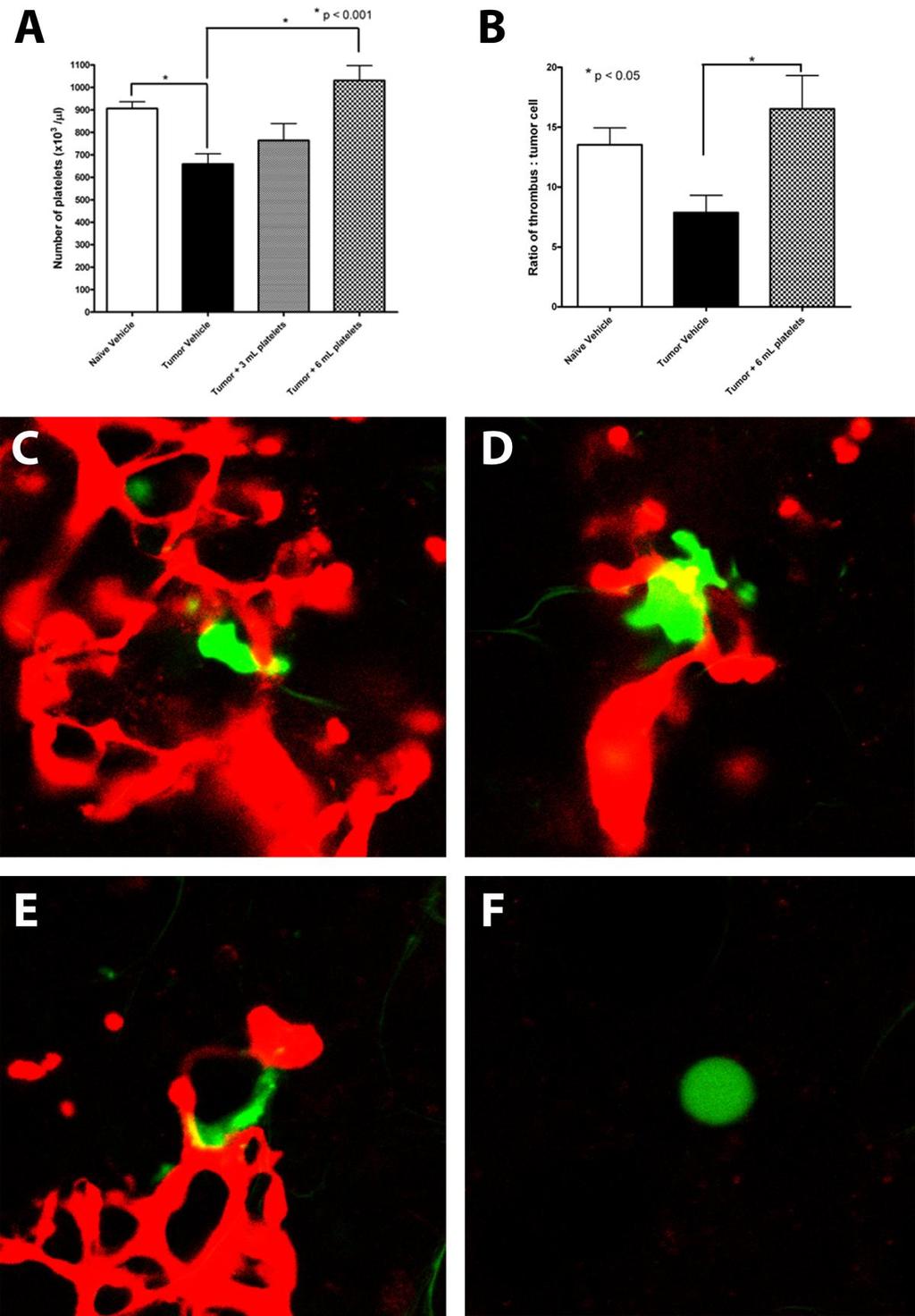
99 80
100 81 Figure 3.5 Reconstitution of platelet number in tumor-bearing animals reestablished lung metastasis formation. Tumor-naïve animals received an i.v. injection of platelet vehicle and tumor-bearing animals received an i.v. injection of platelet vehicle or isolated platelets prior to i.v. B16F10-LacZ cell injection (n = 12/group). Platelet vehicle injection into tumor-naïve animals reduced metastasis number as compared to control (un-injected naïve and primary data are historical controls from Fig 1A) (ANOVA, Tukey s post test, p < 0.05). Vehicle injection into tumor-bearing mice did not further reduce metastasis from that seen previously in tumor-bearing animals (p > 0.05). Platelet injection into tumorbearing mice showed an increase in lung metastasis as compared to vehicle injected tumor-bearing animals (p < 0.05) and restored lung metastasis number to that seen in tumor-naïve animals (p > 0.05). Columns, mean; bars, SE.
101 82 restoring lung metastasis of B16F10-LacZ cells and was able to counteract the effect of the presence of a B16F10 primary tumor (Figure 3.6). 3.5 Discussion In this study, we established that the presence of a B16F10 primary tumor was capable of significantly inhibiting the number and size of lung metastases arising from secondarily injected B16F10-LacZ cells. This inhibition of metastasis was found to be due to a primary-tumor mediated reduction in circulating platelet number, resulting in insufficient thrombus formation in the lung following metastatic cell injection. Restoration of circulating platelet number, through injection of platelets isolated from donor mice, reestablished thrombus formation in primary tumor bearing mice and allowed equivalent metastasis formation in both primary tumor-bearing and tumor-naïve animals. The negative impact of a B16F10 primary tumor on secondary metastasis was somewhat surprising given that the B16 cell line has been reported to stimulate pre-metastatic niche formation in the lung, liver, testis, spleen and kidney 30. Recruitment of BMDC to facilitate formation of a pre-metastatic niche is thought to aid metastasis formation, as VEGFR1 + cell present in the niche increase adhesion and arrest of metastatic tumor cells. Additionally, in vitro analysis found that B16 cells were increasingly mobile in response to VEGFR1 + cells 30. It is possible that B16F10 cells do not stimulate formation of the pre-metastatic niche as seen with the B16 cells used by Kaplan et al. 30, or that the reduction in platelet number in primary tumor-bearing animals is able to inhibit metastasis formation in spite of pre-metastatic niche presence. Development of concomitant tumor resistance has been found to occur via immunogenic (small tumors, immunogenic types) and non-immunogenic mechanisms (large tumors, anti-angiogenic or anti-mitotic mechanisms) 10, 31, 32, but the potential for a primary tumor to both inhibit and promote metastasis through concomitant tumor resistance and pre-metastatic niche formation requires further investigation. Importantly, in this study animals received the second injection of tumor cells when the primary tumor was relatively small (~530 mm 3 ) such that the previously identified anti-angiogenic and anti-mitotic mechanisms of concomitant tumor resistance found to occur after primary tumors reached 2000 mm 3 11
102 83 should not yet be involved. Additionally, immunogenic control is not thought to be responsible for the suppression of B16F10 metastases seen here, as B16 cells were derived from a spontaneously formed tumor and are poorly immunogenic 12. Direct analysis of tumor immunity in a B16 model found that it was induction of CD4 + CD25 + regulatory T cells that was responsible for the lack of prevented the development of concomitant tumor immunity in C57Bl/6 mice 12. It is well established that platelets play an integral role in metastatic establishment Following arrest in the vasculature, tumor cells stimulate thrombus formation at their cell surface 14, 16, 18. The interaction between tumor cells and thrombi provides valuable adhesion contacts 36, growth and survival factors and protection from host immune surveillance 16, 34. In pre-clinical studies, treatment of animals with an anti-coagulant such as heparin 14, 37 or hiruden 17 prior to metastatic cell injection results in significant reduction in pulmonary metastasis 38. Previous work in our laboratory also found that inhibition of serine-protease activity in vivo stabilized tumor cell-associated thrombi and increased B16F10 metastasis formation 14. Activated platelets were found to increase invasiveness of human ovarian cancer cells 39 and tumor cells that are able to stimulate clot formation are more likely to be retained in the pulmonary vasculature as they are more likely to form stable adhesive contacts and spread along the inside of the pulmonary vasculature 15. Induction of thrombocytopenia through injection of bacterial lipopolysaccharide or neuraminidase significantly decreases lung metastasis of both strongly and weakly immunogenic tumor types 40. Interestingly, injection of platelets into thrombocytopenic animals prior to i.v cell injection did not restore metastasis number, potentially due to insufficient platelet injection, as evidence for platelet number rescue was not presented 40. The direct role of platelets in establishment of metastases has been investigated in B16 and fibrosarcoma models with conflicting results. The positive correlation between platelet aggregation and metastatic ability found in the B16F10 cell line did not hold when individual B16F10 clones were generated and tested. 41 Similar clonal investigation with the PAK 17 fibrosarcoma cell line showed that highly metastatic clones require platelet interaction for successful metastasis. This disparity could be due to differences in analysis of platelet aggregating ability 42, but could indicate that the most aggressive B16F10 clones are capable of metastasis regardless of platelet interaction, due
103 84 to other malignant characteristics. For example, those clones that exhibit high metastatic ability without extensive platelet aggregation activity may form more extensive tumor cell-tumor cell associations allowing for similar protection from immune surveillance for the central cells as is provided by thrombus formation 21. Alternatively, these cells may indirectly stimulate platelet aggregation in vivo, which would not be readily detectable in vitro. In general these two studies identified that high platelet aggregating ability is not sufficient for metastasis of non-metastatic cells 41, and indicate that tumor cell-thrombus association is an important determinant in metastatic potential 42. Interestingly, the splenomegaly identified in tumor-bearing animals was associated with tri-lineage extramedullary hematopoiesis, with a preponderance of megakaryocytic precursors. A similar increase in hematopoiesis was not found in the bone marrow of tumor-bearing mice, which is in accordance with normal murine physiologic response. During stress, the majority of (reactive) hematopoiesis is performed in the murine spleen 27. Despite the increase in platelet manufacture in the spleen, the number of circulating platelets was significantly decreased in tumor-bearing mice, indicating that the spleen was unable to replenish the platelets being lost. Several possibilities exist for the cause of the platelet consumption and resulting thrombocytopenia in this case. B16F10 murine melanoma cells express TF on their cell surface 23, 43, therefore the cells within the tumor may activate circulating platelets resulting in thrombus formation at the primary tumor site. B16F10 primary tumors tend be hemorrhagic with a necrotic core void of much secondary structure, and can lead to extensive bleeding with even minor trauma (unpublished observation). In addition, and perhaps most importantly, the presence of a B16F10 primary tumor may stimulate disseminated intravascular coagulation. Disseminated intravascular coagulation in cancer is characterized by activation of the coagulation cascade in the defective tumor vasculature or by pro-coagulant activity on tumor cells, or their membrane components, and is the most common cause of thrombocytopenia in cancer patients 34, 44. Local fibrinogen and platelet consumption then lead to systemic deficiencies and increased clotting time. Although human patients may be somewhat less susceptible to tumorassociated thrombocytopenia (thrombocytosis is the most common blood abnormality in
104 85 cancer patients 34, 35 ), mice are known to compensate poorly for blood or platelet loss and easily develop anemia or thrombocytopenia following a minor insult 27. Interestingly, B16F10 cells have been found to generate large numbers of microvesicles (MV) in vitro; isolation and injection of these MV prior to i.v. injection of B16F10 cells has been linked to increased metastasis 45. Given the cell surface expression of TF on B16F10 cells, 23 it would be anticipated that these MV would also contain TF. Therefore, it is possible that injection of MV two-hours prior to injection of B16F10 cells 45 could result in an increase in tumor-cell associated thrombus formation, leading to an increase in metastasis. In the presence of an ongoing release of TF-containing MV from a B16F10 primary tumor, it would be anticipated that rather than a pro-metastatic effect of the MV, there would be sustained activation of the coagulation cascade leading to platelet exhaustion and/or depletion, as was seen here. Therefore the release of MV from the B16F10 primary tumor may be partially responsible for the significant reduction in platelet numbers. The integral role of coagulation in metastasis formation is further indicated by the effect of anti-coagulant therapy in cancer patients. Preliminary clinical trials have shown a significant increase in overall survival of patients randomized to receive LMWH versus placebo 46. Interestingly, long-term exposure to LMWH in patients with cancer-related thrombosis only increased survival of those patients who did not have metastatic disease at the time of study enrollment 46. Additionally, large cohort analysis of overall probability of death in those patients diagnosed with cancer as compared to those diagnosed with cancer and thromboembolism, found that approximately 20% of patients with cancer alone and 90% of patients with a combined diagnosis succumbed to their disease within 6 months. It has been indicated that these deaths could be attributed to three possible scenarios: 1) fatal recurrent pulmonary thromboembolism; 2) identification of a coagulation disorder may simply be a surrogate marker for more aggressive malignancy; 3) systemic aberrations in the coagulation pathway may result in a more permissive tumor cell-host interaction giving rise to extensive tumor growth resulting in the early death of the patient 46. Given these data, further analysis of modulation of the systemic coagulation pathway in cancer patients as a means to prevent metastasis establishment and premature death is warranted.
105 86 The development of metastases occurs in the midst of a complex interaction between a primary tumor and the host - the full extent of which remains unknown. There is the potential for a primary tumor to both promote metastatic progression through stimulation of pre-metastatic niche formation, as well as to restrict metastasis through concomitant tumor resistance. In this study, we identified a form of concomitant tumor resistance known as athrepsia, which was in this case due to primary tumor-induced thrombocytopenia and the resulting reduction in tumor-thrombus formation, with inhibition of metastasis. Full understanding of the interplay between a primary tumor, the host and forming metastases will be essential to the development of strategies to inhibit metastatic progression, either before or after the surgical resection of the primary tumor.
106 References 1. Chambers AF, Groom AC, MacDonald IC. Dissemination and growth of cancer cells in metastatic sites. Nat Rev Cancer 2002;2: Lorusso G, Ruegg C. The tumor microenvironment and its contribution to tumor evolution toward metastasis. Histochem Cell Biol 2008;130: Clark WH, Jr., Elder DE, Guerry Dt, Braitman LE, Trock BJ, Schultz D, Synnestvedt M, Halpern AC. Model predicting survival in stage I melanoma based on tumor progression. J Natl Cancer Inst 1989;81: Warren BA, Chauvin WJ, Philips J. Blood-borne tumor emboli and their adherance to vessel walls. In: Day SB, Laird Myers, W.P., Stansley, P., Garattini, S. Progress in Research and Therapyed., vol. 5. New York: Raven Press, 1977: Sugarbaker EV, Thornthwaite J, Ketcham AS. Inhibitory effect of a primary tumor on metastasis. In: Day SB, Laird Myers, W.P., Stansley, P., Garattini, S. Progress in Research and Therapyed., vol. 5. New York: Raven Press, 1977: Prehn RT. The inhibition of tumor growth by tumor mass. Cancer Res 1991;51: Gorelik E. Concomitant tumor immunity and the resistance to a second tumor challenge. Adv Cancer Res 1983;39: Bashford EF, Murray, J.A., and Cramer, W. The natural and induced resistance of mice to the growth of cancer. Proceedings of the Royal Society of London. Series B 1907;79: Folkman J. Angiogenesis inhibitors generated by tumors. Mol Med 1995;1: O'Reilly MS, Holmgren L, Shing Y, Chen C, Rosenthal RA, Moses M, Lane WS, Cao Y, Sage EH, Folkman J. Angiostatin: a novel angiogenesis inhibitor that mediates the suppression of metastases by a Lewis lung carcinoma. Cell 1994;79: Franco M, Bustuoabad OD, Di Gianni PD, Meiss RP, Vanzulli S, Buggiano V, Pasqualini CD, Ruggiero RA. Two different types of concomitant resistance induced by murine tumors: morphological aspects and intrinsic mechanisms. Oncol Rep 2000;7: Turk MJ, Guevara-Patino JA, Rizzuto GA, Engelhorn ME, Sakaguchi S, Houghton AN. Concomitant tumor immunity to a poorly immunogenic melanoma is prevented by regulatory T cells. J Exp Med 2004;200:
107 Falanga A, Panova-Noeva M, Russo L. Procoagulant mechanisms in tumour cells. Best Pract Res Clin Haematol 2009;22: Kirstein JM, Graham KC, Mackenzie LT, Johnston DE, Martin LJ, Tuck AB, MacDonald IC, Chambers AF. Effect of anti-fibrinolytic therapy on experimental melanoma metastasis. Clin Exp Metastasis 2009;26: Im JH, Fu W, Wang H, Bhatia SK, Hammer DA, Kowalska MA, Muschel RJ. Coagulation facilitates tumor cell spreading in the pulmonary vasculature during early metastatic colony formation. Cancer Res 2004;64: Palumbo JS, Talmage KE, Massari JV, La Jeunesse CM, Flick MJ, Kombrinck KW, Jirouskova M, Degen JL. Platelets and fibrin(ogen) increase metastatic potential by impeding natural killer cell-mediated elimination of tumor cells. Blood 2005;105: Esumi N, Fan D, Fidler IJ. Inhibition of murine melanoma experimental metastasis by recombinant desulfatohirudin, a highly specific thrombin inhibitor. Cancer Res 1991;51: Palumbo JS, Kombrinck KW, Drew AF, Grimes TS, Kiser JH, Degen JL, Bugge TH. Fibrinogen is an important determinant of the metastatic potential of circulating tumor cells. Blood 2000;96: Palumbo JS, Potter JM, Kaplan LS, Talmage K, Jackson DG, Degen JL. Spontaneous hematogenous and lymphatic metastasis, but not primary tumor growth or angiogenesis, is diminished in fibrinogen-deficient mice. Cancer Res 2002;62: Palumbo JS, Talmage KE, Liu H, La Jeunesse CM, Witte DP, Degen JL. Plasminogen supports tumor growth through a fibrinogen-dependent mechanism linked to vascular patency. Blood 2003;102: Palumbo JS, Talmage KE, Massari JV, La Jeunesse CM, Flick MJ, Kombrinck KW, Hu Z, Barney KA, Degen JL. Tumor cell-associated tissue factor and circulating hemostatic factors cooperate to increase metastatic potential through natural killer cell-dependent and-independent mechanisms. Blood 2007;110: Palumbo JS, Barney KA, Blevins EA, Shaw MA, Mishra A, Flick MJ, Kombrinck KW, Talmage KE, Souri M, Ichinose A, Degen JL. Factor XIII transglutaminase supports hematogenous tumor cell metastasis through a mechanism dependent on natural killer cell function. J Thromb Haemost 2008;6: Kirszberg C, Lima LG, Da Silva de Oliveira A, Pickering W, Gray E, Barrowcliffe TW, Rumjanek VM, Monteiro RQ. Simultaneous tissue factor expression and phosphatidylserine exposure account for the highly procoagulant pattern of melanoma cell lines. Melanoma Res 2009;19:301-8.
108 Trousseau A. In: Bailliere J, ed. Clinique Medicale de l'hotel-dieu de Paris, ed. 2nd, vol. 3 Paris, 1865: Gay LJ, Felding-Habermann B. Contribution of platelets to tumour metastasis. Nat Rev Cancer;11: Goring DR, Rossant J, Clapoff S, Breitman ML, Tsui LC. In situ detection of beta-galactosidase in lenses of transgenic mice with a gamma-crystallin/lacz gene. Science 1987;235: Everds NE. Hematology of the laboratory mouse. In: Fox JG, Davisson, M.T., Quimby, F.W., Barthold, S.W., Newcomer, C.E., Smith, A.L. The mouse is biomedical research, 2nd Edition, 2nd ed., vol. 3. Burlington, MA: Elsevier, Inc, 2007: Musaji A, Vanhoorelbeke K, Deckmyn H, Coutelier JP. New model of transient strain-dependent autoimmune thrombocytopenia in mice immunized with rat platelets. Exp Hematol 2004;32: Rasband WS. ImageJ Bethesda, Maryland, USA: National Institutes of Health, : 30. Kaplan RN, Riba RD, Zacharoulis S, Bramley AH, Vincent L, Costa C, MacDonald DD, Jin DK, Shido K, Kerns SA, Zhu Z, Hicklin D, et al. VEGFR1-positive haematopoietic bone marrow progenitors initiate the pre-metastatic niche. Nature 2005;438: Franco M, Bustuoabad OD, di Gianni PD, Goldman A, Pasqualini CD, Ruggiero RA. A serum-mediated mechanism for concomitant resistance shared by immunogenic and non-immunogenic murine tumours. Br J Cancer 1996;74: Ruggiero RA, Bustuoabad OD, Bonfil RD, Meiss RP, Pasqualini CD. "Concomitant immunity" in murine tumours of non-detectable immunogenicity. Br J Cancer 1985;51: Borsig L. The role of platelet activation in tumor metastasis. Expert Rev Anticancer Ther 2008;8: Green DL, Karpatkin S. Effect of cancer on platelets. Cancer Treat Res 2009;148: Nash GF, Turner LF, Scully MF, Kakkar AK. Platelets and cancer. Lancet Oncol 2002;3: McCarty OJ, Mousa SA, Bray PF, Konstantopoulos K. Immobilized platelets support human colon carcinoma cell tethering, rolling, and firm adhesion under dynamic flow conditions. Blood 2000;96:
109 Stevenson JL, Varki A, Borsig L. Heparin attenuates metastasis mainly due to inhibition of P- and L-selectin, but non-anticoagulant heparins can have additional effects. Thrombosis research 2007;120 Suppl 2:S Niers TM, Bruggemann LW, Klerk CP, Muller FJ, Buckle T, Reitsma PH, Richel DJ, Spek CA, Van Tellingen O, Van Noorden CJ. Differential effects of anticoagulants on tumor development of mouse cancer cell lines B16, K1735 and CT26 in lung. Clin Exp Metastasis 2009;26: Holmes CE, Levis JE, Ornstein DL. Activated platelets enhance ovarian cancer cell invasion in a cellular model of metastasis. Clin Exp Metastasis 2009;26: Kimoto M, Ando K, Koike S, Matsumoto T, Jibu T, Moriya H, Kanegasaki S. Significance of platelets in an antimetastatic activity of bacterial lipopolysaccharide. Clin Exp Metastasis 1993;11: Kimura AK, Mehta P, Xiang JH, Lawson D, Dugger D, Kao KJ, Lee- Ambrose L. The lack of correlation between experimental metastatic potential and platelet aggregating activity of B16 melanoma clones viewed in relation to tumor cell heterogeneity. Clin Exp Metastasis 1987;5: Mahalingam M, Ugen KE, Kao KJ, Klein PA. Functional role of platelets in experimental metastasis studied with cloned murine fibrosarcoma cell variants. Cancer Res 1988;48: Kirszberg C, Rumjanek VM, Monteiro RQ. Assembly and regulation of prothrombinase complex on B16F10 melanoma cells. Thromb Res 2005;115: Kvolik S, Jukic M, Matijevic M, Marjanovic K, Glavas-Obrovac L. An overview of coagulation disorders in cancer patients. Surg Oncol;19:e Lima LG, Chammas R, Monteiro RQ, Moreira ME, Barcinski MA. Tumorderived microvesicles modulate the establishment of metastatic melanoma in a phosphatidylserine-dependent manner. Cancer Lett 2009;283: Kakkar AK. Antithrombotic therapy and survival in cancer patients. Best Pract Res Clin Haematol 2009;22:
110 91 Chapter 4 4 Primary tumor presence restricts metastatic establishment in a murine model of human breast cancer 4.1 Synopsis Breast cancer is the most common cancer and the second most common cause of cancerrelated mortality in Canadian women. These deaths are not due to the presence of a primary tumor however, but are caused by the spread of cancer cells to sites throughout the body. These cells give rise to metastases that are present at the time of diagnosis, or lead to tumor recurrence and patient relapse. Analysis of breast cancer patient data acquired prior to the routine use of adjuvant therapies identified a pattern of breast cancer recurrence that was independent of disease stage at the time of surgery and appears synchronized with the time of tumor resection. It appears that removal of the primary tumor causes a simultaneous initiation of metastatic growth through the release of growth factors during the wound healing process, or that the primary tumor was somehow restricting the growth of metastases and upon tumor resection, metastatic growth is no longer suppressed. Alternatively, formation of a pre-metastatic niche, a cluster of bone marrow host cells that forms a supportive metastatic environment, is initiated by proteins released from a primary tumor. The pre-metastatic niche forms prior to the arrival of metastatic cells and leads to increased tumor cell adhesion and growth. The phenomena of pre-metastatic niche formation and primary tumor suppression of metastasis are contradictory and together they indicate that the interaction between a primary tumor, host systems, and metastatic cells is complex, and remains poorly understood. Previous work in our laboratory has shown that a murine melanoma primary tumor is able to restrict the development of secondary metastasis through alterations in systemic hemostatic factors (Chapter 3). Therefore, we sought to evaluate the effect of an established breast primary tumor on the early development of lung metastases to determine if the metastatic inhibition seen previously also exists in a model of human breast cancer
111 92 In this study, we investigated the effect of a breast primary tumor on the early development of lung metastases and found that tumor bearing animals developed significantly fewer lung metastases than tumor-naïve control animals. Tumor-bearing animals showed an increased number of VEGFR1 + cells in the lung, potentially indicating BMDC recruitment due to expression of OPN. Preliminary data to determine the mechanism of primary tumor-mediated inhibition of metastasis indicates that successful metastasis may depend on activation of the host hemostatic system which may be impaired in the presence of a primary tumor.
112 Introduction Breast cancer is estimated to account for 5400 deaths in Canada in These deaths are not due to the primary tumor, but rather because of the spread of tumor cells from the primary tumor to distant sites 2. Those tumors that have reached a relatively large size and/or have metastasized to several lymph nodes at the time of diagnosis carry a significantly higher risk of recurrence compared early-stage tumors 3. Interestingly, analysis of patient data accumulated prior to the routine use of adjuvant chemotherapy, has identified a two-peak pattern for recurrence 3, 4. When all patients are considered, regardless of tumor stage at diagnosis, there is a sharp peak of recurrences at 18 months post-surgery and another broad peak at 60 months 3. This recurrence pattern was identified in over 12 clinical studies, with varying peak amplitudes, but with an identical timeline 5, 6. More recent clinical data shows that this first peak no longer exists at 18 months, but rather, the addition of adjuvant chemo- and radiation therapy has shifted the major peak of recurrence to approximately 4 years 7. The similar patterns of recurrence in all patient groups regardless of disease stage at the time of surgery may indicate a synchronization of metastatic development upon removal of the primary tumor 5. This synchronization could be due to the release of growth factors during wound healing or a sudden removal of growth suppressive factors, such as angiostatin, with resection of the primary tumor 3, 5. Concomitant tumor resistance, the ability of a primary tumor to restrict the growth of secondary metastases, has been identified in several pre-clinical models of metastasis and is thought to occur through three potential mechanisms 8. First, the primary tumor may prime the immune system to assist clearance of metastatic cells. Second, the primary tumor may restrict the growth of distant metastases through production of anti-angiogenic molecules such as angiostatin or TSP 9. Third the primary tumor may systemically deplete essential host factors, thereby preventing the growth of any other tumors 8. The first hypothesis would not explain the clinical breast cancer data, as cells shed prior to tumor resection are not cleared by the immune system if they are able to cause recurrence at later time points. The second or third hypothesis could explain the clinical recurrence
113 94 pattern however. Removal of the primary tumor would eliminate the growth-suppressive molecule responsible for holding metastases in check 3 or could free up host factors essential for metastatic growth. In both scenarios, removal of the primary tumor would lead to a simultaneous growth initiation of distant metastases due to sudden removal or availability of the key factor(s). Alternatively, intriguing work published by McAllister, et al. 10 showed that a human breast cancer xenograft did not restrict the growth of other tumors or metastases, but rather was capable of stimulating the growth of an otherwise-indolent transformed mammary epithelial cell line (HMLER-HR). This growth stimulation was due to the activation and mobilization of BMDC by OPN secreted from the stimulating primary tumor. The activated BMDC colonized both the stimulating and indolent tumor sites which allowed for supportive tumor stroma development and increased tumor growth 10. As further indication that a primary tumor can have a systemic effect, BMDC stimulation by the presence of a primary tumor has also been described during pre-metastatic niche formation. In this case, BMDC mobilization and colonization of distant normal tissues occurs prior to the arrival of metastatic cells, and these BMDC develop into a supportive environment which aids the arrest and development of metastases The pre-clinical identification of pre-metastatic niche formation conflicts with the observation that a primary tumor may hold metastatic growth in check 6, 7. Together, these phenomena indicate that there is a complex interaction between a primary tumor, secondary metastases and the host environment which remains poorly understood. Previous work in our laboratory has shown that a murine melanoma primary tumor is able to restrict the development of secondary metastasis through alterations in systemic hemostatic factors (Chapter 3). Therefore, we sought to evaluate the effect of an established breast primary tumor on the early development of lung metastases to determine if the metastatic inhibition seen previously also exists in a model of human breast cancer. Using a variant of the human breast carcinoma MDA-MB-231 cell line with increased metastatic ability (231LN), we found that the presence of a mammary fat pad (mfp)
114 95 tumor reduced the number of lung metastases arising from a second intravenous injection. The size of lung metastases that did develop was not affected, indicating that the primary tumor may be affecting initial cell arrest or survival in the lung. Immunohistochemistry analysis of lung tissue found that tumor bearing animals showed increased presence of VEGFR1 + cells, indicating that 231LN cells may be capable of stimulating pre-metastatic niche formation. Despite the presence of the metastasissupporting VEGFR1 + cells, the number of lung metastases present in tumor-bearing animals was suppressed, therefore the inhibition of metastasis by the primary tumor overwhelms the supportive role of VEGFR1 + cells. Preliminary data to identify the mechanism behind the inhibition of metastasis by a primary tumor indicates that in accordance with previous work from our laboratory, successful metastasis may depend on activation of the host hemostatic system which may be impaired in the presence of a primary tumor. 4.3 Materials and Methods Cell culture and transfections MDA-MB-231-luc-D3H2LN (231LN) cells were purchased from Caliper Life Sciences (Hopkinton, MA). They are a variant of the MDA-MB-231 cell line which contain a luciferase-expressing gene and have a propensity to metastasize the lymph nodes, and have a high endogenous OPN production. They were maintained in MEM (Gibco) supplemented with 10% fetal bovine serum (FBS, Sigma, Mississauga, Canada), 1 mm sodium pyruvate (Gibco) and 0.1 mm non-essential amino acids (Gibco) at 37 C with 5% CO 2. Two cell lines were generated from the 231LN cells, one carried no fluorescence marker, the 231LN-scr cells and the other carried a stable red fluorescent tdtomato marker. 231LN-tdTomato cells were generated by nucleofection of the MDA- MB-231-luc-D3H2LN (231LN) with the tdtomato-containing pcdna3.1 vector with a hygromycin resistance gene, using Amaxa nucleofection technology (Amaxa, Koeln, Germany) according to manufacturer s instructions. Briefly, 1 x LN cells were resuspended in Cell Line Nucleofector Kit V (Amaxa), mixed with 2 μg cdna and pulsed with the program X-13, as suggested by the manufacturer. Immediately after nucleofection, cells were transferred into wells containing pre-warmed (37 C) culture
115 96 medium (as above). Forty-eight hours post-nucleofection, cells were selected using 750 μg/ml hygromycin (Gibco), to create a stably tdtomato-transfected cell line. All cells were routinely tested and confirmed to be free of mycoplasma contamination using the Mycoplasma Plus PCR primer set (Stratagene, Cedar Creek, TX) Primary tumor and metastasis assay Each animal used in this assay received a mfp injection of either 231LN-scr cells or cell vehicle, and all animals received an i.v. injection of 231LN-tdTomato to allow for separation of metastases arising from the primary tumor or i.v. injection. 231LN-scr cells were grown to 70-80% confluence, trypsinized and washed in cold Hanks Balanced Salt Solution (HBSS, Invitrogen). For mfp injection, cells were resuspended in a 50% Matrigel Matrix (BD Biosciences) solution with HBSS at a concentration of 4 x 10 7 cells/ml and 50 µl (2 x 10 6 cells) were injected. NIH III (nude-beige, Charles River, Massachusetts) mice were lightly sedated with an intraperitoneal injection of xylazine/ketamine (1.3 mg ketamine and.065 mg xylazine per 20 g body mass) and cells were into the right thoracic mammary fat pad without an incision. As illustrated in Figure 4.1, control animals received a mammary fat pad injection of 50% Matrigel matrix (BD Biosciences) solution with HBSS following the same procedure. Primary tumors were allowed to grow for 28 days, at which point all animals received a second cell injection. Unanesthetized tumor-bearing and tumor-naïve mice received an intravenous (i.v.) injection of 5 x LN-tdTomato cells in 100 l HBSS via the lateral tail vein. Animals were sacrificed 6 days following i.v. injection and lung tissue was isolated and snap-frozen to enable quantification of the number of lung surface metastases using fluorescence Quantification of lung metastasis The total number of red-fluorescent metastases was manually determined by a blinded observer on intact lung lobes using an Olympus Inverted IX81 microscope. Area of individual lung metastases was determined using the Olympus FluoView FV1000 coupled to the IX81 motorized inverted system microscope at the Victoria Research Laboratory confocal microscope core facility. Images were acquired with maintained
116 97 imaging parameters by a blinded observer and area of each metastasis was determined on fluorescent images using ImageJ Histology and Immunohistochemistry Lung tissues were fixed in 10 % neutral buffered formalin and embedded in paraffin. Sections were cut to 4 µm thickness and stained with H&E or with monoclonal rat antimouse VEGFR1 (R&D Systems, Minneapolis Mn; #MAB471,) diluted to 10 µg/ml for one hour at room temperature, after heat-induced epitope retrieval. Secondary detection was achieved by linking to biotinylated polyclonal rabbit anti-rat antibody (Dako, Burlington Ontario; #E0468) diluted to 1:500 for thirty five minutes, followed by labeling with StreptABComplex/HRP (Dako, Burlington Ontario; #K0377) and diaminobenzidine (DAB). Positively-stained cells were counted by a blinded, trained observer on an upright microscope on a 40x objective (Olympus BX45) Western blot analysis of conditioned media To confirm that OPN secretion from 231LN-scr cells did not differ from the parental 231LN cells, conditioned media was collected, concentrated and used in Western blots as previously described 16. Briefly, 231LN and 23LN-scr cells were grown to 70-80% confluency, trypsinized and 5 x 10 5 cells were plated on 100 mm dishes. Plates were incubated at 37 C with 5% CO 2 for 24 hours, then media was removed and plates washed twice with PBS. Five millilitres of OPTIMEM was added to each and plates were returned to incubator for a further 24 hours. OPTIMEM was drawn off and concentrated using Amicon Ultra centrifugal filters with a dalton molecular weight cut off (Millipore; Billerica, MA). Cells were harvested from plates and counted using a hemocytometer. Using the number of cells, volumes of conditioned media were calculated to represent a specified number of cells (1 x 10 5 ), allowing conditioned medium from equal numbers of cells to be loaded for Western blotting. Western blots were carried out using the BioRad Mini Protean II Cell system. Polyacrylamide gels (8%) were loaded with concentrated conditioned media with 5X reducing loading buffer. After electrophoresis, separated proteins were transferred to polyvinylidene fluoride (PVDF) membranes (Amersham, GE Healthcare; Baie d Urfe,
117 98 QC). Membranes were blocked with 5% milk in TBS-T (tris-buffered saline with 0.1% Tween) for one hour and after rinsing with TBS-T, were incubated with anti-opn mab 53 mouse monoclonal antibody (Assay Designs, Enzo Life Sciences; Plymouth, PA) at 1:1000 in TBS-T at 4 C overnight. After rinsing three times with TBS-T, anti-mouse (Amersham) secondary antibody was incubated at room temperature for one hour. After rinsing three times with TBS-T, membranes were incubated with ECL Plus Western Blotting Detection System (Amersham) and exposed to film in a dark room Flow cytometry To compare TF expression between the parental MDA-MB-231 (ATCC), and 231LN-scr and 231LN-tdTomato cell lines, all cells were grown to 70-80% confluency, trypsinized and washed twice with cold flow buffer (PBS + 2% FBS). Cells were resuspended in cold flow buffer (1 x 10 6 cells/ml) and separated into 100 µl aliquots. Cells were incubated with 3 µl anti-tissue factor antibody (FITC-conjugated anti-human tissue factor, 4508CJ American Diagnostica, Stamford, CT) or 4 µl control antibody to account for non-specific binding (FITC-conjugated Mouse IgG1, BD Biosciences, San Jose, CA) for 30 minutes with occasional vortexing. Cells were then washed twice with excess volume PBS, resuspended in cold flow buffer prior to analysis on an EPICS XL- MCL flow cytometer (Beckman Coulter, Mississauga, ON). A minimum of 1 x 10 5 FITC + events were counted per sample. 4.4 Results Primary tumor presence decreases lung metastasis To evaluate the effect of a human breast adenocarcinoma primary tumor on the early development of metastases, we utilized a modified model of experimental metastasis where animals received both a primary and secondary tumor cell injection (Figure 4.1 a). Animals first received a mammary fat pad injection of 231LN-scr cells (primary tumor group) or cell vehicle (tumor-naïve group). After twenty-eight days, when primary tumors had reached ~750 mm 3 (Figure 4.1 b), all animals received a secondary injection of 231LN-tdTomato cells via the lateral tail vein to target cells directly to the lung. The fluorescent label in 231LN-tdTomato cells enabled delineation of metastases arising from
118 99 a b Figure 4.1 Timeline of primary tumor and metastasis injection model. (a) Experimental model used to analyze the effect of a primary tumor on the early development of metastases. Animals first received a mammary fat pad injection of Matrigel (cell vehicle) or 231LN-scr cells suspended in Matrigel. Following primary tumor establishment all animals received an i.v. injection of 231LN-tdTomato cells. Sixdays following i.v. injections all animals were sacrificed and lung and primary tumor tissues were collected. (b) Primary tumor growth of 231LN-scr cells. Animals received an i.v. injection of 231LN-tdTomato cells on day 28 of tumor growth when primary tumors were approximately 750 mm 3.
119 100 the i.v. injection. We found that the presence of a primary tumor significantly inhibited experimental lung metastasis, as six days following secondary injection there were fewer red-fluorescent lung surface metastases in primary tumor-bearing mice as compared to tumor-naïve animals (Figure 4.2 a). There was no difference in the size of those lung metastases that did form in the primary tumor-bearing and tumor-naïve group (Figure 4.2 b). Histological analysis of lung tissue identified single tumor cells and small clusters of tumor cells in both tumor-naïve and tumor-bearing animals (Figure 4.2 c and d), in agreement with the small size of metastases determined using fluorescence microscopy BMDC recruitment to the lung Previous reports have shown that expression of OPN from a primary tumor can stimulate mobilization of BMDC to colonize primary tumor stroma 10. It has also been shown that proteins secreted from a primary tumor can stimulate VEGFR1 + BMDC recruitment to form the pre-metastatic niche, which may lead to increased metastatic cell arrest and growth 12, 17. Therefore, we performed immunohistochemistry to determine if VEGFR1 + cells were present in the lung of primary tumor-bearing or tumor-naïve animals and if these cells were found in association with 231LN-tdTomato cells. Quantification of immunohistochemistry slides found significantly more VEGFR1 + cells in lung tissue of primary tumor-bearing animals as compared to tumor-naïve controls (Figure 4.3 a). Additionally, large clusters of more than fifteen VEGFR1 + cells were found in primary tumor-bearing animals, potentially indicating the presence of a pre-metastatic niche (Figure 4.3 b). These large clusters were not visualized in control lung tissue. Tumor cells were found associated within the clusters of VEGFR1 + cells, either in small groups of breast cancer cells or as single tumor cells in both primary tumor-bearing (Figure 4.3 c) and tumor-naïve animals (Figure 4.3 d) OPN expression by 231LNscr cells To determine if the increased presence of VEFGR1 + cells in the lung was due to secretion of the BMDC-stimulating protein, OPN, Western blot analysis of conditioned media samples was performed. It was found that 231LN-scr cells maintained a similarly-high level of OPN secretion as the 231LN cells after extended in vitro culture (Figure 4.4),
 The presence of a 231LN-scr primary tumor significantly reduced the number of 231LN-tdTomato lung metastases as compared to")
120 101 a b c d Figure 4.2 Primary tumor presence significantly reduced pulmonary metastasis (a) The presence of a 231LN-scr primary tumor significantly reduced the number of 231LN-tdTomato lung metastases as compared to tumor-naïve controls (n = 13-15/group; t-test, p < 0.05). (b) Lung metastases in primary tumor-bearing animals were equivalent in size to their tumor-naïve controls (n = 13-15/group; t-test, p > 0.05). (c) Histological analysis showed isolated tumor cells (arrowhead) or (d) small clusters of tumor cells (outline) in both primary tumor-bearing and tumor-naïve animals. Scale 50 µm.
 Quantification of immunohistochemistry staining for VEGFR1 identified a significant increase in the number of VEGFR1 + cells in the lung of primary tumorbearing animals (n = 8/group, t-test p < 0.")
121 102 a b c d Figure 4.3 Primary tumor presence increases the number of VEGFR1 + cells in the lung. (a) Quantification of immunohistochemistry staining for VEGFR1 identified a significant increase in the number of VEGFR1 + cells in the lung of primary tumorbearing animals (n = 8/group, t-test p < 0.005). Large clusters of more than 15 VEGFR1 + cells were only visualized in primary tumor-bearing mice (b) and tumor cells (arrow heads) were found associated within the clusters of VEGFR1 + cells in both primary tumor-bearing (c) and tumor-naïve animals (d).
122 103 therefore the increased presence of VEGFR1 + cells may be due to BMDC recruitment stimulated by OPN secretion. Interestingly, despite the increased presence of potentially metastasis-promoting VEGFR1 + cells 17, primary tumor-bearing animals showed reduced metastasis (Figure 4.1 a) Tissue Factor expression Previous work in our laboratory has shown that a B16F10 murine melanoma primary tumor inhibits metastatic establishment through depletion of circulating platelet numbers (Chapter 3). High levels of TF on the surface of B16F10 cells 18 results in thrombus formation following arrest of these cells in the lung, which is essential for metastatic spread (Chapter 3). Therefore, expression of TF on 231LN-scr and 231LNtdTomato cells was evaluated by flow cytometry. It was found that in accordance with previously published MDA-MB-231 parental cell data 19, MDA-MB-231 cells (Figure 4.5 a) as well as both the 231LN-scr (Figure 4.5 b) and 231LN-tdTomato (Figure 4.5 c) cell lines indeed express TF, indicating that these cells should be capable of stimulating thrombus formation, and successful thrombus formation may play a key role in the establishment of lung metastases, as has been found previously for MDA-MB Discussion The ability of a primary tumor to impact the development of metastases can take two forms. Through stimulation of a pre-metastatic niche a tumor can increase metastatic establishment, or through concomitant tumor resistance a tumor can prevent successful metastasis. In this study, we determined that the presence of a 231LN-scr mammary fat pad primary tumor significantly inhibited the early establishment of 231LN-tdTomato lung metastases. This inhibition is likely due to a decrease in sustained cell arrest or survival in the lung vasculature rather than an inhibition of growth, as the size of those metastases that did develop was unaltered by the presence of a primary tumor. As has been demonstrated with B16 cells 17, the 231LN-scr primary tumor led to a significant increase in the number of VEGFR1 + cells in the lung, potentially due to OPN secretion and stimulation of BMDC recruitment. The presence of VEGFR1 + cells has previously
123 104 been reported to increase lung metastasis through initiation of pre-metastatic niche formation, yet in this study, the presence of a primary tumor inhibited metastasis. The mechanism behind the significant decrease in metastasis remains unclear, but appears to override the metastasis-stimulating effect of VEGFR1 + cells. As outlined in Chapter 3, the presence of a B16F10 melanoma primary tumor was found to inhibit lung metastasis formation due to a significant reduction in the circulating platelet numbers in tumor-bearing animals. This led to an impairment of tumor cell surface-thrombus formation following i.v. injection of B16F10-LacZ cells. Restoration of the platelet number re-established thrombus formation as well as lung metastasis. Given that the B16F10 18, 231LN-scr, and 231LN-tdTomato cells all express TF on the cell surface and the importance of thrombus formation in successful B16F10 metastasis, it is possible that thrombus formation may play a key role in metastasis of 231LN cells. The presence of a 231LN-scr primary tumor may result in a similar depletion of circulating platelets as seen in the B16F10 model. Analysis of circulating platelet numbers in 231LN-scr tumor bearing animals is ongoing in our laboratory. The inability to form a thrombus during metastasis in the B16F10 model system may lead to an increase in NK-mediated cell killing 20 as C57Bl/6 mice are fully immunocompetent. In the 231LN model however, the NIH III animals do not have functional T-cells, B-cells or NK cells 21, making it improbable that increased immune surveillance in the absence of thrombus formation leads to the reduction in metastasis seen here. Thrombus formation is also known to result in increased stable cell adhesion in the lung 22, 23, therefore the lack of thrombus formation may result in an increased number of cells passing through the lung capillary bed or increased cell death due to anoikis from lack of stable adhesion 24. Unlike other examples of concomitant tumor resistance where the release of antiangiogenic molecules restricts metastatic growth 9, it is not anticipated that inhibition of angiogenesis is responsible for the decrease in metastasis seen here. First, there was no size difference between metastases quantified in the tumor-bearing and tumor-naïve animals. Second, metastases found in both primary tumor-bearing and tumor-naïve animals were very early in development and were less than 1 mm in diameter and therefore have not reached a size where they need to recruit their own blood supply 25, 26.
124 105 Additionally, previous work in a fibroscarcoma model has found a biphasic pattern of concomitant tumor resistance 27. It was found that an immunogenic tumor (MC-C fibrosarcoma) is capable of stimulating increased immune surveillance of metastatic cells when the primary tumor is less than 500 mm 3 and is capable of restricting metastatic growth through inhibition of angiogenesis when the same tumor was greater than 2000 mm Primary tumors between 500 and 1500 mm 3 had no effect on metastatic progression 27. In the 231LN model used here, primary tumors were approximately 750 mm 3 at the time of the second injection and therefore were within the range when no significant effect on metastasis was identified previously. It is unknown if the MC-C fibrosarcoma cell line expresses TF or if it is highly thrombogenic, though a difference in TF expression may account for the differential pattern of inhibition seen between the MC-C and the B16F10 (Chapter 3) and the 231LN-scr cell lines. The MDA-MB-231 cell line has previously been shown to stimulate BMDC migration to tumor sites 10, and to the pre-metastatic niche 12. Expression of OPN from the MDA-MB- 231 cell line was found to mobilize BMDCs and stimulated their colonization of primary tumor sites, which led to growth instigation in an otherwise-indolent cell line 10. Additionally, expression of lysyl oxidase by a MDA-MB-231 primary tumor led to increased collagen IV crosslinking and increased CD11b + myeloid cell recruitment to the lung 12. These essential components of the pre-metastatic niche increased metastatic cell recruitment and tumor cell invasion 12. Here, we found a significant increase in VEGFR1 + cells in the lung of primary tumor-bearing animals, but this increase was unable to promote metastasis, or was unable to overcome the inhibitory influence of the primary tumor. Histological analysis identified the presence of VEGFR1 + cells in both tumor-naïve and primary tumor-bearing animals, and association of these cells with tumor cells in both animal groups. Larger numbers of VEGFR1 + cells and the presence of clusters of more than fifteen VEGFR1 + cells were only identified in primary tumor-bearing animals, potentially indicating the presence of a pre-metastatic niche. This niche formation could be due to expression of OPN from the 231LN-scr primary tumor. Further analysis of

125 LN 231LN-scr 95 kda 70 kda Figure 4.4 Western blot analysis of conditioned media from 231LN and 231LN-scr cells. 23lLN and 231LN-scr cells were cultured in vitro for 4-weeks prior to isolation of conditioned media to determine secreted OPN levels. Control transfection with a scrambled shrna sequence does not appear to have affected OPN expression, as similar levels of OPN secretion were found in the 231LN and 231-scr cell conditioned media. Multiple bands of OPN protein are present between 70 and 95 kda.
, 231LN-scr (b) and 231LN-tdTomato (c) cells express high levels of TF.")
126 107 a b c TF - FITC Log fluorescence Figure 4.5 Flow cytometry analysis of TF expression. Analysis of cell surface expression of TF by flow cytometry showed that all MDA-MB- 231 (a), 231LN-scr (b) and 231LN-tdTomato (c) cells express high levels of TF. Grey curves represent control IgG1 antibody; black curves represent TF-specific antibody.
Part IV Antithrombotics, Anticoagulants and Fibrinolytics
 Part IV Antithrombotics, Anticoagulants and Fibrinolytics "The meaning of good and bad, of better and worse, is simply helping or hurting" Emerson Chapter 16: Blood Coagulation and Fibrinolytic System
Hemostasis and Thrombosis
 Hemostasis Hemostasis and Thrombosis Normal hemostasis is a consequence of tightly regulated processes that maintain blood in a fluid state in normal vessels, yet also permit the rapid formation of a hemostatic
1. The metastatic cascade. 3. Pathologic features of metastasis. 4. Therapeutic ramifications. Which malignant cells will metastasize?
 1. The metastatic cascade 3. Pathologic features of metastasis 4. Therapeutic ramifications Sir James Paget (1814-1899) British Surgeon/ Pathologist Paget s disease of Paget s disease of the nipple (intraductal
Ch. 45 Blood Plasma proteins, Coagulation and Fibrinolysis Student Learning Outcomes: Describe basic components of plasma
 Chapt. 45 Ch. 45 Blood Plasma proteins, Coagulation and Fibrinolysis Student Learning Outcomes: Describe basic components of plasma Inheritance of X-linked gene for Factor VIII hemophilia A Explain the
1.The metastatic cascade. 2.Pathologic features of metastasis. 3.Therapeutic ramifications
 Metastasis 1.The metastatic cascade 2.Pathologic features of metastasis 3.Therapeutic ramifications Sir James Paget (1814-1899) British Surgeon/ Pathologist Paget s disease of bone Paget s disease of the
Tissue repair. (3&4 of 4)
") Tissue repair (3&4 of 4) What will we discuss today: Regeneration in tissue repair Scar formation Cutaneous wound healing Pathologic aspects of repair Regeneration in tissue repair Labile tissues rapid
Tissue repair (3&4 of 4) What will we discuss today: Regeneration in tissue repair Scar formation Cutaneous wound healing Pathologic aspects of repair Regeneration in tissue repair Labile tissues rapid
Neoplasia 18 lecture 8. Dr Heyam Awad MD, FRCPath
 Neoplasia 18 lecture 8 Dr Heyam Awad MD, FRCPath ILOS 1. understand the angiogenic switch in tumors and factors that stimulate and inhibit angiogenesis. 2. list the steps important for tumor metastasis
Inflammatory Cells and Metastasis
 Inflammatory Cells and Metastasis Experimentelle Krebsforschung SS 07 Gerhard Christofori Institute of Biochemistry and Genetics Department of Clinical-Biological Sciences Center of Biomedicine University
UNIT VI. Chapter 37: Platelets Hemostasis and Blood Coagulation Presented by Dr. Diksha Yadav. Copyright 2011 by Saunders, an imprint of Elsevier Inc.
 UNIT VI Chapter 37: Platelets Hemostasis and Blood Coagulation Presented by Dr. Diksha Yadav Hemostasis: Prevention of Blood Loss Vascular constriction Formation of a platelet plug Formation of a blood
Tissue renewal and Repair. Nisamanee Charoenchon, PhD Department of Pathobiology, Faculty of Science
 Tissue renewal and Repair Nisamanee Charoenchon, PhD Email: nisamanee.cha@mahidol.ac.th Department of Pathobiology, Faculty of Science Topic Objectives 1. Describe processes of tissue repair, regeneration
Blood clotting. Subsequent covalent cross-linking of fibrin by a transglutaminase (factor XIII) further stabilizes the thrombus.
 further stabilizes the thrombus.") Blood clotting It is the conversion, catalyzed by thrombin, of the soluble plasma protein fibrinogen (factor I) into polymeric fibrin, which is deposited as a fibrous network in the primary thrombus. Thrombin
Blood clotting It is the conversion, catalyzed by thrombin, of the soluble plasma protein fibrinogen (factor I) into polymeric fibrin, which is deposited as a fibrous network in the primary thrombus. Thrombin
This slide belongs to iron lecture and it is to clarify the iron cycle in the body and the effect of hypoxia on erythropoitein secretion
 This slide belongs to iron lecture and it is to clarify the iron cycle in the body and the effect of hypoxia on erythropoitein secretion Topics of today lectures: Hemostasis Meaning of hemostasis Mechanisms
Hemostasis. Learning objectives Dr. Mária Dux. Components: blood vessel wall thrombocytes (platelets) plasma proteins
 plasma proteins") Hemostasis Learning objectives 14-16 Dr. Mária Dux Components: blood vessel wall thrombocytes (platelets) plasma proteins Hemostatic balance! procoagulating activity anticoagulating activity 1 Thrombocytes
Hemostasis Learning objectives 14-16 Dr. Mária Dux Components: blood vessel wall thrombocytes (platelets) plasma proteins Hemostatic balance! procoagulating activity anticoagulating activity 1 Thrombocytes
Blood coagulation and fibrinolysis. Blood clotting (HAP unit 5 th )
") Blood coagulation and fibrinolysis Blood clotting (HAP unit 5 th ) Vessel injury Antithrombogenic (Favors fluid blood) Thrombogenic (Favors clotting) 3 Major systems involved Vessel wall Endothelium ECM
Blood coagulation and fibrinolysis Blood clotting (HAP unit 5 th ) Vessel injury Antithrombogenic (Favors fluid blood) Thrombogenic (Favors clotting) 3 Major systems involved Vessel wall Endothelium ECM
Seeds and soil theory by Stephen Paget at the end of the XIX century.
 Seeds and soil theory by Stephen Paget at the end of the XIX century. In The Distribution Of Secondary Growths In Cancer Of The Breast Paget presents and analyzes 735 fatal cases of breast cancer, complete
Disseminated Intravascular Coagulation. M.Bahmanpour MD Assistant professor IUMS
 به نام خدا Disseminated Intravascular Coagulation M.Bahmanpour MD Assistant professor IUMS Algorithm for Diagnosis of DIC DIC Score factor score Presence of known underlying disorder No= 0 yes=2 Coagolation
Chapter 19 Blood Lecture Outline
 Chapter 19 Blood Lecture Outline Cardiovascular system Circulatory system Blood 1. distribution 2. regulation 3. protection Characteristics: ph 7.4 38 C 4-6 L Composition: Plasma Formed elements Erythrocytes
Hemodynamic Disorders, Thromboembolic Disease, and Shock
 Hemodynamic Disorders, Thromboembolic Disease, and Shock Kumar et al: Robbins & Cotran Pathologic Basis of Disease 7E Figure 4-1 Factors affecting fluid balance across capillary walls. Capillary hydrostatic
Cancer Biology Course. Invasion and Metastasis
 Cancer Biology Course Invasion and Metastasis 2016 Lu-Hai Wang NHRI Cancer metastasis Major problem: main reason for killing cancer patients, without it cancer can be cured or controlled. Challenging questions:
Blood Lecture Test Questions Set 2 Summer 2012
 Blood Lecture Test Questions Set 2 Summer 2012 1. Leukocytes are attracted to a site of injury or disease by: a. diapedesis b. chemotaxis c. leukocytosis d. heparin e. leukomotosis 2. Leukocytes leave
ACTIVATION OF T LYMPHOCYTES AND CELL MEDIATED IMMUNITY
 ACTIVATION OF T LYMPHOCYTES AND CELL MEDIATED IMMUNITY The recognition of specific antigen by naïve T cell induces its own activation and effector phases. T helper cells recognize peptide antigens through
Ann F. Chambers, PhD. Canada Research Chair in Oncology. Cancer Program Director, Translational Breast Cancer Research Unit London Ontario Canada
 Imaging g cells as they metastasize e Ann F. Chambers, PhD Canada Research Chair in Oncology Distinguished Oncology Scientist, London Regional Cancer Program Director, Translational Breast Cancer Research
Lymphoid System: cells of the immune system. Answer Sheet
 Lymphoid System: cells of the immune system Answer Sheet Q1 Which areas of the lymph node have most CD3 staining? A1 Most CD3 staining is present in the paracortex (T cell areas). This is towards the outside
Heterotypy and Angiogenesis
 Heterotypy and Angiogenesis Tumors are perpetual wounds 1. Normally stroma and epithelia converse at a distance. 2. Juxtaposition of stroma and epithelia is indicative of tissue damage. 4. Activate strategies
Anatomy and Physiology
 Anatomy and Physiology For The First Class 2 nd Semester Thrombocytes = Platelets Thrombocytes = Platelets Blood platelets are non-nucleated disc like cell fragments 2-4 µm in diameter. Platelets are not
Chapter 19. Hemostasis
 Chapter 19 Hemostasis Hemostasis Hemostasis is the cessation of bleeding stopping potentially fatal leaks important in small blood vessels not effective in hemorrhage excessive bleeding from large blood
Topics of today lectures: Hemostasis
 Topics of today lectures: Hemostasis Meaning of hemostasis Mechanisms of hemostasis - Vascular contraction - Platelets plug - Blood coagulation (clotting) - Structure and functions of platelets - Blood
Real-time imaging reveals the single steps of brain metastasis fo mation r
 Real-time imaging reveals the single steps of brain metastasis fo mation r Yvonne Kienast, Louisa von Baumgarten, Martin Fuhrmann, Wolfgang E.F. Klinkert, Roland Goldbrunner, Jochen Herms and Frank Winkler
SIBLINGs, cancer's multifunctional weapons
 SIBLINGs, cancer's multifunctional weapons 6/18/08 Akeila Bellahcène and Vincent Castronovo of the Metastasis Research laboratory of the University of Liège are among the first researchers to have discovered
Primary Exam Physiology lecture 5. Haemostasis
 Primary Exam Physiology lecture 5 Haemostasis Haemostasis Body s response for the prevention and cessation of bleeding. Broadly consists of: Primary Haemostasis - vascular spasm and platlet plug formation
The Role of Microenvironment in the Control of Tumor Angiogenesis
 The Role of Microenvironment in the Control of Tumor Angiogenesis Domenico Ribatti The Role of Microenvironment in the Control of Tumor Angiogenesis Domenico Ribatti Department of Basic Medical Sciences,
10. Which of the following immune cell is unable to phagocytose (a) neutrophils (b) eosinophils (c) macrophages (d) T-cells (e) monocytes
 neutrophils (b) eosinophils (c) macrophages (d) T-cells (e) monocytes") Chapter 2. Acute and chronic inflammation(6): 1. In acute inflammation, which events occur in the correct chronological order? (Remembered from 2000, 2004 exam.) p50 (a) transient vasoconstriction, stasis
Chapter 2. Acute and chronic inflammation(6): 1. In acute inflammation, which events occur in the correct chronological order? (Remembered from 2000, 2004 exam.) p50 (a) transient vasoconstriction, stasis
Physiology of. The Blood hemostasis. By prof. Israa f. jaafar
 Physiology of The Blood hemostasis By prof. Israa f. jaafar Learning objectives Understand the Platelet structure and function Explane the Platelet production Understand the phases of hemostasis: vascular
The Angiopoietin Axis in Cancer
 Ang2 Ang1 The Angiopoietin Axis in Cancer Tie2 An Overview: The Angiopoietin Axis Plays an Essential Role in the Regulation of Tumor Angiogenesis Growth of a tumor beyond a limiting size is dependent upon
Effect of a nutrient mixture on the localization of extracellular matrix proteins in HeLa human cervical cancer xenografts in female nude mice
 Effect of a nutrient mixture on the localization of extracellular matrix proteins in HeLa human cervical cancer xenografts in female nude mice Publication from the Dr. Rath Research Institute Experimental
INFLAMMATION & REPAIR
 INFLAMMATION & REPAIR Lecture 7 Chemical Mediators of Inflammation Winter 2013 Chelsea Martin Special thanks to Drs. Hanna and Forzan Course Outline i. Inflammation: Introduction and generalities (lecture
Tumor Associated Macrophages as a Novel Target for Cancer Therapy
 Tumor mass Tumor Associated Macrophage Tumor Associated Macrophages as a Novel Target for Cancer Therapy This booklet contains forward-looking statements that are based on Amgen s current expectations
Cytokine Arrays Reveal Black Ops Tactics of Tumor-induced Immunosuppression
 Cytokine Arrays Reveal Black Ops Tactics of Tumor-induced Immunosuppression Jarad J Wilson, Ph.D. Technical Support & Marketing Specialist Ruo-Pan Huang, MD, Ph.D. Founder and CEO What are Antibody Arrays?
!!! Oncology for Scientists (RPN 530) Metastasis and Angiogenesis Chapter 13 and 14 Oct. 28 th 2014
 Metastasis and Angiogenesis Chapter 13 and 14 Oct. 28 th 2014") Oncology for Scientists (RPN 530) Metastasis and Angiogenesis Chapter 13 and 14 Oct. 28 th 2014 Department of Cancer Genetics Masashi Muramatsu, Ph.D. About Exam First, think and understand the concepts.
Oncology for Scientists (RPN 530) Metastasis and Angiogenesis Chapter 13 and 14 Oct. 28 th 2014 Department of Cancer Genetics Masashi Muramatsu, Ph.D. About Exam First, think and understand the concepts.
Cytokines, adhesion molecules and apoptosis markers. A comprehensive product line for human and veterinary ELISAs
 Cytokines, adhesion molecules and apoptosis markers A comprehensive product line for human and veterinary ELISAs IBL International s cytokine product line... is extremely comprehensive. The assays are
What are blood clots?
 What are blood clots? Dr Matthew Fay GP Principal The Willows Medical Practice- Queensbury GPwSI and Co-Founder Westcliffe Cardiology Service GP Partner Westcliffe Medical Group Created 5/31/18 Dr. Matthew
Hemodynamic Disorders, Thrombosis, and Shock. Richard A. McPherson, M.D.
 Hemodynamic Disorders, Thrombosis, and Shock Richard A. McPherson, M.D. Edema The accumulation of abnormal amounts of fluid in intercellular spaces of body cavities. Inflammation and release of mediators
PHM142 Lecture 4: Platelets + Endothelial Cells
 PHM142 Lecture 4: Platelets + Endothelial Cells 1 Hematopoiesis 2 Platelets Critical in clotting - activated by subendothelial matrix proteins (e.g. collagen, fibronectin, von Willebrand factor) and thrombin
Oral Anticoagulant Drugs
 Oral Anticoagulant Drugs Spoiled sweet clover caused hemorrhage in cattle(1930s). Substance identified as bishydroxycoumarin. Initially used as rodenticides, still very effective, more than strychnine.
Healing & Repair. Tissue Regeneration
 Healing & Repair Dr. Srikumar Chakravarthi Repair & Healing: Are they same? Repair :Regeneration of injured cells by cells of same type, as with regeneration of skin/oral mucosa (requires basement membrane)
Hemostasis and. Blood Coagulation
 Hemostasis and Blood Coagulation Events in Hemostasis The term hemostasis means prevention of blood loss. Whenever a vessel is severed or ruptured, hemostasis is achieved by several mechanisms: (1) vascular
number Done by Corrected by Doctor Maha Shomaf
 number 21 Done by Ahmad Rawajbeh Corrected by Omar Sami Doctor Maha Shomaf Ability to Invade and Metastasize The metastatic cascade can be subdivided into two phases: 1-invasion of ECM and vascular dissemination:
Matrixnedbrydning ved
 Matrixnedbrydning ved cancerinvasion -Biokemiske aspekter Ekstracellulære Proteaser og endocytosereceptorer Niels Behrendt The Finsen Laboratory Rigshospitalet Copenhagen Nødvendige begivenheder i metastasering
-Hashim ahmed is the one who wrote this sheet. I just edited it according to our record.
 * Subjects of this lecture : - Hemostasis - Platelets, general information, their ultrastructure and role in hemostasis. - Definitions: Thrombus, Embolus, Arteriosclerosis and Atherosclerosis. *NOTE: Prof
Chapter 2 Interactions of Normal Tissues and Systems with Metastatic Cells: Impact on Location, Survival and Growth
 Chapter 2 Interactions of Normal Tissues and Systems with Metastatic Cells: Impact on Location, Survival and Growth Jennifer M. Kirstein and Ann F. Chambers 2.1 Introduction Tumor formation is not a cell
Page 1 of 6 THROMBOCYTES
 Page 1 of 6 THROMBOCYTES Platelets are not cells in the strict sense. About one-fourth the diameter of a lymphocyte, they are cytoplasmic fragments of extraordinarily large cells (up to 60 µm in diameter)
HEME 10 Bleeding Disorders
 HEME 10 Bleeding Disorders When injury occurs, three mechanisms occur Blood vessels Primary hemostasis Secondary hemostasis Diseases of the blood vessels Platelet disorders Thrombocytopenia Functional
CANCER IMMUNOPATHOLOGY. Eryati Darwin Faculty of Medicine Andalas University
 CANCER IMMUNOPATHOLOGY Eryati Darwin Faculty of Medicine Andalas University Padang 18 Mei 2013 INTRODUCTION Tumor: cells that continue to replicate, fail to differentiate into specialized cells, and become
Chapter 19: Cardiovascular System: Blood
 Chapter 19: Cardiovascular System: Blood I. Functions of Blood A. List and describe the seven major homeostatic functions of blood: 1. 2. 3. 4. 5. 6. 7. II. Plasma A. Composition 1. It is a fluid consisting
Oncolytic Virotherapy: Targeting Cancer Stem Cells
 Oncolytic Virotherapy: Targeting Cancer Stem Cells Cancer Stem Cells (CSCs) or Cancer Initiating Cells (CICs) A consensus of five defining criteria has been established to affirm the existence of CICs:
In vitro scratch assay: method for analysis of cell migration in vitro labeled fluorodeoxyglucose (FDG)
") In vitro scratch assay: method for analysis of cell migration in vitro labeled fluorodeoxyglucose (FDG) 1 Dr Saeb Aliwaini 13/11/2015 Migration in vivo Primary tumors are responsible for only about 10%
In vitro scratch assay: method for analysis of cell migration in vitro labeled fluorodeoxyglucose (FDG) 1 Dr Saeb Aliwaini 13/11/2015 Migration in vivo Primary tumors are responsible for only about 10%
PHASES OF HAEMOSTASIS
 HAEMOSTASIS Maintains the integrity of a closed, highpressure circulatory system after vascular damage Vessel Wall Injury events in the vessel wall and in the blood which seal breach Delicate balance exists
ACTIVATION AND EFFECTOR FUNCTIONS OF CELL-MEDIATED IMMUNITY AND NK CELLS. Choompone Sakonwasun, MD (Hons), FRCPT
, FRCPT") ACTIVATION AND EFFECTOR FUNCTIONS OF CELL-MEDIATED IMMUNITY AND NK CELLS Choompone Sakonwasun, MD (Hons), FRCPT Types of Adaptive Immunity Types of T Cell-mediated Immune Reactions CTLs = cytotoxic T lymphocytes
ACTIVATION AND EFFECTOR FUNCTIONS OF CELL-MEDIATED IMMUNITY AND NK CELLS Choompone Sakonwasun, MD (Hons), FRCPT Types of Adaptive Immunity Types of T Cell-mediated Immune Reactions CTLs = cytotoxic T lymphocytes
Haemostasis & Coagulation disorders Objectives:
 Haematology Lec. 1 د.ميسم مؤيد علوش Haemostasis & Coagulation disorders Objectives: - Define haemostasis and what are the major components involved in haemostasis? - How to assess the coagulation status?
Branch of medicine that deals with blood, its formation and disorders is called. Three main functions of cardiovascular system are,, and.
 Chapter 19 The Blood Human body must maintain a balance called. Body fluid inside the cells is called fluid; that outside is called or fluid. Two major fluid networks that help in connecting cells are
Invasion And Metastasis. Wirsma Arif Harahap Surgical Oncologist Surgery Department Andalas Medical Schoool
 Invasion And Metastasis Wirsma Arif Harahap Surgical Oncologist Surgery Department Andalas Medical Schoool Biology of tumor growth The natural history of malignant tumors can be divided into four phase:
Chapter 11. Lecture and Animation Outline
 Chapter 11 Lecture and Animation Outline To run the animations you must be in Slideshow View. Use the buttons on the animation to play, pause, and turn audio/text on or off. Please Note: Once you have
Blood consists of red and white blood cells suspended in plasma Blood is about 55% plasma and 45% cellular elements Plasma 90% water 10% dissolved
 Bio 100 Guide 21 Blood consists of red and white blood cells suspended in plasma Blood is about 55% plasma and 45% cellular elements Plasma 90% water 10% dissolved inorganic ions, proteins, nutrients,
stem cell products Basement Membrane Matrix Products Rat Mesenchymal Stem Cell Growth and Differentiation Products
 stem cell products Basement Membrane Matrix Products Rat Mesenchymal Stem Cell Growth and Differentiation Products Stem Cell Qualified Extracellular Matrix Proteins Stem cell research requires the finest
L iter diagnostico di laboratorio nelle coagulopatie congenite emorragiche
 L iter diagnostico di laboratorio nelle coagulopatie congenite emorragiche Armando Tripodi Angelo Bianchi Bonomi Hemophilia and Thrombosis Center Dept. of Clinical Sciences and Community Health University
Chapter 19 Cardiovascular System Blood: Functions. Plasma
 Chapter 19 Cardiovascular System Blood: Functions 19-1 Plasma Liquid part of blood. Colloid: liquid containing suspended substances that don t settle out of solution 91% water. Remainder proteins, ions,
Chapter 3. Haemostatic abnormalities in patients with liver disease
 Chapter 3 Haemostatic abnormalities in patients with liver disease Ton Lisman, Frank W.G. Leebeek 1, and Philip G. de Groot Thrombosis and Haemostasis Laboratory, Department of Haematology, University
Agenda. Components of blood. Blood is Fluid Connective Tissue. Blood: General functions
 Agenda Chapter 19: Blood Major functions Major Components Structure of RBCs and WBCs ABO Blood Types, and Rh Factor Lab 34.1 and Blood Typing Blood: General functions Transport of dissolved gases, nutrients,
Uncovering the mechanisms of wound healing and fibrosis
 Any Questions??? Ask now or contact support support@sabiosciences.com 1-888-503-3187 International customers: SABio@Qiagen.com Uncovering the mechanisms of wound healing and fibrosis Webinar related questions:
VWF other roles than hemostasis. Summary 1: VWF & hemostasis synthesis 11/4/16. Structure/function relationship & functions kDa.
 VWF other roles than hemostasis Len$ng PJ, Casari C et al JTH 2012 Summary 1: VWF & hemostasis synthesis Structure/function relationship & functions (HMWM) 20.000kDa multimerization propeptide FVIII GPIb
Disorders of Cell Growth & Neoplasia. Lecture 4 Molecular basis of cancer
 General Pathology VPM 152 Disorders of Cell Growth & Neoplasia Lecture 4 Molecular basis of cancer Enrique Aburto Apr 2010 Skin tumor in a 10-year-old Rottweiler. Considering the external appearance and
Coagulation Disorders. Dr. Muhammad Shamim Assistant Professor, BMU
 Coagulation Disorders Dr. Muhammad Shamim Assistant Professor, BMU 1 Introduction Local Vs. General Hematoma & Joint bleed Coagulation Skin/Mucosal Petechiae & Purpura PLT wound / surgical bleeding Immediate
NEUTROPHIL, BASOPHIL, EOSINOPHIL, AND PLATELETS SURFACE RECEPTORS
 LECTURE: 15 Title NEUTROPHIL, BASOPHIL, EOSINOPHIL, AND PLATELETS SURFACE RECEPTORS LEARNING OBJECTIVES: The student should be able to: Determine the relative percentages in blood for the various types
Subject Index. Bcl-2, apoptosis regulation Bone marrow, polymorphonuclear neutrophil release 24, 26
 Subject Index A1, apoptosis regulation 217, 218 Adaptive immunity, polymorphonuclear neutrophil role 31 33 Angiogenesis cancer 178 endometrium remodeling 172 HIV Tat induction mechanism 176 inflammatory
Supplemental Material
 Supplemental Material Supplementary Fig. 1. EETs stimulate primary tumor growth. a) Schematic presentation of genetic and pharmacological tools used to manipulate endogenous EET levels. b) Endothelial
Bleeding and Haemostasis. Saman W.Boskani HDD, FIBMS Maxillofacial Surgeon
 Bleeding and Haemostasis Saman W.Boskani HDD, FIBMS Maxillofacial Surgeon 1 Beeding Its escaping or extravasation of blood contents from blood vessels Types: - Arterial - Venous - Capillary Differences
Chapter 14. Blood. Blood Volume. Blood Composition. Blood
 Blood connective tissue transports vital substances maintains stability of interstitial fluid distributes heat Chapter 14 Blood Blood Cells form mostly in red bone marrow red blood cells white blood cells
Role of Inflammation in Pulmonary Hypertension
 Role of Inflammation in Pulmonary Hypertension K. R. Stenmark University of Colorado Denver, USA Prominent Fibroproliferative Changes are Observed in the Lung Vasculature of Infants With Pulmonary Arterial
Outline Anti-coagulant and anti-thrombotic drugs Haemostasis and Thrombosis Year 3 Dentistry
 Outline Anti-coagulant and anti-thrombotic drugs Year 3 Dentistry Professor Yotis Senis Cellular Haemostasis y.senis@bham.ac.uk I. Haemostasis and II. Coagulation and anti-coagulants III. Platelets and
PRP Basic Science. Platelets. Definition of PRP 10/4/2011. Questions that this talk aims to answer
 PRP Basic Science Peter J. Moley, MD Hospital for Special Surgery October 5, 2011 Questions that this talk aims to answer 1. What is PRP? 2. What blood components are NOT in PRP? 3. What are the active
La Trombosi Arteriosa
 La Trombosi Arteriosa Prof. Giovanni Davì Medicina Interna Chieti Platelet activation and thrombosis Harrison 19 edizione Platelets are essential for primary hemostasis and repair of the endothelium They
Chapter 1 Introduction
 Chapter 1 Introduction There are several disorders which carry an increased risk of thrombosis, clots that interfere with normal circulation, including: venous thromboembolism (VTE), comprising both deep
EARLY INFLAMMATORY RESPONSES TO VASCULAR DEVICES
 EARLY INFLAMMATORY RESPONSES TO VASCULAR DEVICES JAMES M. ANDERSON, MD, PhD DISTINGUISHED UNIVERSITY PROFESSOR DEPARTMENTS OF PATHOLOGY, MACROMOLECULAR SCIENCE, AND BIOMEDICAL ENGINEERING CASE WESTERN
TITLE: Role of ADAM15 in Tumor/Endothelial Interactions Prostate Cancer Regression
 AD Award Number: W81XWH-07-1-0030 TITLE: Role of ADAM15 in Tumor/Endothelial Interactions Prostate Cancer Regression PRINCIPAL INVESTIGATOR: Mark L. Day CONTRACTING ORGANIZATION: University of Michigan
PATHOBIOLOGY OF NEOPLASIA
 PATHOBIOLOGY OF NEOPLASIA Department of Pathology Gadjah Mada University School of Medicine dr. Harijadi Blok Biomedis, 6 Maret 2009 [12] 3/17/2009 1 The pathobiology of neoplasia Normal cells Malignant
Role of Cancer Microenvironment in Metastasis: Focus on Colon Cancer
 Cancer Microenvironment (2008) 1:69 83 DOI 10.1007/s12307-008-0007-2 REVIEW PAPER Role of Cancer Microenvironment in Metastasis: Focus on Colon Cancer Stéphanie Gout & Jacques Huot Received: 22 December
Contents 1 Introduction Structure and function of the skin Wound definition and implications 2 Phases and processes during healing Inflammatory phase
 Mathematical Models for Wound Healing Events Part 1: Biological background E. Javierre 1, F. J. Vermolen 2, P. Moreo 1,3, J. M. García-Aznar 1,3, M. Doblaré 1,3 1 Centro de Investigación Biomédica en Red
Biomarker Discovery: Prognosis and Management of Chronic Diabetic Foot Ulcers
 Biomarker Discovery: Prognosis and Management of Chronic Diabetic Foot Ulcers Joseph Colasurdo, BS Dr. William M. Scholl College of Podiatric Medicine July 29, 2017 S Disclosure of Conflicts of Interest
DISSEMINATED INTRAVASCULAR COAGULATION (DIC) Pichika Chantrathammachart MD Division of Hematology, Department of Medicine Ramathibodi Hospital
 Pichika Chantrathammachart MD Division of Hematology, Department of Medicine Ramathibodi Hospital") DISSEMINATED INTRAVASCULAR COAGULATION (DIC) Pichika Chantrathammachart MD Division of Hematology, Department of Medicine Ramathibodi Hospital Disseminated intravascular coagulation (DIC) Disseminated
DISSEMINATED INTRAVASCULAR COAGULATION (DIC) Pichika Chantrathammachart MD Division of Hematology, Department of Medicine Ramathibodi Hospital Disseminated intravascular coagulation (DIC) Disseminated
Chapter 1. General introduction
 Chapter 1 General introduction 8 Haemostasis All organs and tissues of higher organisms are provided with nutrients and oxygen through the bloodstream. The bloodstream is an extensive vascular system that
The Cardiovascular System: Blood
 C h a p t e r 11 The Cardiovascular System: Blood PowerPoint Lecture Slides prepared by Jason LaPres Lone Star College - North Harris Introduction to the Cardiovascular System A circulating transport system
Chapter 3, Part A (Pages 37-45): Leukocyte Migration into Tissues
: Leukocyte Migration into Tissues") Allergy and Immunology Review Corner: Chapter 3, Part A (pages 37-45) of Cellular and Molecular Immunology (Seventh Edition), by Abul K. Abbas, Andrew H. Lichtman and Shiv Pillai. Chapter 3, Part A (Pages
Allergy and Immunology Review Corner: Chapter 3, Part A (pages 37-45) of Cellular and Molecular Immunology (Seventh Edition), by Abul K. Abbas, Andrew H. Lichtman and Shiv Pillai. Chapter 3, Part A (Pages
Generation of post-germinal centre myeloma plasma B cell.
 Generation of post-germinal centre myeloma. DNA DAMAGE CXCR4 Homing to Lytic lesion activation CD38 CD138 CD56 Phenotypic markers Naive Secondary lymphoid organ Multiple myeloma is a malignancy of s caused
THE BIOLOGY OF PLATELET-GEL THERAPY
 THE BIOLOGY OF PLATELET-GEL THERAPY The synopsis of normal healing includes a well known sequence of coordinated phases. The unique process leading to healing is ontologically partitioned in three sequential
Vascular Endothelial Growth Factor Expression in Oral Epithelial Dysplasia and Oral Squamous Cell Carcinoma
 OMPJ Vascular Endothelial Growth Factor Expression in Oral Epithelial Dysplasia 10.5005/jp-journals-10037-1003 and Oral Squamous Cell Carcinoma ORIGINAL RESEARCH Vascular Endothelial Growth Factor Expression
Introduction: 年 Fas signal-mediated apoptosis. PI3K/Akt
 Fas-ligand (CD95-L; Fas-L) Fas (CD95) Fas (apoptosis) 年 了 不 度 Fas Fas-L 力 不 Fas/Fas-L T IL-10Fas/Fas-L 不 年 Fas signal-mediated apoptosis 度降 不 不 力 U-118, HeLa, A549, Huh-7 MCF-7, HepG2. PI3K/Akt FasPI3K/Akt
DECLARATION OF CONFLICT OF INTEREST. No conflicts of interest
 DECLARATION OF CONFLICT OF INTEREST No conflicts of interest University Heart Centre Tübingen Angiogenic actions of platelets Meinrad Gawaz, MD, FESC Tübingen, Germany ESC 2011 Paris GPIb GPIb GPVI TxA2
Role of Inflammatory and Progenitor Cells in Pulmonary Vascular Remodeling: Potential Role for Targeted Therapies. Traditional Hypothesis Stress
 3/1/212 Role of Inflammatory and Progenitor Cells in Pulmonary Vascular Remodeling: Potential Role for Targeted Therapies K.R. Stenmark University of Colorado Denver, CO 845 Prominent Fibroproliferative
12 Dynamic Interactions between Hematopoietic Stem and Progenitor Cells and the Bone Marrow: Current Biology of Stem Cell Homing and Mobilization
 Table of Contents: PART I: Molecular and Cellular Basis of Hematology 1 Anatomy and Pathophysiology of the Gene 2 Genomic Approaches to Hematology 3 Regulation of Gene Expression, Transcription, Splicing,
Moath Darweesh. Omar Sami. Saleem Khreisha. 1 P a g e
 7 Moath Darweesh Omar Sami Saleem Khreisha 1 P a g e -First of all, I want to give a quick revision to simplify the whole hemostasis mechanism, it will be much easier here with me. Enjoy (you can skip