AN ABSTRACT OF THE THESIS OF
|
|
|
- Harry Wilson
- 5 years ago
- Views:
Transcription
1 AN ABSTRACT OF THE THESIS OF Hui Qian for the Master of Science. in Biology presented on July 19, Title: Development of a Cocktail Therapy against Human Malignant Melanoma by Combining Autophagy Inhibitors and Vemurafenib Thesis Chair: Abstract approved: (Thesis Advisor Signature) Abstract Metastatic melanoma, a malignancy originating from pigment-producing melanocytes, is the most aggressive form of skin cancer. Most patients with metastasized melanoma harbor the activating mutation gene, BRAF V600E. Vemurafenib (PLX4032), a BRAF inhibitor (BRAFi), develops drug resistance in the patients. Autophagy is a self-salvaging mechanism for cells to deal with different stresses and promotes cancer cell survival and growth. However, the role autophagy contributes to resistance to anti-tumor drugs is not well studied. In this research, we determine the effects of autophagy inhibitors, as well as a combination of autophagy inhibitors and Vemurafenib, on cell viability and cell migration. Autophagy inhibitors significantly reduced cell viability and inhibited cell
2 migration of human malignant melanoma cells and displayed an additive cytotoxicity with PLX4032 in both Vemurafenib sensitive and resistant melanoma cells. Moreover, autophagy inhibitors alone, or combined with Vemurafenib, induced apoptosis in both Vemurafenib sensitive and resistant melanoma cells. We further examined the signaling pathways that autophagy inhibitors may employ to exert cytotoxic effect and overcome BRAFi resistance. Autophagy inhibitors alone activated Erk and worked synergistically with BRAFi to inhibit autophagy by reducing the expression of Atg12. In conclusion, our results suggest that a combination of autophagy inhibitors and BRAF would be a potential therapeutic strategy to treat melanoma and to overcome the drug resistance in the melanoma cells. Keywords: Melanoma, BRAF V600E, Autophagy inhibitor, BRAF inhibitor, Combination therap
3 Development of a Cocktail Therapy against Human Malignant Melanoma by Combining Autophagy Inhibitors and Vemurafenib A Thesis Presented to The Department of Biological Sciences EMPORIA STATE UNIVERSITY In Partial Fulfillment of the Requirement for the Degree Master of Science By Hui Qian July 2017
4 Approved by the Department Chair Committee Member Committee Member Dean of the Graduate School and Distance Education ii
5 ACKNOWLEDGEMENTS I would like to express my deepest appreciation to my advisor, Dr. Yixin Yang, for his valuable guidance, encouragement, and friendship throughout my graduate study. His intellectual guidance and keen insight in creative research provided me with a solid foundation on which I will build my future scientific career. I would also like to thank my thesis committee members, Dr. Scott Crupper and Dr. John Richard Schrock, for their kindness to serve on my committee. I am thankful for their suggestions on this thesis. I would like to thank my lab mates, Younan Ma, Chunmiao Yu, Yao Yan, Changkun Hu, Huiyun Sun, Chris Alderman, and Hongjun Wang, for their assistance in the lab and their understanding throughout my graduate study. Special thanks to my parents and my husband, Jianzheng Wu, who receive my deepest gratitude for their education, dedication, encouragement and endless love. Finally, I would like to thank Biological Sciences department and the Graduate School at Emporia State University for their financial support on my research project. iii
6 PREFACE The organization of this thesis references the publication style of Molecular Carcinogenesis. iv
7 TABLE OF CONTENTS PAGE ACKNOWLEGEMENTS...iii PREFACE... iv TABLE OF CONTENTS.....v LIST OF FIGURES vi CAPTURE Introduction.. 1 Materials and Methods...14 Results Discussion Literature Cited..69 v
8 LIST OF FIGURES PAGE Figure 1: Structure of PLX Figure 2: Structure of Bafilomycin A Figure 3: Structure of hydroxychloroquine Figure 4: Structure of SBI Figure 5: Bafilomycin A1 inhibited cell viability and had an additive effect with PLX4032 in reducing cell viability in melanoma cells Figure 6: Hydroxychloroquine inhibited cell viability and had an additive effect with PLX4032 in reducing cell viability in melanoma cells.. 23 Figure 7: SBI inhibited cell viability and had an additive effect with PLX4032 in reducing cell viability in melanoma cells..25 Figure 8: Effect of autophagy inhibitors, BA-1, SBI and HCQ, or effect of combination treatments on cell migration of PLX4032 sensitive melanoma cells 30 Figure 9: Figure 9. PLX4032 and autophagy inhibitors induced apoptosis in melanoma cells Figure 10: The expression of phosphorylated Braf and non-phosphorylated Braf in melanoma cells.. 46 Figure 11: The expression of phosphorylated Erk and non-phosphorylated Erk in melanoma cells...49 vi
9 Figure 12: The expression of phosphorylated Akt and non-phosphorylated Akt in melanoma cells...53 Figure 13: The expression of LC3B in melanoma cells 57 Figure 14: The expression of Atg12 in melanoma cells 61 vii
10 1 I. Introduction Metastatic melanoma, a malignancy tumor originated from pigment-producing melanocytes, is the most aggressive form of skin cancers. BRAF is a serine threonine protein kinase which controls cell proliferation and growth through the Ras-Raf-MEK- ERK signaling pathway. Approximately 50% of melanomas contain hyper-active BRAF V600E mutations which lead to the continual activation of the BRAF downstream protein MEK [1]. BRAF is an attractive target for the development of anti-melanoma drug therapies. Vemurafenib (PLX4032, Plexxikon) (Figure 1) [2], an FDA (Food and Drug Administration) approved melanoma drug, is a potent inhibitor for the BRAF V600E mutation. Vemurafenib significantly inhibits the continual activation of the BRAF V600E kinase, and blocks the proliferation and growth of the melanoma cells that harbor the BRAF V600E mutation [3,4]. Clinical trials have shown that after a 7-month treatment with Vemurafenib, a complete or partial tumor regression is observed in most of the metastatic melanoma patients harboring the BRAF V600E mutation [5]. This result indicates that the chronic antitumor drug therapy using Vemurafenib results in a drug resistance to BRAF inhibitors. The development of drug resistance in melanoma cells appears inevitable in patients and presents a significant clinical challenge [6-9]. To overcome the drugs resistance to BRAF inhibitors, combination therapy has been exploited. In this research, we propose the autophagy proteins as potential molecular targets in designing a combination cocktail therapy. Autophagy is a process where cells eat and recycle (digest) themselves to deal with various stresses such as nutrient
11 2 deprivation, hypoxia, and exposure to toxic substances [10]. Autophagy involves sequestration, cell degradation by lysosomes, and the recycling of cellular organelles, proteins and free fatty acids [11,12]. Autophagy has been proposed to play two seemingly contradictory roles in cancer. Autophagy can act as tumor suppressors in cancer cells by leading to type II apoptosis, autophagic cell death [12,13]. The autophagic apoptosis is an alternative pathway of type I apoptosis and usually occurs in the anti-type I apoptotic cells [14]. On the other hand, since it is a self-salvaging mechanism for cells to handle various stresses, this autophagy promotes cell survival and tumor growth [12,15]. Moreover, under certain conditions, cancer cells develop drug resistance to chemotherapy by employing autophagy as a survival strategy [16-18]. It was reported that autophagy markers of microtubule-associated protein light chain 3 (LC3) and BECN1 were expressed at a higher level in human colon adenoma cell lines with the BRAF V600E mutation. In addition, the autophagy inhibitor 3-Methyladenine (3-MA) and Vemurafenib can work synergistically to sensitize BRAF V600E mutant colorectal cell lines to Vemurafenib; it has been shown that autophagy contributes to the survival of the AKT inhibitor resistance cells [19,20]. A previous study also showed that patients who have a higher level autophagy induced by BRAF inhibitor have significantly lower rates of response to the Vemurafenib and have a shorter survival time [21]. Thus, autophagy inhibitors can help an anti-melanoma drug achieve a better therapeutic effect. Therefore, an autophagy inhibitor may be a valuable constituent of a cocktail therapy for melanoma. The course of autophagy is initiated by the ULK1-Atg13-FIP200 complex [22]. During autophagy, the double-membraned autolysosomes, formed from the fusion of
12 3 autophagosomes and lysosomes, are responsible for depredating and recycling the cell internal material of the cells [23,24]. There are two ubiquitylation-like modifications that are indispensable for autophagosome formation, Atg12-conjugation and LC3- modification [25]. The formation of the preautophagosome requires Atg12-conjugation involvement. The lipidated form of LC3 has to be converted from the inactive form LC3- I to the active form LC3-II, which is bound to the outer membrane of autophagosomes as a typical autophagosome marker [25-27]. Increasing evidence suggests that the PI3K/AKT/mTOR signaling pathway contributes to the drug resistance of the metastatic melanoma [28]. Additional studies suggest that when the activation of PI3 kinase is inhibited, the activity of MEK, which is a BRAF downstream protein, is also inhibited both in vitro and in vivo [29-31]. The melanoma cells with the acquired drug resistance develop the alternative pathway PI3K/AKT/mTOR, which circumvents the BRAF to activate the downstream proteins MEK and keeps driving cell growth [32]. Additionally, the PI3K/AKT/mTOR pathway plays a key role in autophagy regulation [33]. The mammalian target of rapamycin (mtor) is a protein kinase that regulates the signals between cell growth and the nutrient starvation which induces the autophagy [34]. mtor is composed of two constitutive complexes, mtorc1 and mtorc2 [35]. mtorc1 is a primary suppressor of autophagy, regulating autophagy by refraining the essential AMPK-promoted phosphorylation of ULK1[33,36]. In this way, the activated PI3K/AKT/mTOR will suppress autophagy [37]. There are several autophagy inhibitors available for this study. Bafilomycin A1 (BA-1), Hydroxychloroquine (HCQ), and SBI (SBI), all can inhibit the different steps of
13 4 autophagy. Bafilomycin A1 is a selective inhibitor for the vacuolar-type ATPase (V- ATPase) (Figure 2) [23]. It blocks the translocation of H +, resulting in the increased concentrations of H + in the cytosol of BA-1 treated cells [38,39]. As an autophagy inhibitor, BA-1 inhibits the autophagy process by impairing the fusion between autophagosomes and lysosomes [40]. Moreover, BA-1 shows ability to elicit cell death by disrupting the mitochondrial membrane potential and inducing the Bax-dependent apoptosis [23,41]. Hydroxychloroquine (HCQ), as a weak base, neutralizes the acidic environment in lysosomes and increases the ph of lysosomes, inhibiting autophagy by disrupting the lysosomal function of lysosomal enzymes (Figure 3) [42,43]. The reported studies also indicate that the antitumor efficacy of HCQ is caused by triggering apoptosis via activating p53 [41,44-46]. Another autophagy inhibitor that we selected is SBI which is a specific inhibitor of ULK1 kinase and inhibits the initial step of autophagy (Figure 4) [47,48]. Combining SBI and the mtor inhibitor has been shown to have a synergistic effect increasing apoptosis in tumor cells [48]. Although a few drugs for treating malignant melanoma have been developed and approved by the FDA, resistance to those drugs has been developed by malignant melanoma cells. There is therefore a pressing need to develop new therapy regimens to overcome drug resistance in treatments of melanoma and to extend the survival time of patients. The aim of this research is to investigate the increased cytotoxic effects of the cocktail therapy and to elucidate the underlying molecular mechanisms by which the autophagy inhibitors overcome the resistance of melanoma cell to Vemurafenib. Specifically, this work will examine how the autophagy inhibitors affect the efficacy of
14 5 the current anti-melanoma drug (Vemurafenib) in Vemurafenib-sensitive cell lines and Vemurafenib-resistant cell lines and what mechanisms autophagy inhibitors employ in exerting cytotoxic effects on melanoma cell lines that are resistance to BRAF inhibitors. This study will lead to the development of more effective cocktail therapies for fighting against malignant melanoma with drug resistance.
15 Figure 1. Structure of PLX4032 6
16 7
17 Figure 2. Structure of Bafilomycin A1 8
18 9
19 Figure 3. Structure of hydroxychloroquine 10
20 11
21 Figure 4. Structure of SBI
22 13
23 14 II. Materials and Methods 2.1 Cell lines, Cell culture, and Reagents Human malignant melanoma cell lines SK-MEL-28 (ATCC HTB-72 ) and A2058 ((ATCC CRL )) were purchased from American Type Culture Collection (ATCC, Manassas, VA, USA). SK-MEL-28 was cultured in Eagle s Minimum Essential Medium, supplemented with 10% fetal bovine serum (FBS) (Hyclone, Logan, UT, USA), 0.1% penicillin/streptomycin (Fisher Bioreagents, Pittsburgh, PA, USA), and L- Glutamine. A2058 was maintained in Dulbecco s Modified Eagle s Medium, supplemented with 10% FBS and 0.1% penicillin/streptomycin (Fisher Bioreagents, Pittsburgh, PA, USA). L-Glutamine and all of the media were purchased from Sigma (St. Louis, MO, USA). Human malignant melanoma cell lines A375 and the A375- Vemurafenib Resistant cell line (A375VR) were provided by Dr. David A. Proia (Synta Pharmaceuticals Corp., Lexington, Massachusetts). The cell culturing protocols of these two cell lines were described by J. Acquaviva et al. [49]. BRAF inhibitor PLX4032 and autophagy inhibitor hydroxychloroquine (HCQ) were purchased from Selleck Chemicals (Houston, TX, USA). The autophagy inhibitors Bafilomycin A1 (BA-1) and SBI (SBI) were purchased from Cayman Chemical Company (Ann Arbor, MI, USA). 2.2 Cell viability assay
24 15 Cell viability was examined by the MTS ((3-(4,5-dimethylthiazol-2-yl)-5-(3- carboxymethoxyphenyl)-2-(4-sulfophenyl)-2h-tetrazolium)), colorimetric assay. Tetrazolium reagents in MTS (Promega Corporation, Madison, USA) can be reduced to an aqueous soluble formazan by NAD(P)H from metabolically active cells. Their cell viability can be evaluated by the intensity of absorbance of the purple solution. Cells were seeded into a 96-well plate for 24 hours, then treated with PLX4032 (500nM for sensitive cell lines, 5 μm for Vemurafenib resistant cell lines), BA-1 (250 nm), HCQ (25 μm), SBI (5 μm), and combination treatment of PLX4032 and autophagy inhibitors, respectively. After a 48-hour incubation period, 10 μl of MTS reagent and 90 μl cell media were added to each well. The 96-well plate was placed in a 37 C incubator for 2 hours, and the absorbance of the purple solution was measured by the Spectrophotometer (Fisher Scientific, Pittsburgh, PA, USA) at 490 nm. Three independent experiments were performed and the results are reported as mean ± S.D. 2.3 Wound healing assay To perform a wound-healing assay, cells were first trypsinized after 48-hour transfection and seeded into a 24-well plate and allowed to grow up to 90% confluency. Then, an artificial wound was created using a 200 μl pipet tip (Fisherbrand, Pittsburgh, PA) in the center of the confluent cell monolayer. Hanks Balanced Salt solution (Sigma, St. Louis, MO, USA) was used to wash off cell debris in each well. The cells were treated with PLX4032, autophagy inhibitors and combination treatments of PLX4032 and autophagy inhibitors for an additional 48 hours. Finally, photographs were taken using an inverted microscope to examine the area of cells that migrated into the wound surface. ImageJ
25 16 (National Institutes of Health, Bethesda, Maryland, USA) was used to analyze the healing areas. 2.4 Apoptosis analysis The melanoma cells were treated with PLX4032 (500nM for sensitive cell lines, 5 μm for Vemurafenib resistant cell lines), BA-1 (250 nm), HCQ (25 μm) and SBI (5 μm) and combination with PLX4032 for each of the autophagy inhibitors. After 48 hours, cell apoptosis was determined by flow cytometry using the Annexin V-FITC-PI (propidium iodide) dual staining kit (BioLegend, San Diego, CA) according to the manufacturer s instruction. Briefly, cells were harvested, washed with binding buffer, and re-suspended in staining buffer at a final density of cells/ml. The cell suspensions were stained with Annexin V-FITC and PI and analyzed by the Accuri C6 Flow Cytometer System (Accuri Cytometers Inc., Ann Arbor, MI, USA). 2.5 Western blot analysis After 48 hours treatment, the treated cells were harvested and lysed by lysis buffer (Cell Signaling Technology, Danvers, MA, USA) on ice for 30 minutes. The lysed cells were centrifuged, and loading buffer (Bio-Rad Company, Hercules, CA) was added to the obtained suspended solution at the ratio of 1:1. Western Blot was performed as described by H. Qian et al. [50]. The BRAF, p-braf, AKT, p-akt, ERK, p-erk, LC3B, ATG12 and glyceraldehyde-3-phosphate dehydrogenase (GAPDH) proteins were probed by primary antibodies (Cell Signaling Technology, Danvers, MA, USA). The protein bands were developed by ClarityTM Western ECL Substrate (Bio-Rad, Hercules, CA) and
26 17 detected by FluorChemTM E system (Protein Simple, Santa Clara, CA). The signals were analyzed by ImageJ (National Institutes of Health, Bethesda, Maryland, USA). 2.6 Statistical analysis Data were analyzed by Student T-test, One-way Analysis of Variance (ANOVA), and Tukey s post-test to evaluate the statistical differences among the multiple-groups with SPSS statistics software version 19.0 (IBM Corp., Armonk, NY). The P critical value P (α= 0.05) was considered to represent statistically significant differences among groups. All experimental data were reported as mean ± S.D.
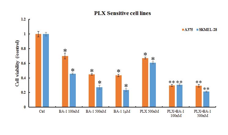
27 18 III. Results 3.1 Autophagy inhibitors exhibited potent cytotoxicity against human malignant melanoma cells and have a combined effect with PLX4032. Based on the fact that 50% of melanomas contain hyperactive BRAF V600E mutations, and that autophagy can help cancer cells survive under certain conditions, I hypothesized that if we combine the BRAF inhibitors and the autophagy inhibitors to reduce both the activity of autophagy and the activity of the BRAF in melanoma cells at the same time, we should expect a stronger cytotoxic effect on melanoma cells. To test this hypothesis, an MTS viability assay was employed to examine if the combination of the BRAF and the autophagy inhibitors can inhibit the proliferation of the melanoma cells. In this study, PLX4032 (Vemurafenib) was selected as the BRAF inhibitor because it is an FDAapproved drug for melanoma cells with BRAF mutations. Three different autophagy inhibitors, namely BA-1, SBI and HCQ, were used in this study to test the effect of autophagy on cell viability. Each treatment group was tested in four different cell lines: two PLX4032 sensitive cell lines (A375 and SK-MEL-28), and two PLX4032 resistant cell lines (A375VR and A2058). The cell viability assay showed that the autophagy inhibitor BA-1 significantly reduced the viability of the PLX4032 sensitive cell lines compared to the control group at various concentrations (Figure 5A), and BA-1 is more effective than PLX4032 in decreasing the viability of melanoma cells, suggesting that autophagy is used as a cell survival strategy in these cell lines. When both BRAF inhibitor PLX4032 and autophagy inhibitor BA-1 were added into the melanoma cells, cell viability decreased even more compared to the PLX4032 or BA-1 alone treatment at
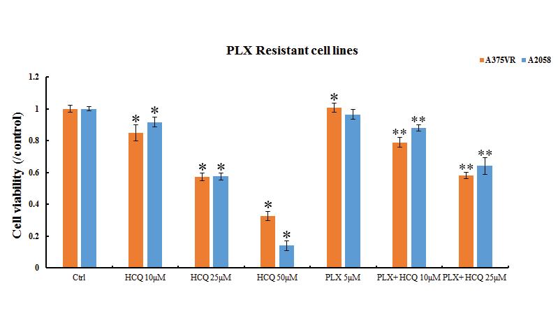
28 19 comparable concentrations, indicating that the combination of PLX4032 and BA-1 increased the cytotoxic effect on the PLX4032 sensitive cells lines. Since the development of drug resistance is common in melanoma cells, which significantly reduces the efficiency of the current therapy relying on single drugs such as PLX4032, we investigated if the autophagy inhibitor can show a similar cytotoxic effect in the PLX4032 resistant cell lines. To do this, we treated the PLX4032 resistant cell lines, A375VR, and A2058 with autophagy inhibitors, and performed the MTS assay to measure the viability of these two cell lines. As we can see in Figure 5B, while A375VR and A2058 are resistant to the PLX4032 treatment, the viability of these two cells lines was significantly reduced by the BA-1 treatment. To rule out the possibility that the cytotoxic effect is specific for BA-1, and to further confirm that the inhibition of autophagy is a major factor in reducing the viability of the melanoma cells, we examined the anti-melanoma effect of another two autophagy inhibitors that are different from BA-1 in the way they inhibit autophagy. SBI inhibits the initiating step of autophagy. The complete autophagy process requires fusion between autophagosomes and lysosomes to become the autolysosomes. BA-1 inhibits the fusion between autophagosomes and lysosomes. HCQ can neutralize H+ in lysosomes, blocking the fusion between autophagosomes and lysosomes. HCQ, similar to BA-1, significantly reduced the viability of the PLX4032 sensitive cell lines, and the combination of PLX4032 and HCQ showed a stronger inhibitory effect on cell viability (Figure 6A). As expected, HCQ significantly reduced the cell viability of the PLX4032 resistant cell lines as well (Figure 6B). Similar effects were also observed for the other autophagy inhibitor
29 20 SBI for both PLX4032 sensitive and resistant cell lines (Figures 7A, B, and C). These results suggest that autophagy is used as a survival strategy for the melanoma cells, and the inhibition of autophagy can reduce cell viability of the melanoma cells.
30 21 Figure 5. Bafilomycin A1 inhibited cell viability and has an additive effect with PLX4032 in reducing cell viability in PLX4032 sensitive melanoma cell lines (A. SK- MEL-28 and A375) and PLX4032 resistant melanoma cell lines (B. A375VR and A2058). Effect of Bafilomycin A1 on cell viability of PLX4032 sensitive melanoma cell lines (A. SK-MEL-28 and A375) and PLX4032 resistant melanoma cell lines (B. A375VR and A2058). Melanoma cells were determined by MTS assay after 48 hours treatment with Bafilomycin A1 at a final concentration of 100 nm, 500 nm, and 1 M. Every experiment was repeated five times with quadruplicate reactions in each independent experiment. The results are represented as mean ± SD. Asterisks indicate a significant difference between the Bafilomycin A1 treatment groups and the control group (p<0.05). Double asterisks indicate a significant difference between PLX4032- Bafilomycin A1 combination groups and PLX4032 group (PLX4032 at 500 nm in SK-MEL-28 and A375; PLX4032 at 5 μm in A375VR and A2058) (p<0.05).

31 22 A. B.
32 23 Figure 6. Hydroxychloroquine inhibited cell viability has a combined effect with PLX4032 in reducing cell viability in PLX4032 sensitive melanoma cell lines (A. SK- MEL-28 and A375) and PLX4032 resistant melanoma cell lines (B. A375VR and A2058). Effect of hydroxychloroquine on cell viability of PLX4032 sensitive melanoma cell lines (A. SK-MEL-28 and A375) and PLX4032 resistant melanoma cell lines (B. A375VR and A2058). Melanoma cells were determined by MTS assay after 48 hours treatment with hydroxychloroquine at a final concentration of 10 M, 25 M, and 50 M. Every experiment was repeated five times with quadruplicate reactions in each independent experiment. The results were represented as mean ± SD. Asterisks indicate a significant difference between the hydroxychloroquine treatment groups and the control group (p<0.05). Double asterisks indicate a significant difference between PLX4032- hydroxychloroquine combination groups and PLX4032 group (PLX4032 at 500 nm in SK-MEL-28 and A375; PLX4032 at 5 μm in A375VR and A2058) (p<0.05).

33 24 A. B.
34 25 Figure 7. SBI inhibited cell viability and had a combined effect with PLX4032 in reducing cell viability in PLX4032 sensitive melanoma cell lines (A. SK-MEL-28 and A375) and PLX4032 resistant melanoma cell lines (B. A375VR and A2058). Effect of SBI on cell viability of PLX4032 sensitive melanoma cell lines (A. SK-MEL-28 and A375) and PLX4032 resistant melanoma cell lines (B. A375VR and A2058). Melanoma cells were determined by MTS assay after 48 hours treatment with SBI at final concentrations of 2.5 M, 5 M, and 10 M in A375, A375VR, and A2058. However, IC50* of the SK-MEL-28 cell line is higher than in the other three cell lines. Every experiment was repeated five times with quadruplicate reactions in each independent experiment. The results were represented as mean ± SD. Asterisks indicate a significant difference between the SBI treatment groups and the control group (p<0.05). Double asterisks indicate a significant difference between PLX4032-SBI combination groups and the PLX4032 groups (PLX4032 at 500 nm in SK-MEL-28 and A375; PLX4032 at 5 μm in A375VR and A2058) (p<0.05). * The IC50 is the concentration of an inhibitor where the cell viability is reduced to 50%.
35 26 A. B.

36 C. 27
37 Autophagy inhibitors have an inhibitory effect on cell migration and have a combined inhibitory effect with PLX4032. Similar to other cancer cells, the melanoma cells not only have a high proliferation rate but also have a strong ability to migrate, which is critical for metastasis. As we already show by the MTS assay the autophagy inhibitors and the combination treatment can inhibit the proliferation; I was curious about whether the autophagy inhibitor can reduce the ability of melanoma cells to migrate. To do this, we test the inhibitors in a wellestablished cell migration assay, known as the wound-healing assay. In the woundhealing assay, an artificial wound area is created in the adhesive cell culture as described in the Materials and Methods. Since the cells in the wound area are all removed, the only way for the cells to heal this wound area is that the cells that are outside of the wound area migrate into the wound and eventually heal the wound. Cells with a stronger migration ability will cover more wound area given the same amount of incubation time. As shown in Figures 8A and B, the black dots are cells and the white rectangular shape in the center is the wound area. For the control group without any treatment, the A375 melanoma cells migrated into the wound area and covered almost the entire wound area after 48 hours. However, when the cells were treated with PLX nm, BA nm, SBI 5 M, or HCQ 25 M, their migration ability was significantly reduced as evidenced by the lesser number of cells that migrated into the wound area.
38 29 We observed similar effects on cell ability to migrate in SK-MEL-28 (Figures 8A and B), A2058 (Figures 8E and F) and A375VR (Figures 8G and H) cell lines after treatment with PLX4032 (500 nm at PLX4032 sensitive cell lines and 5 M at PLX4032 resistant cell lines), BA-1, SBI, and HCQ. When we combined the autophagy inhibitors with PLX4032 to treat the cells, the combination treatments showed a more potent inhibitory effect on the migration ability of the melanoma cells than did PLX4032 alone or autophagy alone, though the degrees of inhibition were different among each combination treatment in these cell lines. Collectively, these findings indicated that autophagy inhibitors inhibited the cell migration as well as the cell proliferation of the tested melanoma cells. These results further suggest that a combination of autophagy inhibitors and BRAF inhibitors would be a potential therapeutic strategy to treat melanoma and overcome the drug resistance in the melanoma cells.
39 30 Figure 8. Effect of autophagy inhibitors, BA-1, SBI and HCQ, and effect of combination treatments on cell migration of PLX4032 sensitive melanoma cell lines (A. and B. SK- MEL-28; C. and D. A375) and PLX4032 resistant melanoma cell lines (E. and F. A2058; G. and H. A375VR). Cell migration assay was performed as described in Materials and Methods. After cells were treated with PLX4032, autophagy inhibitors, BA-1, SBI and HCQ, and combination treatments of PLX4032 and autophagy inhibitors, the cells were incubated for an additional 48 hours. The inhibition of cell migration ability was measured by the reduction of cells in the wound area. The results are presented as the bar graphs. Every experiment was done in triplicate and the results are presented as the mean ± SD (except for the SK- MEL-28 cell line). An asterisk indicates a mathematically significant difference between the treatment groups and the control group (p<0.05). Double asterisks indicate a significant difference between the PLX4032-autophagy inhibitor combination groups and the PLX4032 group (PLX4032 at 500 nm in SK-MEL-28 and A375; PLX4032 at 5 μm in A375VR and A2058) (p<0.05).


40 A. 31
41 B. 32

42 C. 33
43 D. 34

44 E. 35
45 F. 36
46 G. 37
47 H. 38
48 Autophagy inhibitors induced cell apoptosis in melanoma cells and have a combined effect with PLX4032 to induce apoptosis in melanoma cells. I further explored the underlying mechanisms by which the autophagy inhibitors reduce the cell proliferation of the melanoma cells. Since apoptosis is one of the major events that regulate cell death, I decided to examine the apoptosis in these melanoma cells after treatment to see if these autophagy inhibitors, together with PLX4032, induce apoptosis. After treating the melanoma cells with autophagy inhibitors, or PLX4032, or both, the cells were stained with PI and Annexin V-FITC, which was then followed by flow cytometry analysis. In the data analysis plot, the quadrant was drawn based on the negative and positive control group; cells falling into the right quadrants are cells that undergo apoptosis. As shown in Figures 9A and B, the percentage of apoptotic cells in the control group was 27.5%, whereas in the PLX4032 treatment, it was 43.2%. The BA- 1 treatment was 86.8% and the combination of BA-1 with PLX4032 treatment was 90.3%. The SBI treatment was 47.7%, and the combination of SBI with PLX4032 treatment was 68.2%. The HCQ treatment was 49.9%, and the combination of HCQ with PLX4032 was 81.5%. The increase in the number of apoptotic cells after treatment indicated that the autophagy inhibitor and PLX4032 induced apoptosis in the A375 cells. Similar effects were also observed for A375 (Figures 9C and D), A2058 (Figures 9E and H), and A375VR (Figures 9G and H) cell lines. This suggests a common mechanism where the autophagy inhibitors and PLX4032 inhibit the cell proliferation by inducing apoptosis in the melanoma cells.
49 40 Figure 9. PLX4032 and autophagy inhibitors induced apoptosis in melanoma cells Cells were treated with PLX4032, BA-1, SBI, HCQ and with combinations of PLX4032 and autophagy inhibitors at indicated concentrations for 48 hours. SK-MEL-28 cells (A), A375 (C), A2058 (E), and A375VR (G) cells were then stained with Annexin V-FITC and PI, and the fluorescence intensity was measured by flow cytometry. The percent of Annexin-V positive cells is represented in the bar graphs for SK-MEL-28 cells (B), A375 (D), A2058 (F), and A375VR (H). This experiment was performed three times and is reported as mean ± SD. An asterisk indicates a significant difference between the treatments groups and the control group (p<0.05). Double asterisks indicate a significant difference between PLX4032-autophagy inhibitor combination groups and the PLX4032 groups (PLX4032 at 500 nm in SK-MEL-28 and A375; PLX4032 at 5 μm in A375VR and A2058) (p<0.05).

50 41 A. B.

51 42 C. D.
52 43 E. F.

53 G. 44 H.
54 Effect of autophagy inhibitors and PLX4032 on activation or inactivation of signaling pathways in melanoma cells Autophagy inhibitors alone have no effect on up-regulating phosphorylated Braf, while PLX4032 enhances the expression of phosphorylated Braf in PLX4032 resistant melanoma cell lines In SK-MEL-28 cells, PLX4032 at 500 nm did not inhibit the activation of Braf (Figure 10A). BA-1 and the combination of BA-1 and PLX4032 did not affect the activation of Braf. Similarly, hydroxychloroquine and the combination of hydroxychloroquine and PLX4032 did not affect the activation of Braf. However, even though SBI did not affect the activation of Braf, SBI and PLX4032 together to decrease the activation of Braf. In the PLX-4032 resistant cell line A2058, PLX4032, a specific Braf inhibitor, paradoxically stimulated the activation of Braf (Figure 10B). In addition, all three autophagy inhibitors BA-1, SBI and hydroxychloroquine stimulated the activation of Braf. Surprisingly, BA-1 and hydroxychloroquine individually inhibited the activation of Braf caused by PLX4032. In another PLX4032 resistant cell line A375AR, similarly to the A2058 cell line, PLX4032 paradoxically stimulated the activation of Braf (Figure 10C). Each of the three autophagy inhibitors BA-1, SBI and hydroxychloroquine stimulated the activation of Braf. Also, similar to A2058, the three autophagy inhibitors BA-1, SBI, and hydroxychloroquine individually inhibited the activation of Braf induced by PLX4032.
55 46 Figure 10. The expression of phosphorylated Braf and non-phosphorylated Braf in SK- MEL-28, A2058 and A375VR cell lines. Western-blot analysis of p-braf and Braf activity in three melanoma cell lines treated with PLX4032, BA-1, SBI, HCQ, and combinations of PLX4032 and autophagy inhibitors at indicated concentrations for 48 h were performed as described in Materials and Methods in three independent experiments. The representative data are shown. GAPDH was used as the internal loading control.


56 47 A. B. C.
57 Autophagy inhibitors alone activate phosphorylated Erk, while PLX4032 suppresses the expression of phosphorylated Erk in PLX4032 melanoma cell lines In SK-MEL-28, PLX4032 successfully abrogated the activation of Erk, which is a downstream signaling molecule in the Ras-Raf-Mek-Erk signaling pathway (Figure 11A). However, the autophagy inhibitors BA-1 and hydroxychloroquine remarkably stimulated the activation of Erk. PLX4032 reduced the activation of Erk by BA-1 or hydroxychloroquine. In contrast to BA-1 and hydroxychloroquine, SBI exhibited an inhibitory effect on Erk activation, which can explain the inhibitory effect of SBI on the cell viability of SK-MEL-28 cells. In another PLX4032-sensitive cell line A375, similar to the SK-MEL-28 cell line, BA-1 remarkably stimulated the activation of Erk, and PLX4032 was not able to quench the activation by BA-1 (Figure 11B). Also similar to SK-MEL-28, SBI exhibited an inhibitory effect on Erk activation. Hydroxychloroquine markedly reduced the activation of Erk. In both the PLX-4032 resistant cell line A2058 and A375AR, similar to the PLX4032-sensitive cell lines, PLX4032 successfully abrogated the activation of Erk, and BA1 remarkably stimulated the activation of Erk, in which the stimulatory effect was not inhibited by PLX4032 (Figures 11C and D). In the A375AR cell line, both SBI and hydroxychloroquine strongly enhanced the activation of Erk, and the stimulatory effect was not reduced by PLX4032. In the A2058 cell line, hydroxychloroquine remarkably stimulated the activation of Erk; however, the activation was abolished by PLX4032 (Figures 11C and D).
58 49 Figure 11. The expression of phosphorylated Erk and non-phosphorylated Erk in SK- MEL-28, A375, A2058 and A375VR cell lines. Western-blot analysis of p-erk and Erk activity in four melanoma cell lines treated with PLX4032, BA-1, SBI, HCQ and in combinations of PLX4032 and autophagy inhibitors at indicated concentrations for 48 h were performed as described in Materials and Methods in three independent experiments. The representative data are shown. GAPDH was used as the internal loading control.


59 50 A. B. C.

60 D. 51
61 PLX4032 has no effect on phosphorylated Akt, while PLX4032 and autophagy inhibitors work synergistically to reduce the phosphorylated Akt in PLX4032- sensitive cell lines. In both PLX4032 sensitive cell lines SK-MEL-28 and A375, PLX4032 did not seem to affect the activation of Akt (Figures 12A and B). However, PLX4032 worked synergistically with BA-1, SBI or hydroxychloroquine to abolish the activation of Akt in the A375AR cell line. Both SBI and hydroxychloroquine individually reduced the activation of AKT in both SK-MEL-28 and A375 cell lines. As in A375 cell lines, the combination of SBI and PLX4032 synergistically inhibited the Akt activation (Figure 12B). In both PLX4032 resistant cell lines A2058 and A375AR, PLX4032 stimulated the activation of Akt (Figures 12C and D). BA-1 and hydroxychloroquine displayed dramatically different effects on the two PLX4032 resistance cell lines. While BA-1 and hydroxychloroquine remarkably reduced the activation of Akt in the A375AR cells, they stimulated the expression of Akt in the A2058 cells (Figures 12C and D). It is noteworthy that while both PLX4032 and BA-1 alone increased the activation of Ark, the combination of these two compounds markedly reduced the activation of Akt, which is currently hard to explain.
62 53 Figure 12. The expression of phosphorylated Akt and non-phosphorylated Akt in SK- MEL-28, A375, A2058 and A375VR cell lines. Western-blot analysis of p-akt and Akt activity in four melanoma cell lines treated with PLX4032, BA-1, SBI, HCQ and in combinations of PLX4032 and autophagy inhibitors at indicated concentrations for 48 h were performed as described in Materials and Methods in three independent experiments. The representative data are shown. GAPDH was used as the internal loading control.


63 54 A. B. C.


64 D. 55
65 Autophagy inhibitors BA-1 and hydroxychloroquine suppress the fusion of autophagosomes and lysosomes in autophagy, indicating the enhanced expression of LC3B. Whereas PLX4032 attenuates the inhibitory effect induced by BA-1 or hydroxychloroquine. In all four cell lines, regardless of PLX4032 sensitivity, BA-1 and hydroxychloroquine remarkably increased the level of LC3B, implying that both compounds induced the autophagy (Figure 13). PLX4032 almost completely abolished the stimulatory effect of BA-1 on the two forms of LC3B (LC3B I and LC3B II) in the A2058 cell line, implying a strong inhibitory effect of PLX4032 on autophagy induction induced by BA-1 in A2058 cells (Figure 13C). In SK-MEL-28 cells, PLX4032 significantly reduced the level of LC3B II but not LC3B I; however, it abolished the LC3B I but not LC3B II in the A375 cell line. Since LC3B II is converted from LC3B I and is directly involved in the formation of autophagosomes, PLX4032 may inhibit the autophagy induced by BA-1 more effectively in SK-MEL-28 than in A375 (Figures 13A and B). In addition, PLX4032 significantly reduced the LC3B I form that was increased by hydroxychloroquine in the A375 cell line (Figure 13B).
66 57 Figure 13. The expression of LC3B in the SK-MEL-28, A375, A2058 and A375VR cell lines. Western-blot analysis of LC3B activity in four melanoma cell lines treated with PLX4032, BA-1, SBI, HCQ, and combinations of PLX4032 and autophagy inhibitors at the indicated concentrations for 48 h were performed as described in Materials and Methods in three independent experiments. Representative data are shown. GAPDH was used as the internal loading control.


67 58 A. B. C.

68 D. 59
69 Autophagy inhibitors and PLX4032 work synergistically to reduce the expression of ATG12 in both PLX4032-sensitive and PLX4032-resistant melanoma cells. In the SK-MEL-28 cell line, PLX4032 remarkably reduced the level of ATG12 (Figure 14). In addition, with one exception where the combination of SBI and PLX4032 increased the ATG12, PLX4032 synergistically worked with each of the three autophagy inhibitors BA-1, SBI, and hydroxychloroquine to reduce the level of ATG12 in all the four cell lines, suggesting that these combinations inhibited autophagy induction. BA-1 increased the level of ATG12 in both the SK-MEL-28 and A375 cell lines, suggesting its pro-autophagy effect in PLX4032 sensitive cell lines (Figures 14A and B). However, BA- 1 remarkably reduced the level of ATG12 in the A375AR and A2058 cell lines, suggesting its anti-autophagy effect in PLX resistance cell lines (Figures 14C and D). SBI markedly reduced the level of ATG12 in both the SK-MEL-28 and A375 cell lines, suggesting it has an anti-autophagy effect in PLX4032 sensitive cell lines. However, SBI remarkably increased the level of ATG12 in the A375AR and A2058 cell lines, suggesting it has a pro-autophagy effect in PLX4032 resistant cell lines. Hydroxychloroquine markedly reduced the level of AG12 in both the PLX4032 sensitive A375 cells and PLX4032 resistant A375AR cells, suggesting that hydroxychloroquine may inhibit autophagy (Figures 14B and D).
70 61 Figure 14. The expression of ATG12 in the SK-MEL-28, A375, A2058 and A375VR cell lines. Western-blot analysis of ATG12 activity in four melanoma cell lines treated with PLX4032, BA-1, SBI, HCQ, and the combinations of PLX4032 and autophagy inhibitors at indicated concentrations for 48 h were performed as described in Materials and Methods in three independent experiments. Representative data are shown. GAPDH was used as the internal loading control.


71 62 A. B. C. D.
72 63 IV. Discussion After the BRAF mutations were identified in the melanoma cells, this opened a new chapter for developing targeted therapies to treat melanoma. The BRAF inhibitors were discovered to be very effective drugs to inhibit the progression of the metastasized melanoma in patients with the BRAF V600E mutation [1]. However, this BRAF-inhibitor therapy was soon challenged by the drug resistance developed in the melanoma patients, with the melanoma recurring and becoming resistant to the BRAF inhibitors in most patients [50]. In an effort of overcome this drug resistance, researchers tried to combine MEK (a downstream protein of BRAF) inhibitors and BRAF inhibitors in therapy; however, resistance to these two inhibitors still developed in the melanoma patients [51]. Since BRAF and MEK are in the same signaling pathway that is dysregulated in melanoma cells, it is important to identify a new mechanism that the melanoma cells employ to survive. Recent studies have suggested that autophagy might be a self-salvaging strategy for some cancer cells to survive. The most important function of autophagy is degrading and recycling unwanted intracellular components [52]. Autophagy is a double-edged sword in cancer progression. Whether autophagy is a negative regulator or a positive regulator in cancer cells survivability depends on the progression stages of tumors, the extracellular conditions, and the therapeutic treatments [53]. Autophagy can eliminate the damaged cells that are affected by early-stage cancer cells, which could then block the growth of cancer cells [52]. This autophagy activation is favorable when found in several types of radiation and chemotherapy treated cancer cells, and it is suggested that autophagy could
73 64 remove the damaged cells and avoid the apoptosis that is triggered by damaged organelles [54]. In addition, a study showed that the Beclin-1 gene, which is required for the autophagosome formation, is lost in several types of breast cancer, leading to an inactivation of autophagy [55]. Furthermore, metformin suppresses the growth of the melanoma tumor by activating autophagy and enhancing apoptosis [56]. However, there is other evidence showing that the autophagy is important for maintaining cell health and could help cancer cells survive. For instance, suppressing the etoposide-induced autophagy can elevate the cell death and enhance the anti-tumor activity of etoposide in hepatoma G2 cells [57] and cervical carcinoma cells [58]. Autophagy is also found to inhibit cisplatin-induced apoptosis in human glioma cells [59] and inhibit Sulforaphaneinduced apoptosis in human colon cancer cells [60]. Based on these findings, we propose that autophagy could also play a cytoprotective role in the melanoma cells. Cell viability assay in this study showed that autophagy inhibitors significantly reduced the viability of both the PLX sensitive and resistant melanoma cell lines, suggesting the autophagy contributes to the survival of the melanoma cells. In addition, when we combined the PLX4032 and autophagy inhibitors, the viability of the tested melanoma cells decreased even more. Besides the cytotoxic effect on the viability, we also found that the migration ability, an important factor that contributes to invasiveness and metastasis [61-63] of the melanoma cells, was significantly reduced by autophagy inhibitors alone and the combination treatments of autophagy inhibitors and PLX4032. This result is in agreement with a recent study reporting autophagy inhibitor SBI downregulates migration ability in gastric
74 65 adenocarcinoma cells [64]. Some researchers propose that autophagy could increase the motility of some cells by degrading the focal adhesions and paxillin that adhere cells to their extracellular matrix [65-67]. Though it is still unclear how autophagy regulates the mobility in the melanoma cells, our data suggest that autophagy inhibitors in combination with BRAF inhibitors could be a more effective therapeutic strategy for melanoma because this combination not only suppresses the proliferation but also inhibits cell migration in both PLX4032 resistant as well as PLX4032 sensitive melanoma cell lines. Exploring the mechanism by which the autophagy inhibitors reduce viability of the melanoma cells is important for understanding how autophagy inhibitors exert cytotoxicity on melanoma cells and for developing any cocktail combination therapy against melanoma. More and more evidence has emerged to support the notion that there is a cross-talk between apoptosis and autophagy [68]. Our results show that the autophagy inhibitors, BA-1, HCQ, and SBI, can increase the apoptosis in both the PLX4032 sensitive and resistant melanoma cells. This finding is supported by other studies showing that BA-1 not only attenuates cytoprotective autophagy but also accumulates autophagosomes to trigger the Bax-dependent apoptosis in cerebellar granule neurons and lymphoblastic leukemia [23,38]. Unlike BA-1 that up-regulates the pro-apoptotic protein Bax, hydroxychloroquine induces the apoptosis by down-regulating the anti-apoptotic protein Bcl-2 [69,70]. Our data and other studies consistently suggest that inducing apoptosis is a common mechanism through which autophagy inhibitors alone or combined with PLX4032 inhibit the cell proliferation in both PLX4032 sensitive and PLX4032 resistant melanoma cells.
75 66 A puzzling result in this study was that PLX4032, a specific inhibitor of Braf, paradoxically increased the level of phosphorylated Braf in PLX4032 resistant melanoma cell lines. It is noteworthy that the same concentration of PLX4032 suppressed the expression of phosphorylated Erk in both PLX4032-sensitive and resistant melanoma cells. Therefore, even though there exists a feedback loop that activates Braf in response to PLX4032, which contributes to resistance to PLX4032, PLX4032 is still able to inactivate Erk by an alternative pathway. In addition, autophagy inhibitors alone activated phosphorylated Erk; however, in the combination treatments, PLX4032 attenuates the expression of phosphorylated Erk produced by autophagy inhibitors. The other interesting result that we observed is that autophagy inhibitors and PLX4032 work synergistically to reduce the expression of ATG12 in both PLX4032-sensitive and PLX4032-resistant melanoma cells. ATG12-conjugation is a required modification for formation of pre-autophagosomes [25]. Therefore, PLX4032 and autophagy inhibitors synergistically inhibited autophagy. Moreover, PLX4032 also alleviated the higher level of LC3B induced by BA-1 and hydroxychloroquine, suggesting that PLX4032 potently inhibit autophagy. BRAF is a proto-oncogene that can lead to cancer if mutated. Approximately 50% of patients with melanoma have been found with this activating mutation in their BRAF genes [1]. Vemurafenib, a BRAF inhibitor, works well in suppressing the tumor growth in the first 4 months; however, a resistance to Vemurafenib usually develops in patients after 5 months [50]. Some research shows that the autophagy and the BRAF signaling
76 67 pathway communicate with each other, and both play important roles in cancer initiation and development. For instance, metformin was shown to inhibit the growth of a melanoma tumor by activating the autophagy process [56]. Two recent studies posed the possibility of using autophagy inhibitors to delay or inhibit the emergence of drug resistance that appears in BRAF mutant cells [19,21]. Both studies illustrated that increased apoptotic cells were observed in BRAF inhibitor resistance cells treated by the combination of autophagy inhibitors and a BRAF inhibitor [19,21]. G. Maria et al. reported that the Beaf-Mek-Eek signaling pathway enhanced the expression of the LC3 protein, an autophagy marker, in the human colon adenoma cells with BRAF mutation, suggesting that autophagy is activated in these cells [19]. The expression level of the autophagic marker is decreased by the BRAF inhibitor, PLX4720 [19]. Moreover, PLX4720 and the autophagy inhibitor, 3-MA, have a synergistic effect to overcome the resistance of the colorectal cancer cells with the mutated BRAF [19]. Another study suggested that PLX4032-resistant melanoma cells had higher level of autophagy compared to the baseline, and cytoprotective autophagy is induced by BRAF and BRAF/MEK inhibitors, and inhibiting autophagy led to increased cell death induced by PLX4032 [21]. In conclusion, we further confirmed that autophagy plays a cytoprotective role in melanoma cells, and autophagy inhibitors in melanoma cells can significantly reduce cell viability. The current study demonstrates that the autophagy inhibitors or combination of autophagy inhibitors and BRAF inhibitors suppressed the cytoprotective autophagy in both BRAF mutant cells and BRAF inhibitor resistant cells through turning on Bax/Bcl-2
AN ABSTRACT OF THE THESIS OF. in Biology presented on July 19, Title: The Anti-melanoma Effects of Heat Shock Protein Inhibitors.
 AN ABSTRACT OF THE THESIS OF Yao Yan for the Master of Science (name of student) (degree) in Biology presented on July 19, 2017 Title: The Anti-melanoma Effects of Heat Shock Protein Inhibitors Thesis
A particular set of insults induces apoptosis (part 1), which, if inhibited, can switch to autophagy. At least in some cellular settings, autophagy se
, which, if inhibited, can switch to autophagy. At least in some cellular settings, autophagy se") A particular set of insults induces apoptosis (part 1), which, if inhibited, can switch to autophagy. At least in some cellular settings, autophagy serves as a defence mechanism that prevents or retards
A particular set of insults induces apoptosis (part 1), which, if inhibited, can switch to autophagy. At least in some cellular settings, autophagy serves as a defence mechanism that prevents or retards
HCC1937 is the HCC1937-pcDNA3 cell line, which was derived from a breast cancer with a mutation
 SUPPLEMENTARY INFORMATION Materials and Methods Human cell lines and culture conditions HCC1937 is the HCC1937-pcDNA3 cell line, which was derived from a breast cancer with a mutation in exon 20 of BRCA1
Part-4. Cell cycle regulatory protein 5 (Cdk5) A novel target of ERK in Carb induced cell death
 A novel target of ERK in Carb induced cell death") Part-4 Cell cycle regulatory protein 5 (Cdk5) A novel target of ERK in Carb induced cell death 95 1. Introduction The process of replicating DNA and dividing cells can be described as a series of coordinated
Part-4 Cell cycle regulatory protein 5 (Cdk5) A novel target of ERK in Carb induced cell death 95 1. Introduction The process of replicating DNA and dividing cells can be described as a series of coordinated
For the rapid, sensitive and accurate measurement of apoptosis in various samples.
 ab14082 500X Annexin V-FITC Apoptosis Detection Reagent Instructions for Use For the rapid, sensitive and accurate measurement of apoptosis in various samples. This product is for research use only and
TITLE: Improved Therapy for Breast Cancer by Inhibiting Autophagy. CONTRACTING ORGANIZATION: Trustees of Dartmouth College Hanover, NH
 AD Award Number: W81XWH-07-1-0684 TITLE: Improved Therapy for Breast Cancer by Inhibiting Autophagy PRINCIPAL INVESTIGATOR: Alan Eastman CONTRACTING ORGANIZATION: Trustees of Dartmouth College Hanover,
Muse Assays for Cell Analysis
 Muse Assays for Cell Analysis Multiple Assay Outputs for Cell Analysis Cell Health Cell Signalling Immunology Muse Count & Viability Kit Muse Cell Cycle Kit Muse Annexin V & Dead Cell Kit Muse Caspase
Li et al. Journal of Experimental & Clinical Cancer Research (2018) 37:108
 37:108") Li et al. Journal of Experimental & Clinical Cancer Research (2018) 37:108 https://doi.org/10.1186/s13046-018-0774-7 CORRECTION Correction to: Novel smac mimetic APG- 1387 elicits ovarian cancer cell killing
Li et al. Journal of Experimental & Clinical Cancer Research (2018) 37:108 https://doi.org/10.1186/s13046-018-0774-7 CORRECTION Correction to: Novel smac mimetic APG- 1387 elicits ovarian cancer cell killing
The Annexin V Apoptosis Assay
 The Annexin V Apoptosis Assay Development of the Annexin V Apoptosis Assay: 1990 Andree at al. found that a protein, Vascular Anticoagulant α, bound to phospholipid bilayers in a calcium dependent manner.
RayBio Annexin V-FITC Apoptosis Detection Kit
 RayBio Annexin V-FITC Apoptosis Detection Kit User Manual Version 1.0 May 25, 2014 (Cat#: 68FT-AnnV-S) RayBiotech, Inc. We Provide You With Excellent Support And Service Tel:(Toll Free)1-888-494-8555 or
Supporting Information
 Supporting Information Identification of Novel ROS Inducers: Quinone Derivatives Tethered to Long Hydrocarbon Chains Yeonsun Hong,, Sandip Sengupta,, Wooyoung Hur, *, Taebo Sim,, * KU-KIST Graduate School
Instructions for Use. APO-AB Annexin V-Biotin Apoptosis Detection Kit 100 tests
 3URGXFW,QIRUPDWLRQ Sigma TACS Annexin V Apoptosis Detection Kits Instructions for Use APO-AB Annexin V-Biotin Apoptosis Detection Kit 100 tests For Research Use Only. Not for use in diagnostic procedures.
Supplementary Information
 Supplementary Information Figure S1. Int6 gene silencing efficiency. (A) Western Blot analysis of Int6 expression at different times after sirna transfection. Int6 expression is strongly silenced in Int6
Overexpression of Mcl-1 confers resistance to BRAFV600E inhibitors alone and in combination with MEK1/2 inhibitors in melanoma
 Overexpression of Mcl-1 confers resistance to BRAFV600E inhibitors alone and in combination with MEK1/2 inhibitors in melanoma The Harvard community has made this article openly available. Please share
Impact factor: Reporter:4A1H0019 Chen Zi Hao 4A1H0023 Huang Wan ting 4A1H0039 Sue Yi Zhu 4A1H0070 Lin Guan cheng 4A1H0077 Chen Bo xuan
 Curcumin Protects Neonatal Rat Cardiomyocytes against High Glucose-Induced Apoptosis via PI3K/Akt Signalling Pathway Wei Yu,1,2 Wenliang Zha,1 Zhiqiang Ke,1 Qing Min,2 Cairong Li,1 Huirong Sun,3 and Chao
p47 negatively regulates IKK activation by inducing the lysosomal degradation of polyubiquitinated NEMO
 Supplementary Information p47 negatively regulates IKK activation by inducing the lysosomal degradation of polyubiquitinated NEMO Yuri Shibata, Masaaki Oyama, Hiroko Kozuka-Hata, Xiao Han, Yuetsu Tanaka,
Annexin V-FITC Apoptosis Detection Kit
 ab14085 Annexin V-FITC Apoptosis Detection Kit Instructions for Use For the rapid, sensitive and accurate measurement of Apoptosis in living cells (adherent and suspension). View kit datasheet: www.abcam.com/ab14085
Problem Set 8 Key 1 of 8
 7.06 2003 Problem Set 8 Key 1 of 8 7.06 2003 Problem Set 8 Key 1. As a bright MD/PhD, you are interested in questions about the control of cell number in the body. Recently, you've seen three patients
Introduction: 年 Fas signal-mediated apoptosis. PI3K/Akt
 Fas-ligand (CD95-L; Fas-L) Fas (CD95) Fas (apoptosis) 年 了 不 度 Fas Fas-L 力 不 Fas/Fas-L T IL-10Fas/Fas-L 不 年 Fas signal-mediated apoptosis 度降 不 不 力 U-118, HeLa, A549, Huh-7 MCF-7, HepG2. PI3K/Akt FasPI3K/Akt
Annexin V-PE Apoptosis Detection Kit
 Annexin V-PE Apoptosis Detection Kit Catalog Number KA0716 100 assays Version: 02 Intended for research use only www.abnova.com Table of Contents Introduction... 3 Background... 3 General Information...
AWARD NUMBER: W81XWH TITLE: An in Vivo shrna-drug Screen to Identify Novel Targeted Therapy Combinations for KRAS Mutant Cancers
 AWARD NUMBER: W81XWH-12-1-0420 TITLE: An in Vivo shrna-drug Screen to Identify Novel Targeted Therapy Combinations for KRAS Mutant Cancers PRINCIPAL INVESTIGATOR: Ryan B. Corcoran, M.D., Ph.D. CONTRACTING
- 1 - Cell types Monocytes THP-1 cells Macrophages. LPS Treatment time (Hour) IL-6 level (pg/ml)
 IL-6 level (pg/ml)") Supplementary Table ST1: The dynamic effect of LPS on IL-6 production in monocytes and THP-1 cells after GdA treatment. Monocytes, THP-1 cells and macrophages (5x10 5 ) were incubated with 10 μg/ml of
Supplementary Table ST1: The dynamic effect of LPS on IL-6 production in monocytes and THP-1 cells after GdA treatment. Monocytes, THP-1 cells and macrophages (5x10 5 ) were incubated with 10 μg/ml of
MTS assay in A549 cells
 Project: VIGO MTS assay in A549 cells Detection of cell viability/activity AUTHORED BY: DATE: Cordula Hirsch 20.01.2014 REVIEWED BY: DATE: Harald Krug 10.04.2014 APPROVED BY: DATE: DOCUMENT HISTORY Effective
Enzyme-coupled Receptors. Cell-surface receptors 1. Ion-channel-coupled receptors 2. G-protein-coupled receptors 3. Enzyme-coupled receptors
 Enzyme-coupled Receptors Cell-surface receptors 1. Ion-channel-coupled receptors 2. G-protein-coupled receptors 3. Enzyme-coupled receptors Cell-surface receptors allow a flow of ions across the plasma
ab Lysosome/Cytotoxicity Dual Staining Kit
 ab133078 Lysosome/Cytotoxicity Dual Staining Kit Instructions for Use For studying lysosome function at the cellular level. This product is for research use only and is not intended for diagnostic use.
A. Generation and characterization of Ras-expressing autophagycompetent
 Supplemental Material Supplemental Figure Legends Fig. S1 A. Generation and characterization of Ras-expressing autophagycompetent and -deficient cell lines. HA-tagged H-ras V12 was stably expressed in
p53 and Apoptosis: Master Guardian and Executioner Part 2
 p53 and Apoptosis: Master Guardian and Executioner Part 2 p14arf in human cells is a antagonist of Mdm2. The expression of ARF causes a rapid increase in p53 levels, so what would you suggest?.. The enemy
Apoptosis Mediated Cytotoxicity of Curcumin Analogues PGV-0 and PGV-1 in WiDr Cell Line
 Apoptosis Mediated Cytotoxicity of Curcumin Analogues PGV-0 and PGV-1 in WiDr Cell Line Endah Puji Septisetyani, Muthi Ikawati, Barinta Widaryanti and Edy Meiyanto* ) Cancer Chemoprevention Research Center,
SUPPLEMENTARY INFORMATION
 Figure S1 Induction of non-apoptotic death of SV40-transformed and primary DKO MEFs, and DKO thymocytes. (A-F) STS-induced non-apoptotic death of DKO MEF. (A, B) Reduced viability of DKO MEFs after exposure
C-Phycocyanin (C-PC) is a n«sjfc&c- waefc-jduble phycobiliprotein. pigment isolated from Spirulina platensis. This water- soluble protein pigment is
 is a n«sjfc&c- waefc-jduble phycobiliprotein. pigment isolated from Spirulina platensis. This water- soluble protein pigment is") ' ^Summary C-Phycocyanin (C-PC) is a n«sjfc&c- waefc-jduble phycobiliprotein pigment isolated from Spirulina platensis. This water- soluble protein pigment is of greater importance because of its various
' ^Summary C-Phycocyanin (C-PC) is a n«sjfc&c- waefc-jduble phycobiliprotein pigment isolated from Spirulina platensis. This water- soluble protein pigment is of greater importance because of its various
RayBio Annexin V-Cy5 Apoptosis Detection Kit
 RayBio Annexin V-Cy5 Apoptosis Detection Kit User Manual Version 1.0 Mar 20, 2014 (Cat#: 68C5-AnnV-S) RayBiotech, Inc. We Provide You With Excellent Support And Service Tel:(Toll Free)1-888-494-8555 or
RCPA Research Award Final Progress Review
 RCPA Research Award 2010-2011 Final Progress Review Name: Dr Craig Wallington-Beddoe Degree/Institution/Year: PhD, The University of Sydney, Year 2 Research Project Title: New Therapeutic Strategies for
Annexin V-Cy3 Apoptosis Detection Kit
 ab14142 Annexin V-Cy3 Apoptosis Detection Kit Instructions for Use For the rapid, sensitive and accurate measurement of apoptosis in various samples. This product is for research use only and is not intended
Introduction. Cancer Biology. Tumor-suppressor genes. Proto-oncogenes. DNA stability genes. Mechanisms of carcinogenesis.
 Cancer Biology Chapter 18 Eric J. Hall., Amato Giaccia, Radiobiology for the Radiologist Introduction Tissue homeostasis depends on the regulated cell division and self-elimination (programmed cell death)
PUMA gene transfection can enhance the sensitivity of epirubicin-induced apoptosis of MCF-7 breast cancer cells
 PUMA gene transfection can enhance the sensitivity of epirubicin-induced apoptosis of MCF-7 breast cancer cells C.-G. Sun 1 *, J. Zhuang 1 *, W.-J. Teng 1, Z. Wang 2 and S.-S. Du 3 1 Department of Oncology,
IN VITRO HORMESIS EFFECTS OF SODIUM FLUORIDE ON KIDNEY CELLS OF THREE-DAY-OLD MALE RATS
 292 Tang, An, Du, Zhang, Zhou 292 IN VITRO HORMESIS EFFECTS OF SODIUM FLUORIDE ON KIDNEY CELLS OF THREE-DAY-OLD MALE RATS Qin-qing Tang, a Xiao-jing An, a Jun Du, a Zheng-xiang Zhang, a Xiao-jun Zhou a
6. TNF-α regulates oxidative stress, mitochondrial function and autophagy in neuronal cells
 6. TNF-α regulates oxidative stress, mitochondrial function and autophagy in neuronal cells 6.1 TNF-α induces mitochondrial oxidative stress in SH-SY5Y cells. The dysregulation of mitochondria and oxidative
Leucine Deprivation Reveals a Targetable Liability
 Cancer Cell, 19 Supplemental Information Defective Regulation of Autophagy upon Leucine Deprivation Reveals a Targetable Liability of Human Melanoma Cells In Vitro and In Vivo Joon-Ho Sheen, Roberto Zoncu,
8. CHAPTER IV. ANTICANCER ACTIVITY OF BIOSYNTHESIZED SILVER NANOPARTICLES
 8. CHAPTER IV. ANTICANCER ACTIVITY OF BIOSYNTHESIZED SILVER NANOPARTICLES 8.1. Introduction Nanobiotechnology, an emerging field of nanoscience, utilizes nanobased-systems for various biomedical applications.
Understanding Signaling Pathways by Modifying Sensitivity to PLX4720 in B-RAF V600E Melanoma
 Understanding Signaling Pathways by Modifying Sensitivity to PLX4720 in B-RAF V600E Melanoma Muska Hassan NCI-ICBP Summer Fellow Broad Institute of MIT and Harvard: Cancer Program Mentor: Cory Johannessen,
AMPK Phosphorylation Assay Kit
 AMPK Phosphorylation Assay Kit Catalog Number KA3789 100 assays Version: 02 Intended for research use only www.abnova.com Table of Contents Introduction... 3 Intended Use... 3 Background... 3 Principle
NF-κB p65 (Phospho-Thr254)
") Assay Biotechnology Company www.assaybiotech.com Tel: 1-877-883-7988 Fax: 1-877-610-9758 NF-κB p65 (Phospho-Thr254) Colorimetric Cell-Based ELISA Kit Catalog #: OKAG02015 Please read the provided manual
Assay Biotechnology Company www.assaybiotech.com Tel: 1-877-883-7988 Fax: 1-877-610-9758 NF-κB p65 (Phospho-Thr254) Colorimetric Cell-Based ELISA Kit Catalog #: OKAG02015 Please read the provided manual
THE ROLE OF ALTERED CALCIUM AND mtor SIGNALING IN THE PATHOGENESIS OF CYSTINOSIS
 Research Foundation, 18 month progress report THE ROLE OF ALTERED CALCIUM AND mtor SIGNALING IN THE PATHOGENESIS OF CYSTINOSIS Ekaterina Ivanova, doctoral student Elena Levtchenko, MD, PhD, PI Antonella
B16-F10 (Mus musculus skin melanoma), NCI-H460 (human non-small cell lung cancer
, NCI-H460 (human non-small cell lung cancer") Electronic Supplementary Material (ESI) for ChemComm. This journal is The Royal Society of Chemistry 2017 Experimental Methods Cell culture B16-F10 (Mus musculus skin melanoma), NCI-H460 (human non-small
Electronic Supplementary Material (ESI) for ChemComm. This journal is The Royal Society of Chemistry 2017 Experimental Methods Cell culture B16-F10 (Mus musculus skin melanoma), NCI-H460 (human non-small
Quantitative Data Analysis Assignment Sample Newessays.co.uk
 Absorbance Quantitative Data Analysis Assignment Sample Newessays.co.uk Part A: Results of the Study Is there a difference of curve profile between the MTT assay and the cell number? What do the different
Kinase Inhibitor p21 WAF1/CIP1 in Apoptosis and Autophagy
 Pivotal Role of the Cyclin-dependent Kinase Inhibitor p21 WAF1/CIP1 in Apoptosis and Autophagy Keishi Fujiwara, Shigeru Daido, Akitsugu Yamamoto, Ryuji Kobayash, Tomohisa Yokoyama, Hiroshi Aok, Eiji Iwado,
Proteomic profiling of small-molecule inhibitors reveals dispensability of MTH1 for cancer cell survival
 Supplementary Information for Proteomic profiling of small-molecule inhibitors reveals dispensability of MTH1 for cancer cell survival Tatsuro Kawamura 1, Makoto Kawatani 1, Makoto Muroi, Yasumitsu Kondoh,
Supplementary Figure 1. BMS enhances human T cell activation in vitro in a
 Supplementary Figure 1. BMS98662 enhances human T cell activation in vitro in a concentration-dependent manner. Jurkat T cells were activated with anti-cd3 and anti-cd28 antibody in the presence of titrated
The Past, Present, and Future of Acute Myeloid Leukemia
 The Past, Present, and Future of Acute Myeloid Leukemia Carter T. Davis, MD Hematology-Oncology Fellow Duke University Health System September 10, 2016 Overview Overview of Acute Myeloid Leukemia Review
SUPPLEMENTAL MATERIAL. Supplementary Methods
 SUPPLEMENTAL MATERIAL Supplementary Methods Culture of cardiomyocytes, fibroblasts and cardiac microvascular endothelial cells The isolation and culturing of neonatal rat ventricular cardiomyocytes was
Cell Migration and Invasion Assays INCUCYTE LIVE-CELL ANALYSIS SYSTEM. Real-time automated measurements of cell motility inside your incubator
 Cell Migration and Invasion Assays INCUCYTE LIVE-CELL ANALYSIS SYSTEM Real-time automated measurements of cell motility inside your incubator See the whole story Real-time cell motility visualization and
Annexin V-Cy3 Apoptosis Detection Reagent
 ab14143 Annexin V-Cy3 Apoptosis Detection Reagent Instructions for Use For the rapid, sensitive and accurate measurement of apoptosis in various samples This product is for research use only and is not
Multi-Parameter Apoptosis Assay Kit
 Multi-Parameter Apoptosis Assay Kit Catalog Number KA1335 5 x 96 assays Version: 05 Intended for research use only www.abnova.com Table of Contents Introduction... 3 Background... 3 Principle of the Assay...
In Vitro Studies of the Aurora-Kinase Inhibitor MLN8237 in Prostate Cancer Cell Lines
 In Vitro Studies of the Aurora-Kinase Inhibitor MLN837 in Prostate Cancer Cell Lines Nicole V. Julian Mentor: Ana Aparicio, M.D. Special Thanks to Brittany Kleb Department of Genitourinary Medical Oncology
HEK293 cells transfected with human MATE1, MATE2-K, or vector control were established by
 SUPPLEMENTAL DIGITAL CONTENT METHODS In Vitro Metformin Transport Studies Effect of Dolutegravir on Metformin Transport by MATE1 and MATE2-K HEK293 cells transfected with human MATE1, MATE2-K, or vector
Protocol for A-549 VIM RFP (ATCC CCL-185EMT) TGFβ1 EMT Induction and Drug Screening
 TGFβ1 EMT Induction and Drug Screening") Protocol for A-549 VIM RFP (ATCC CCL-185EMT) TGFβ1 EMT Induction and Drug Screening Introduction: Vimentin (VIM) intermediate filament (IF) proteins are associated with EMT in lung cancer and its metastatic
Protocol for A-549 VIM RFP (ATCC CCL-185EMT) TGFβ1 EMT Induction and Drug Screening Introduction: Vimentin (VIM) intermediate filament (IF) proteins are associated with EMT in lung cancer and its metastatic
Glucose Uptake-Glo Assay
 TECHNICAL MANUAL Glucose Uptake-Glo Assay Instructions for Use of Products J1341, J1342, and J1343 Revised 2/17 TM467 Glucose Uptake-Glo Assay All technical literature is available at: www.promega.com/protocols/
UNIVERSITY OF MEDICINE AND PHARMACY CRAIOVA PhD SCHOOL. PhD THESIS
 UNIVERSITY OF MEDICINE AND PHARMACY CRAIOVA PhD SCHOOL PhD THESIS THE IMPORTANCE OF TUMOR ANGIOGENESIS IN CEREBRAL TUMOR DIAGNOSIS AND THERAPY ABSTRACT PhD COORDINATOR: Prof. univ. dr. DRICU Anica PhD
Supplementary data Supplementary Figure 1 Supplementary Figure 2
 Supplementary data Supplementary Figure 1 SPHK1 sirna increases RANKL-induced osteoclastogenesis in RAW264.7 cell culture. (A) RAW264.7 cells were transfected with oligocassettes containing SPHK1 sirna
Cover Page. The handle holds various files of this Leiden University dissertation.
 Cover Page The handle http://hdl.handle.net/1887/23854 holds various files of this Leiden University dissertation. Author: Marel, Sander van der Title: Gene and cell therapy based treatment strategies
Effect of starvation-induced autophagy on cell cycle of tumor cells
 [Chinese Journal of Cancer 27:8, 102-108; August 2008]; 2008 Sun Yat-Sen University Cancer Center Basic Research Paper Effect of starvation-induced autophagy on cell cycle of tumor cells Jun-Na Ge, 1 Dan
SUPPLEMENTARY INFORMATION
 DOI: 10.1038/ncb3311 A B TSC2 -/- MEFs C Rapa Hours WCL 0 6 12 24 36 pakt.s473 AKT ps6k S6K CM IGF-1 Recipient WCL - + - + - + pigf-1r IGF-1R pakt ps6 AKT D 1 st SILAC 2 nd SILAC E GAPDH FGF21 ALKPGVIQILGVK
The effect of insulin on chemotherapeutic drug sensitivity in human esophageal and lung cancer cells
 The effect of insulin on chemotherapeutic drug sensitivity in human esophageal and lung cancer cells Published in: Natl Med J China, February 10, 2003; Vol 83, No 3, Page 195-197. Authors: JIAO Shun-Chang,
Islet viability assay and Glucose Stimulated Insulin Secretion assay RT-PCR and Western Blot
 Islet viability assay and Glucose Stimulated Insulin Secretion assay Islet cell viability was determined by colorimetric (3-(4,5-dimethylthiazol-2-yl)-2,5- diphenyltetrazolium bromide assay using CellTiter
Addicted to Autophagy: Ph+ B-ALL May Acquire Imatinib-resistance and Enhanced Malignancy. through a Highly-active Autophagy Pathway.
 Addicted to Autophagy: Ph+ B-ALL May Acquire Imatinib-resistance and Enhanced Malignancy through a Highly-active Autophagy Pathway by Nohea Arkus A Thesis Presented in Partial Fulfillment of the Requirements
RAS Genes. The ras superfamily of genes encodes small GTP binding proteins that are responsible for the regulation of many cellular processes.
 ۱ RAS Genes The ras superfamily of genes encodes small GTP binding proteins that are responsible for the regulation of many cellular processes. Oncogenic ras genes in human cells include H ras, N ras,
PLX7486 Background Information October Candidate for CRUK Combinations Alliance
 PLX7486 Background Information October 2015 Candidate for CRUK Combinations Alliance 1 Oct 2015 Plexxikon s Development Pipeline Compound Target Cancer Indication Stage of Development Pre- IND Ph1 Ph2
IN VITRO ANTICANCER ACTIVITY OF FLOWER EXTRACTS OF COUROUPITA GUIANENSIS
 CHAPTER 3 IN VITRO ANTICANCER ACTIVITY OF FLOWER EXTRACTS OF COUROUPITA GUIANENSIS 3. INTRODUCTION Plants are the basic source of knowledge of modern medicine. Almost all the parts of the plant, namely
Cell Lysis Buffer. Catalog number: AR0103
 Cell Lysis Buffer Catalog number: AR0103 Boster s Cell Lysis Buffer is a ready-to-use Western blot related reagent solution used for efficient extraction of total soluble protein in nondenatured state
Normal RAS-RAF (MAPK) pathway signaling
 pathway signaling") BRAF-Mutations in Melanomas L. Mazzucchelli Istituto Cantonale di Patologia, Locarno 77. Annual Meeting Swiss Society of Pathology, Lucerne 2011 Sponsored by Roche Pharma Switzerland Melanoma has increased
BRAF-Mutations in Melanomas L. Mazzucchelli Istituto Cantonale di Patologia, Locarno 77. Annual Meeting Swiss Society of Pathology, Lucerne 2011 Sponsored by Roche Pharma Switzerland Melanoma has increased
Johannes F Fahrmann and W Elaine Hardman *
 Fahrmann and Hardman Lipids in Health and Disease 2013, 12:36 RESEARCH Open Access Omega 3 fatty acids increase the chemo-sensitivity of B-CLL-derived cell lines EHEB and and of B-PLL-derived cell line
In vitro scratch assay: method for analysis of cell migration in vitro labeled fluorodeoxyglucose (FDG)
") In vitro scratch assay: method for analysis of cell migration in vitro labeled fluorodeoxyglucose (FDG) 1 Dr Saeb Aliwaini 13/11/2015 Migration in vivo Primary tumors are responsible for only about 10%
In vitro scratch assay: method for analysis of cell migration in vitro labeled fluorodeoxyglucose (FDG) 1 Dr Saeb Aliwaini 13/11/2015 Migration in vivo Primary tumors are responsible for only about 10%
Supplementary Materials
 Supplementary Materials Figure S1. MTT Cell viability assay. To measure the cytotoxic potential of the oxidative treatment, the MTT [3-(4,5-dimethylthiazol- 2-yl)-2,5-diphenyl tetrazolium bromide] assay
Supplementary Materials and Methods
 Supplementary Materials and Methods Immunoblotting Immunoblot analysis was performed as described previously (1). Due to high-molecular weight of MUC4 (~ 950 kda) and MUC1 (~ 250 kda) proteins, electrophoresis
Supplemental Figures:
 Supplemental Figures: Figure 1: Intracellular distribution of VWF by electron microscopy in human endothelial cells. a) Immunogold labeling of LC3 demonstrating an LC3-positive autophagosome (white arrow)
Essential Medium, containing 10% fetal bovine serum, 100 U/ml penicillin and 100 µg/ml streptomycin. Huvec were cultured in
 Supplemental data Methods Cell culture media formulations A-431 and U-87 MG cells were maintained in Dulbecco s Modified Eagle s Medium. FaDu cells were cultured in Eagle's Minimum Essential Medium, containing
Supporting Information
 Copyright WILEY-VCH Verlag GmbH & Co. KGaA, 69469 Weinheim, Germany, 212. Supporting Information for Adv. Funct. Mater., DOI:.2/adfm.2122233 MnO Nanocrystals: A Platform for Integration of MRI and Genuine
FOXO Reporter Kit PI3K/AKT Pathway Cat. #60643
 Data Sheet FOXO Reporter Kit PI3K/AKT Pathway Cat. #60643 Background The PI3K/AKT signaling pathway is essential for cell growth and survival. Disruption of this pathway or its regulation has been linked
Annexin V-FITC Apoptosis Detection Kit with SYTOX
 ab14086 Annexin V-FITC Apoptosis Detection Kit with SYTOX Instructions for Use For the rapid, sensitive and accurate measurement of apoptosis in living cells. This product is for research use only and
MTC-TT and TPC-1 cell lines were cultured in RPMI medium (Gibco, Breda, The Netherlands)
") Supplemental data Materials and Methods Cell culture MTC-TT and TPC-1 cell lines were cultured in RPMI medium (Gibco, Breda, The Netherlands) supplemented with 15% or 10% (for TPC-1) fetal bovine serum
Supplemental data Materials and Methods Cell culture MTC-TT and TPC-1 cell lines were cultured in RPMI medium (Gibco, Breda, The Netherlands) supplemented with 15% or 10% (for TPC-1) fetal bovine serum
TITLE: The Importance of Autophagy in Breast Cancer Development and Treatment
 AD Award Number: W81XWH-06-1-0557 TITLE: The Importance of Autophagy in Breast Cancer Development and Treatment PRINCIPAL INVESTIGATOR: Jin-Ming Yang, Ph.D. CONTRACTING ORGANIZATION: University of Medicine
Department of General Surgery, The Third People s Hospital of Dalian, Dalian Medical University, Dalian, Liaoning, China,
 JBUON 2018; 23(2): 340-345 ISSN: 1107-0625, online ISSN: 2241-6293 www.jbuon.com E-mail: editorial_office@jbuon.com ORIGINAL ARTICLE Boswellic acid exerts potent anticancer effects in HCT-116 human colon
Key words: apoptosis, beta-amyrin, cell cycle, liver cancer, tritepenoids
 JBUON 2018; 23(4): 965-970 ISSN: 1107-0625, online ISSN: 2241-6293 www.jbuon.com E-mail: editorial_office@jbuon.com ORIGINAL ARTICLE Antitumor effects of beta-amyrin in Hep-G2 liver carcinoma cells are
Autophagy induction by low-dose cisplatin: The role of p53 in autophagy
 248 Autophagy induction by low-dose cisplatin: The role of p53 in autophagy KYUNG-HWA CHO 1, JI-HYE PARK 1, KANG-BEOM KWON 2, YOUNG-RAE LEE 2, HONG-SEOB SO 2, KANG-KYOO LEE 3, SAM-YOUN LEE 4, SUN-ROCK
Corporate Medical Policy
 Corporate Medical Policy BRAF Gene Mutation Testing to Select Melanoma or Glioma Patients File Name: Origination: Last CAP Review: Next CAP Review: Last Review: braf_gene_mutation_testing_to_select_melanoma_or_glioma_patients_for_targeted_
BG-4, a bioactive peptide from Momordica charantia, promotes apoptosis in ovarian cancer cells
 University of Tennessee, Knoxville Trace: Tennessee Research and Creative Exchange University of Tennessee Honors Thesis Projects University of Tennessee Honors Program 5-2018 BG-4, a bioactive peptide
Nimbolide inhibits pancreatic cancer growth and metastasis through ROS-mediated
 Nimbolide inhibits pancreatic cancer growth and metastasis through ROS-mediated apoptosis and inhibition of epithelial-to-mesenchymal transition Ramadevi Subramani a, Ph.D., Elizabeth Gonzalez b, MS.,
Annexin V APC Assay Kit
 Annexin V APC Assay Kit Item No. 601410 www.caymanchem.com Customer Service 800.364.9897 Technical Support 888.526.5351 1180 E. Ellsworth Rd Ann Arbor, MI USA TABLE OF CONTENTS GENERAL INFORMATION 3 Materials
FOCUS SubCell. For the Enrichment of Subcellular Fractions. (Cat. # ) think proteins! think G-Biosciences
 think proteins! think G-Biosciences") 169PR 01 G-Biosciences 1-800-628-7730 1-314-991-6034 technical@gbiosciences.com A Geno Technology, Inc. (USA) brand name FOCUS SubCell For the Enrichment of Subcellular Fractions (Cat. # 786 260) think
169PR 01 G-Biosciences 1-800-628-7730 1-314-991-6034 technical@gbiosciences.com A Geno Technology, Inc. (USA) brand name FOCUS SubCell For the Enrichment of Subcellular Fractions (Cat. # 786 260) think
Supporting Information Nitric oxide releasing photoresponsive nanohybrids as excellent therapeutic agent for cervical cancer cell lines
 upporting Information itric oxide releasing photoresponsive nanohybrids as excellent therapeutic agent for cervical cancer cell lines Priya udhesh a, Kaviyarasan Tamilarasan a, Palaniappan Arumugam a and
Assessing Autophagy with the guava easycyte Benchtop Flow Cytometer
 Application Note Assessing Autophagy with the guava easycyte Benchtop Flow Cytometer Mark Santos, Kevin Su, Luke Armstrong, Angelica Olcott, Jason Whalley, and Matthew Hsu EMD Millipore Introduction Autophagy
Assays for Immuno-oncology Research Real-time automated measurements of immune and tumor cell dynamics within your incubator
 INCUCYTE LIVE-CELL ANALYSIS SYSTEM Assays for Immuno-oncology Research Real-time automated measurements of immune and tumor cell dynamics within your incubator See what your cells are doing and when they
Phospho-AKT Sampler Kit
 Phospho-AKT Sampler Kit E 0 5 1 0 0 3 Kits Includes Cat. Quantity Application Reactivity Source Akt (Ab-473) Antibody E021054-1 50μg/50μl IHC, WB Human, Mouse, Rat Rabbit Akt (Phospho-Ser473) Antibody
Human non small cell lung cancer cells can be sensitized to camptothecin by modulating autophagy
 INTERNATIONAL JOURNAL OF ONCOLOGY Human non small cell lung cancer cells can be sensitized to camptothecin by modulating autophagy YI-HAN CHIU 1*, SHIH-HSIEN HSU 2*, HSIAO-WEI HSU 2,3, KUO-CHIN HUANG 4,
SUPPLEMENT. Materials and methods
 SUPPLEMENT Materials and methods Cell culture and reagents Cell media and reagents were from Invitrogen unless otherwise indicated. Antibiotics and Tet-certified serum were from Clontech. In experiments
Cellular Screening in 2D and 3D. Identification of cancer specific modulators. ELRIG.de, March 2013
 Cellular Screening in 2D and 3D Identification of cancer specific modulators ELRIG.de, March 2013 Introduction International FP7 program Aim is the identification of novel compounds with anti-cancer properties
Antitumor activity of the ERK inhibitor SCH against BRAF mutant, NRAS mutant and wild-type melanoma
 Wong et al. Molecular Cancer 2014, 13:194 RESEARCH Open Access Antitumor activity of the ERK inhibitor SCH722984 against BRAF mutant, NRAS mutant and wild-type melanoma Deborah JL Wong 1, Lidia Robert
Data Sheet TIGIT / NFAT Reporter - Jurkat Cell Line Catalog #60538
 Data Sheet TIGIT / NFAT Reporter - Jurkat Cell Line Catalog #60538 Background: TIGIT is a co-inhibitory receptor that is highly expressed in Natural Killer (NK) cells, activated CD4+, CD8+ and regulatory
Electronic Supporting Information for
 Electronic Supplementary Material (ESI) for RSC Advances. This journal is The Royal Society of Chemistry 2015 Electronic Supporting Information for Rhodamine based Turn-On Fluorescent Probe for Pb(II)
2-Deoxyglucose Assay Kit (Colorimetric)
") 2-Deoxyglucose Assay Kit (Colorimetric) Catalog Number KA3753 100 assays Version: 01 Intended for research use only www.abnova.com Table of Contents Introduction... 3 Background... 3 General Information...
2-Deoxyglucose Assay Kit (Colorimetric) Catalog Number KA3753 100 assays Version: 01 Intended for research use only www.abnova.com Table of Contents Introduction... 3 Background... 3 General Information...