Cultured Skin Fibroblasts in Storage Disorders
|
|
|
- Lora Lindsey
- 6 years ago
- Views:
Transcription

1 Cultured Skin Fibroblasts in Storage Disorders An Analysis of Ultrastructural Features E. Kamensky, MD, M. Philippart, MD, P. Cancilla, MD and S. P. Frommes, BS Electron microscopic studies were performed on cultured fibroblasts from patients with metachromatic leukodystrophy, Fabry's, Gaucher's, Niemann-Pick's (Type A and C), Sanfilippo's (Type A and B) disease, chondroitin-4-sulfate mucopolysaccharidosis, lipofuscinosis (Spielmeyer-Vogt's disease) and ceroid-lipofuscinosis (Batten's disease with curvilinear bodies). Specific cytoplasmic inclusions with a limiting membrane were identified in Fabry's disease, Niemann-Pick syndrome, chondroitin-4-sulfate mucopolysaccharidosis and Sanfilippo's Type B disease. In Fabry's disease, the lipid inclusions tended to form stacks of parallel and concentric membranes. In Niemann-Pick syndrome, the lipid inclusions were made of wavy, loosely packed membranes. In chondroitin-4-sulfate mucopolysaccharidosis and Sanfilippo B, the lysosomes were enlarged and contained a reticular matrix with little electron-dense material. No specific ultrastructural changes were observed in Gaucher's, Sanfilippo's (Type A) disease, metachromatic leukodystrophy (sulfatidosis) and Batten's disease (Am J Pathol 73:59-80, 1973). THE STORAGE DISORDERS are characterized histologically by the appearance of a variety of cytoplasmic inclusions. These inclusion bodies are generally regarded as lysosomes which have retained substances because of the deficiency of a specific enzyme. Several specific enzyme deficiencies have been recognized in the sphingolipidoses and Hers has developed the concept of inborn lysosomal disorders as a base for understanding the protean family of diseases included in the general designation of the storage disorders.' We have been utilizing the skin fibroblast system in metabolic studies of the sphingolipidoses, mucopolysaccharidoses and lipofuscinoses. Most ultrastructural studies of these disorders have been done directly on tissue obtained from the liver or nervous system. Since cultured skin fibroblasts have become a valuable system for metabolic stuldies 2 and From the Departments of Pathology, Pediatrics, Neurology and Psychiatry, The Mental Retardation Center, The Neuropsychiatric Institute, Los Angeles, Calif. Supported by funds from the Department of Mental Hygiene, State of California, The Mental Retardation Program, NPI, University of California at Los Angeles and Research CGrants NB 06938, HD 04612, MCH 927, HD 00345, HD and NS from the National Institutes of Health. Accepted for publication June 1, Address reprint requests to Dr. M. Philippart, Mental Retardation Center, The Neuropsychiatric Institute, Los Angeles, CA

2 60 KAMENSKY ET AL American Journal of Pathology will be utilized more in the future, the present study was undertaken to examine the cells and their inclusions from cultures and to compare them with earlier studies of cells derived from other tissues. Materials and Methods Skin was obtainied from the arm by punch biopsy. There were 16 patients with the conditions listed in Table 1. Several patients have been the subject of previous reports as indicated below. Thirteen fibroblasts lines were entirely processed in our laboratories. Two lines were received from Dr. E. Neufeld (one line each from Sainfilippo Type A and B) as nonifrozen subeultures in Eagle's Miniimal Essential Medium containing 10% fetal calf serum; they were lhanid-carried from Washington, DC to Los Angeles. The third line (Niemainni-Pick Type A), as a nonfrozen subculture in medium F 10 (Grand Islanid Biological Co) conitaininig 15% of fetal calf serum, was air-mailed by Dr. J. Leroy from B3elgium. Control biopsies were obtained from subjects who were investigated as possible heterozygotes but were found to hlave acid hydrolase levels within1 the normal ranige. Fibroblasts derived from the skin after no more than 3 or 4 subcultures, within 6 to 8 weeks after obtaining the skin biopsy, were grown up to conifluenice in plastic T-flasks (250 ml) with Eagle's Minimum Essential Medium conitaininlg 15% fetal calf serum and 0.6% glutaminie. This is a well-established culture medium which we have suceessfully used for growing about 100 different fibroblasts lines. These lines, including those which are the subject of the present report, have been tested for acid hydrolase activity. For each line at least six of the following activities have been determilled: arylsulfatase A and B, " methylumbelliferyl sulfatase,4 (- and [3-galactosidase,5 n- and f-glucosidase,75 a- and f-n-acetylglucosaminidase,i a- anld O-N-acetylgalactosaminidase,7 acid phosphatase,7 hyalur-onidase, (t-glucuronidase,5 cerebrosidase,11 lactosylceramide-(-galactosidase,'1' phosphodiesterases I and IV.ll,12 The metabolic turinover of all these fibroblasts lines has been extensively studied by following incorporation and decay of 1-'4C-acetate for at least 6 weeks after a 2-day pulse. Experiments with 14 C-labeled galaietose, mannlose, N-acetylmanniiosaminie and N- acetylneuriaminic acid, :12 P-phosphate, and :15S-sulfate have also been condtucted onl most of these lines. No morphologic evidence for Mycoplasma contamination was ever encountered in short-term culturles used for the priesenit study. Chromosome studies were not undertaken on most of otur lines, but ouir standard sshort-term ctulture conditions are not known to induce chromosomal abbnormalities. Cell pellets were obtained by trypsinization and centrifugation, and were prefixed in 3% glutaraldehyde in phosphate btuffer for 2 hours at 4 C, rinsed in the same buffer and then postfixed in 1% buiffered osmitum tetroxide for 2 houirs at 4 C. After rapid dehydiration in a graded series of ethanol solutions, the cells were embedded in Epon 812 anid placed in an oven at 60 C for at least 2 days. The blocks were theni cut with glass knives for tlhiek (1 i) sections whichl were stained with toluidine blue to select suitable areas for electi-on miciroscopy. Liver aind brain tissue obtainied by biopsy was fixed in 3% buffer-ed glutar-aldehyde and processed as described above. Ultrathin sections were obtained with an LKI3 uiltriamicrotome using a diamondi knife, stained with uranyl acetate and lead citralte and examined with a Siemen's electron microsecope. Patient Data The first patient with Fabry's Disease'8 w.ls suic-essfuilly translanted for rel inisufficiency. He was deficien t in (x-galactosilase and ceraumide trihexosidase. Calactose turnover was impaired in fibroblasts (ulttured from hiis skin.14 The

3 Vol. 73, No. 1 October 1973 FIBROBLASTS IN STORAGE DISORDERS 61 Table 1-Summary of Patients Abnormal electron microscopic features of possible diagnostic Diseases Numberof patients significance Fabry 3 + Sulfatidosis (metachromatic 1 leukodystrophy) Gaucher 1 Nieman-Pick TypeA 2 + TypeC 1 + M ucopolysaccharidosis Sanfilippo A 2 Sanfilippo B 1 + Chondroitin-4-sulfate 1 + Lipofuscinosis 2 + Ceroid-lipofuscinosis 2 second patient with Fabry's disease came from a large, previously unreported family with affected males distributed among four generations. He was also deficient in a-galactosidase and had high levels of ceramide trihexosides in blood and urine.15 The third patient, a female, was an obligatory carrier, being the daughter of an affected male and the niece of our first patient.'3 Her skin fibroblasts were cloned and two cell populations, with and without deficient a-galactosidase activity, were obtained. The patient with sulfatidosis had an increased excretion of sulfatide, a deficiency of arylsulfatase A and an accumulation of sulfatide in a biopsied sural nerve.'r The patient with Gaucher's disease had Gaucher cells in her bone marrow, deficient 3-glucosidase activity in her urine and cultured fibroblasts and increased glucosyl ceramide in her plasma.", The family of the first patient with Niemann-Pick A has been the subject of a detailed report, including the autopsy of a sibling.1" Cultured skin fibroblasts were markedly deficient in sphingomyelinase. The seconid patient with Niemann-Pick Type A excreted excessive amounts of sphingomyelin in his urine and had a severe deficiency of sphingomyelinase in his cultur-ed fibroblasts, where an increased amount of sphingomyelin was also documented. The patient with Niemann-Pick Type C was diagnosed on the basis of foam cells in liver biopsy and bone marrow, normal urinary sphingomyelin, normal sphingomyelinase and abnormal phosphodiesterase activity in her skin fibroblasts.17 The first patient with Sanfilippo Type A was diagnosed on the basis of an abnormal urinary excretion of heparan sulfate, absence of skeletal lesioni, and normal activity of a-n-acetylglucosaminidase. The second patienit with Sanfilippo Type A was studied by Dr. E. Neufeld who demonstrated a deficienicy in heparan sulfate sulfatase activity in cultured fibroblasts."' Alpha-N-acetylglucosaminidase was normal in these cells. The patient with Sanfilippo Type B who was shown by Dr. Neufeld to have a normal heparan sulfate sulfatase activity was severely deficient in a- N-acetylglucosaminidase. Similar findings have been reported independently by O'Brien.! The patient with chondroitin-4-sulfate mucopolysaccharidosis has been reported previously."' No enzyme deficiency has been detected, although the absence of cross-correction with authentic Hurler lines2'1 might be interpreted as evidence for an a-l-iduronidase deficiency. The first patient with lipofusciniosis
 were diagnosed by the demonstration of curvilinear bodies in a brain biopsy.")
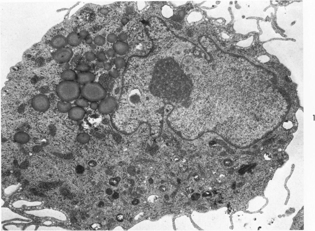
4 62 KAMENSKY ET AL American Journal of Pathology (Spielmeyer-Vogt) was diagnosed by brain biopsy. An abnormal lipid turnover has been documented in the brain explant but not in fibroblast culture.21'22 The second patient was a clinically affected sister. The two patients with ceroidlipofuscinosis (Batten's Disease) were diagnosed by the demonstration of curvilinear bodies in a brain biopsy. No enzyme deficiencies have been detected so far in the patients with lipofuscinosis or ceroid-lipofuscinosis. Results Normal Cultures Fibroblasts derived from normal subjects were characterized in our tissue culture system by an irregular outline with thin and varying sized cytoplasmic processes covered by a cytoplasmic membrane. The cytoplasm contained typical organelles, including mitochondria, smooth and rough surfaced endoplasmic reticulum, ribosomal aggregates, Golgi substance and filaments (Figure 1). A variety of homogeneous or vacuolated dense bodies were occasionally present in small numbers in the cytoplasm (Figures 1 and 2). Most of the dense bodies were limited by a single membrane. The heterogeneity of the cytoplasmic inclusions and the complexity of some individual bodies were evident in this material. Since we were primarily interested in the appearance of these structures in storage disorders, the variability of these structures emphasized the need for careful comparisons between cells derived from patients with a disorder and those obtained from subjects without disease under strictly similar conditions. The nucleus was frequently irregular in outline and often contained a distinct nucleolus (Figure 1). Fabry's Disease In Fabry's disease, trihexosyl ceramide accumulates as a result of an a-galactosidase deficiency. Lipid inclusions with similar morphologic characteristics have been described in a variety of tissues.2325 The lipid aggregates were generally limited by a membrane and may vary in size from 0.1 to 10 [t in diameter. Variation in stirulcture of the inclusions was present, but the most typical pattern consisted of parallel, concentric or interdigitating lamellae with a periodicity in the range of 40 to 50 A. Such inclusions were present in skin biopsies directly processed for electron microscopy (Figure 3). Acid phosphatase activity was demonstrated in these stiructures. In cultured fibroblasts from 3 patients with Fabry's disease we observed typical inclusions (Figure 4) similar to those found in vivo (Figure 3). The cytoplasmic bodies varied in size and shape, but they were generally surroutnded by a unit membrane. They were composed

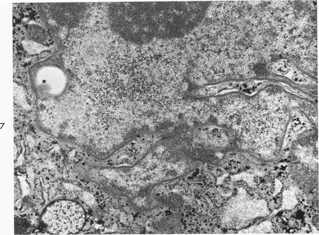
5 Vol. 73, No. 1 FIBROBLASTS IN STORAGE DISORDERS 63 October 1973 of stacks or circularly disposed lamellae with alternating dark and light bands with a regular period between 40 and 50 A. The space between the lamellar aggregates contained electron-dense granular material. In some cells, electron-lucent and vacuolated cytoplasmic bodies similar to those found in fibroblasts from normal subjects were commonly identified (Figure 5). The endoplasmic reticulum was more prominent than that observed in cells from normal subjects, and the cisternae were often dilated. Mitochondria and fibrils appeared normal. Cultured fibroblasts from a female carrier of Fabry's disease contained typical inclusions, but there were fewer inclusions in this instance than in male patients with the disease. Metachromatic Leukodystrophy (Sulfatidosis) In metachromatic leukodystrophy, lamellated or prismatic cytoplasmic inclusions have been described in a variety of tissues In the nervous system the inclusions may be composed of membranebound spherules organized into lamellar structures with a periodicity of around 60 A. Resibois has analyzed the two types of inclusions and found average dimensions of 60 A for the prismatic type and 75 A for the leaflet type of inclusion."} Cultured fibroblasts from a patient with metachromatic leukodystrophy contained large numbers of reticulated foamy vacuoles and several signet-ring and empty vacuoles (Figure 6). These structures were similar to those observed occasionally in fibroblasts from normal subjects. Structures identical to those described in tissues from patients with metachromatic leukodystrophy were not found. Gaucher's Disease Characteristic tubular structures have been described in a variety of organs in Gaucher's disease. The tubular elements measure up to 300 A in diameter and are contained in cytoplasmic bodies which measure up to 2 ti in diameter.31 Coarse clumps of chromatin applied to the inner surface of the nuclear membrane were a distinctive nuclear feature. There was fre(luent variability in the appearance of cytoplasmic structures such as mitochondria and the endoplasmic reticulum. No characteristic cytoplasmic bodies were evident in our material. The nuclei of the fibroblasts were often folded and the nuclear membranes were irregular in outline, but coarse slumping of the chromatin along the nuclear membrane was not fouind, and the nucleoli appeared normal (Figure 7). The endoplasmic reticulum was sometimes dilated and contained a fine flocculent material (Figure 7).

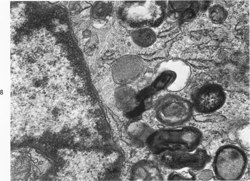
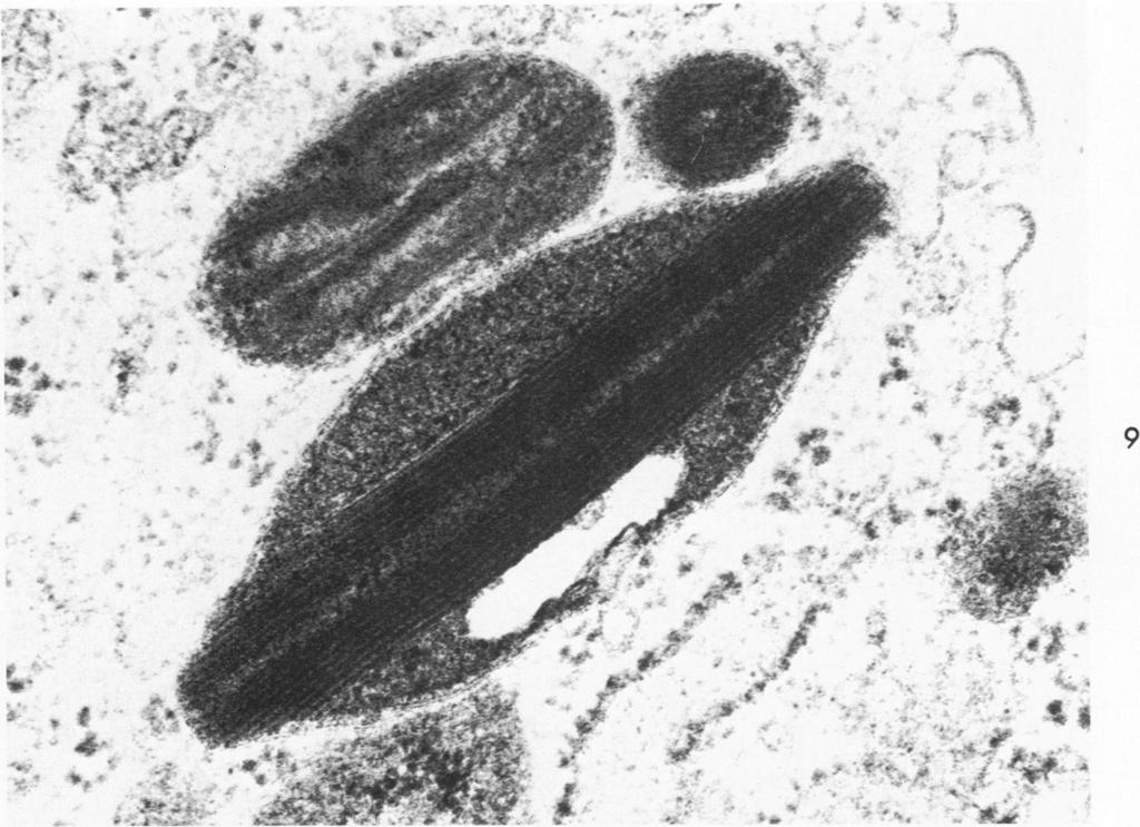
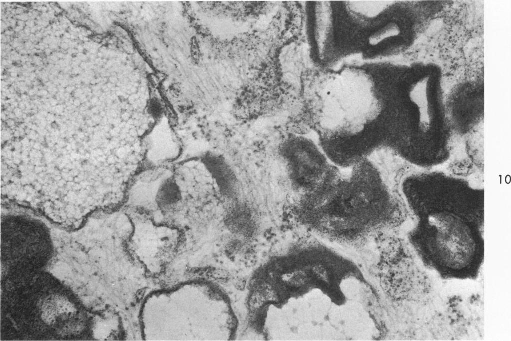
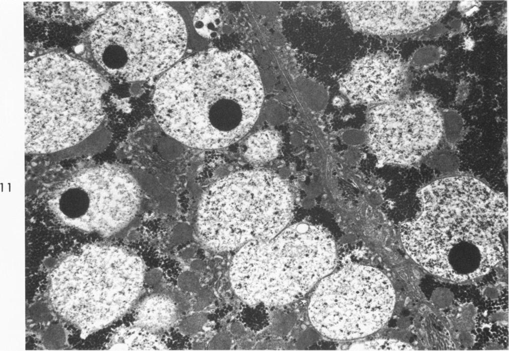
6 64 KAMENSKY ET AL American Journal of Pathology Niemann-Pick's Syndrome In Niemann-Pick's disease Type A (Crocker's classification 32) lamellated structures have been found in many different cell types. In liver,33 brain and spleen,34 the cytoplasm was filled with loose concentric lamellar figures around an electron-lucent center. In cultured fibroblasts from a clinical variant of Type A,'6 there were large numbers of cytoplasmic inclusion bodies which varied in size and shape and were surrounded by a unit membrane (Figure 8). Some of these structures had a granular content, but most of them contained loosely packed, wavy, concentric lamellar figures. The lamellae were often present at one pole of the inclusion, while larger inclusions were often filled with lamellae. Other lamellar inclusions were associated with a dense granular matrix and vacuolated and reticulated inclusions similar to those observed in cells from normal subjects were also observed. Fibroblasts from another patient with Type A disease contained numerous lamellar inclusions. The folding of the limiting membrane surrounding the inclusion body and their fusiform shape suggested a rigidity of the closely stacked elongated structures within the inclusion (Figure 9). The number of lamellae varied in these structures and often groups of lamellae were separated by a finely granular matrix. In a case of Niemann-Pick's disease Type C, the cytoplasmic inclusion bodies were very irregular in shape. The lamellar bodies were more compact than those seen in the first example of this disorder, and they were frequently found as a compact cluster of membranes within the boundaries of a vacuole (Figure 10). Foamy and reticulated inclusions were also present (Figure 10), and in some cells the Golgi substance and dilated cisternae of the endoplasmic reticulum were prominent. Mucopolysaccharidoses Sanfilippo's Disease condition have recently been characterized.6'18 Two variants of this numerous vacuoles within the cytoplasm. The vacuoles were lined by Examination of hepatocytes in 1 patient with Sanfilippo A revealed a single membrane and contained an electron-lucent finely reticulated matrix and a circumscribed electron-dense core (Figure 11). The cytoplasm of the liver cells contained abundant glycogen and the mitochondria were similar to those described in other examples of the mucopolysaccharidoses.3536 In the cultured fibroblasts from this patient, mostly small vacuoles
, although biochemical analysis 37 of")
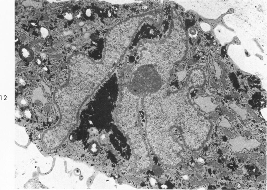
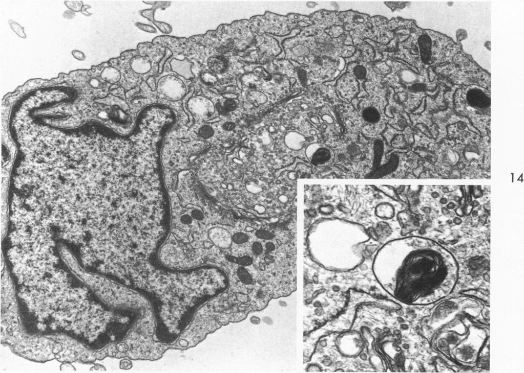
7 Vol. 73, No. 1 FIBROBLASTS IN STORAGE DISORDERS 65 October 1973 with indistinct or ill-defined contents were found (Figure 12). There were no electron-dense circumscribed cores within these vacuoles. Much of the cytoplasm was obscured by dense aggregates of glycogen which were more extensive than any found in the other disorders or normal subjects (Figure 12), although biochemical analysis 37 of these lines did not reveal an increase in the glycogen level. Nonspecific cytoplasmic structures were encountered which included lipid and partly vacuolated dense bodies. The endoplasmic reticulum was dilated and contained a finely granular material (Figure 12). In cultured fibroblasts from a patient with Sanfilippo B, there was abundant glycogen and numerous finely reticulated or vacuolated bodies with dense core regions (Figure 13 and inset). Chondroitin-4-Sulfate Mucopolysaccharidosis Electron micrographs of the liver in this patient "' were indistinguishable from those described in the example of Sanfilippo's disease (Figure 11). The cytoplasm of the hepatocytes was vacuolated and the vacuoles were lined by a single membrane and contained finely reticulated material and electron-dense circumscribed cores. The fibroblasts from this patient contained a variety of inclusion bodies none of which was similar to those described in the hepatocytes. The most distinctive inclusion body contained stacks of lamellated membranes often attached to one portion of the surrounding limiting membrane (Figure 14 and inset). Other inclusions were vacuolated with finely dispersed reticulated or granular material. Vacuoles were present in the Golgi region, and the endoplasmic reticulum was dilated and filled with granular material (Figure 14). Lipofuscinosis (Spielmeyer-Vogt) and Ceroid-Lipofuscinosis Typical lipofuscin bodies with acid hydrolase activity (Batten's Disease) were present in the neurons from a cerebral biopsy from this patient. The inclusions within the perikaryon were numerous and appeared in greater numbers than would be expected for the patient's age (Figure 15). A large number of amorphous lipid inclusions and dense bodies with a finely granular matrix were distributed within the cytoplasm of the cultured fibroblasts. Although there was vacuolization of some of the dense bodies, no typical lipofuscin bodies or structures identical to the neuronal inclusions described above were found (Figure 16). Polyribosomes were present in the cytoplasm and mitochondria were unremarkable. Fibroblasts obtained from the affected sister of this patient contained numerous identical nonspecific inclusions. None of the
 were indistinguishable from normal controls and thus they were clearly different from the two lipofuscinosis lines.")
8 66 KAMENSKY ET AL American Journal of Pathology controls exhibited such a large number of lipid inclusions. Fibroblasts from 2 unrelated patients with ceroid-lipofuscinosis (Batten's disease) were indistinguishable from normal controls and thus they were clearly different from the two lipofuscinosis lines. Discussion The types of inclusion bodies found in the cytoplasm of cultured fibroblasts are similar to those reported in other cell types. They are usually limited by a unit membrane which is fre(luently underlined by a clear halo typical of lysosomes.3x Except for glycogen granules, the occurrence of material lying free in the cytoplasm was infrequent. Acid hydrolase activity has been demonstrated within inclusion bodies from various storage disorders.4,2931 ' When the lysosomal matrix is markedly dilated, enzyme activity is only demonstrable near the limiting membrane.35 Culture conditions are known to have an influence on the ultrastructural features of cells. For example, the number of lysosomes may increase in older cells or diminish after the medium is changed.4042 Cells grown in a medium containing 50% human cord serum may have dilatation of the rough endoplasmic reticulum and an increase in the size and number of lysosomes.43 The serum may supply needed nutrients to the culture, but it may also be potentially toxic when used in such large concentrations (50% rather than 15% in the present study). The difference between sufficient and excessive concentration of serum has not been established and may vary from one line to another. As a result, it was not possible to interpret the significance of the slight dilatation of the endoplasmic reticulum occasionally observed in our study. Lipids, such as cholesterol, in the serum have repressed lipid synthesis significantly in cultured fibroblasts 44 and l)rain explants.' Morphologic abnormalities detectable by the most sensitive technic presently available are still inexact. For example, subtle alteration in membrane structutre involving targets for hormonal signal are not detectable and may well cause significant abnormalities of synthesis or catabolism. The diagnosis of a specific storage disorder cannot always be determined by an ultrastructural examination of cult-tred fibroblasts because, as this study has illustrated, specific inclusions may not be present in conditions such as Gaucher's disease and metachromatic leukodystrophy. This situation was not unexpected, since generally it is only cells which synthesize an undegradable substance that will presenit with enlarged lysosomes. Synthetic enzymes represent one of the bio-

9 Vol. 73, No. 1 FIBROBLASTS IN SIORAGE UISUKULDR 67 October o%---ao%r- r%ir%e%r%r%rlr!bc- -7 chemical expressions of cell differentiation. Accordingly, the distribution of products such as hemoglobin or blood-group substances is restricted to a limited number of cells. On the other hand, lysosomal hydrolases are ubiquitous, although there may be differences in their concentrations in some instances. This difference between synthetic and degrading enzymes may have an evolutionary advantage, since it would protect cells from being overwhelmed by exogenous undigestible material. Cells which synthesize only small amounts of a substrate might still be capable of degrading it properly by utilizing the residual activity of a genetically deficient enzyme. This may be the case in fibroblasts from patients with Gaucher's disease, which have the ability to synthesize glucosyl ceramide as well as complex derivatives of the lipid.14 The lack of an ultrastructural abnormality in metachromatic leukodystrophy may be an example of the actual absence of an undegradable substrate because fibroblasts do not synthesize sulfatides from radioactive precursors.14 Studies in which there is experimental overloading of tissue cultures may help to circumvent this problem.3" The type of inclusion observed in storage disorders may be classified as either electron-lucent or electron-dense. Combination of both electron-lucent and electron-dense material are also observed. Electrondense inclusions can be amorphous, granular or lamellar in appearance. Different combinations of granules and lamellae are commonly observed with some indication that granules may be preliminary steps towards the lamellar formation 4r (Figure 4). The morphologic appearance of glycogen 44 might be misleading, however, as attested to by the lack of an increased glycogen content in cells from Sanfilippo disease (Figures 11-13). Further work is needed to characterize the observed granules. Lamellae have generally been associated with the presence of lipids, although it has been demonstrated that myelin lamellae maintained their morphologic appearance after lipid extraction.47 Glycolipids such as trihexosyl ceramide give rise to rigid-appearing, sometimes crystal-like lamellae (Figures 3-5). GM, or GM., ganglioside storage has been associated with regular but less rigid lamellae.4 Sphingomyelin is the only type of phospholipid storage presently known. A loose type of lamella has been repeatedly associated with that type of storage 3'3"34 (Figures 8-10). Neutral lipids such as triglycerides give rise to amorphous, moderately electron-dense inclusions 4" (Figure 16). Electron-lucent vacuoles have been typically observed in various types of mucopolysaccharidosis 4" (Figures 12, 13 and 14). Smaller and less numerous but similar vacuoles have been observed in normal controls

10 68 KAMENSKY ET AL American Journal of Pathology as well as in a variety of storage disorders. Since "metachromatic granules" 5" and increased mucopolysaccharide content on biochemical analysis 51 have been described in the same instances, it is reasonable to speculate that the large reticulated electron-lucent vacuoles might represent metachromatic granules containing increased amounts of mucopolysaccharide. As a corollary to this hypothesis, mucopolysaccharide might be normally involved in the lysosomal function. Many of the ultrastructural features of the inclusion bodies in storage disorders are nonspecific and may represent an exaggerated appearance of normal organelles. This study indicates that, in some of the storage disorders, unique types of inclusion bodies may be found in fibroblasts as well as in other cell types such as in the liver and nervous system. Since storage substances tend to be chemically heterogeneous,'5 caution must be used before absolute values are given to ultrastructural features. References 1. Hers HG: Iniborn lysosomal diseases. Gastroeniteirology 48: , eamn AG: Cell ctulttur-e in inherited disease with some notes onl genetic heterogenieity. N Engi J Med 286: , Baum H, Dodgsoni KS, Spencer B: The assay of arylsulphlatases A and B in humani turine. Clini Clhim Acta 4: , Guilbault GG, Hiesermani J: Fluorometric stubstrate for sulfatase and lipase. Anial Chem 41: , Vain Hoof F, Hers HG: The abnor-malities of lysosomal enizymes in mtucopolysaccharidoses. Eur J Biochem 7:34-44, O'Brieni JS: Sanifilippo syndrome: profounid deficienicy of alpha-acetylglucosaminidase activity in organs aind skini fibroblasts from Type-B patients. Proe Natl Acad Sci USA 69: , Callahani JW, Lassila EL, Deni Tanidt W, Philippairt M. Alpha-N-acetylgalactosaminiidase: isolationi, properties anid distr-ibuition of the hulman enzyme. Biochem Med 7: , Margolis RU, Mar-golis RK, Sanitella R, Ather-toni DM: The lhyalturolidase of br-ain. J Neur-ochlem 19: , oweni DMI, Radini NS: Cer-ebr oside galactosidase: a method for determinationi anid a comparison witlh other lysosomal elzymes in developing r-at braini. J Neturochem 16: , ensauide I, Philippart XI, Hildebranid J: Alpha-galactosidic configulration in cer-amide trihexoside synitlhesized by r-at spleeni homogellate. B3iochem Med 6: , Blrightwell R, Tappel AL: Suibcelltiular distribuitionis anid properties of rtlt liver- phosplhodiesterases. Arch 13iochem B3iophlys 124: , Philippairt XI: Unipuiblislhed data 13. Philippairt NI, Fr-anklini SS, Gordoni, A: Reversal of ani inbornl splingolipidosis (Fabry's disease) by kidniey tranisplaitation. Anin Intern Nled 77: , 1972
 galactose incorporation into skin fibroblast in glycolipid storage disorders (sulfatidosis, Fabry's, Gaucher's, and Hurler's disease).")
11 Vol. 73, No. 1 FIBROBLASTS IN STORAGE DISORDERS 69 October Philippart M: (14C) galactose incorporation into skin fibroblast in glycolipid storage disorders (sulfatidosis, Fabry's, Gaucher's, and Hurler's disease). Proceedings of the Society for Pediatric Research, Eighty-first Annual Meeting, 1971, p Philippart M: Glycolipid, mucopolysaccharide and carbohydrate distribution in tissues, plasma and urine from glycolipidoses and other disorders: complex nature of the accumulated substances, Glycolipids, Glycoproteins, and Mucopolysaccharides of the Nervous System. Edited by V Zambotti, G Tettamanti, M Arrigoni. New York, Plenum Publishing Corporation, 1972, pp Martin JJ, Philippart, M, Van Hauwaert J, Callahan JW, Deberdt R: Neimann-Pick's disease (Crocker's Group A) with late onset and pigmentary degeneration similar to Hallervorden-Spatz syndrome. Arch Neurol 27:45-51, Callahan JW, Lassila EL, Philippart, M: Phosphodiesterases in normal and Niemann-Pick tissues. Trans Am Soc Neurochem 4:61, Neufeld EF, Barton RW, Cantz M, Derge JG, Hall CW, Kresse H, Scott JF: Deficiency of specific proteins in the inborn errors of mucopolysaccharide metabolism, Sphingolipids, Sphingolipidosis and Allied Disorders. Edited by B Volk, Aronson. New York, Plenum Press, 1972, pp Philippart M, Sugarman GI: Chondroitin-4-sulfate mucopolysaccharidosis. Lancet 2:854, Neufeld E: Personal communication 21. Philippart M: (14C) Incorporationi into brain explants from lipofuscinosis and sulfatidosis. Third International Meeting of the International Society for Neurochemistry, Budapest, 1971, p Philippart, M: Abnormal lipid turnover in culttured brain cells from lipofuscinosis, Fourth Symposium onl Batteni's Disease. In press 23. Sweeley CC, Klionsky B, Krivit, W, Desniick Rj: Fabry's disease: glycosphingolipid lipidosis, The Metabolic Basis of Inherited Disease, Third edition. Edited by JB Stanbury, JB Wynigaardeni, DS Fredrickson. New York, McGraw-Hill Book Company, 1972, pp Hashimoto K, Gross BG, Lever WF: Anigiokeratoma corporis diffusum (Fabry): histochemical anid EM stuidies of the skin. J Invest Dermatol 44: , vanl Mullem PJ, Ruiter M: Finie structur e of the skin in angiokeratoma corporis diffusum (Fabry's Disease). J Pathol 101: , Terry RD, Suzuki K, Weiss M: B3iopsy study in three cases of metachromatic leukodystrophy. J Neuropathol Exp Neurol 25: , Fogelson MH, Gonatas, NK, Rorke, LB, Spiro, A: Oligodendroglial lamellar inielusions. Arch Neurol 19: , Liu, HM: Ultrastructure of central nervous system lesions in metachromatic leukodystrophy with special reference to morphogenesis. J Neuropathol Exp Neurol 27: , Resibois AE: Microscopic study of metachromatic leukodystrophy: lysosomal natur-e of the inlclusionls. Acta Neuropathol 13: , Resibois A: Electron microscopic studies of metachromatic leucodystrophy. IV. Liver and kidnley alterationis. Pathol Eur 6: , Hibbs RG, Ferranis VJ, Cipriaino PR, Tardiff KJ: A histochemical and electron microscopic study of Gaucher cells. Arch Pathol 89: , 1970

12 70 KAMENSKY ET AL American Journal of Pathology 32. Crocker AC: The cerebral defect in Tay-Sachs disease and Nieman-Pick disease. J Neurochem 7:69-80, Volk BW, Wallace, BJ: The liver in lipidosis: an electron microscopic and histochemical study. Am J Pathol 49: , Kamoshita S, Aron AM, Suzuki K, Suzuki S: Infantile Niemann-Pick disease: a chemical study with isolation and characterization of membranous cytoplasmic bodies and myelin. Am J Dis Child 117: , Wallace BJ, Kaplan D, Adachi M, Schneck L, Volk BV: Mucopolysaccharidosis type III: morphologic and biochemical studies of two siblings with Sanfilippo syndrome. Arch Pathol 82: , Van Hoof F, Hers HG: L'ultrastructure du foie moses. Rev Intern Hepatol 17: , 1967 dans certaines thesauris- 37. Angelini C, Engel AG, Titus JL: Adult acid maltase deficiency: abnormalities in fibroblasts cultured from patients. N Engl J Med 287: , Daems WT, Van Rijssel TD: The fine structure of the peribiliary bodies of mouse liver tissue. J Ultrastruct Res 5: , 1961 dense 39. Tallman FJ, Brady RO, Suzuki K: Enzymic activities associated with membranous cytoplasmic bodies and isolated brain lysosomes. J Neurochem 18: , Coelho-Maciera A, Garcia-Ciralt E, Adrian M: Changes in lysosomal associated structures in human fibroblasts kept in resting phase. Proc Soc Exp Biol Med 138: , Cirelli, E. Beitrag zur Anat 76:25-34, 1970 Ultrastruktur menslicher Fibroblasten in vitro. Acta 42. Robbins E, Levine, EM, Eagle, H: Morphologic ehanges accompanying senescence of culture human diploid cells. J Exp Med 131: , Brown CA, Diaper, P: An electron-microscope study of rat fibroblasts infected with Mycobacterituni lepraemuriunm. J Pathol 102:21-26, Bailey JM: Lipid metabolism in cultured cells. IV. Lipid biosynthesis in serum anid synthetic growth media. Bioehim 13iophys Acta 125: , Stern J, Novikoff AB, Terry, RD: The induction of sulfatide, ganglioside and cerebroside storage in organized nervous system culture.'8 pp Baudhuin P, Hers HG, Loeb, H: An electron microscopic and biochemical study of type II glycogenosis. Lab Invest 13: , Napolitano L, Lebaron F, Scaletti J: Preservation of myelin lamellar structure in the absence of lipidd J Cell Biol 34: , Stein 0, Stein Y: Electronmicroscopic autoradiography of :HH-glycerol labeled lipid in ethanol induced fatty liver. Exp Cell Res 42: , Kenyon KR, Quigley HA, Hussels, IE, Wyllie RG: The systemic mucopolysaccharidoses: ultrastructural and histochemical studies of conjunctiva and skin. Am J Ophthalmol 73: , Dorfman A, Matalon, R: The mucopolysaccharidoses, The Metabolic Basis of Inherited Disease, Third edition. Edited by JB Stanbury, JB Wyngaarden, DS Fredrickson. New York, McGraw-Hill Book Company, 1972, pp

13 Vol. 73, No. 1 FIBROBLASTS IN STORAGE DISORDERS 71 October Matalon R, Dorfman A: Acid mucopolysaccharides in cultured human fibroblasts. Lancet 2: , 1969 Acknowledgments We thank Drs. G. Carpentier, Stanley S. Franklin, Arthur Gordon, Jules Leroy, Edward A. Loeb, Jean-Jacques Martin, Elizabeth F. Neufeld, Richard Schain, Gerald Sugarman and Larry L. Tibbles for referring patients or providing skin fibroblast cultures; Robert Sparkes and Mary Katz for cloning Fabry fibroblasts; Robert Neerhout for bone marrow examinations and W. R. Den Tandt for glycogen determinations.
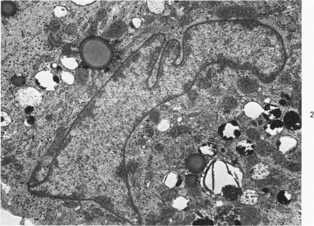
. Fig 2-Normal cultured fibroblast.")
14 72 KAMENSKY ET AL American Journal of Pathology Legends for Figures Fig 1-Normal cultured fibroblast with typical organelles: mitochondria, smooth and rough endoplasmic reticulum, ribosomes, Golgi structure, various dense and vacuolated bodies. The nucleolus is apparent (Uranyl acetate and lead citrate, x 10,500). Fig 2-Normal cultured fibroblast. The cytoplasm is filled with a large number of homogeneous or vacuolated dense bodies (Uranyl acetate and lead citrate, x 16,800). Fig 3-Fibroblast from a skin biopsy of a patient with Fabry's disease. The cytoplasm is filled with lamellar inclusions, surrounded by a unit membrane, few foamy vacuoles. Outside of the cell, bundles of collagen fibers are seen (Uranyl acetate and lead citrate, x 64,800). Fig 4-Cultured fibroblast from a patient with Fabry's disease. In the cytoplasm the inclusion bodies are surrounded by a unit membrane. These bodies are composed of stacks of parallel or concentric lamellae with alternating dark and light bands (Uranyl acetate and lead citrate, x 60,000). Fig 5-Cultured fibroblast from a patient with Fabry's disease. In addition to characteristic inclusions shown in Figure 3, there are typical organelles and inclusions which are also similar but larger and more numerous than those found in normal cultured fibroblasts (Uranyl acetate and lead citrate, x 18,400). Fig 6-Cultured fibroblasts from a patient with metachromatic leukodystrophy. Similar vacuoles with a reticulated, foamy matrix may be encountered in normal senescent fibroblasts. In the largest vacuoles present, the distended matrix becomes difficult to detect but almost never disappears entirely. A number of vacuoles have a signet-ring appearance (Uranyl acetate and lead citrate, x 11,500). Fig 7-Cultured fibroblast from patient with Gaucher's disease. The nucleus appears normal; the endoplasmic reticulum is dilated and contains flocculent material (Uranyl acetate and lead citrate, x 27,600). Fig 8-Cultured fibroblast from a patient with Niemann-Pick's disease, Type A. Inclusions surrounded by a unit membrane contain a granular core and loose, wavy, concentric lamellae (Uranyl acetate and lead citrate, x 32,000). Fig 9-Cultured fibroblast from another patient with Niemann-Pick's disease, Type A. In addition to inclusions similar to those shown in Figure 8, another type was present. The elongated shape of these inclusion bodies suggests a rigidity of the lamellae which are separated by a granular matrix (Uranyl acetate and lead citrate, x 100,000). Fig 10-Cultured fibroblast from a patient with Niemann-Pick's disease, Type C. This picture shows compact clusters of membranes within vacuoles, foamy reticulated inclusions and dilated endoplasmic reticulum (Uranyl acetate and lead citrate, x 64,400). Fig 11-Liver biopsy of a patient with Sanfilippo's disease. In the hepatocytes, vacuoles surrounded by a unit membrane contain a fine reticular matrix with a circumscribed electron-dense core and scattered granules of varying density. Abundant glycogen and mitochondria with indistinct cristae are also recognizable (Uranyl acetate and lead citrate, x 11,500). Fig 12-Cultured fibroblast from the same patient with Sanfilippo, Type A disease. Small vacuoles and dense glycogen clumps are present. The endoplasmic reticulum is dilated by a fine granular material (Uranyl acetate and lead citrate, x 11,500). Fig 13-Cultured fibroblast from a patient with Sanfilippo, Type B disease. Abundant glycogen and numerous large reticular vacuolated bodies were found (Uranyl acetate and lead citrate, x 8800). Inset-Details from the same micrograph which shows the reticular bodies with a signet-ring appearance (Uranyl acetate and lead citrate, x 22,000). Fig 14-Cultured fibroblast from a patient with chondroitin-4-sulphate mucopolysaccharidosis. The cytoplasm is filled with lamellar or granular inclusions. The Golgi structure is vacuolated, and the endoplasmic reticulum is dilated and filled with a granular material (Uranyl acetate and lead citrate, x 12,000). Inset-Detail showing the lamellar body, and vacuoles in the golgi structure (Uranyl acetate and lead citrate, x 15,500). Fig 15-Brain biopsy from a patient with lipofuscinosis. This picture demonstrates lipofuscin bodies with acid phosphatase activity (Uranyl acetate and lead citrate, x 25,600). Fig 16-A cultured fibroblast from the same patient as Figure 15. Tightly packed triglyceride-like inclusions and dense bodies with a granular matrix are seen (Uranyl acetate and lead citrate, x 28,800).
15 .4 I 2


16 e > i*7ee0 ;;;*.? lj *' '-S at h;-;3n<-sjix W x>v '-emr1 j ~ ~~~9 I, 3 4


17 6 ;$ '2),?.t -.0 -'r,' :x4"*
18 7 8
19 .dr-4f - i 41~~~~~~~~~~~~~~~~~~~~~~~~~~~~f sw 4 / /; ~~~~~~~~~~~~~~~~~~~~~~~~~~~~~C..:..w _ oi.s,am* L--"' I I V '.
20 1 1 12

21 13 NJ-1 14


22 15 V t , V. I 4;- dktv T. -17 % IF Al 4jv, 11.%,- w -O :1,,I..0 16
METABOLISM OF ACYLGLYCEROLS AND SPHINGOLIPDS. Ben S. Ashok MSc.,FAGE.,PhD., Dept. of Biochemistry
 METABOLISM OF ACYLGLYCEROLS AND SPHINGOLIPDS Ben S. Ashok MSc.,FAGE.,PhD., Dept. of Biochemistry STORAGE AND MEMBRANE LIPIDS STORAGE LIPIDS Mainly as triacylglycerols (triglycerides) in adipose cells Constitute
Cell Overview. Hanan Jafar BDS.MSc.PhD
 Cell Overview Hanan Jafar BDS.MSc.PhD THE CELL is made of: 1- Nucleus 2- Cell Membrane 3- Cytoplasm THE CELL Formed of: 1. Nuclear envelope 2. Chromatin 3. Nucleolus 4. Nucleoplasm (nuclear matrix) NUCLEUS
The role of the laboratory in diagnosing lysosomal disorders
 The role of the laboratory in diagnosing lysosomal disorders Dr Guy Besley, formerly Willink Biochemical Genetics Unit, Manchester Children s Hospital, Manchester M27 4HA, UK. Lysosomal disorders What
(From The Rockefeller Institute) Materials and Methods. Observations with the Electron Microscope
 Materials and Methods. Observations with the Electron Microscope") ELECTRON MICROSCOPE STUDY OF THE DEVELOPMENT OF THE PAPILLOMA VIRUS IN THE SKIN OF THE RABBIT* BY ROBERT S. STONE,~ M.D., RICHARD E. SHOPE, M.D., DAN H. MOORE, P,~.D. (From The Rockefeller Institute) PLATES
ELECTRON MICROSCOPE STUDY OF THE DEVELOPMENT OF THE PAPILLOMA VIRUS IN THE SKIN OF THE RABBIT* BY ROBERT S. STONE,~ M.D., RICHARD E. SHOPE, M.D., DAN H. MOORE, P,~.D. (From The Rockefeller Institute) PLATES
Chapter 2 Cell. Zhou Li Prof. Dept. of Histology and Embryology
 Chapter 2 Cell Zhou Li Prof. Dept. of Histology and Embryology The inner life of the cell Ⅰ. Plasma membrane (Plasmalemma) 1.1 The structure Unit membrane: inner layer 3-layered structure outer layer mediat
1. Diagnosis of Lysosomal Storage Disorders in Australia. 2. Comparison of Incidence/prevalence of lysosomal storage diseases in different country
 List of Tables: 1. Diagnosis of Lysosomal Storage Disorders in Australia 2. Comparison of Incidence/prevalence of lysosomal storage diseases in different country 3. Relative frequency of LSD in Portugal
Fine Structure of the Normal Trigeminal Ganglion in the Cat and Monkey*
 Fine Structure of the Normal Trigeminal Ganglion in the Cat and Monkey* DAVID S. MAXWELL, PH.D. Principal Contributor and Leader of Discussion HE inclusion of animal material m a y be justified as a means
The Cytoplasm Li Shulei Department of Histology & Embryology
 The Cytoplasm Li Shulei lishulei@tom.com Department of Histology & Embryology Cell components Cytoplasm Plasma membrane Organelles Cytoplasmic deposits Cytoskeleton Cytosol ( Matrix ) Nucleus Plasma membrane
Basophilic. Basophilic structures are stained by basic dyes: Mnemonic: Basophilic = Blue
 Cell Overview Basophilic Basophilic structures are stained by basic dyes: Basic dyes are positive Basophilic structures are negative (ex. DNA, RNA, ribosomes, RER) Mnemonic: Basophilic = Blue Acidophilic
Metabolism of acylglycerols and sphingolipids. Martina Srbová
 Metabolism of acylglycerols and sphingolipids Martina Srbová Types of glycerolipids and sphingolipids 1. Triacylglycerols function as energy reserves adipose tissue (storage of triacylglycerol), lipoproteins
Clinical Approach to Diagnosis of Lysosomal Storage Diseases
 Clinical Approach to Diagnosis of Lysosomal Storage Diseases M. Rohrbach, MD, PhD FMH Pädiatrie und FMH Medizinische Genetik Abteilung Stoffwechsel Universitätskinderklinik Zürich Lysosomal storage disorders
SBI3U7 Cell Structure & Organelles. 2.2 Prokaryotic Cells 2.3 Eukaryotic Cells
 SBI3U7 Cell Structure & Organelles 2.2 Prokaryotic Cells 2.3 Eukaryotic Cells No nucleus Prokaryotic Cells No membrane bound organelles Has a nucleus Eukaryotic Cells Membrane bound organelles Unicellular
ELECTRON MICROSCOPIC STUDIES ON EQUINE ENCEPHALOSIS VIRUS
 Onderstepoort]. vet. Res. 40 (2), 53-58 (1973) ELECTRON MICROSCOPIC STUDIES ON EQUINE ENCEPHALOSIS VIRUS G. LECATSAS, B. J. ERASMUS and H. J. ELS, Veterinary Research Institute, Onderstepoort ABSTRACT
The Cell Organelles. Eukaryotic cell. The plasma membrane separates the cell from the environment. Plasma membrane: a cell s boundary
 Eukaryotic cell The Cell Organelles Enclosed by plasma membrane Subdivided into membrane bound compartments - organelles One of the organelles is membrane bound nucleus Cytoplasm contains supporting matrix
basic unit structure and function
 Chapter 3 Cells Introduction The cell is the basic unit of structure and function in living things. Cells vary in their shape, size, and arrangements, but all cells have similar components with a particular
Cell Structure. Present in animal cell. Present in plant cell. Organelle. Function. strength, resist pressure created when water enters
 Cell Structure Though eukaryotic cells contain many organelles, it is important to know which are in plant cells, which are in animal cells and what their functions are. Organelle Present in plant cell
Cell Structure & Function. Source:
 Cell Structure & Function Source: http://koning.ecsu.ctstateu.edu/cell/cell.html Definition of Cell A cell is the smallest unit that is capable of performing life functions. http://web.jjay.cuny.edu/~acarpi/nsc/images/cell.gif
Cell morphology. Cell organelles structure and function. Chapter 1: UNIT 1. Dr. Charushila Rukadikar
 UNIT 1 Cell morphology Cell organelles structure and function Chapter 1: Dr. Charushila Rukadikar Assistant Professor Department Of Physiology ZMCH, Dahod Physiology The science that is concerned with
Conjunctival ultrastructure in mucolipidosis III (pseudo-hurler polydystrophy)
") Conjunctival ultrastructure in mucolipidosis III (pseudo-hurler polydystrophy) Harry A. Quigley and Morton F. Goldberg Conjunctiva! biopsies from two sibs with mucolipidosis III and from their clinically
Conjunctival ultrastructure in mucolipidosis III (pseudo-hurler polydystrophy) Harry A. Quigley and Morton F. Goldberg Conjunctiva! biopsies from two sibs with mucolipidosis III and from their clinically
Cell Structure and Function. Biology 12 Unit 1 Cell Structure and Function Inquiry into Life pages and 68-69
 Cell Structure and Function Biology 12 Unit 1 Cell Structure and Function Inquiry into Life pages 45 59 and 68-69 Assignments for this Unit Pick up the notes/worksheet for this unit and the project There
Ultrastructural Study of Human Natural Killer CNK) Cell*)
 Cell*)") Hiroshima Journal of Medical Sciences Vol. 31, No. 1, March, 1982 HJIM 31-6 31 Ultrastructural Study of Human Natural Killer CNK) Cell*) Yoshinori KAWAGUCHI, Eishi KITTAKA, Yoshito TANAKA, Takeo TANAKA
Hiroshima Journal of Medical Sciences Vol. 31, No. 1, March, 1982 HJIM 31-6 31 Ultrastructural Study of Human Natural Killer CNK) Cell*) Yoshinori KAWAGUCHI, Eishi KITTAKA, Yoshito TANAKA, Takeo TANAKA
New aspect of hepatic nuclear glycogenosis
 J. clin. Path. (1968), 21, 19 New aspect of hepatic nuclear glycogenosis in diabetes1 F. CARAMIA, F. G. GHERGO, C. BRANCIARI, AND G. MENGHINI From the Institute of General Pathology, University of Rome,
Basic Structure of a Cell
 Basic Structure of a Cell 1 Introduction to Cells Cells are the basic units of organisms Cells can only be observed under microscope Basic types of cells: Animal Cell Plant Cell Bacterial Cell 2 Number
AN ELECTRON-MICROSCOPIC STUDY OF THE STARCH-CONTAINING PLASTIDS IN THE FERN TODEA BARBARA
 J. Cell Sci. 4, 211-221 (1969) 211 Printed in Great Britain AN ELECTRON-MICROSCOPIC STUDY OF THE STARCH-CONTAINING PLASTIDS IN THE FERN TODEA BARBARA H. M. SMITH* AND D. S. SMITHf Department of Biology,
Eukaryotic Cell Structures
 Comparing the Cell to a Factory Eukaryotic Cell Structures Structures within a eukaryotic cell that perform important cellular functions are known as organelles. Cell biologists divide the eukaryotic cell
Lysosomes. Gr: lysis solution, soma body. Membrane bounded vesicles. Usually round ovoid or irregular electron dense bodies m.
 Lysosomes Gr: lysis solution, soma body Membrane bounded vesicles Usually round ovoid or irregular electron dense bodies 0.05 0.5 m. Lysosomes No. varies from a few to several hundred per cell, in different
7-2 : Plasma Membrane and Cell Structures
 7-2 : Plasma Membrane and Cell Structures Plasma Membrane of aveolar sac But first... Let s Review What is cell theory? Light microscopes vs. electron microscopes Prokaryotic vs. eukaryotic Basic Cell
7-2 : Plasma Membrane and Cell Structures
 7-2 : Plasma Membrane and Cell Structures Plasma Membrane of aveolar sac But first... Let s Review What is cell theory? Light microscopes vs. electron microscopes Prokaryotic vs. eukaryotic Basic Cell
Intercellular Matrix in Colonies of Candida
 JouRNAL OF BAcTEROLOGY, Sept. 1975, p. 1139-1143 Vol. 123, No. 3 Copyright 0 1975 American Society for Microbiology Printed in U.S.A. ntercellular Matrix in Colonies of Candida K. R. JOSH, J. B. GAVN,*
10/13/11. Cell Theory. Cell Structure
 Cell Structure Grade 12 Biology Cell Theory All organisms are composed of one or more cells. Cells are the smallest living units of all living organisms. Cells arise only by division of a previously existing
Organelles Found in a Generalized Animal Cell
 Organelles Found in a Generalized Animal Cell 1. Cell Membrane 2. Cytoplasm 3. Nucleus 4. Nuclear Membrane 5. Nucleoplasm 6. Nucleolus 7. Chromosomes 8. Vacuole 9. Ribosomes 10. Rough Endoplasmic Reticulum
Cell Physiology
 Cell Physiology 21-10-2018 1 The two major parts of a typical cell are the nucleus and the cytoplasm. The nucleus is separated from the cytoplasm by a nuclear membrane, and the cytoplasm is separated from
By: Brooke Sheppard
 By: Brooke Sheppard What is a Cell? Cells are the basic structure of life for all organisms. Cells are microscopic, which means we can only view cells under a microscope. There are animal cells and plant
Some Observations on the Fine Structure of the Goblet Cells. Special Reference to the Well-Developed Agranular Endoplasmic Reticulum
 Okajimas Folia Anat. Jpn., 58(4-6) : 583-594, March 1982 Some Observations on the Fine Structure of the Goblet Cells in the Nasal Respiratory Epithelium of the Rat, with Special Reference to the Well-Developed
Cell Structure and Organelles SBI4U 2016/10/14
 Cell Structure and Organelles SBI4U 2016/10/14 Inside the cell These are generalizations, not rules! Everything inside the cell membrane besides the nucleus is called the cytoplasm; The liquid is known
ataxia, head tremors and mild inappentence, was given palliative care
 2013-5-2 Cerebellum, Spleen-Raccoon Ahmed M. Abubakar BOVINE PATHOLOGY CONTRIBUTING INSTITUTION :College of Veterinary Medicine UC Davies Signalment: Wild-caught juvenile male raccoon, ( Procyon lotor)
STUDIES OF THE HUMAN UNFERTILIZED TUBAL OVUM*t
 FERTILITY AND STERILITY Copyright @ 1973 by The Williams & Wilkins Co. Vol. 24, No.8, August 1973 Printed in U.S.A. STUDIES OF THE HUMAN UNFERTILIZED TUBAL OVUM*t C. NORIEGA, M.D., AND C. OBERTI, M.D.
Initially, the patients did not receive extra vitamin E except for a very
 EFFECT OF VITAMIN E ON MEMBRANES OF THE INTESTINAL CELL BY I. MOLENAAR, F. A. HOMMES, W. G. BRAAMS, AND H. A. POLMAN CENTER FOR MEDICAL ELECTRON MICROSCOPY AND DEPARTMENT OF PEDIATRICS, UNIVERSITY OF GRONINGEN,
NUCLEOLUS CELL MEMBRANE MITOCHONDRIA CELL WALL CHLOROPLAST NUCLEAR MEMBRANE VACOULE NUCLEUS ORGANELLE CARD ORGANELLE CARD ORGANELLE CARD
 CELL MEMBRANE NUCLEOLUS CELL WALL MITOCHONDRIA NUCLEAR MEMBRANE CHLOROPLAST NUCLEUS VACOULE CENTRAL VACOULE LEUCOPLAST CHROMOPLAST LYSOSOME CYTOPLASM CYTOSKELETON CENTRIOLE RIBOSOME CHROMATIN ROUGH ENDOPLASMIC
Organelles. copyright cmassengale 1
 Organelles copyright cmassengale 1 Organelles Very small (Microscopic) Perform various functions for a cell Found in the cytoplasm May or may not be membrane-bound 2 Animal Cell Organelles Nucleolus Nucleus
A&P 1 Cellular Anatomy, Division & Mitosis - Pre-Lab Exercises
 A&P 1 Cellular Anatomy, Division & Mitosis - Pre-Lab Exercises Have someone in your group read the following out loud, while the others read along: In this "Pre-lab Guide", we will be going over some of
The Fine Structure of the Epithelial Cells of the Mouse Prostate* II. Ventral Lobe Epithelium
 Published Online: 1 June, 1960 Supp Info: http://doi.org/10.1083/jcb.7.3.511 Downloaded from jcb.rupress.org on September 28, 2018 The Fine Structure of the Epithelial Cells of the Mouse Prostate* II.
Structures in Cells. Cytoplasm. Lecture 5, EH1008: Biology for Public Health, Biomolecules
 Structures in Cells Lecture 5, EH1008: Biology for Public Health, Biomolecules Limian.zheng@ucc.ie 1 Cytoplasm Nucleus Centrioles Cytoskeleton Cilia Microvilli 2 Cytoplasm Cellular material outside nucleus
Essentials of Anatomy and Physiology, 9e (Marieb) Chapter 3 Cells and Tissues. Short Answer. Figure 3.1
 Chapter 3 Cells and Tissues. Short Answer. Figure 3.1") Essentials of Anatomy and Physiology, 9e (Marieb) Chapter 3 Cells and Tissues Short Answer Figure 3.1 Using Figure 3.1, match the following: 1) The illustration of simple cuboidal epithelium is. Answer:
Essentials of Anatomy and Physiology, 9e (Marieb) Chapter 3 Cells and Tissues Short Answer Figure 3.1 Using Figure 3.1, match the following: 1) The illustration of simple cuboidal epithelium is. Answer:
Structure. Lysosomes are membrane-enclosed organelles. Hydrolytic enzymes. Variable in size & shape need
 Lysosomes Structure Lysosomes are membrane-enclosed organelles Hydrolytic enzymes Variable in size & shape need Degrade material taken up from outside and inside the cell Variable in size and shape Lysosomal
Ultrastructure of Connective Tissue Cells of Giant African Snails Achatina fulica (Bowdich)
") Kasetsart J. (Nat. Sci.) 36 : 285-290 (2002) Ultrastructure of Connective Tissue Cells of Giant African Snails Achatina fulica (Bowdich) Viyada Seehabutr ABSTRACT The connective tissue sheath of cerebral
Kasetsart J. (Nat. Sci.) 36 : 285-290 (2002) Ultrastructure of Connective Tissue Cells of Giant African Snails Achatina fulica (Bowdich) Viyada Seehabutr ABSTRACT The connective tissue sheath of cerebral
A. Major parts 1. Nucleus 2. Cytoplasm a. Contain organelles (see below) 3. Plasma membrane (To be discussed in Cellular Transport Lecture)
 3. Plasma membrane (To be discussed in Cellular Transport Lecture)") Lecture 5: Cellular Biology I. Cell Theory Concepts: 1. Cells are the functional and structural units of living organisms 2. The activity of an organism is dependent on both the individual and collective
Lecture 5: Cellular Biology I. Cell Theory Concepts: 1. Cells are the functional and structural units of living organisms 2. The activity of an organism is dependent on both the individual and collective
Chapter 3 Cell Structures & Functions
 Biology 12 Name: Cell Biology Per: Date: Chapter 3 Cell Structures & Functions Complete using BC Biology 12, pages 62-107 Diagnostic Questions (mark using the answer key on page 527) 1. 2. 3. 4. 9. What
Structures in Cells. Lecture 5, EH1008: Biology for Public Health, Biomolecules.
 Structures in Cells Lecture 5, EH1008: Biology for Public Health, Biomolecules Limian.zheng@ucc.ie 1 Cytoplasm Nucleus Centrioles Cytoskeleton Cilia Microvilli 2 Cytoplasm Cellular material outside nucleus
Abdallah Q& Razi. Faisal
 27 & Ahmad Attari م ح م د ي وس ف Abdallah Q& Razi Faisal Sphingophospolipids - The backbone of sphingophospholipids is sphingosine, unlike glycerophospholipids with a glycerol as the backbone. Which contains
6. What surrounds the nucleus? How many membranes does it have?
 Biology-R track Study Guide: 7.2 Cell Structure Cell Organization 1. What are the 2 major parts that you can divide the eukaryotic cell into? 2. What part is the fluid portion of the cell outside the nucleus?
Title. Author(s)SONODA, Mitsuo; KOBAYASHI, Kosaku. CitationJapanese Journal of Veterinary Research, 18(3): 125- Issue Date DOI. Doc URL.
SONODA, Mitsuo; KOBAYASHI, Kosaku. CitationJapanese Journal of Veterinary Research, 18(3): 125- Issue Date DOI. Doc URL.") Title PLASMACYTOID CELLS OF CANINE PERIPHERAL BLOOD IN ELE Author(s)SONODA, Mitsuo; KOBAYASHI, Kosaku CitationJapanese Journal of Veterinary Research, 18(3): 125- Issue Date 1970-09 DOI 10.14943/jjvr.18.3.125
Title PLASMACYTOID CELLS OF CANINE PERIPHERAL BLOOD IN ELE Author(s)SONODA, Mitsuo; KOBAYASHI, Kosaku CitationJapanese Journal of Veterinary Research, 18(3): 125- Issue Date 1970-09 DOI 10.14943/jjvr.18.3.125
First to View Cells. copyright cmassengale
 CELL THEORY All living things are made of cells Cells are the basic unit of structure and function in an organism (basic unit of life) Cells come from the reproduction of existing cells (cell division)
Ocular pathology of GM2 gangliosidosis
 Brit. J. Ophthal. (I973) 579 514 Ocular pathology of GM2 gangliosidosis Type 2 (Sandhoff's disease) ALEC GARNER Institute of Ophthalmology, University of London The introduction in recent years of more
CELLS.
 CELLS http://www.aimediaserver.com/studiodaily/harvard/harvard.swf INTERESTING FACTS The longest cells in the human body are the motor neurons. They can be up to 1.37 meters long and go from the spinal
Human Epithelial Cells
 The Cell Human Epithelial Cells Plant Cells Cells have an internal structure Eukaryotic cells are organized Protective membrane around them that communicates with other cells Organelles have specific jobs
MULTIPLE CHOICE. Choose the one alternative that best completes the statement or answers the question.
 Exam Name MULTIPLE CHOICE. Choose the one alternative that best completes the statement or answers the question. 1) All of the following are synthesized along various sites of the endoplasmic reticulum
Chapter 7 Notes. Section 1
 Chapter 7 Notes Section 1 Cells Cells remained out of sight during most of human history until the invention of the first microscopes. It was not until the mid 1600s that scientists began to use microscopes
Eukaryotic cells are essentially two envelope systems. Nuclear materials are separated from cytoplasm by nuclear membrane. Complex structure Also
 Dr. Gugale Pritesh Ramanlal M.Sc., Ph.D., B.Ed., D.M.L.T. Email id - pritesh.gugale09@gmail.com Contact numbernumber- 8446475310 Eukaryotic cells are essentially two envelope systems. Nuclear materials
number Done by Corrected by Doctor
 number 26 Done by حسام أبو عوض Corrected by Zaid Emad Doctor فيصل الخطيب 1 P a g e A small note about phosphatidyl inositol-4,5-bisphosphate (PIP2) before moving on: This molecule is found in the membrane
Cell Theory All living matter is composed of one or more The cell is the structural and functional unit of life All cells come from pre-existing cell
 Cell Theory All living matter is composed of one or more The cell is the structural and functional unit of life All cells come from pre-existing cell Prokeryotic Bacteria or archaea Cell wall, small circular
CELL (PLASMA) MEMBRANE
 MEMBRANE") CELL (PLASMA) MEMBRANE Separates cell from its environment Controls substances that enter and leave the cell Selectively permeable allows certain substances to pass COMPOSITION Composed of 2 layers of
CELL (PLASMA) MEMBRANE Separates cell from its environment Controls substances that enter and leave the cell Selectively permeable allows certain substances to pass COMPOSITION Composed of 2 layers of
CELL STRUCTURE AND FUNCTION. Chapter 7
 CELL STRUCTURE AND FUNCTION Chapter 7 WARM UP EXERCISE Please complete the pretest that you picked up as you came in. LIFE IS CELLULAR Robert Hooke- coined the term cells The Cell Theory All living things
Plants, Animals, Fungi and Protists have Eukaryotic Cell(s)
") Cell Structure Plants, Animals, Fungi and Protists have Eukaryotic Cell(s) Plant Cell Animal Cell straight edges curved edges Cell Organization cytoplasm cell membrane Eukaryotic cells have 3 major parts:
Cell Structure Plants, Animals, Fungi and Protists have Eukaryotic Cell(s) Plant Cell Animal Cell straight edges curved edges Cell Organization cytoplasm cell membrane Eukaryotic cells have 3 major parts:
SHORT ANSWER. Write the word or phrase that best completes each statement or answers the question.
 SHORT ANSWER. Write the word or phrase that best completes each statement or answers the question. Figure 2.1 Using Figure 2.1, match the following: 1) Rough endoplasmic reticulum 1) 2) Nucleolus 2) 3)
:1c.c :& Preliminary and Short Report GRANULE FORMATION IN THE LANGERHANS CELL* structure with rounded ends and a striated lamella
 THE JOURNAL OF INVESTIGATIVE DERMATOLOGY Copyright 1566 by The Williams & Wilkins Co. Vol. 7, No. 5 Printed in U.S.A. Preliminary and Short Report GRANULE FORMATION IN THE LANGERHANS CELL* ALVIN S. ZELICKSON,
Cell Structure. Cells. Why are cells so small? 9/15/2016. Schleiden and Schwann proposed Cell Theory in
 Cell Structure Cells Cells are sacs of fluid that are reinforced by proteins and surrounded by membranes. Inside the fluid float organelles. Organelles: structures inside the cell that are used for metabolic
2. scanning electron microscope vs. transmission electron microscope. nucleus, nuclear envelope, nucleolus, ribosomes
 Honors Biology Unit 2 Chapter 4 A TOUR OF THE CELL 1. light microscope 2. scanning electron microscope vs. transmission electron microscope 3. surface area to volume ratio 4. prokaryotic cell vs. animal
Starch grains - excess sugars
 (a) Membrane system - site of light reactions (photosynthesis) - chlorpophyll pigments - enzymes - electron carriers - flattened, fluid-filled sacs (called thylakoids which are stacked to form grana) -
(From the Division q[ Human Genetics, Department of Medicine, CorneU University Medical College, New York 10021) Materials and Methods
 Materials and Methods") OYSTER CILIARY INHIBITION BY CYSTIC FIBROSIS CULTURE MEDIUM* BY B. SHANNON DANES ANn ALEXANDER G. BEARN (From the Division q[ Human Genetics, Department of Medicine, CorneU University Medical College,
OYSTER CILIARY INHIBITION BY CYSTIC FIBROSIS CULTURE MEDIUM* BY B. SHANNON DANES ANn ALEXANDER G. BEARN (From the Division q[ Human Genetics, Department of Medicine, CorneU University Medical College,
Cell Theory Vocabulary Flashcards
 Mr. Powner Biology Cell Theory Vocabulary Flashcards Instructions: Cut out the flashcards from the following pages. The following word list is the vocabulary for studying cell theory. Write each word on
BME NEUROSCIENCE PRINCIPLES OF NEURAL SCIENCE 1 ST SEMESTER GRADUATE COURSE HYOUNG F. KIM
 BME NEUROSCIENCE PRINCIPLES OF NEURAL SCIENCE 1 ST SEMESTER GRADUATE COURSE HYOUNG F. KIM BASIC CONCEPT OF CELL BIOLOGY CELL & NEURON What are the differences? BASIC CONCEPT OF CELL 1. What are the cells
Cell Structure Text Ref Pg 4-7, 63-81
 Cell Structure Text Ref Pg 4-7, 63-81 The Cellular Basis of Life Before people had a scientific explanation for where life came from, they believed in the theory of spontaneous generation, where organisms
(A) Cell membrane (B) Ribosome (C) DNA (D) Nucleus (E) Plasmids. A. Incorrect! Both prokaryotic and eukaryotic cells have cell membranes.
 Cell membrane (B) Ribosome (C) DNA (D) Nucleus (E) Plasmids. A. Incorrect! Both prokaryotic and eukaryotic cells have cell membranes.") High School Biology - Problem Drill 03: The Cell No. 1 of 10 1. Which of the following is NOT found in prokaryotic cells? #01 (A) Cell membrane (B) Ribosome (C) DNA (D) Nucleus (E) Plasmids Both prokaryotic
High School Biology - Problem Drill 03: The Cell No. 1 of 10 1. Which of the following is NOT found in prokaryotic cells? #01 (A) Cell membrane (B) Ribosome (C) DNA (D) Nucleus (E) Plasmids Both prokaryotic
Date of commencement: February Principal Investigator Dr. Jayesh J. Sheth CASE RECORD FORM
 ICMR-FRIGE-MULTICENTRIC LSDs Project Foundation for Research in Genetics & Endocrinology [FRIGE], FRIGE House, Jodhpur Gam road, Satellite, Ahmedabad-380015 Tel no: 079-26921414, Fax no: 079-26921415 E-mail:
Yara shwabkeh. Osama Alkhader. Heba Kalbouneh
 2 Yara shwabkeh Osama Alkhader Heba Kalbouneh CELL OVERVIEW -Note ; the important thing is to know how the organelles appear under the microscope - the stains we usually use in Histology are composed of
Ultrastructural studies of human cutaneous nerve
 J. clin. Path. (1965), 18, 188 Ultrastructural studies of human cutaneous nerve with special reference to lamellated cell inclusions and vacuole-containing cells MARJORE J. EVANS, J. B. FNEAN, AND A. L.
The endoplasmic reticulum is a network of folded membranes that form channels through the cytoplasm and sacs called cisternae.
 Endoplasmic reticulum (ER) The endoplasmic reticulum is a network of folded membranes that form channels through the cytoplasm and sacs called cisternae. Cisternae serve as channels for the transport of
MEMBRANE LIPIDS I and II: GLYCEROPHOSPHOLIPIDS AND SPHINGOLIPIDS
 December 6, 2011 Lecturer: Eileen M. Lafer MEMBRANE LIPIDS I and II: GLYCEROPHOSPHOLIPIDS AND SPHINGOLIPIDS Reading: Stryer Edition 6: Chapter 26 Images: All images in these notes were taken from Lehninger,
Section 7 2 Eukaryotic Cell Structure
 Section 7 2 Eukaryotic Cell Structure (pages 174 181) Key Concept What are the functions of the major cell structures? Comparing a Cell to a Factory (page 174) 1. What is an organelle? 2. Label the structures
The Cell. Biology 105 Lecture 4 Reading: Chapter 3 (pages 47 62)
") The Cell Biology 105 Lecture 4 Reading: Chapter 3 (pages 47 62) Outline I. Prokaryotic vs. Eukaryotic II. Eukaryotic A. Plasma membrane transport across B. Main features of animal cells and their functions
The Cell Biology 105 Lecture 4 Reading: Chapter 3 (pages 47 62) Outline I. Prokaryotic vs. Eukaryotic II. Eukaryotic A. Plasma membrane transport across B. Main features of animal cells and their functions
Don t Freak Out. Test on cell organelle on Friday!
 Cell Structure 1 Don t Freak Out Test on cell organelle on Friday! This test should be a buffer test and help raise your overall test score. All information will come from this week! 2 Cells Provide Compartments
Membranous Organelles
 Membranous Organelles 1. Membrane-limited organelles 2. Golgi apparatus 3. Lysosomes 4. Peroxisomes (microbodies) 5. Secretory vesicles (granules) 6. Coated vesicles Membrane-limited organelles Endoplasmic
I-Cell Disease: Biochemical Studies
 JULES G. LEROY, MAE WAN HO, MONICA C. MACBRINN, KLAUS ZIELKE, JACK JACOB, AND JOHN S. O'BRIEN 132 ] Pediat. Res. 6: 52-5 (12) Acid hydrolases lipids I-cell disease lysosome I-Cell Disease: Biochemical
EDUCATIONAL COMMENTARY MORPHOLOGIC ABNORMALITIES IN LEUKOCYTES
 EDUCATIONAL COMMENTARY MORPHOLOGIC ABNORMALITIES IN LEUKOCYTES Educational commentary is provided through our affiliation with the American Society for Clinical Pathology (ASCP). To obtain FREE CME/CMLE
UNIT 2: Cells Chapter 3: Cell Structure and Function
 UNIT 2: Cells Chapter 3: Cell Structure and Function I. Cell Theory (3.1) A. Early studies led to the development of the cell theory 1. Discovery of Cells a. Robert Hooke (1665)-Used compound microscope
ON THE PRESENCE OF A CILIATED COLUMNAR EPITHELIAL CELL TYPE WITHIN THE BOVINE CERVICAL MUCOSA 1
 ON THE PRESENCE OF A CILIATED COLUMNAR EPITHELIAL CELL TYPE WITHIN THE BOVINE CERVICAL MUCOSA 1 R. I. Wordinger, 2 J. B. Ramsey, I. F. Dickey and I. R. Hill, Jr. Clemson University, Clemson, South Carolina
Most mammalian cells are located in tissues where they are surrounded by a complex extracellular matrix (ECM) often referred to as connective tissue.
 often referred to as connective tissue.") GLYCOSAMINOGLYCANS Most mammalian cells are located in tissues where they are surrounded by a complex extracellular matrix (ECM) often referred to as connective tissue. The ECM contains three major classes
GLYCOSAMINOGLYCANS Most mammalian cells are located in tissues where they are surrounded by a complex extracellular matrix (ECM) often referred to as connective tissue. The ECM contains three major classes
Cells & Cell Organelles
 Cells & Cell Organelles The Building Blocks of Life AP Biology 2008-2009 Types of cells bacteria cells Prokaryote - no organelles Eukaryotes - organelles animal cells plant cells Cell size comparison Animal
Blood Cells. Dr. Sami Zaqout. Dr. Sami Zaqout Faculty of Medicine IUG
 Blood Cells Dr. Sami Zaqout Blood Blood Blood cells (45%) Erythrocytes Platelets Leukocytes Plasma (55%) Hematocrit tubes with blood Composition of Plasma Plasma Aqueous solution (90%) Substances (10%)
Biology 12 Cell Structure and Function. Typical Animal Cell
 Biology 12 Cell Structure and Function Typical Animal Cell Vacuoles: storage of materials and water Golgi body: a series of stacked disk shaped sacs. Repackaging centre stores, modifies, and packages proteins
Biology. Dr. Khalida Ibrahim
 Biology Dr. Khalida Ibrahim BONE TISSUE Bone tissue is a specialized form of connective tissue and is the main element of the skeletal tissues. It is composed of cells and an extracellular matrix in which
Silver-Impregnation of the Golgi Complex in Epididymal Epithelial Cells of Mice
 CELL STRUCTURE AND FUNCTION 8, 339-346 (1984) C by Japan Society for Cell Biology Silver-Impregnation of the Golgi Complex in Epididymal Epithelial Cells of Mice Ikuo Yamaoka, Sumie Katsuta and Yoshimi
*Need a summary just not covered yet!
 *Need a summary just not covered yet! Organelle Pupil Organelle Pupil Nucleus KO N Microtubules + centrioles LF Endoplasmic reticulum ED Plant cell wall OC Ribosomes KL Chloroplasts RK Golgi apparatus
Cell Structure and and Function Chapter 4
 Cell Structure and Function Chapter 4 Robert Hooke (1635-1703) 1703) Discovered cells by studying the cork layer of bark from an oak tree. Found cells when studied tree stems, roots, and leaves. Antony
Chapter 3: Cytology. Cytology is the study of cells. Cells are the basic units of life. We are made up of trillions of cells.
 PLEASE NOTE THAT THE ITEMS IN THE TEXT THAT ARE HIGHLIGHTED IN YELLOW ARE THOSE THAT ARE TOUCHED ON IN THE READING ASSIGNMENT (PAGES 90-99) AND IN THE LECTURE. ESPECIALLY KNOW THIS MATERIAL FOR THE FIRST
Biology 2.4 AS Introduction Cells and cell processes. Introduction Cells and cell processes
 Biology 2.4 AS 91156 Demonstrate understanding of life processes at the cellular level Externally assessed 4 credits Copy correctly Up to 3% of a workbook Copying or scanning from ESA workbooks is subject
CELL PART OF THE DAY. Chapter 7: Cell Structure and Function
 CELL PART OF THE DAY Chapter 7: Cell Structure and Function Cell Membrane Cell membranes are composed of two phospholipid layers. Cell membrane is flexible, not rigid The cell membrane has two major functions.
Renata Schipp Medical Biology Department
 Renata Schipp Medical Biology Department Deffinition of cell The cell is the smallest structural and functional unit of all known living organisms The cell was discovered by Robert Hooke in 1665 and also
RESEARCH COMMUNICATION TISSUE CULTURE STUDIES ON A SUSPECTED LYSOSOMAL STORAGE DISEASE IN ABYSSINIAN CATS
 Onderstepoort f. vet. Res., 47, 121-127 (1980) RESEARCH COMMUNICATION TISSUE CULTURE STUDIES ON A SUSPECTED LYSOSOMAL STORAGE DISEASE IN ABYSSINIAN CATS A. LUClA LANGE, Department of Pathology, Faculty