Group X secreted phospholipase A2 induces lipid droplet formation and prolongs breast cancer cell survival
|
|
|
- Sharyl Parrish
- 6 years ago
- Views:
Transcription

1 Molecular Cancer This Provisional PDF corresponds to the article as it appeared upon acceptance. Fully formatted PDF and full text (HTML) versions will be made available soon. Group X secreted phospholipase A2 induces lipid droplet formation and prolongs breast cancer cell survival Molecular Cancer 2013, 12:111 doi: / Anja Pucer (anja.pucer@ijs.si) Vesna Brglez (vesna.brglez@ijs.si) Christine Payré (payre@ipmc.cnrs.fr) Jo e Punger ar (joze.pungercar@ijs.si) Gérard Lambeau (lambeau@ipmc.cnrs.fr) Toni Petan (toni.petan@ijs.si) ISSN Article type Research Submission date 4 July 2013 Acceptance date 24 September 2013 Publication date 27 September 2013 Article URL This peer-reviewed article can be downloaded, printed and distributed freely for any purposes (see copyright notice below). Articles in Molecular Cancer are listed in PubMed and archived at PubMed Central. For information about publishing your research in Molecular Cancer or any BioMed Central journal, go to For information about other BioMed Central publications go to Pucer et al. This is an open access article distributed under the terms of the Creative Commons Attribution License ( which permits unrestricted use, distribution, and reproduction in any medium, provided the original work is properly cited.
2 Group X secreted phospholipase A 2 induces lipid droplet formation and prolongs breast cancer cell survival Abstract Background Anja Pucer 1 anja.pucer@ijs.si Vesna Brglez 1 vesna.brglez@ijs.si Christine Payré 2 payre@ipmc.cnrs.fr Jože Pungerčar 1 joze.pungercar@ijs.si Gérard Lambeau 2 lambeau@ipmc.cnrs.fr Toni Petan 1* * Corresponding author toni.petan@ijs.si 1 Department of Molecular and Biomedical Sciences, Jožef Stefan Institute, Ljubljana, Slovenia 2 Institut de Pharmacologie Moléculaire et Cellulaire, CNRS et Université de Nice Sophia Antipolis, UMR 6097, Sophia Antipolis Valbonne, France Alterations in lipid metabolism are inherent to the metabolic transformations that support tumorigenesis. The relationship between the synthesis, storage and use of lipids and their importance in cancer is poorly understood. The human group X secreted phospholipase A 2 (hgx spla 2 ) releases fatty acids (FAs) from cell membranes and lipoproteins, but its involvement in the regulation of cellular FA metabolism and cancer is not known. Results Here we demonstrate that hgx spla 2 induces lipid droplet (LD) formation in invasive breast cancer cells, stimulates their proliferation and prevents their death on serum deprivation. The effects of hgx spla 2 are shown to be dependent on its enzymatic activity, are mimicked by oleic acid and include activation of protein kinase B/Akt, a cell survival signaling kinase. The hgx spla 2 -stimulated LD biogenesis is accompanied by AMP-activated protein kinase (AMPK) activation, up-regulation of FA oxidation enzymes and the LD-coating protein
3 perilipin 2, and suppression of lipogenic gene expression. Prolonged activation of AMPK inhibited hgx spla 2 -induced LD formation, while etomoxir, an inhibitor of FA oxidation, abrogated both LD formation and cell survival. The hgx spla 2 -induced changes in lipid metabolism provide a minimal immediate proliferative advantage during growth under optimal conditions, but they confer to the breast cancer cells a sustained ability to resist apoptosis during nutrient and growth factor limitation. Conclusion Our results identify hgx spla 2 as a novel modulator of lipid metabolism that promotes breast cancer cell growth and survival by stimulating LD formation and FA oxidation. Keywords Secreted phospholipase A 2, Breast cancer, Cell survival, Apoptosis, Lipid droplets, Fatty acid oxidation, Lipid signaling, Etomoxir, Carnitine palmitoyltransferase 1, AMP-activated protein kinase Background Tumor cells display progressive, oncogene-driven alterations in the metabolic pathways that supply energy and biosynthetic intermediates to enable their survival, growth and proliferation [1]. At the core of this metabolic reprogramming is a shift towards macromolecular biosynthesis, based largely on the use of mitochondrial metabolites as anabolic precursors, and supported by changes in lipid synthesis, degradation and signaling [2-4]. The attainment of a lipogenic phenotype, characterized by the increased dependence of cancer cells on de novo fatty acid (FA) synthesis, is typical of many cancer cells [2]. The transformed properties of tumor cells can also depend on lipolytic remodeling [3,5] and FA oxidation [6-10]. The biochemical mechanisms governing the transformations of lipid metabolism in cancer cells, in particular the relationships between lipid synthesis, storage and use, and their importance in the neoplastic process are still largely unknown. Identifying the factors responsible for the modulation of lipid metabolism and signaling in cancer is important for understanding the disease and for devising more rational preventive and therapeutic approaches. Secreted phospholipases A 2 (spla 2 s) are lipolytic enzymes that act on membrane glycerophospholipids to liberate free FAs (FFAs) and lysophospholipids by catalyzing the hydrolysis of their sn-2 ester bond [11]. These low-molecular mass, disulphide-rich and Ca 2+ - dependent enzymes are secreted from a variety of cells and act in autocrine or paracrine manners on cell membranes and other extracellular phospholipids, including lipoprotein particles, surfactant and dietary lipids, microbial membranes and microvesicles [12]. The nine active spla 2 enzymes known in humans display different tissue expression patterns and specific enzymatic preferences for binding to different types of phospholipid membranes, suggesting distinct biological roles for each spla 2 [13,14]. The multitude of cellular effects of the released FAs and lysophospholipids, and of their numerous bioactive metabolites, further explain their involvement in a variety of physiological processes and diseases, including lipid digestion and remodeling, acute and chronic inflammatory diseases, cardiovascular diseases, reproduction and host defense against infections [12]. Recent studies have implicated various spla 2 s in cancer and metabolic disorders [15].
4 Aberrant expression of various spla 2 s in cancer cells has been associated with the pathology of colorectal, breast, gastric and prostate cancers [16,17]. The most studied group IIA spla 2 has been proposed to have a pro-tumorigenic role in prostate [18] and esophageal cancer [19], but an anti-tumorigenic role in gastric cancer [20]. Its role in colorectal cancer is still controversial [12,16,21,22]. The involvement of spla 2 s in cancer and other diseases has been investigated in relation to their ability to release arachidonic acid (AA) from cell membranes and stimulate, either directly or in coordination with the cytosolic group IVA PLA 2 (cpla 2 α), the production of eicosanoids, including the mitogenic prostaglandin E2 (PGE2) [12,14,23]. Several studies have suggested a tumor-promoting role for the group III and X spla 2 s in colorectal cancer, based on their ability to stimulate PGE2 synthesis and cell proliferation [24,25]. However, the human group X (hgx) spla 2 stimulates colon cancer cell proliferation by a mechanism that is dependent on the released FFAs and lysophospholipids, but not on its potent stimulation of PGE2 synthesis [26]. The underlying mechanisms of the action of hgx spla 2 and other spla 2 enzymes in different cancers are not known and confirmation of their functional contribution to tumorigenesis awaits further studies. The group X spla 2 displays the greatest potency among mammalian spla 2 s in hydrolyzing the phosphatidylcholine (PC)-rich extracellular leaflet of mammalian plasma membranes and of lipoprotein particles [12,13]. Besides AA, the enzyme also releases numerous other monounsaturated and polyunsaturated FFAs, which could influence lipid metabolism and tumorigenesis in a variety of ways [3,7,12,27,28]. FFAs can be remodeled into membrane phospholipids, catabolized through mitochondrial FA oxidation or esterified into triacylglycerols (TAGs) and stored in lipid droplets (LDs) [29-31]. Several enzymes regulating FFA availability through synthesis, such as fatty acid synthase (FAS) and acetyl- CoA carboxylase (ACC) [2], and through lipolysis [3,5] have been clearly associated with cancer. In addition, there is increasing evidence for an important role for mitochondrial FA oxidation in tumorigenesis [6,8-10]. Interestingly, several recent reports have revealed that group X spla 2 affects lipid metabolism in various physiological and pathophysiological settings, including steroid hormone synthesis in adrenal glands [32], lipid digestion in the gut and diet-induced obesity [33]. Its recently proposed role in adipogenesis in mice has been associated with down-regulation of the expression of several genes important for lipid synthesis and adipogenesis, including sterol regulatory element-binding protein-1 (SREBP-1) and FAS [34]. Additionally, the group X spla 2 hydrolyzes serum low-density lipoprotein (LDL) and stimulates lipid accumulation and foam cell formation from macrophages [12]. The possible associations between spla 2 s and basic lipid metabolism, such as fatty acid oxidation and synthesis, TAG synthesis and lipolysis, in the context of cell fate and tumorigenesis have, however, not been explored. Altered lipid metabolism, including lipogenesis, β-oxidation and phospholipid remodeling, contributes to the transformed phenotype of breast cancer [2,4,35]. The involvement of spla 2 s in breast cancer has not been studied, and there are only a few reports correlating the increased expression of group IIA spla 2 with advanced cancer and decreased patient survival [17,36]. The aim of this study was to determine whether hgx spla 2 affects breast cancer cell growth and survival, and to delineate the underlying mechanism of action. We show for the first time that hgx spla 2 induces LD formation in the highly tumorigenic MDA-MB-231 breast cancer cells in an enzyme activity-dependent manner, thereby stimulating cell proliferation and significantly prolonging cell survival under serum deprivation-induced stress. Our results suggest that FFAs, in particular oleic acid (OA), released from membrane phospholipids by the action of hgx spla 2, are in large part
5 responsible for LD biogenesis and cell survival. We also demonstrate that the mechanism of hgx-induced cell survival and lipid accumulation is associated with alterations in the expression of key lipogenic and β-oxidation enzymes, and modulation of AMP-activated protein kinase (AMPK) and protein B/Akt kinase signaling pathways. The pro-tumorigenic effects induced by hgx spla 2 were abolished by etomoxir, suggesting a critical role for β- oxidation in hgx-induced LD formation and cell survival in breast cancer cells. Results hgx spla 2 stimulates proliferation and prolongs serum-free survival of MDA-MB-231 cells in an enzyme activity-dependent manner In order to determine whether hgx spla 2 affects the growth of breast cancer cells, we measured the proliferation rate of MDA-MB-231 cells treated with hgx spla 2. Addition of recombinant hgx spla 2 (10 nm) stimulated the proliferation of quiescent, serum-deprived MDA-MB-231 cells (Figure 1A). The effect was completely abolished by the spla 2 inhibitor varespladib [37,38], suggesting a dependence on spla 2 enzyme activity. Importantly, the enzyme also displayed a mitogenic effect at sub-nanomolar concentrations in proliferating MDA-MB-231 cells grown in the presence of 10% FBS (Additional file 1: Figure S1A). To confirm the dependence on enzymatic activity of hgx-induced stimulation of cell proliferation, we treated starved MDA-MB-231 cells with a range of concentrations of recombinant wild-type mouse group X (mgx) spla 2 and with its active site mutant mgx H48Q that possesses less than 0.1% of the wild-type enzyme activity [26]. The use of the mouse ortholog of hgx spla 2 is justified since both enzymes display very similar enzymatic characteristics on cell membranes and mitogenic activities on colon cancer cells [13,26]. The results (Additional file 1: Figure S1B) confirm the role of enzyme activity in the mitogenic effect of the group X spla 2 enzyme, since mgx spla 2 induced a similar increase in the rate of cell proliferation as the human enzyme, whereas its catalytically inactive H48Q mutant did not induce significant changes in MDA-MB-231 cell proliferation. The inability of the mutant to stimulate cell proliferation also excludes a potential mitogenic action of a low-level contaminating agent, such as lipopolysaccharide (LPS), which may be present in bacterially expressed recombinant spla 2 s.
6 Figure 1 hgx spla 2 stimulates the proliferation and prevents serum withdrawalinduced cell death of breast cancer cells in an enzymatic activity-dependent manner. After serum deprivation for 48 h (A) or 24 h (B) MDA-MB-231 cells were treated with recombinant hgx (10 nm) in serum-free medium containing 0.1% FAF BSA for 24 h (A) or 96 h (B) in the presence or absence of the spla 2 inhibitor varespladib (Var) at a final concentration of 50 µm. MDA-MB-231 cells grown for 24 h in complete culture medium were transiently transfected with empty vector and plasmids encoding the wild-type hgx or catalytic-site mutant hgx(h48q) (C, D). The cells were then cultured in complete medium in the presence or absence of varespladib for an additional 48 h (C). Alternatively, the cells were washed at 24 h post-transfection, and incubated in serum-free medium containing 0.05% FAF BSA for an additional 96 h (D). Breast cancer cell lines were exposed to prolonged serum-deprivation without medium renewal, as described in Methods, and treated with 10 nm recombinant hgx in the presence or absence of 50 µm varespladib (E). Cell proliferation (A, C) was determined using the EdU incorporation assay on fixed cells with additional 7-AAD staining for accurate quantification of cells in the S-phase of the cell cycle. The nucleoside analog EdU was added to a final concentration of 10 µm for the last 6 h of treatment. Cell death (B, D, E) was analyzed using the TMRM/YO-PRO-1 assay and the percentage of TMRM negative and YO-PRO-1 positive (late apoptotic) cells within the population was used for final analyses. Note that the effects of hgx on cell proliferation and survival were absent when cells were transfected with the enzymatically-impaired mutant hgx(h48q) and when the spla 2 inhibitor varespladib was added to culture media. The resulting values are means ± SD of at least two experiments performed in duplicate. Results that are statistically significant over control samples are indicated (*, P < 0.05; **, P < 0.01; ***, P < 0.001; one-way ANOVA with Bonferroni adjustment). Since the positive effect of hgx spla 2 on MDA-MB-231 cell proliferation was more prominent when the cells were serum-starved, we questioned whether the apparent mitogenic effect of hgx spla 2 could be the result of an increase in cell survival under conditions of serum and nutrient limitation. Indeed, when MDA-MB-231 cells were serum-starved for 24 h and then incubated with recombinant hgx spla 2, in the absence of serum and without medium renewal for the next 96 h, there was a 2-fold reduction in the percentage of late apoptotic cells in treated cells relative to untreated controls (Figure 1B) and a corresponding 2-fold increase in the number of healthy adherent cells (Additional file 1: Figure S1C). This robust anti-apoptotic effect was completely prevented by inhibition of the enzyme with varespladib, suggesting that the ability of hgx spla 2 to prevent MDA-MB-231 cell death during prolonged serum withdrawal is dependent on the products of its hydrolysis. Together, the above results using exogenously added spla 2 show that hgx spla 2 can act from the extracellular milieu to exert a pro-survival effect via its enzymatic activity. Exogenously added and ectopically expressed spla 2 s may act by different mechanisms, leading to different cellular responses [14]. In contrast to the recombinant enzyme, natural hgx is glycosylated in mammalian cells and releases FFAs from intracellular membranes during its secretion [39]. To confirm that cell-derived hgx spla 2 also has a positive effect on MDA-MB-231 cell growth and/or survival, we performed gain-of-function experiments by transiently expressing hgx spla 2 and its catalytically inactive H48Q mutant. The expression and secretion of active hgx spla 2 protein from transiently transfected cells was confirmed with a highly sensitive enzymatic assay using [ 3 H]-labelled E. coli membranes [39]. Sub-nanomolar amounts of the enzyme ranging from 0.2 nm to 0.5 nm (corresponding to ng/10 6 cells) in the period h after transfection were secreted in the extracellular medium from cells grown both in the presence and absence of serum
7 (Additional file 2: Table S2). Most of the enzyme was secreted from the cells, since only about 1% of total hgx spla 2 was detected in cell lysates 72 h after transfection (data not shown). Cells transiently expressing hgx spla 2 displayed higher proliferation rates (Figure 1C) and were significantly more resistant to serum withdrawal-induced cell death (Figure 1D) than control cells. The mitogenic and the pro-survival effects were not observed in cells expressing the H48Q mutant of hgx spla 2 and were completely abrogated by addition of the spla 2 inhibitor varespladib to the culture media. It is important to emphasize that hgx spla 2, both secreted from transfected MDA-MB-231 cells and the exogenously added recombinant protein (Additional file 1: Figure S1A), was biologically active at very low subnanomolar to nanomolar concentrations, which correspond to the putative endogenous concentrations of hgx spla 2 suggested from the amounts determined in mouse tissues (0.3 nm in sera and 1 10 ng mgx/mg tissue protein; [40]). Thus, transiently expressed hgx spla 2 is secreted from MDA-MB-231 cells in an active form and, through the products of its phospholipolytic activity, it stimulates cell proliferation and confers resistance to serum withdrawal-induced cell death. Since spla 2 s may have opposing effects on cell growth in different cancer cells [17], we next asked whether hgx also prevents cell death in other breast cancer cells with different tumorigenic properties. Interestingly, hgx spla 2 did not significantly affect the survival of the non-tumorigenic basal MCF-10A cells or of the weakly tumorigenic, estrogen receptor (ER) positive luminal MCF7 cells (Figure 1E). Further, it displayed a slight negative effect on the survival of the ER negative and HER2 positive SK-BR-3 cells. A weak, but statistically significant pro-survival effect, similar to that observed in the basal ER negative MDA-MB-231 cells, was observed in the weakly tumorigenic, ER positive luminal T-47D cells. Thus, hgx spla 2 displays a differential ability to protect breast cancer cells from cell death, and of the cell lines tested, the effect was most prominent in the most tumorigenic and highly invasive triple negative MDA-MB-231 cell line. hgx spla 2 prevents serum withdrawal-induced cell death by stimulating LD formation in MDA-MB-231 cells One of the most important observations from our flow cytometry analyses of MDA-MB-231 cells treated with hgx spla 2 was the significant augmentation of cell granularity, inferred from the increase in the side scatter (SSC) parameter (Additional file 3: Figure S2). Such changes in cell morphology can be the result of extensive accumulation of neutral lipid in LDs, cytoplasmic organelles present in almost all cell types [31]. LDs not only store triglycerides and cholesterol esters to provide fuel and biosynthetic substrates, but can also prevent lipotoxicity and affect cell metabolism, growth and survival [3,31]. OA is a known inducer of LD formation in various cell types. It is also one of the most abundant FFAs incorporated into PC in cell membranes, including those of MDA-MB-231 cells [41], and is one of the major products of hgx spla 2 activity on mammalian cells [12]. Importantly, exogenously added OA has been shown to induce LD accumulation in MDA-MB-231 cells, stimulate proliferation in serum-free media and prevent cell death induced by serum withdrawal [27,42]. Thus, we speculated that the pro-survival effect of hgx spla 2 may be associated with LD formation stimulated by FFAs, including OA, released by hgx spla 2 enzymatic hydrolysis of breast cancer cell membranes. In fact, recombinant hgx spla 2 induced a gradual enzymatic activity-dependent increase in LD content in serum-starved MDA-MB-231 cells (Figures 2A and 2B) during the 96 h survival experiment (Figure 1B). The induction of LD formation was much more significant in proliferating MDA-MB-231 cells, as evidenced by flow cytometry and fluorescence microscopy analyses of Nile red
8 stained cells (Figures 2C and 2D). The hgx-induced increase in neutral lipid staining in proliferating cells corresponded to the increase in TAG amount (Figure 2E), demonstrating that hgx stimulates TAG synthesis and incorporation into LDs. The higher level of hgxinduced LDs in proliferating cells than in serum-starved, quiescent cells is in line with the predominant anabolic metabolism [1,4] and the higher availability of phospholipid substrates for hgx spla 2 in proliferating cells, including cell membranes and serum lipoproteins [12,43]. Most of the LD formation in proliferating MDA-MB-231 cells occurred within the first 24 h of treatment and the level of accumulated LDs reached maximal values after 48 h of incubation with the enzyme (Figure 2C). This could explain the modest and saturable positive effect of hgx on MDA-MB-231 cell proliferation in serum-containing media (Additional file 1: Figure S1A) and its augmentation on serum-deprived cells (Figure 1A). Figure 2 hgx induces TAG synthesis and LD formation in MDA-MB-231 cells in an enzymatic activity-dependent manner. (A) Quiescent MDA-MB-231 cells were treated with recombinant hgx (10 nm) in serum-free medium and neutral lipid accumulation was determined at indicated time points, using Nile red staining and flow cytometry. (B) Quiescent MDA-MB-231 cells were treated with hgx (10 nm) for 96 h in serum-free medium in the presence or absence of varespladib (Var; 50 µm) and stained with Nile red. (C) MDA-MB-231 cells were grown in complete culture medium in the presence of different concentrations of hgx and stained with Nile red at the indicated time points. (D) MDA-MB- 231 cells were grown in complete culture medium in the presence of hgx (1 nm) for 48 h and TAG content was determined in cell lysates as described in Methods. The TAG content of hgx-treated cells was significantly higher than in that of control cells (**, P = ; Student s t-test). (E) MDA-MB-231 cells were grown in complete culture medium in the presence of hgx (1 nm) for 48 h. The cells were fixed, stained with Nile red to visualize LDs (green) and DAPI to visualize nuclei (blue). The image shown is representative of two experiments. Values on the graphs are means ± SD of at least two independent experiments performed in duplicate. Results that are statistically significant over control samples are indicated (**, P < 0.01; ***, P < 0.001; one-way ANOVA with Bonferroni adjustment). The energy and building blocks stored in LDs during growth in serum may be used gradually by the cell to support cell growth and survival depending on its needs [3]. It was thus reasonable to investigate whether hgx-induced LDs accumulated in proliferating cells would provide a survival advantage to the cells on serum withdrawal and removal of the enzyme. To this end, MDA-MB-231 cells were treated with hgx spla 2 for 48 h in the presence of serum (LD accumulation phase), and the lipid content and cell survival then measured during the subsequent 96 h period of serum-free incubation in the absence of spla 2 and without medium renewal (LD consumption phase). The LDs formed by the action of hgx spla 2 were consumed during the subsequent serum starvation phase (Figure 3A) and, importantly, the cells, which now contained large amounts of pre-formed LDs, displayed a remarkable survival rate even in the absence of the enzyme (Figure 3B).
9 Figure 3 LDs formed by the action of hgx convey a survival advantage to MDA-MB- 231 cells during serum deprivation. (A) MDA-MB-231 cells were grown in complete culture medium in the presence of hgx for 48 h (1 nm). The cells were washed, spla 2 -free and serum-free medium containing 0.02% BSA was added, and the cells incubated further without medium renewal. Neutral lipid content was measured by Nile red staining at the indicated time points during starvation. Note that LD content gradually decreased during serum-starvation and that cells that contained pre-formed hgx-induced LDs displayed an increased survival rate after 96 h of serum-starvation, as determined by the TMRM/YO-PRO- 1 apoptosis assay (B). (C) Breast cancer cell lines were grown in complete culture medium with hgx (10 nm) in the presence or absence of varespladib (Var; 50 µm) for 24 h and cellular LD content was measured by Nile red staining. Values on the graphs are means ± SD of at least two experiments performed in duplicate and results that are statistically significant over control samples are indicated (*, P < 0.05; ***, P < 0.001; one-way ANOVA with Bonferroni adjustment). To strengthen the proposal that LDs are responsible for the effects of hgx on breast cancer cell survival, we next investigated whether the differential ability of hgx spla 2 to stimulate cell survival in different breast cancer cells (Figure 1E) corresponds to its LD-inducing potency in a particular cell line. While hgx was observed to induce a very significant increase in LD accumulation in MDA-MB-231 and T-47D cells (Figure 3C), which were both protected from cell death by hgx during starvation (Figure 1E), it induced only a modest increase in LD formation in MCF7 cells and did not affect their survival upon serum withdrawal. A lipogenic phenotype has been suggested for MDA-MB-231 cells, but not for MCF7 cells, indicating that the ability of hgx spla 2 to induce LDs is dependent on the capacity of a particular cell line to synthesize and store large amounts of TAGs [42,44]. These results strongly support the proposal that the LDs formed due to the enzymatic action of hgx spla 2, in both rapidly proliferating and quiescent cells, are responsible for the increased survival of breast cancer cells during serum deprivation. Clearly, rather than conferring an immediate proliferative advantage to the cells grown under optimal conditions, hgx-induced changes in lipid accumulation are associated with a pro-survival, anti-apoptotic mechanism that is triggered under conditions of nutrient and growth factor limitation. The ability of hgx spla 2 to hydrolyze PC-rich membranes is important for the stimulation of LD formation and its pro-survival action The different human and venom spla 2 s exhibit specific and subtle substrate preferences towards phospholipid head-groups and acyl-chains at the sn-2 position, which result from their distinct membrane-binding affinities to various phospholipid interfaces and also from subtle differences in their active sites [13,45,46]. Since hgx spla 2 is known for its high binding affinity and efficient hydrolysis of PC-rich cell membranes [12,45], we assumed that its potency in inducing LD formation and cell survival in MDA-MB-231 cells could depend on this property. Accordingly, two other spla 2 s with similarly high affinities for PC-rich surfaces coupled with the ability to release FFAs from mammalian cells, the human group V (hgv) spla 2 [13] and the ammodytoxin A (AtxA) V31W mutant, a neurotoxic snake venom spla 2 [46], induced significant LD formation and protected MDA-MB-231 cells from starvation-induced cell death in an enzymatic activity-dependent manner (Figures 4A and 4B). In contrast, the human group IIA enzyme, which does not bind well to or hydrolyze the PC-rich plasma membranes of mammalian cells [45], did not induce LD formation or support cell survival. Thus, the ability of an spla 2 to bind with high affinity and hydrolyze cell membrane phospholipids is essential for the induction of LD formation and for breast cancer
10 cell survival. These results provide an additional proof that the hydrolytic products released from MDA-MB-231 cell membranes by hgx spla 2 are responsible for the formation of LDs, which in turn confer resistance to serum withdrawal-induced apoptosis. Figure 4 The effects of hgx are reproduced by other PC-hydrolyzing spla 2 s. (A) Quiescent MDA-MB-231 cells were treated with the indicated concentrations of recombinant hgiia, hgv and AtxA(V31W) in combination with varespladib (Var; 50 µm) in serum-free medium for 96 h. Cellular neutral lipid content was then determined by Nile red staining (A) and the percentage of apoptotic cells within the population was measured in parallel samples by the TMRM/YO-PRO-1 assay (B). The hgv and AtxA(V31W) spla 2 s, similar to hgx in their ability to hydrolyze PC-rich membranes [13,46], induced LD formation and prolonged cell survival in an enzymatic activity-dependent manner, while hgiia did not induce LD formation or prolong survival. Values on the graphs are means ± SD of three experiments performed in duplicate. Results that are statistically significant over control samples are indicated (**, P < 0.01; ***, P < 0.001; one-way ANOVA with Bonferroni adjustment). Oleic acid is an important mediator of hgx-induced LD formation and cell survival in MDA-MB-231 breast cancer cells It has been shown that exogenously added OA and other monounsaturated FAs induce LD formation and stimulate the growth and survival of various cells, including breast cancer cells [27,29]. Furthermore, short-term exposure of MDA-MB-231 cells to high micromolar concentrations of exogenous OA leads to accumulation of TAG, increased lipolysis and a long-term resistance to serum withdrawal-induced apoptosis [42]. The patterns of lipid accumulation (Figure 3C) and of cell survival changes (Figure 1E) induced by hgx spla 2 are very similar to that reported previously where exogenous OA stimulated TAG synthesis and protected MDA-MB-231 and T-47D, but not MCF7 and MCF-10A, cells from serum starvation-induced cell death [42]. This suggests that the effects of hgx spla 2 may be mediated by OA. We next confirmed that OA is readily released from the membranes of adherent MDA-MB-231 cells by hgx spla 2 (Additional file 4: Figure S3) and demonstrated that exogenously added OA and recombinant hgx spla 2 display similar abilities to induce LD formation in the three cell lines tested (Figure 5A). In contrast to hgx spla 2, however, OA prevented cell death only in MDA-MB-231 cells (Figure 5B). This lack of a pro-survival effect of OA in T-47D cells, despite their relatively high levels of LDs, indicates that other spla 2 hydrolytic products are involved in the anti-apoptotic activity of hgx, in particular in T-47D cells. Since Hardy et al. have shown [27,28,47] that OA stimulates MDA-MB-231 cell proliferation by activating the phosphatidylinositol 3-kinase (PI3K)/Akt pathway, we asked whether hgx also activates Akt kinase. In fact, both hgx and OA increased the level of Akt phosphorylation at Ser-473 in starved MDA-MB-231 cells (Figure 5C). Interestingly, the dynamics of Akt activation were different with hgx maximally activating Akt after 30 min, while OA was less effective and reached a similar level only after 2 h of incubation (Figure 5D). Nevertheless, this supports the idea that OA mediates, at least in part, the effects of hgx spla 2 and that activated Akt supports the anti-apoptotic and metabolic alterations caused by hgx spla 2 [1]. Clearly, OA, as one of the most abundant FAs released from cell membranes by the action of hgx, may be one of the dominant lipid mediators of the effects of hgx on MDA-MB-231 cells by being the major FFA feeding the pathway of TAG synthesis and LD formation and also triggering pro-survival signaling.
11 Figure 5 Recombinant hgx spla 2 and OA activate the pro-survival Akt kinase and display similar effects in breast cancer cells. (A, B) MDA-MB-231, T-47D and MCF7 cell lines were grown in serum-free medium for 96 h in the presence of the indicated concentrations of OA complexed with 0.5% FAF BSA. LD content was determined by Nile red staining (A) and cell death was assessed in parallel samples using the TMRM/YO-PRO-1 apoptosis assay (B). (C) MDA-MB-231 cells were made quiescent by 48 h serum starvation, then washed and incubated with hgx (10 nm) or OA (100 µm) in serum-free medium for the times indicated. Cell lysates were analyzed for the presence of Ser473-phosphorylated Akt (p-akt) kinase, total Akt protein and β-actin loading control by immunoblotting. The amount of p-akt was normalized to total Akt protein and was quantified relative to untreated controls at each specific time point (D). Values on the graphs are means ± SD of at least two experiments performed in duplicate and results that are statistically significant over control samples are indicated (*, P < 0.05; **, P < 0.01; ***, P < 0.001; one-way ANOVA with Bonferroni adjustment). We have shown previously that hgx spla 2 releases a complex mixture of mitogenic products from colon cancer cells, including AA, OA and linoleic acid, lysophosphatidic acid (LPA), and eicosanoids [26]. To assess the involvement in the effects of hgx spla 2 of several signaling pathways activated by these products [23,47], we tested a range of pharmacological inhibitors for their ability to interfere with hgx-induced LD formation and survival. The cpla 2 α inhibitors AZ-1 [48] and pyrrolidine-2 did not suppress the stimulating effect of hgx on LD formation in proliferating cells (Additional file 5: Figure S4A). Pyrrolidine-2 increased both basal and hgx-induced LD formation and was slightly toxic to starving MDA-MB-231 cells, but did not affect the pro-survival activity of hgx (Additional file 5: Figure S4B). The non-selective cyclooxygenase (COX) inhibitor indomethacin only slightly inhibited LD formation, but did not affect hgx-induced cell survival (Additional file 4: Figures S3A and S3B). The mammalian target of rapamycin (mtor) pathway may be activated by PI3K/Akt signaling, as well as by AA [49], to alter lipid metabolism and stimulate anabolic growth in breast cancer cells [50]. The mtor inhibitor rapamycin marginally reduced hgx-induced LD formation (Additional file 5: Figure S4A), indicating that mtor may participate in, but is not critical for, hgx-induced LD formation. Finally, results with two inhibitors of the autotaxin (ATX)-LPA axis, S32826 and BrP-LPA [51,52] (Additional file 5: Figure S4A), indicate that LPA signaling is not necessary for the effects of hgx. These results, therefore, suggest that, while cpla 2 α activity and the ATX-LPA axis are not involved in the effects of hgx, COX-mediated AA-metabolism and mtor signaling may contribute to, but are not critical for, the LD-promoting and pro-survival activities of hgx in MDA-MB-231 cells. These results further strengthen the suggestion that, of the various hydrolytic products released by hgx spla 2 from cell membranes, OA plays a very important role in LD formation, lipid metabolism alterations and pro-survival signaling in MDA-MB-231 breast cancer cells. Etomoxir suppresses hgx-induced LD accumulation and cell survival in serum-starved MDA-MB-231 cells It has been shown recently that β-oxidation contributes to tumorigenesis [35,53] and may protect cancer cells from starvation-induced cell death [6,8-10]. Additionally, it has been suggested that it complements LD accumulation as a mechanism of preventing lipotoxicity in cells exposed to high levels of exogenous FAs [31]. We therefore hypothesized that β- oxidation could be important for cell survival in hgx-treated cells and that it may contribute through either one or both of the following mechanisms: β-oxidation of FFAs released by
12 spla 2 membrane hydrolysis and/or liberated from LDs through lipolysis. We therefore sought to determine whether pharmacological modulators of β-oxidation would affect the positive effects of hgx on MDA-MB-231 cell survival and LD formation. Etomoxir is an irreversible inhibitor of carnitine palmitoyltransferase 1 (CPT1), the rate-limiting enzyme in β-oxidation that transports activated FAs across the mitochondrial membrane [54]. It effectively suppresses β-oxidation in various cells and tissues [6,9,54], including MDA-MB- 231 breast cancer cells [28]. Remarkably, when MDA-MB-231 cells were incubated for 96 h in serum-free medium in the presence of low, non-toxic concentrations of etomoxir (20 µm), the increase in LD accumulation (Figure 6A) and the anti-apoptotic activity (Figure 6B and Additional file 6: Figure S5) of hgx spla 2 were completely abolished. This strengthens the above conclusions that the prolonged survival of serum-deprived MDA-MB-231 cells is dependent on the hgx-induced LDs. Surprisingly, without affecting the levels of neutral lipids in control cells, etomoxir significantly reduced accumulation of LDs in well-fed, proliferating MDA-MB-231 cells treated with hgx for 48 h (Figure 6C), when β-oxidation is expected to be minimal [1]. This was unexpected, since etomoxir treatment typically leads to a compensatory increase in TAG accumulation [54], presumably reflecting an attempt of the cell to minimize the lipotoxicity of accumulating FFAs in the cytosol [31]. This suggests that etomoxir may also suppress the pro-survival action of hgx in starved cells by reducing hgxinduced LD formation. To confirm that β-oxidation is critical for cell survival enabled by hgx-induced LDs, we tested the ability of etomoxir to alter the survival of serum-deprived MDA-MB-231 cells that actively consume pre-formed LDs (Figure 3A) and in which β- oxidation is presumably highly active. LD breakdown was effectively blocked by etomoxir (Figure 6D), even leading to increased levels of neutral lipids, indicating that, by blocking β- oxidation, etomoxir also suppressed LD lipolysis and that any additional FFAs are channeled towards TAG synthesis [55]. Importantly, etomoxir not only abolished the positive effect of LDs on cell survival, but also induced cell death in both control cells and in cells with preformed droplets (Figure 6E), strongly suggesting that β-oxidation is necessary for cell survival during starvation. Thus, while non-toxic concentrations of etomoxir suppressed hgx-induced LD formation and cell survival in quiescent cells, higher concentrations of the inhibitor prevented LD consumption and abolished their anti-apoptotic effect when added to cells with pre-formed LDs. Figure 6 Etomoxir, an inhibitor of β-oxidation, suppresses hgx-induced LD biogenesis and cell survival. (A, B) Quiescent MDA-MB-231 cells were treated with hgx (10 nm) in serum-free medium containing 0.02% FAF BSA for 96 h in the presence or absence of etomoxir (Eto; 20 µm) and bezafibrate (Bez; 500 µm). Etomoxir, but not bezafibrate, prevented both hgx-induced LD formation (A) and cell survival (B). (C) MDA-MB-231 cells were grown in complete medium for 24 h and then treated with hgx (1 nm) in complete medium for 48 h in the presence or absence of either etomoxir (20 µm) or bezafibrate (500 µm). Etomoxir significantly reduced the level of hgx-induced LDs in proliferating cells, while bezafibrate stimulated LD formation in the absence of hgx (#, P < 0.05; one-way ANOVA with Bonferroni adjustment) and also supported hgx-induced LD formation. (D, E) MDA-MB-231 cells were pre-treated with hgx (10 nm) for 48 h in complete cell culture medium in order to form LDs; the cells were then washed (0 h) and incubated in serum-free and hgx-free medium for an additional 48 h in the presence or absence of either etomoxir (250 µm) or bezafibrate (500 µm). The levels of LDs remaining after serum deprivation (48 h) were determined by Nile red staining, normalized to control samples and compared with those at the beginning of starvation (0 h). Both etomoxir and bezafibrate significantly prevented LD breakdown when present in the starvation medium (D). (E, F) MDA-MB-231 cells were treated as described in (D) and the percentage of apoptotic cells determined by the
13 TMRM/YO-PRO-1 apoptosis assay following 48 h (E) or 96 h (F) serum deprivation. Etomoxir (250 µm) alone significantly increased the percentage of apoptotic cells in samples with preformed LDs, and also in hgx-untreated cells (E). The presence of bezafibrate did not abolish the increase in the survival rate in samples with hgx-induced pre-formed LDs. Values on the graphs are means ± SD of at least two experiments performed in duplicate. Results that are statistically significant over control samples are indicated (*, P < 0.05; **, P < 0.01; ***, P < 0.001; one-way ANOVA with Bonferroni adjustment). On the other hand, bezafibrate, a pan-peroxisome proliferator-activated receptor (PPAR) agonist and an activator of mitochondrial biogenesis and β-oxidation [3,6,56], did not affect hgx-induced accumulation of LDs or cell survival in starved cells (Figures 6A and 6B), but caused a slight increase in both basal and hgx-induced LD accumulation levels in proliferating cells (Figure 6C). During the LD consumption phase in cells with pre-formed LDs, bezafibrate suppressed LD breakdown, even inducing further accumulation of LDs in both control and hgx-treated cells, but did not prevent the pro-survival effect of the LDs (Figures 6D and 6F). Accordingly, besides stimulating mitochondrial biogenesis and β- oxidation, including the expression of CPT1 [54], PPARα activation has also been shown to induce TAG accumulation [57]. Thus, bezafibrate stimulates LD accumulation and effectively prevents net LD breakdown in starving MDA-MB-231 cells, but does not block hgx-induced LD formation or cell survival, most probably due to its ability to stimulate β- oxidation as well. This is in line with the suggestion that active β-oxidation contributes to LD formation and is necessary for cell survival in hgx-treated MDA-MB-231 cells. Collectively, the results of these experiments using pharmacological modulators of FA metabolism confirm that the pro-survival effect of hgx in MDA-MB-231 cells depends on its ability to stimulate LD formation. They also support a hypothesis that β-oxidation contributes to the process of hgx-induced LD biogenesis in MDA-MB-231 cells, regardless of their metabolic and proliferative status, and is critical for the effect of hgx-induced LDs on cell survival during starvation. hgx spla 2 alters the expression of major lipid metabolism genes Since our results suggest that the pro-survival effect of hgx in MDA-MB-231 cells depends on neutral lipid accumulation and β-oxidation, we sought whether hgx may affect the expression of major lipid metabolism and LD-associated genes. Using qpcr, we analyzed the expression of a set of 38 selected genes involved in FA activation (ACSL1, ACSL3, ACSL4, ACSL5, ACSL6), FA oxidation (CPT1A, ACADL, ACADVL, HADHA, HADHB) and synthesis (ACACA, ACACB, FASN, SCD), TAG synthesis (GPAM, GPAT2, AGPAT6, AGPAT9, DGAT1, DGAT2) and lipolysis (PNPLA2, MGLL), cholesterol metabolism (HMGCR, SOAT1, CAV1), LD-associated proteins (PLIN1, PLIN2, PLIN3, PLIN4, PLIN5, CIDEB), lipid metabolism transcription factors (SREBF1, PPARA, PPARG, RXRB), lysophosphatidylcholine (LPC) esterification (LPCAT1, LPCAT2) and FA uptake (SCL27A1). No alterations in the expression levels of these genes were found in serum-deprived MDA- MB-231 cells treated with hgx for 96 h (data not shown). However, there were significant changes in the expression of several genes (Figure 7A) when MDA-MB-231 cells were treated with hgx for 48 h in the presence of serum to induce maximal LD biogenesis (Figure 2C), then serum-deprived for 24 h in the absence of hgx to allow high levels of LD lipolysis (Figure 3A). Eight time-points were analyzed to search for possible correlations between the time-course of net LD accumulation in proliferating cells and LD consumption in starved cells with changes in gene expression. A significant decrease was detected in the expression of genes for the lipogenic transcription factor sterol regulatory element-binding protein 1
14 (SREBP-1) and the key FA synthesis enzymes ACC1, FAS and stearoyl-coa desaturase 1 (SCD-1) [2,4,29] (Figure 7A). Small, but significant decreases were also detected for the long-chain acyl-coa synthetase 3 (ACSL3) and the hydroxymethylglutaryl-coenzyme A reductase (HMGCR) enzymes. Interestingly, changes in the expression of most of the lipogenic genes were first observed at the 48 h time-point, when MDA-MB-231 cells had typically accumulated their maximal level of LDs, but were greatest 12 h after the cells were switched to serum-free media. The basal expression of the genes encoding for FAS, SCD-1 and SREBP-1 was elevated at these time-points, suggesting that hgx acts on MDA-MB-231 cells to suppress their induction, most probably due to a rising need for de novo lipid synthesis. On the other hand, there was a significant increase in the mrna levels of two key β-oxidation enzymes, CPT1A and very long-chain acyl-coa dehydrogenase (VLCAD) the first enzyme in the β-oxidation cycle [58]. In contrast to the lipogenic genes, the expression of the two β-oxidation genes was augmented by hgx after only 24 h of cell growth and was further increased at the beginning of the starvation period. Interestingly, there was no alteration in the basal expression levels of these genes, suggesting that their expression is not regulated by serum deprivation. Furthermore, the mrna level of the LD-coating protein perilipin 2 (PLIN2; Figure 7A), that promotes LD formation and regulates lipolysis in different cells [3,57,59], was also higher in hgx-treated cells. Its mrna levels were significantly elevated after only 12 h of incubation of proliferating MDA-MB-231 cells with hgx; they reached maximal levels 6 h after serum withdrawal and decreased steadily over the final 18 h of starvation, suggesting a correlation between the amount of LDs and perilipin 2 mrna levels in MDA-MB-231 cells. The hgx-induced alterations in gene expression were confirmed at the protein level for the first enzymes in FA synthesis and β-oxidation, ACC1 and VLCAD, respectively, corroborating the qpcr results (Figures 7B and 7C). Collectively, these results strongly suggest that proliferating MDA-MB-231 cells respond to the products of hgx phospholipolysis first by up-regulating perilipin 2, which supports LD formation [3], followed very closely by an increase in the expression of the major β-oxidation enzymes, CPT1 and VLCAD, suggesting an augmentation of the rates of β-oxidation [60]. When the amount of accumulated LDs reaches its maximal levels (Figure 2C), and after serum withdrawal, when LDs are rapidly consumed (Figure 3A), the induction of the expression of lipogenic genes, in particular the ones encoding SREBP-1, ACC1, FAS and SCD, is significantly repressed, while expression of the key β-oxidation enzymes, CPT1 and VLCAD, reaches maximal levels. Clearly, the hgx-induced LD accumulation in MDA-MB- 231 cells is accompanied by significant changes in the expression of major lipid metabolism genes, indicative of an increase in β-oxidation and LD formation, as well as a reciprocal decrease in de novo FA and cholesterol synthesis. Figure 7 hgx alters the expression levels of genes involved in lipid metabolism. MDA- MB-231 cells were grown in complete culture medium in the presence of hgx (1 nm) for 48 h. The medium was removed and cells were washed and incubated for an additional 24 h in serum-free and hgx-free medium containing 0.02% FAF BSA. Cell lysates were prepared at the time points indicated and the relative expression levels of genes involved in lipid metabolism were determined by means of qpcr (A). The relative protein expression levels of ACC1 and VLCAD were analyzed by immunoblotting (B) at the indicated time-points. (C) Protein band intensities were analyzed by densitometry and normalized to loading control (βactin) values at each time-point. Values on the graphs are means ± SD of three experiments performed in duplicate and results that are statistically significant over control samples are indicated (*, P < 0.05; **, P < 0.01; ***, P < 0.001; one-way ANOVA with Bonferroni adjustment).
15 hgx-induced LD formation is associated with activation of AMPK AMPK is a central metabolic sensor and reciprocal regulator of cellular metabolism. It blocks anabolic and activates energy producing processes in response to low energy states of the cell [61]. Activation of AMPK increases β-oxidation and TAG lipolysis [62] and inhibits FA and TAG synthesis [61,63]. Importantly, AMPK increases cancer cell growth and survival during energy stress by altering FA metabolism [6,8,60]. An increase in the amount of Thr172- phosphorylated AMPKα (p-ampkα) in proliferating MDA-MB-231 cells was observed after 48 h of growth in the presence of recombinant hgx spla 2 or exogenous OA (Figures 8A and 8B). This showed that the effects of hgx on LD formation and cell survival are associated with the activation of AMPK. In line with their ability to suppress hgx-induced LD formation (Figures 6C and Additional file 5: Figure S4A), etomoxir and the non-selective ACS inhibitor triacsin C prevented the increase in p-ampkα levels induced by hgx. Bezafibrate, on the other hand, increased the basal level of activated AMPK and hgx did not further elevate p-ampkα levels, in keeping with its effects on LD formation (Figure 6C). These results suggest that the levels of p-ampkα correlate with the amount of hgx-induced LDs. In support of this, LD accumulation reached peak levels after 48 h in hgx-treated proliferating MDA-MB-231 cells (Figure 2C), suggesting that the increase in AMPK activation may be a consequence of extensive TAG synthesis and LD formation [29]. These results therefore point to the effects of hgx on LD formation and cell survival being associated with a regulatory mechanism involving AMPK. Further, timely activation of AMPK, leading to blockade of LD formation may be crucial for preventing excessive energy consumption in rapidly proliferating MDA-MB-231 cells treated with hgx. To substantiate this view, we asked whether prolonged activation of AMPK would prevent the LD formation induced by hgx. Activating AMPK with the AMP-analog 5-aminoimidazole-4-carboxamide ribonucleoside (AICAR) [6] (Figures 8A and 8B) completely abolished hgx-induced LD formation in both proliferating (Figure 8C) and in starved MDA-MB-231 cells (Figure 8D), indicating that AMPK activation indeed blocks hgx-induced LD biogenesis. This is in line with the complete blockade of lipid synthesis caused by AICAR in MDA-MB-231 cells [64]. It further raised the question as to whether the suppression of LD biogenesis by AICAR would abolish the positive effect of hgx on cancer cell survival during serum deprivation. We found that prolonged treatments with AICAR reduced the basal level of dying cells in the starving MDA-MB-231 cell population to a level similar to that observed with hgx itself (Figure 8E), thus effectively masking the positive effect of hgx. The effect of AICAR accords with the recently reported role for AMPK in enabling cancer cell survival during energy stress by suppressing lipogenesis and activating β-oxidation [6,8]. It is therefore also consistent with the proposed importance of hgx-induced alterations in FA metabolism for the survival of hgx-treated MDA-MB-231 cells. Thus, prolonged activation of AMPK by AICAR in MDA-MB-231 cells prevents hgx-induced lipid accumulation by blocking LD biogenesis in both proliferating and starved cells, suggesting that the role of AMPK may indeed be to suppress TAG synthesis and LD formation in hgx-treated cells.
16 Figure 8 hgx-induced LD accumulation is associated with activation of AMPKα. (A) MDA-MB-231 cells were treated with OA (100 µm) and hgx (1 nm) in complete culture medium for 48 h in the presence of etomoxir (Eto; 20 µm), triacsin C (TrC; 2 µm), AICAR (500 µm) or bezafibrate (Bez; 500 µm). Free OA was incubated in complete culture medium for 1 h before addition to the cells. Cell lysates were analyzed for the presence of Thr172- phosphorylated AMPKα (p-ampkα), total AMPKα and β-actin loading control by immunoblotting and densitometry. The amounts of p-ampkα obtained from three separate experiments were normalized to total AMPKα protein levels and quantified relative to untreated controls (B). (C) Cells were treated as in (A). Prolonged incubation (48 h) of proliferating MDA-MB-231 cells with AICAR (500 µm) abolished the hgx-induced (1 nm) LD formation. Cellular LD content was determined by Nile red staining. (D, E) Serumstarved MDA-MB-231 cells were treated with hgx (10 nm) in serum-free medium containing 0.02% FAF BSA for 96 h in the presence or absence of AICAR (500 µm). After 96 h, cellular LD content was determined by Nile red staining, showing that AICAR prevented hgx-induced LD formation (D). Cell survival was assessed with the TMRM/YO- PRO-1 apoptosis assay, indicating that AICAR alone has a pro-survival effect in MDA-MB- 231 cells, thus effectively masking the positive effect exerted by hgx. Values on the graphs are means ± SD of at least two experiments performed in duplicate and results that are statistically significant over control samples are indicated (*, P < 0.05; **, P < 0.01; ***, P < 0.001; one-way ANOVA with Bonferroni adjustment). Discussion We have demonstrated here that hgx spla 2 -mediated phospholipid hydrolysis induces LD formation and alters lipid metabolism in triple negative breast cancer cells, stimulating their proliferation and prolonging cell survival during serum deprivation. Several mammalian spla 2 s have been shown to stimulate cell proliferation in cancer cells [18,19,22,25]. An antiapoptotic role in growth factor-deprived cells has been shown for the group IIA in kidney fibroblasts [65], and for the group III and X spla 2 s in neuronal cultures [66,67]. The enzymatic activity-dependent mechanisms reported [18,22,65] have usually been associated with the pleiotropic actions of AA-derived eicosanoids [25] and rarely with lysophospholipids [26,66]. The involvement of signaling pathways triggered by spla 2 receptors on the cell surface has been also suggested in some cases [68,69]. However, the exact molecular mechanisms involved in the effects of spla 2 on cell fate have not been elucidated nor has the relevance of these activities in mammalian pathophysiology been delineated [14]. This is not surprising given the differential tissue expression patterns of spla 2 s, the vast variety of extracellular target membranes and the plethora of bioactive products that are released from cell membranes in response to the action of spla 2 s [12,26]. hgx spla 2 has been shown to induce colon cancer cell proliferation by releasing a complex mixture of mitogenic FAs, lysophospholipids and eicosanoids, but, surprisingly, the proliferative effects of hgx spla 2 were not dependent on the mitogenic activity of AAderived prostaglandin or LPC-derived LPA signaling [26]. Information about the possible involvement of spla 2 s in the modulation of cellular metabolism, rather than direct bioactive lipid signaling, is only beginning to emerge [15]. Here we show that hgx spla 2 acts through the products of its hydrolysis and induces significant alterations in fatty acid metabolism and storage in breast cancer cells. These changes result primarily in prevention of serum withdrawal-induced cell death rather than in stimulation of cell proliferation. The previously reported mitogenic effects of spla 2 s in colon cancer and other cells were also modest [18,19,22,25,26], suggesting that the positive effects of spla 2 s on cell proliferation could, at
17 least in some of these studies, be in fact a consequence of underlying changes in basic lipid metabolism and a pro-survival action, which is most evident under stressful conditions for the cell. Several lines of evidence demonstrate that the effects of hgx spla 2 on breast cancer cells are dependent on its enzymatic activity. First, the effects of recombinant hgx spla 2 on MDA-MB-231 cell proliferation (Figure 1A), cell survival (Figure 1B) and LD formation (Figure 2B) were prevented by the potent spla 2 inhibitor varespladib. Secondly, it also prevented the effects of ectopically expressed hgx spla 2. The transient expression of its catalytically impaired H48Q mutant did not affect MDA-MB-231 cell proliferation or survival upon serum withdrawal (Figures 1C and 1D). Further, the fact that varespladib, an inhibitor with low cell membrane permeability, completely prevents the actions of exogenous and ectopically expressed hgx argues for an extracellular action of the enzyme. Thirdly, while the potency of the recombinant mgx spla 2 to stimulate cell proliferation was similar to that of the human enzyme, its H48Q mutant did not induce a significant change in MDA- MB-231 cell proliferation rate (Additional file 1: Figure S1B). Fourthly, two other spla 2 enzymes, hgv spla 2 [13] and a neurotoxic snake venom spla 2, AtxA(V31W) [46], each with high activity on mammalian cell membranes, prevented cell death and induced LD formation in a varespladib sensitive manner. The hgv spla 2 enzyme was less effective than hgx in inducing these cellular effects, which is consistent with previous results showing a better ability of the latter enzyme to act on plasma membranes of mammalian cells and release free FAs, such as arachidonic acid [13,45]. In contrast, the hgiia spla 2 enzyme, known for its inability to bind to PC-rich membranes and act on intact mammalian cells [45], was unable to induce LD formation or prevent MDA-MB-231 cell death. These facts lead to the conclusion that the LD formation and prevention of cell death induced by hgx in MDA- MB-231 cells are dependent on the ability of hgx to bind to and hydrolyze phospholipid membranes. Numerous studies have shown that hgx spla 2 is the most potent of the mammalian spla 2 enzymes in hydrolyzing PC-rich phospholipid vesicles, plasma membranes and lipoprotein particles, thus releasing large amounts of lysophospholipids and unsaturated FAs, including oleic, linoleic and arachidonic acids [12,13,26,43]. Thus, lipoprotein particles are an important target for hgx spla 2 hydrolysis during cell culture in the presence of serum; however, the major source of lipid for hgx-induced LD generation in serum-deprived cells must be the cell membranes of MDA-MB-231 cells. hgx spla 2 may act directly on the plasma membrane of the cells and/or on microvesicles being actively released and recycled by MDA-MB-231 cells [70], as well as on apoptotic cells during starvation [71]. Regardless of the source of lipid, the results of this study indicate that of the products typically released upon hgx spla 2 membrane hydrolysis OA is largely responsible for the metabolic and signaling alterations that support its pro-tumorigenic effects. Exogenous OA is known to induce a PI3K/Akt-dependent proliferation, stimulate LD formation and prevent serum withdrawal-induced apoptosis in MDA-MB-231 cells [27,28,42,47]. hgx spla 2 is shown in the present work to stimulate cell proliferation and increase the survival of serum-deprived MDA-MB-231 cells (Figures 1A and 5C and 5D). Further, exogenous hgx and OA are both shown to activate AMPK in proliferating cells (Figures 8A and 8B), strongly suggesting that OA is one of the major mediators of the pro-tumorigenic effects of hgx. Importantly, the effects of OA are not restricted to breast cancer cells, since there is ample evidence that OA feeds into the TAG synthesis pathway and stimulates LD formation, cell growth and survival in different non-adipose cells, even channeling saturated FAs to TAGs to prevent their apoptotic effects [31,55]. In cells exposed to excess lipids, the removal of FFAs through
18 increased TAG accumulation and β-oxidation appears to be a general cellular response to the lipotoxic effects of FA overload [31]. Thus, besides promoting TAG synthesis, OA also prevented palmitate-induced apoptosis in skeletal muscle cells by stimulating β-oxidation through elevation of the expression of CPT1, activation of AMPK and repression of the activity of ACC [72]. Similarly, hgx significantly increased the levels of two important β- oxidation enzymes, CPT1A and VLCAD, in MDA-MB-231 cells (Figure 7), in parallel with the high rate of LD formation, activation of AMPK (Figures 8A and 8B) and suppression of the induction of lipogenic enzymes, including ACC1 (Figure 7). Nevertheless, it is highly likely that, besides OA, other products of hgx phospholipid hydrolysis contribute to its effects in breast cancer cells, either by feeding metabolic pathways or by triggering cell signaling to various degrees [73]. Our results indicate that cpla 2 α activation and LPA signaling (Additional file 5: Figure S4) are not important for the effects of hgx on MDA- MB-231 cells. However, the ability of rapamycin and indomethacin to partially suppress hgx-induced LD formation points to a possible role for AA in supporting LD formation through mtor activation [49] and COX-dependent prostaglandin synthesis, respectively. Nevertheless, the contribution of AA-mediated signaling mechanisms to the changes in lipid metabolism induced by hgx spla 2 in MDA-MB-231 cells is clearly minimal. Altogether, the results presented in this study suggest that FFAs, in particular OA, liberated from membrane phospholipids by the enzymatic activity of hgx spla 2 are responsible for the observed alterations in lipid metabolism and the pro-survival effects induced by hgx in MDA-MB-231 breast cancer cells. LDs, the intracellular neutral lipid storehouses until recently regarded as inert energy depots, are now regarded as complex organelles not only involved in the metabolic regulation of lipolysis and lipogenesis, but also in cell survival, apoptosis and cancer [3,5,31]. hgx spla 2 induced robust TAG synthesis and LD formation in proliferating MDA-MB-231 cells (Figures 2C and 2E), but the effects on cell proliferation were modest (Additional file 1: Figure S1A). On the other hand, although LD formation was less pronounced in serumdeprived cells, the increase in cell proliferation (Figure 1A) and, in particular, the reduction in apoptosis (Figure 1B) were more significant. This suggests a mechanism by which the formed LDs provide energy, building blocks or signaling molecules to sustain cell survival during energy stress [3,5,31]. Consistent with this, although the LDs accumulated in hgxtreated proliferating cells exhibited a minimal immediate proliferative effect (Figures 1A and Additional file 1: Figure S1A), they conferred to the cells a marked survival advantage during long-term starvation in the absence of the spla 2 (Figure 3B). The hgx-induced LD accumulation was accompanied by increased levels of perilipin 2 mrna, while a decrease in its transcriptional level was observed 24 h after the cells were switched to serum-free medium (Figure 7A). This is in line with its suggested role in promoting TAG accumulation and blocking lipolysis [3,59], as well as with the reported correlation between TAG amount and perilipin 2 expression [57]. Since the transcription of β-oxidation genes was elevated almost in parallel with that of perilipin 2, it is conceivable that the FFAs released by hgx from membrane phospholipids are immediately partitioned between β-oxidation and TAG synthesis, which may contribute to cell survival by minimizing FFA toxicity [55]. However, since hgx-induced LDs were sufficient to prevent cell death in the absence of the spla 2 (Figure 3B), the FFAs released following LD lipolysis are probably also involved in the hgx-induced changes in cell metabolism and survival [3,5]. Indeed, a cycle of FFA esterification and TAG lipolysis was required for FA-induced PPAR-mediated signaling responsible for mitochondrial gene expression and oxidative phosphorylation in cardiomyocytes [74]. Furthermore, PPAR activation by lipolytic FFAs modulated mitochondrial gene expression in brown adipose tissue, matching FA oxidation with supply
19 [75]. In line with this, the hgx-induced alterations in gene expression were augmented when proliferating cells were switched to serum-free and spla 2 -free medium (Figure 7A), suggesting that they form the basis for the metabolic adaptations that enable the positive effects of hgx on cell survival. Under these conditions, the pro-survival effects of the preformed LDs were abolished if high concentrations of etomoxir were used to block β- oxidation and LD breakdown (Figures 6D and 6E), suggesting that TAG lipolysis followed by β-oxidation is critical for the pro-survival effects of hgx-induced LDs in MDA-MB-231 cells. There is increasing evidence that CPT1 activity and β-oxidation contribute to the metabolic adaptations that enable cancer cell growth and survival [7]. Accelerated β-oxidation protects cancer cells from cell death induced by starvation or matrix detachment [6,8,10,35] by contributing ATP and generating NADPH to counteract the accumulation of ROS during metabolic stress [7-9,35]. Furthermore, the ability of etomoxir to block the positive effect of hgx on cancer cell survival (Figure 6) is in line with recent studies showing that etomoxirmediated inhibition of β-oxidation leads to a reduction in cancer cell proliferation and increased sensitivity to cell death [9,35,53]. Additionally, apoptosis-induced mitochondrial damage leads to LD formation due to inhibition of β-oxidation and increased de novo lipid synthesis [76]. The opposite alterations in FA oxidation and synthesis induced by hgx spla 2 in MDA-MB-231 cells may thus counteract the apoptosis-related changes and avert cell death. Therefore, the increased levels of CPT1A and VLCAD in hgx-treated cells, together with the ability of etomoxir to abrogate hgx-induced cell survival and induce cell death in starved MDA-MB-231 cells, strongly suggest that β-oxidation, and in particular CPT1 activity, is necessary for the positive effects of hgx on MDA-MB-231 cell proliferation and survival following serum withdrawal. The central metabolic regulator AMPK responds to energy stress by suppressing ATPconsuming processes, including FA, cholesterol and TAG synthesis [61,63], while stimulating ATP-producing processes, such as glycolysis, mitochondrial biogenesis and β- oxidation [3,8,61]. The acute effects of AMPK activation in most cell types include a direct inactivation of ACC, leading to suppression of FA synthesis, and also to a reciprocal stimulation of CPT1 activity and β-oxidation due to reduction in malonyl-coa levels [61]. Reduced expression and activity of AMPK have been found in many cancers, including primary breast tumors [77]. A metabolic tumor suppressor role has been demonstrated recently for AMPK in lymphoma, where it negatively regulates the Warburg effect and limits cancer cell growth [78]. However, AMPK can also support cancer cell survival and invasiveness [8,60], suggesting that its role in cancer is dependent on the cancer cell type and the pathophysiological context [8,79]. In this study, we show that the activity of hgx spla 2 in invasive breast cancer cells leads to the activation of AMPK, suggesting that the kinase supports the pro-tumorigenic metabolic alterations induced by hgx spla 2. Elevated phosphorylation of AMPK was detected in hgx-treated cells after 48 h of cell proliferation (Figures 8A and 8B) when neutral lipid accumulation reached maximal levels (Figure 2C) and the gene expression changes were significant (Figure 7A). Furthermore, etomoxir and triacsin C, which both attenuated hgx-induced LD formation, also prevented hgx-induced AMPK activation (Figures 8A and 8B). This suggests that the energy stress caused by rapid cell growth and proliferation combined with extensive FA activation, TAG synthesis and LD biogenesis in hgx-treated MDA-MB-231 cells leads to AMPK activation [29]. Accordingly, by mimicking cellular low energy status and inducing a several-fold higher increase in the level of phosphorylated AMPK relative to hgx (Figures 8A and 8B), the AMPK activator AICAR completely prevented hgx-induced LD formation (Figure 8C). This is consistent
20 with the previously reported strong cytostatic effect of AICAR on MDA-MB-231 cells caused by suppression of DNA, protein and lipid synthesis [64]. It is thus possible that one of the important roles of AMPK in hgx-treated cells is to restore the energy balance by preventing further LD formation, by suppressing TAG synthesis, by phosphorylating glycerol-3-phosphate acyltransferase (GPAT) [63], and by stimulating lipolysis, presumably by activating adipose triglyceride lipase (ATGL/PNPLA2) [3,62], as well as β-oxidation. Apart from these immediate effects on lipid metabolism, the observed long-lasting transcriptional adaptations induced by hgx in MDA-MB-231 cells (Figure 7A) could also be mediated by AMPK. Namely, AMPK blocks SREBP-1 activity by direct phosphorylation [80] or through inhibition of mtor [50], thus suppressing the transcription of its target genes, including ACACA, FASN and SCD [4], but also lowering SREBP-1 expression by reducing its auto-loop regulation [80]. Additionally, hgx-released polyunsaturated FAs may directly suppress the expression of SREBP-1 and its target genes [34], including FASN and SCD, whose inhibition has been shown to induce AMPK activation [4]. Also, elevated AMPK activity may induce the expression and activity of peroxisome proliferator-activated receptor-γ co-activator 1α (PGC1α) [7,61] to stimulate mitochondrial biogenesis and the transcription of β-oxidation genes, such as those encoding CPT1A and VLCAD. Similarly to the effects of hgx in MDA-MB-231 cells, increased rates of β-oxidation associated with AMPK phosphorylation, elevation of CPT1A mrna and a decrease in lipogenesis due to inactivation of ACC have recently been implicated in the adipocyte-induced survival and metastasis of ovarian cancer cells [60]. Importantly, it has been shown that activation of AMPK in cancer cells during energy stress enables cell survival by blocking lipid synthesis through inactivation of ACC and elevating β-oxidation-dependent NADPH production to restore the redox balance [8]. Our results indicate that AMPK activation also supports survival of MDA-MB-231 cells, since AICAR displayed a strong anti-apoptotic effect in these cells (Figure 8E). Thus, the activation of AMPK by hgx in proliferating cells implicates AMPK in the coordination of the adaptation of MDA-MB-231 cell metabolism to the FAs derived from hgx membrane hydrolysis. Its association with the hgx spla 2 - induced LD formation and cell survival, however, remains to be confirmed. Our results with etomoxir and bezafibrate, modulators of β-oxidation, suggest that β- oxidation supports the process of hgx-induced LD biogenesis in MDA-MB-231 cells, regardless of their metabolic and proliferative status (Figures 6A and 6C). It is, however, not clear how β-oxidation can support LD formation. Presumably, elevated β-oxidation may provide ATP and NADPH [7] for the energetically expensive process of LD formation [29], which, besides TAG synthesis, also requires alterations in FA, cholesterol and phospholipid synthesis and remodeling [30]. Although the simultaneous activity of FA synthesis and oxidation is controversial [7], a high β-oxidation flux could contribute to the cytosolic pool of acetyl-coa molecules for de novo FA synthesis. Thus, despite the increased level of FFAs released by the spla 2 from phospholipids and from TAGs through lipolysis, a low level of FA synthesis is probably still necessary for maintaining the proper FA composition of cell membranes and the membranes of LDs, in particular in proliferating cells [1,30]. Additionally, hgx may stimulate a cycle of FA esterification and lipolysis, as suggested for OA in MDA-MB-231 cells [29,42]. Since FA/TAG cycling requires high ACS activity, at the expense of ATP, to provide a continuous supply of FA-CoA, it may also contribute to the observed hgx-induced activation of AMPK [29]. In line with this, besides etomoxir, the ACS inhibitor triacsin C also partially blocked hgx-induced LD formation (Additional file 5: Figure S4) and AMPK activation (Figures 8A and 8B) in proliferating cells. We may thus speculate that, by supplying FFAs, hgx stimulates β-oxidation that in turn supports the anabolic branch of FA/TAG cycling, resulting in net LD accumulation and thus filling the LD
21 energy reserves that can be used to support cell survival. Interestingly, recent studies revealing that mitochondria form contact sites with nascent LDs and participate in phospholipid and TAG synthesis during their biogenesis [30] are in line with a possible association between β-oxidation and LD formation. It is thus likely that hgx spla 2 modulates the balance between the catabolic branch of glycerolipid metabolism, including TAG lipolysis and β-oxidation, and the anabolic processes, such as de novo FA synthesis, phospholipid remodeling and TAG synthesis. spla 2 phospholipid hydrolysis, which may feed a variety of lipids into both branches, would thus induce metabolic alterations that lead to net LD accumulation and enable the pro-survival activity of hgx in MDA-MB-231 cells during prolonged serum deprivation. The metabolic transformations induced by hgx spla 2 in the highly invasive breast cancer cells, that include increased accumulation of cytosolic LDs, up-regulated β-oxidation and suppressed lipogenesis, resemble the effects of omental fat pad adipocytes that provide lipids for ovarian cancer cells to enable their growth and survival at the metastatic site [60]. Interestingly, when MDA-MB-231 and T-47D cells were exposed to the same primary human fat pad adipocytes they also accumulated large amounts of LDs and displayed increased invasive properties [60]. This suggests that the effects of hgx spla 2 identified in this study could be pathophysiologically relevant. hgx spla 2 may be secreted not only from breast cancer cells, but also from different cells in the tumor microenvironment, including inflammatory cells [12] and adipocytes [34], at primary tumor sites or at lipid-rich metastatic sites. It may then act in an autocrine or paracrine manner on cellular and extracellular phospholipids [12] to alter the availability of FFAs and induce metabolic transformations in cancer cells to support their survival, growth and metastatic potential. Furthermore, alterations in lipid metabolism and lipid accumulation within LDs in non-adipose tissue have been recognized as a major risk factor for the development of cancer and also other chronic diseases, such as metabolic syndrome, cardiovascular disease and diabetes [4,31]. Thus, the present study raises the possibility that modulation of cellular lipid metabolism by hgx and other spla 2 s may also contribute to some of these debilitating diseases. Conclusions Several spla 2 s have been previously shown to affect the fate of cancer and other cells, however, their mechanisms of action at the cellular level are still unclear, have not been causally linked to eicosanoid or other lipid signaling, and have never been related to lipid droplets or alterations in basic lipid metabolism. We show in this study that hgx spla 2, through the products of its enzymatic activity, induces LD formation and alters lipid metabolism in triple negative breast cancer cells, stimulating their proliferation and prolonging cell survival during growth factor deprivation. We provide evidence that the protumorigenic effects of hgx are associated with activation of AMPK, suppression of lipogenesis and activation of β-oxidation, which is critical for the survival of hgx-treated MDA-MB-231 cells, most probably by contributing to the restoration of the energy and redox balance [1,7]. The results also suggest the intriguing possibility that hgx-induced elevated β- oxidation also supports the anabolic branch of FA/TAG cycling, leading to a net LD accumulation that in turn enables prolonged cell survival. Finally, the ability of hgx spla 2 to act as a modulator of basic lipid metabolism and cancer cell survival is established. This could have important implications in elucidating the role of hgx and other spla 2 s, such as hgv and hgiii, in cancer and human pathophysiology in general.
22 Materials and methods Materials Cell cultures (MDA-MB-231, T-47D, MCF7, SK-BR-3, MCF-10A) and culture media (RPMI-1640, MEM, McCoy s 5A) were from ATCC (USA). Mammary epithelial cell growth medium (MEGM) was from Lonza (USA) and fetal bovine serum (FBS), Dulbecco s phosphate-buffered saline (DPBS), TrypLe Select and Opti-MEM from Life Technologies (USA). Varespladib (LY315920) was from Selleck Chemicals (USA) and fatty acid-free (FAF) BSA (#A7511), Nile red, tetramethylrhodamine, methyl ester (TMRM), etomoxir, bezafibrate and S32826 from Sigma-Aldrich (USA). YO-PRO-1 iodide was from Life Technologies (USA); oleic acid and rapamycin were from Merck (Germany), 5- aminoimidazole-4-carboxamide ribonucleoside (AICAR) and pyrrolidine-2 (also called pyrrophenone) were from Cayman Chemical (USA), BrP-LPA was from Tebu-Bio (France), indomethacin and triacsin C were from Enzo Life Sciences (Switzerland). The phospho- AMPKα (Thr172) mab (#2535), AMPKα mab (#2603), acetyl-coa carboxylase mab (#3676), phospho-akt (Ser473) mab (#4060), Akt (pan) mab (#4691) were from Cell Signaling Technology (USA). The SREBP-1 (#sc-367) and VLCAD antibodies (#sc ) were from Santa Cruz Biotechnology (USA), β-actin antibody (NB ) was from Novus Biologicals (UK). AZ-1 was provided by Prof. Michael H. Gelb (University of Washington, Seattle, USA) and corresponds to compound 22 in Connolly et al. [48]. The recombinant wild-type mammalian group IIA, V and X spla 2 s, the catalytically inactive mutant (H48Q) of mouse group X spla 2 and the V31W mutant of the snake venom spla 2 AtxA were prepared as described [13,46,81]. All other chemicals were of at least analytical grade and purchased from Sigma-Aldrich (USA) and Serva (Germany). Cell lines and culture conditions The MDA-MB-231 and T-47D cell lines were cultured in RPMI-1640 medium supplemented with 10% FBS, and with 0.2 Units/ml of bovine insulin (Sigma-Aldrich, USA) in the case of the T-47D cell line. MCF7 cells were cultured in MEM with 10% FBS and 0.01 mg/ml bovine insulin, SK-BR-3 cells in McCoy's 5A medium supplemented with 10% FBS and the MCF-10A cell line in MEGM in the presence of 100 ng/ml cholera toxin (Sigma-Aldrich, USA) and without the supplement GA-1000 (Lonza, USA). In experiments using serumdeprived cells, FBS was replaced by % FAF BSA. Pharmacological agents were added to cell culture media at an appropriate concentration 1 h prior to the addition of recombinant spla 2 and were present in the media for the duration of the treatment. The spla 2 inhibitor varespladib was incubated with the enzyme in the appropriate medium at a concentration of 50 µm for 15 min and the mixture then added to cells. In experiments longer than 48 h, culture media was replenished by adding an aliquot from the stock inhibitor solution. Oleic acid was complexed to 0.5% FAF BSA or 10% FBS in culture medium before addition to cell culture. Real-time quantitative PCR (qpcr) Cells were seeded in 6-well plates at a concentration of cells/well. 24 h later they were treated with 1 nm hgx in complete culture medium and incubated for an additional 48 h. The cells were washed with DPBS and incubated for an additional 48 h in serum-free medium containing 0.02% FAF BSA and harvested at desired time-points. Total RNA was
23 extracted from cell lysates using TRIzol reagent (Life Technologies, USA) according to the manufacturer s instructions and quantified using a NanoDrop Spectrophotometer (Thermo Scientific, Rockford, USA). RNA quality was assessed using an Agilent 2100 Bioanalyzer (USA). First strand cdna was synthesized from 1 µg of RNA using the High Capacity cdna Reverse Transcription Kit with RNase Inhibitor (Life Technologies, USA) and random primers, according to the manufacturer s instructions. qpcr reactions were carried out for all genes of interest and two reference genes (Additional file 2: Table S1) in each sample using LightCycler 480 SYBR Green I Master (Roche Applied Science, Germany) chemistry on a LightCycler 480 instrument (Roche Applied Science, Germany). All reactions were performed in a total volume of 5 µl and contained 10 ng RNA equivalent cdna and 250 nm of each set of primers. Thermal cycles were set at 95 C for 10 min, followed by 45 cycles of 95 C for 10 s, 60 C for 15 s and 72 C for 20 s. No template control reactions were included in the assays. PCR efficiencies were at least 80% for all primer pairs and a single melting peak was observed for each primer pair. Relative gene expression was calculated upon normalization to two reference genes and corrected for primer-specific PCR efficiency as described previously [82]. Transient transfection The full-length cdna coding for hgx spla 2 [83] was cloned into the pcdna3.1/d-v5-his- TOPO expression vector (Life Technologies, USA) according to manufacturer s instructions. The hgx H48Q mutant was generated using the QuikChange II Site-Directed Mutagenesis Kit (Agilent Technologies, USA) following manufacturer s instructions. For transient transfection, MDA-MB-231 cells were seeded in 24-well plates at a concentration of cells/well and incubated for 24 h in complete culture medium. Cells were transfected with 0.8 µg/well of plasmid DNA using 1.6 µl/well Lipofectamine 2000 (Life Technologies, USA) according to manufacturer s instructions. Cell proliferation was measured 48 h after transfection. For determination of cell survival after serum-deprivation, cells were washed twice with serum-free medium containing 0.05% FAF BSA 24 h post-transfection, incubated in the same medium for an additional 96 h and analyzed using the TMRM/YO-PRO-1 cell death assay. Cell proliferation assay Cells were plated in complete medium in 24-well culture plates at cells per well. After 24 h the medium was replaced with serum-free medium containing 0.1% BSA and the cells incubated for 48 h. Quiescent cells were then treated for 24 h with 10 nm hgx in serum-free medium with 0.1% BSA. The 5-ethynyl-2 -deoxyuridine (EdU) nucleoside analog was added at a final concentration of 10 µm for the last 6 h of cell treatment. Floating and attached cells were harvested together and stained with Click-iT EdU Alexa Fluor 488 Flow Cytometry Assay Kit (Life Technologies, USA) according to manufacturer's instructions. RNase A (Sigma-Aldrich, USA) was added to a final concentration of 200 µg/ml and cellular DNA was stained with 7-AAD (Life Technologies, USA) added to a final concentration of 10 µg/ml for 1 h. Samples were analyzed on a FACSCalibur flow cytometer equipped with a 488-nm Ar-ion laser using the CellQuest software (Becton Dickinson, USA). The logarithmic Alexa 488 fluorescence signal was collected using the FL-1 filter (530/30) and linear 7-AAD fluorescence signal was collected using the FL-3 filter (650LP). Samples were prepared in duplicate with analysis on events per sample.
24 TMRM/YO-PRO-1 apoptosis assay For survival assays, cells were seeded in 24-well culture plates at a concentration of cells/well (MDA-MB-231, T-47D), cells/well (MCF-10A) or cells/well (MCF7, SK-BR-3). After 24 h, cells were placed in their respective serum-free media with 0.02% FAF BSA for an additional 24 h, and treated with spla 2 and effectors in serum-free medium with 0.02% FAF BSA for an additional 96 h (MDA-MB-231), 120 h (MCF7, MCF- 10A, SK-BR-3) or 168 h (T-47D) and the cells harvested for analysis. To test the effect of pre-formed LDs on cell survival, MDA-MB-231 cells were plated in 24-well culture plates at a concentration of cells/well. Twenty-four hours later, the medium was discarded and 1 nm hgx in complete culture medium was added for an additional 48 h. hgx was removed by washing the cells twice with DPBS, the cells serum-starved in the presence of 0.02% FAF BSA for an additional 96 h and then harvested for analysis. The percentage of apoptotic cells was determined by TMRM/YO-PRO-1 staining using an adapted version of the protocol described previously [84]. Floating and adherent cells were combined, pelleted, resuspended in 100 µl of 150 nm TMRM solution in DPBS and incubated for 15 min in the dark at room temperature. YO-PRO-1 iodide was added to a final concentration of 50 nm for an additional 10 min. The cell suspension was diluted with 200 µl 0.1% BSA in DPBS and analyzed by flow cytometry. The YO-PRO-1 and TMRM fluorescence signals were collected using FL-1 (530/30) and FL-3 (650LP) filters, respectively. TMRM negative and YO-PRO-1 positive cells were considered apoptotic. Samples were prepared in duplicate and analyzed on events per sample. Nile red staining of lipid droplets Cells were seeded in complete culture medium in 24-well plates at a concentration of cells/well (MDA-MB-231), cells/well (T-47D) or 10 5 cells/well (MCF7). Twentyfour hours later, cells were treated with hgx in complete culture medium and incubated for at least 24 h prior to Nile red staining. For LD analysis of serum-deprived cells, the cells were treated as described above for the apoptosis assay. Cells were harvested, and the pellet resuspended in 500 µl of 1 µg/ml Nile red solution in DPBS and incubated in the dark for 10 min. Immediately after staining, the cells were analyzed by flow cytometry. The logarithmic fluorescence signal was collected using the FL-1 filter (530/30). Samples were prepared in duplicate and analyzed on events per sample. Triglyceride assay MDA-MB-231 cells were seeded on 10-cm plates at a concentration of cells/well, grown for 24 h and treated with 1 nm hgx in complete culture medium for the next 48 h. The cells were harvested using a cell scraper, pelleted and cell lysates prepared for analysis of TAG content using the Triglyceride Fluorometric Assay Kit (Cayman Chemicals, USA) according to manufacturer s instructions. Fluorescence microscopy MDA-MB-231 cells were seeded on glass coverslips in 6-well plates at a density of cells/well, grown for 24 h and treated with 1 nm hgx in complete culture medium for the next 48 h. The cells were washed with DPBS and fixed with 4% paraformaldehyde in DPBS for 30 min, washed again, and stained with 1 µg/ml Nile red solution in DPBS for 10 min.
25 After an additional washing step with DPBS, they were mounted on microscope slides using ProLong Gold Antifade Reagent with DAPI (Life Technologies, USA). The images were acquired using a Zeiss Axio Observer Z1 inverted microscope with a plan apochromatic objective (40, 0.95 NA; Carl Zeiss, Germany), using 470/40 excitation and 525/50 emission filters for the Nile red signal and G365 excitation and 445/50 emission filters for the DAPI signal. Immunoblot analysis Cell lysates were prepared by scraping adherent cells into 2 reducing protein-loading buffer (0.125 M Tris HCl, ph 6.8; 4% SDS, 0.02% bromophenol blue, 20% glycerol, 100 mm DTT; Protein Loading Buffer Pack, Thermo Scientific, USA) with the addition of Halt Phosphatase Inhibitor Cocktail (Thermo Scientific, USA) and EDTA-Free Halt Protease Inhibitor Cocktail (Thermo Scientific, USA). The proteins were denatured by heating at 95 C for 10 min and total protein content determined using the Pierce 660 nm Protein Assay in the presence of 50 mm Ionic Detergent Compatible Reagent (Thermo Scientific, USA), using BSA as standard. Aliquots of 5 15 µg of total protein were separated on a 7.5% or 10% SDS- PAGE gel and proteins transferred to a polyvinylidene difluoride (PVDF) membrane (Millipore, USA). After a 1 h-blocking step in 5% BSA in TBS or in 1% Western Blocking Reagent in TBS (Roche Applied Science, Germany), the membrane was incubated overnight at 4 C with gentle shaking in a solution of appropriately diluted primary antibody in 2.5% or 5% BSA in TBS/0.1% Tween-20 (TBST) or in 0.5% Western Blocking Reagent in TBS. After three washing steps in TBST, membranes were incubated with horseradish peroxidase (HRP)-conjugated secondary antibodies (Cell Signaling Technology or Jackson ImmunoResearch Laboratories, USA). After three additional washing steps the proteins bands were visualized using Lumi-Light Western Blotting Substrate (Roche Applied Science, Germany) and Amersham Hyperfilm ECL films (GE Healthcare, UK). Band density was quantified by densitometric analysis using ImageJ software (National Institute of Health, USA). Statistical analysis Data are presented as means ± SD. Prism software (GraphPad Software, USA) was used for statistical analysis, using Student s t-test and one-way ANOVA with Bonferroni adjustment for multiple comparisons. P values lower than 0.05 were considered statistically significant. Abbreviations AA, Arachidonic acid; ACC, Acetyl-CoA carboxylase; ACS, Acyl-CoA synthetase; ACSL3, Long-chain acyl-coa synthetase 3; AICAR, 5-aminoimidazole-4-carboxamide ribonucleoside; AMPK, AMP-activated protein kinase; ATX, Autotaxin; AtxA, Ammodytoxin A; COX, Cyclooxygenase; cpla 2 α, Cytosolic group IVA PLA 2 ; CPT1, Carnitine O-palmitoyltransferase 1; CPT1A, The liver isoform of CPT1; EdU, 5-ethynyl-2 - deoxyuridine; FA, Fatty acid; FAF, Fatty acid-free; FAS, Fatty acid synthase; LD, Lipid droplet; LPA, Lysophosphatidic acid; LPC, Lysophosphatidylcholine; OA, Oleic acid; PC, Phosphatidylcholine; PI3K, Phosphatidylinositol 3-kinase; PLA 2, Phospholipase A 2 ; PPAR, Peroxisome proliferator-activated receptor; SCD-1, Stearoyl-CoA desaturase 1; spla 2, Secreted phospholipase A 2 ; SREBP-1, Sterol regulatory element-binding protein-1; TAG, Triacylglycerol; TMRM, Tetramethylrhodamine, methyl ester; VLCAD, Very long-chain
26 acyl-coa dehydrogenase. spla 2 enzymes are abbreviated with a lowercase letter indicating the species of origin (h, human; m mouse) and with uppercase letters denoting the spla 2 group (GIIA, GV, GX). Competing interests The authors declare that they have no competing interests. Authors contributions AP performed most of the experiments, VB performed qpcr analyses, cell survival and LD experiments with spla 2 s other than hgx, and the oleic acid release analyses. CP, TP and GL prepared recombinant spla 2 s. AP, VB and CP performed enzymatic assays on E. coli membranes. TP conceived the study and coordinated its design. TP and AP interpreted the results and wrote the manuscript. JP and GL participated in the coordination of the study. AP, VB, JP and GL participated in interpretation of the results and finalized the manuscript. All authors read and approved the final manuscript. Acknowledgements We are grateful to Dr. Roger H. Pain for critical reading of the manuscript, Dr. Borut Jerman for preparation of recombinant hgx, Franja Božič for preparation of recombinant hgiia, Prof. Michael H. Gelb for kindly providing the inhibitor AZ-1 and Dr. Jasmina Kožar Logar for scintillation counter measurements of oleic acid release. We thank Minca Ferlin, Veronika Ramovš, Maruša Rajh and Monika Biasizzo for their technical help. This work was supported by grants P and J from the Slovenian Research Agency, by the French-Slovene partnership project BI-FR/12-13-PROTEUS-006, by a short-term French Government Scholarship to A.P. and by CNRS, the Association for International Cancer Research and the Fondation ARC pour la recherche sur le cancer to G.L. References 1. Ward PS, Thompson CB: Metabolic reprogramming: a cancer hallmark even Warburg did not anticipate. Cancer Cell 2012, 21: Menendez JA, Lupu R: Fatty acid synthase and the lipogenic phenotype in cancer pathogenesis. Nat Rev Cancer 2007, 7: Zechner R, Zimmermann R, Eichmann TO, Kohlwein SD, Haemmerle G, Lass A, Madeo F: FAT SIGNALS - lipases and lipolysis in lipid metabolism and signaling. Cell Metab 2012, 15: Santos CR, Schulze A: Lipid metabolism in cancer. FEBS J 2012, 279: Nomura DK, Long JZ, Niessen S, Hoover HS, Ng S-W, Cravatt BF: Monoacylglycerol lipase regulates a fatty acid network that promotes cancer pathogenesis. Cell 2010, 140:49 61.
27 6. Buzzai M, Bauer DE, Jones RG, DeBerardinis RJ, Hatzivassiliou G, Elstrom RL, Thompson CB: The glucose dependence of Akt-transformed cells can be reversed by pharmacologic activation of fatty acid beta-oxidation. Oncogene 2005, 24: Carracedo A, Cantley LC, Pandolfi PP: Cancer metabolism: fatty acid oxidation in the limelight. Nat Rev Cancer 2013, 13: Jeon S-M, Chandel NS, Hay N: AMPK regulates NADPH homeostasis to promote tumour cell survival during energy stress. Nature 2012, 485: Pike LS, Smift AL, Croteau NJ, Ferrick DA, Wu M: Inhibition of fatty acid oxidation by etomoxir impairs NADPH production and increases reactive oxygen species resulting in ATP depletion and cell death in human glioblastoma cells. Biochim Biophys Acta 1807, 2011: Zaugg K, Yao Y, Reilly PT, Kannan K, Kiarash R, Mason J, Huang P, Sawyer SK, Fuerth B, Faubert B, Kalliomäki T, Elia A, Luo X, Nadeem V, Bungard D, Yalavarthi S, Growney JD, Wakeham A, Moolani Y, Silvester J, Ten AY, Bakker W, Tsuchihara K, Berger SL, Hill RP, Jones RG, Tsao M, Robinson MO, Thompson CB, Pan G, et al: Carnitine palmitoyltransferase 1C promotes cell survival and tumor growth under conditions of metabolic stress. Genes Dev 2011, 25: Dennis EA, Cao J, Hsu Y-H, Magrioti V, Kokotos G: Phospholipase A 2 enzymes: physical structure, biological function, disease implication, chemical inhibition, and therapeutic intervention. Chem Rev 2011, 111: Murakami M, Taketomi Y, Sato H, Yamamoto K: Secreted phospholipase A 2 revisited. J Biochem 2011, 150: Singer AG, Ghomashchi F, Le Calvez C, Bollinger J, Bezzine S, Rouault M, Sadilek M, Nguyen E, Lazdunski M, Lambeau G, Gelb MH: Interfacial kinetic and binding properties of the complete set of human and mouse groups I, II, V, X, and XII secreted phospholipases A 2. J Biol Chem 2002, 277: Lambeau G, Gelb MH: Biochemistry and physiology of mammalian secreted phospholipases A 2. Annu Rev Biochem 2008, 77: Murakami M, Lambeau G: Emerging roles of secreted phospholipase A 2 enzymes: an update. Biochimie 2013, 95: Mounier CM, Wendum D, Greenspan E, Fléjou J-F, Rosenberg DW, Lambeau G: Distinct expression pattern of the full set of secreted phospholipases A 2 in human colorectal adenocarcinomas: spla 2 -III as a biomarker candidate. Br J Cancer 2008, 98: Scott KF, Sajinovic M, Hein J, Nixdorf S, Galettis P, Liauw W, de Souza P, Dong Q, Graham GG, Russell PJ: Emerging roles for phospholipase A 2 enzymes in cancer. Biochimie 2010, 92:
28 18. Sved P, Scott KF, McLeod D, King NJC, Singh J, Tsatralis T, Nikolov B, Boulas J, Nallan L, Gelb MH, Sajinovic M, Graham GG, Russell PJ, Dong Q: Oncogenic action of secreted phospholipase A 2 in prostate cancer. Cancer Res 2004, 64: Mauchley D, Meng X, Johnson T, Fullerton DA, Weyant MJ: Modulation of growth in human esophageal adenocarcinoma cells by group IIA secretory phospholipase A 2. J Thorac Cardiovasc Surg 2010, 139: Ganesan K, Ivanova T, Wu Y, Rajasegaran V, Wu J, Lee MH, Yu K, Rha SY, Chung HC, Ylstra B, Meijer G, Lian KO, Grabsch H, Tan P: Inhibition of gastric cancer invasion and metastasis by PLA2G2A, a novel beta-catenin/tcf target gene. Cancer Res 2008, 68: Fijneman RJA, Bade LK, Peham JR, van de Wiel MA, van Hinsbergh VWM, Meijer GA, O'Sullivan MG, Cormier RT: Pla2g2a attenuates colon tumorigenesis in azoxymethanetreated C57BL/6 mice; expression studies reveal Pla2g2a target genes and pathways. Cell Oncol 2009, 31: Belinsky GS, Rajan TV, Saria EA, Giardina C, Rosenberg DW: Expression of secretory phospholipase A 2 in colon tumor cells potentiates tumor growth. Mol Carcinog 2007, 46: Wang D, DuBois RN: Eicosanoids and cancer. Nat Rev Cancer 2010, 10: Morioka Y, Ikeda M, Saiga A, Fujii N, Ishimoto Y, Arita H, Hanasaki K: Potential role of group X secretory phospholipase A 2 in cyclooxygenase-2-dependent PGE2 formation during colon tumorigenesis. FEBS Lett 2000, 487: Murakami M, Masuda S, Shimbara S, Ishikawa Y, Ishii T, Kudo I: Cellular distribution, post-translational modification, and tumorigenic potential of human group III secreted phospholipase A 2. J Biol Chem 2005, 280: Surrel F, Jemel I, Boilard E, Bollinger JG, Payré C, Mounier CM, Talvinen KA, Laine VJO, Nevalainen TJ, Gelb MH, Lambeau G: Group X phospholipase A 2 stimulates the proliferation of colon cancer cells by producing various lipid mediators. Mol Pharmacol 2009, 76: Hardy S, Langelier Y, Prentki M: Oleate activates phosphatidylinositol 3-kinase and promotes proliferation and reduces apoptosis of MDA-MB-231 breast cancer cells, whereas palmitate has opposite effects. Cancer Res 2000, 60: Hardy S, El-Assaad W, Przybytkowski E, Joly E, Prentki M, Langelier Y: Saturated fatty acid-induced apoptosis in MDA-MB-231 breast cancer cells. A role for cardiolipin. J Biol Chem 2003, 278: Prentki M, Madiraju SRM: Glycerolipid metabolism and signaling in health and disease. Endocr Rev 2008, 29: Brasaemle DL, Wolins NE: Packaging of fat: an evolving model of lipid droplet assembly and expansion. J Biol Chem 2012, 287:
29 31. Greenberg AS, Coleman RA, Kraemer FB, McManaman JL, Obin MS, Puri V, Yan Q-W, Miyoshi H, Mashek DG: The role of lipid droplets in metabolic disease in rodents and humans. J Clin Invest 2011, 121: Shridas P, Bailey WM, Boyanovsky BB, Oslund RC, Gelb MH, Webb NR: Group X secretory phospholipase A 2 regulates the expression of steroidogenic acute regulatory protein (StAR) in mouse adrenal glands. J Biol Chem 2010, 285: Sato H, Isogai Y, Masuda S, Taketomi Y, Miki Y, Kamei D, Hara S, Kobayashi T, Ishikawa Y, Ishii T, Ikeda K, Taguchi R, Ishimoto Y, Suzuki N, Yokota Y, Hanasaki K, Suzuki-Yamamoto T, Yamamoto K, Murakami M: Physiological roles of group X-secreted phospholipase A 2 in reproduction, gastrointestinal phospholipid digestion, and neuronal function. J Biol Chem 2011, 286: Li X, Shridas P, Forrest K, Bailey W, Webb NR: Group X secretory phospholipase A 2 negatively regulates adipogenesis in murine models. FASEB J 2010, 24: Carracedo A, Weiss D, Leliaert AK, Bhasin M, de Boer VCJ, Laurent G, Adams AC, Sundvall M, Song SJ, Ito K, Finley LS, Egia A, Libermann T, Gerhart-Hines Z, Puigserver P, Haigis MC, Maratos-Flier E, Richardson AL, Schafer ZT, Pandolfi PP: A metabolic prosurvival role for PML in breast cancer. J Clin Invest 2012, 122: Mannello F, Qin W, Zhu W, Fabbri L, Tonti GA, Sauter ER: Nipple aspirate fluids from women with breast cancer contain increased levels of group IIA secretory phospholipase A 2. Breast Cancer Res Treat 2008, 111: Magrioti V, Kokotos G: Phospholipase A2 inhibitors for the treatment of inflammatory diseases: a patent review (2010 present). Expert Opin Ther Pat 2013, 23: Rosenson RS: Phospholipase A 2 inhibition and atherosclerotic vascular disease: prospects for targeting secretory and lipoprotein-associated phospholipase A 2 enzymes. Curr Opin Lipidol 2010, 21: Jemel I, Ii H, Oslund RC, Payré C, Dabert-Gay A-S, Douguet D, Chargui K, Scarzello S, Gelb MH, Lambeau G: Group X secreted phospholipase A 2 proenzyme is matured by a furin-like proprotein convertase and releases arachidonic acid inside of human HEK293 cells. J Biol Chem 2011, 286: Eerola LI, Surrel F, Nevalainen TJ, Gelb MH, Lambeau G, Laine VJO: Analysis of expression of secreted phospholipases A 2 in mouse tissues at protein and mrna levels. Biochim Biophys Acta 2006, 1761: Dória ML, Cotrim CZ, Simões C, Macedo B, Domingues P, Domingues MR, Helguero LA: Lipidomic analysis of phospholipids from human mammary epithelial and breast cancer cell lines. J Cell Physiol 2013, 228: Przybytkowski E, Joly E, Nolan CJ, Hardy S, Francoeur A-M, Langelier Y, Prentki M: Upregulation of cellular triacylglycerol - free fatty acid cycling by oleate is associated
30 with long-term serum-free survival of human breast cancer cells. Biochem Cell Biol 2007, 85: Pruzanski W, Lambeau L, Lazdunsky M, Cho W, Kopilov J, Kuksis A: Differential hydrolysis of molecular species of lipoprotein phosphatidylcholine by groups IIA, V and X secretory phospholipases A 2. Biochim Biophys Acta 2005, 1736: Antalis CJ, Arnold T, Rasool T, Lee B, Buhman KK, Siddiqui RA: High ACAT1 expression in estrogen receptor negative basal-like breast cancer cells is associated with LDL-induced proliferation. Breast Cancer Res Treat 2010, 122: Bezzine S, Koduri RS, Valentin E, Murakami M, Kudo I, Ghomashchi F, Sadilek M, Lambeau G, Gelb MH: Exogenously added human group X secreted phospholipase A 2 but not the group IB, IIA, and V enzymes efficiently release arachidonic acid from adherent mammalian cells. J Biol Chem 2000, 275: Petan T, Križaj I, Gelb MH, Pungerčar J: Ammodytoxins, potent presynaptic neurotoxins, are also highly efficient phospholipase A 2 enzymes. Biochemistry 2005, 44: Hardy S, St-Onge GG, Joly E, Langelier Y, Prentki M: Oleate promotes the proliferation of breast cancer cells via the G protein-coupled receptor GPR40. J Biol Chem 2005, 280: Connolly S, Bennion C, Botterell S, Croshaw PJ, Hallam C, Hardy K, Hartopp P, Jackson CG, King SJ, Lawrence L, Mete A, Murray D, Robinson DH, Smith GM, Stein L, Walters I, Wells E, Withnall WJ: Design and synthesis of a novel and potent series of inhibitors of cytosolic phospholipase A 2 based on a 1,3-disubstituted propan-2-one skeleton. J Med Chem 2002, 45: Wen Z-H, Su Y-C, Lai P-L, Zhang Y, Xu Y-F, Zhao A, Yao G-Y, Jia C-H, Lin J, Xu S, Wang L, Wang X-K, Liu A-L, Jiang Y, Dai Y-F, Bai X-C: Critical role of arachidonic acid-activated mtor signaling in breast carcinogenesis and angiogenesis. Oncogene 2013, 32: Yecies JL, Manning BD: mtor links oncogenic signaling to tumor cell metabolism. J Mol Med 2011, 89: Ferry G, Moulharat N, Pradère J-P, Desos P, Try A, Genton A, Giganti A, Beucher- Gaudin M, Lonchampt M, Bertrand M, Saulnier-Blache J-S, Tucker GC, Cordi A, Boutin JA: S32826, a nanomolar inhibitor of autotaxin: discovery, synthesis and applications as a pharmacological tool. J Pharmacol Exp Ther 2008, 327: Zhang H, Xu X, Gajewiak J, Tsukahara R, Fujiwara Y, Liu J, Fells JI, Perygin D, Parrill AL, Tigyi G, Prestwich GD: Dual activity lysophosphatidic acid receptor panantagonist/autotaxin inhibitor reduces breast cancer cell migration in vitro and causes tumor regression in vivo. Cancer Res 2009, 69: Samudio I, Harmancey R, Fiegl M, Kantarjian H, Konopleva M, Korchin B, Kaluarachchi K, Bornmann W, Duvvuri S, Taegtmeyer H, Andreeff M: Pharmacologic inhibition of fatty
31 acid oxidation sensitizes human leukemia cells to apoptosis induction. J Clin Invest 2010, 120: Ceccarelli SM, Chomienne O, Gubler M, Arduini A: Carnitine palmitoyltransferase (CPT) modulators: a medicinal chemistry perspective on 35 years of research. J Med Chem 2011, 54: Listenberger LL, Han X, Lewis SE, Cases S, Farese RV, Ory DS, Schaffer JE: Triglyceride accumulation protects against fatty acid-induced lipotoxicity. Proc Natl Acad Sci U S A 2003, 100: Peters JM, Shah YM, Gonzalez FJ: The role of peroxisome proliferator-activated receptors in carcinogenesis and chemoprevention. Nat Rev Cancer 2012, 12: Olofsson S-O, Boström P, Andersson L, Rutberg M, Perman J, Borén J: Lipid droplets as dynamic organelles connecting storage and efflux of lipids. Biochim Biophys Acta 2009, 1791: Houten SM, Wanders RJA: A general introduction to the biochemistry of mitochondrial fatty acid β-oxidation. J Inherit Metab Dis 2010, 33: Bickel PE, Tansey JT, Welte MA: PAT proteins, an ancient family of lipid droplet proteins that regulate cellular lipid stores. Biochim Biophys Acta 2009, 1791: Nieman KM, Kenny HA, Penicka CV, Ladanyi A, Buell-Gutbrod R, Zillhardt MR, Romero IL, Carey MS, Mills GB, Hotamisligil GS, Yamada SD, Peter ME, Gwin K, Lengyel E: Adipocytes promote ovarian cancer metastasis and provide energy for rapid tumor growth. Nat Med 2011, 17: Hardie DG, Ross FA, Hawley SA: AMPK: a nutrient and energy sensor that maintains energy homeostasis. Nat Rev Mol Cell Biol 2012, 13: Ahmadian M, Abbott MJ, Tang T, Hudak CSS, Kim Y, Bruss M, Hellerstein MK, Lee H- Y, Samuel VT, Shulman GI, Wang Y, Duncan RE, Kang C, Sul HS: Desnutrin/ATGL is regulated by AMPK and is required for a brown adipose phenotype. Cell Metab 2011, 13: Wendel AA, Lewin TM, Coleman RA: Glycerol-3-phosphate acyltransferases: rate limiting enzymes of triacylglycerol biosynthesis. Biochim Biophys Acta 2009, 1791: Swinnen JV, Beckers A, Brusselmans K, Organe S, Segers J, Timmermans L, Vanderhoydonc F, Deboel L, Derua R, Waelkens E, De Schrijver E, Van de Sande T, Noël A, Foufelle F, Verhoeven G: Mimicry of a cellular low energy status blocks tumor cell anabolism and suppresses the malignant phenotype. Cancer Res 2005, 65: Zhang Y, Lemasters J, Herman B: Secretory group IIA phospholipase A 2 generates anti-apoptotic survival signals in kidney fibroblasts. J Biol Chem 1999, 274:
32 66. Masuda S, Yamamoto K, Hirabayashi T, Ishikawa Y, Ishii T, Kudo I, Murakami M: Human group III secreted phospholipase A 2 promotes neuronal outgrowth and survival. Biochem J 2008, 409: Arioka M, Cheon S-H, Ikeno Y, Nakashima S, Kitamoto K: A novel neurotrophic role of secretory phospholipases A 2 for cerebellar granule neurons. FEBS Lett 2005, 579: Fonteh AN, Marion CR, Barham BJ, Edens MB, Atsumi G, Samet JM, High KP, Chilton FH: Enhancement of mast cell survival: a novel function of some secretory phospholipase A 2 isotypes. J Immunol 2001, 167: Saegusa J, Akakura N, Wu C-Y, Hoogland C, Ma Z, Lam KS, Liu F-T, Takada YK, Takada Y: Pro-inflammatory secretory phospholipase A 2 type IIA binds to integrins alphavbeta3 and alpha4beta1 and induces proliferation of monocytic cells in an integrin-dependent manner. J Biol Chem 2008, 283: Antonyak MA, Li B, Boroughs LK, Johnson JL, Druso JE, Bryant KL, Holowka DA, Cerione RA: Cancer cell-derived microvesicles induce transformation by transferring tissue transglutaminase and fibronectin to recipient cells. Proc Natl Acad Sci U S A 2011, 108: Nelson J, Gibbons E, Pickett KR, Streeter M, Warcup AO, Yeung CH-Y, Judd AM, Bell JD: Relationship between membrane permeability and specificity of human secretory phospholipase A 2 isoforms during cell death. Biochim Biophys Acta 1808, 2011: Henique C, Mansouri A, Fumey G, Lenoir V, Girard J, Bouillaud F, Prip-Buus C, Cohen I: Increased mitochondrial fatty acid oxidation is sufficient to protect skeletal muscle cells from palmitate-induced apoptosis. J Biol Chem 2010, 285: Guijas C, Pérez-Chacón G, Astudillo AM, Rubio JM, Gil-de-Gómez L, Balboa MA, Balsinde J: Simultaneous activation of p38 and JNK by arachidonic acid stimulates the cytosolic phospholipase A 2 -dependent synthesis of lipid droplets in human monocytes. J Lipid Res 2012, 53: Haemmerle G, Moustafa T, Woelkart G, Büttner S, Schmidt A, van de Weijer T, Hesselink M, Jaeger D, Kienesberger PC, Zierler K, Schreiber R, Eichmann T, Kolb D, Kotzbeck P, Schweiger M, Kumari M, Eder S, Schoiswohl G, Wongsiriroj N, Pollak NM, Radner FPW, Preiss-Landl K, Kolbe T, Rülicke T, Pieske B, Trauner M, Lass A, Zimmermann R, Hoefler G, Cinti S, et al: ATGL-mediated fat catabolism regulates cardiac mitochondrial function via PPAR-α and PGC-1. Nat Med 2011, 17: Mottillo EP, Bloch AE, Leff T, Granneman JG: Lipolytic products activate peroxisome proliferator-activated receptor (PPAR) α and δ in brown adipocytes to match fatty acid oxidation with supply. J Biol Chem 2012, 287: Boren J, Brindle KM: Apoptosis-induced mitochondrial dysfunction causes cytoplasmic lipid droplet formation. Cell Death Differ 2012, 19:
33 77. Hadad SM, Baker L, Quinlan PR, Robertson KE, Bray SE, Thomson G, Kellock D, Jordan LB, Purdie CA, Hardie DG, Fleming S, Thompson AM: Histological evaluation of AMPK signalling in primary breast cancer. BMC Cancer 2009, 9: Faubert B, Boily G, Izreig S, Griss T, Samborska B, Dong Z, Dupuy F, Chambers C, Fuerth BJ, Viollet B, Mamer OA, Avizonis D, DeBerardinis RJ, Siegel PM, Jones RG: AMPK is a negative regulator of the Warburg effect and suppresses tumor growth in vivo. Cell Metab 2013, 17: Hardie DG: The LKB1-AMPK pathway friend or foe in cancer? Cancer Cell 2013, 23: Li Y, Xu S, Mihaylova MM, Zheng B, Hou X, Jiang B, Park O, Luo Z, Lefai E, Shyy JY- J, Gao B, Wierzbicki M, Verbeuren TJ, Shaw RJ, Cohen RA, Zang M: AMPK phosphorylates and inhibits SREBP activity to attenuate hepatic steatosis and atherosclerosis in diet-induced insulin-resistant mice. Cell Metab 2011, 13: Rouault M, Le Calvez C, Boilard E, Surrel F, Singer A, Ghomashchi F, Bezzine S, Scarzello S, Bollinger J, Gelb MH, Lambeau G: Recombinant production and properties of binding of the full set of mouse secreted phospholipases A 2 to the mouse M-type receptor. Biochemistry 2007, 46: Hellemans J, Mortier G, De Paepe A, Speleman F, Vandesompele J: qbase relative quantification framework and software for management and automated analysis of real-time quantitative PCR data. Genome Biol 2007, 8:R Cupillard L, Koumanov K, Mattéi MG, Lazdunski M, Lambeau G: Cloning, chromosomal mapping, and expression of a novel human secretory phospholipase A 2. J Biol Chem 1997, 272: Wlodkowic D, Skommer J, Pelkonen J: Brefeldin A triggers apoptosis associated with mitochondrial breach and enhances HA14-1- and anti-fas-mediated cell killing in follicular lymphoma cells. Leuk Res 2007, 31: Additional files Additional_file_1 as PDF Additional file 1: Figure S1 hgx and mgx stimulate the proliferation of breast cancer cells in an enzymatic activity-dependent manner. (A) MDA-MB-231 cells were cultured in complete medium in the presence of indicated concentrations of hgx for 72 h. (B) After serum deprivation for 48 h, MDA-MB-231 cells were treated with recombinant mgx, or its enzymatically-impaired mutant H48Q, at the indicated concentrations in serum-free medium containing 0.1% BSA for 24 h. Cell proliferation (A, B) was determined using the EdU incorporation assay on fixed cells with additional 7-AAD staining. The nucleoside analog EdU was added to a final concentration of 10 µm for the last 4 h (A) or 6 h (B) of treatment. (C) After serum deprivation for 24 h MDA-MB-231 cells were treated with recombinant hgx (10 nm) in serum-free medium containing 0.02% FAF BSA for 96 h in the presence or absence of the pan-spla 2 inhibitor varespladib (Var) at a final concentration of 50 µm. After 96 h, the adherent cells were washed and the number of viable cells was determined by
34 trypan blue exclusion using a hemocytometer. Values are means ± SD of three experiments and results that are statistically significant over control samples are indicated (*, P < 0.05; one-way ANOVA with Bonferroni adjustment). Additional_file_2 as DOCX Additional file 2 Table S1. Primers used in qpcr analysis. Table S2. Determination of hgx spla 2 enzymatic activity in culture media of transfected MDA-MB-231 cells. MDA-MB- 231 cells grown for 24 h in complete culture medium were transiently transfected with empty vector and plasmids encoding the wild-type hgx or catalytic-site mutant hgx(h48q). The cells were then cultured in complete medium for an additional 72 h (FBS). Alternatively, the cells were washed 24 h post transfection and incubated in serum-free medium containing 0.05% FAF BSA for an additional 48 h (BSA). The concentration of hgx secreted in the culture medium at indicated time points was determined with the in vitro spla 2 enzymatic assay using [ 3 H]oleic acid-radiolabeled E. coli membranes as described in the Supplemental Method. Abbreviations: nd, not detected. Additional_file_3 as PDF Additional file 3: Figure S2 Treatment of proliferating MDA-MB-231 cells with hgx results in increased cell granularity. MDA-MB-231 cells were grown in complete medium for 24 h, then treated with hgx (100 nm) in complete medium for 48 h. The cells were harvested, resuspended in DPBS and their morphology analyzed by flow cytometry. Forward scatter (FSC) and side scatter (SSC) parameters were analyzed revealing a considerable increase in mean cell granularity (SSC) of hgx-treated cells (B) in comparison with control cells (A), indicating accumulation of cytoplasmic LDs. A representative scatter diagram is shown. Additional_file_4 as PDF Additional file 4: Figure S3 hgx spla 2 releases oleic acid from MDA-MB-231 cells. MDA-MB-231 cells were labeled with [ 3 H]OA and grown in complete medium in the presence of hgx (1 nm) for 24 h. [ 3 H]OA release to the medium was determined as described in Supp. Methods. Values on the graph are means ± SD of three independent experiments performed in duplicate. Statistical significance is indicated (**, P = ; Student's t-test). Additional_file_5 as PDF Additional file 5: Figure S4 The effects of various pharmacological agents on hgx-induced changes in LD formation and survival of MDA-MB-231 cells. (A) MDA-MB-231 cells were grown in complete medium for 24 h then treated with hgx (1 nm) in complete medium for 48 h in the presence or absence of indomethacin (Indo; 50 µm), pyrrolidine-2 (Pyr-2; 5 µm), AZ-1 (10 µm), rapamycin (Rapa; 1 µm), S32826 (10 µm), BrP-LPA (10 µm), triacsin C (TrC; 2 µm). Levels of LDs were determined by Nile red staining and normalized to control samples. LD content was significantly greater after hgx treatment, in spite of the presence of inhibitors. Indomethacin, rapamycin and triacsin C significantly attenuated the hgx-induced increase in LD content. Pyrrolidine-2 alone caused an increase in LD content over control values (#, P < 0.001; one-way ANOVA with Bonferroni adjustment) and, in combination with hgx, even potentiated its ability to induce LDs. (B) Quiescent MDA-MB-231 cells were treated with hgx (10 nm) in serum-free medium containing 0.02% FAF BSA for 96 h in the presence or absence of indomethacin (50 µm) and of pyrrolidine-2 (1 µm). The percentage of apoptotic cells was determined by the TMRM/YO-PRO-1 apoptosis assay. Indomethacin and pyrrolidine-2 did not attenuate the hgx-induced pro-survival effect,
35 though pyrrolidine-2 exerted some mild toxicity in serum-starved cells. Values are means ± SD of at least two experiments and results that are statistically significant over control samples are indicated (*, P < 0.05; **, P < 0.01; ***, P < 0.001; one-way ANOVA with Bonferroni adjustment). Additional_file_6 as PDF Additional file 6: Figure S5 Non-toxic concentrations of etomoxir prevent the hgx-induced pro-survival effect. Serum-starved MDA-MB-231 cells were treated with hgx (10 nm) in serum-free medium containing 0.02% FAF BSA for 96 h in the presence or absence of different concentrations of etomoxir (Eto) as indicated. The percentage of apoptotic cells was determined by the TMRM/YO-PRO-1 apoptosis assay. The pro-survival effect of hgx was abolished in the presence of 20 µm and 50 µm concentrations of etomoxir, which were not toxic to control MDA-MB-231 cells. Values are means ± SD of three experiments performed in duplicate and results that are statistically significant over control samples are indicated (*, P < 0.05; ***, P < 0.001; one-way ANOVA with Bonferroni adjustment).
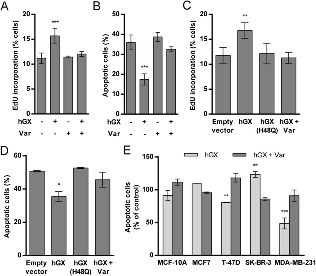
36 Figure 1

37 A B C D Control hgx E Figure 2
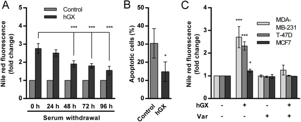
38 Figure 3
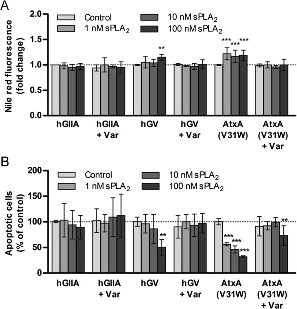
39 Figure 4
 Akt β-actin D Figure")
40 A B C OA 30 min 1 h 2 h 4 h hgx p-akt (Ser473) Akt β-actin D Figure 5
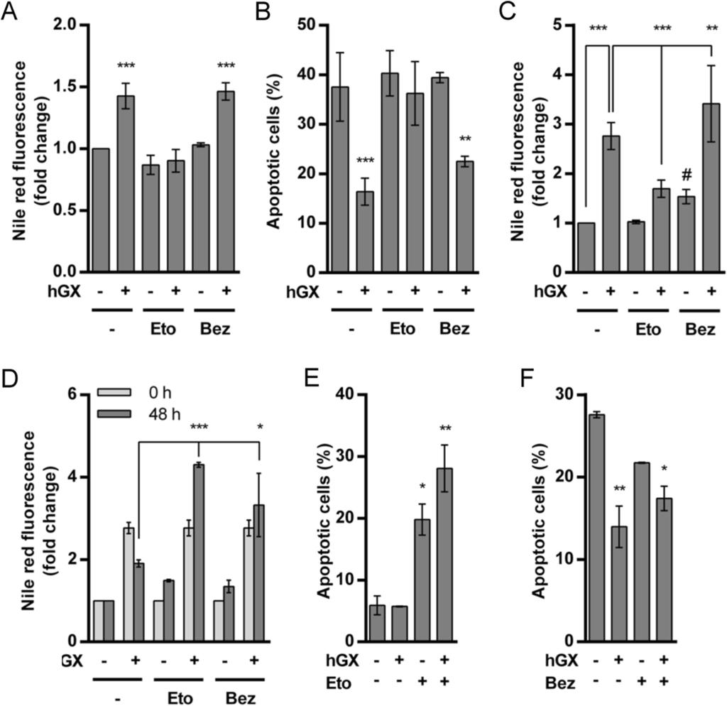
41 Figure 6
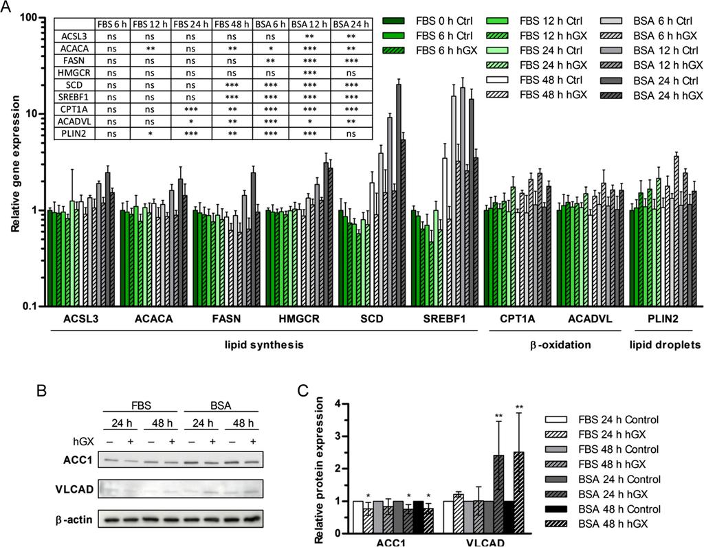
42 Figure 7
 AMPKα β-actin B C D E Figure 8")
43 A - OA Eto TrC AICAR Bez hgx AMPKα (Thr172) AMPKα β-actin B C D E Figure 8
LIPID METABOLISM. Sri Widia A Jusman Department of Biochemistry & Molecular Biology FMUI
 LIPID METABOLISM Sri Widia A Jusman Department of Biochemistry & Molecular Biology FMUI Lipid metabolism is concerned mainly with fatty acids cholesterol Source of fatty acids from dietary fat de novo
Chapter 26 Biochemistry 5th edition. phospholipids. Sphingolipids. Cholesterol. db=books&itool=toolbar
 http://www.ncbi.nlm.nih.gov/sites/entrez? db=books&itool=toolbar 1 The surface of a soap bubble is a bilayer formed by detergent molecules 2 Chapter 26 Biochemistry 5th edition phospholipids Sphingolipids
Lipid Metabolism. Catabolism Overview
 Lipid Metabolism Pratt & Cornely, Chapter 17 Catabolism Overview Lipids as a fuel source from diet Beta oxidation Mechanism ATP production Ketone bodies as fuel 1 High energy More reduced Little water
number Done by Corrected by Doctor Faisal Al-Khatibe
 number 24 Done by Mohammed tarabieh Corrected by Doctor Faisal Al-Khatibe 1 P a g e *Please look over the previous sheet about fatty acid synthesis **Oxidation(degradation) of fatty acids, occurs in the
Biochemistry: A Short Course
 Tymoczko Berg Stryer Biochemistry: A Short Course Second Edition CHAPTER 27 Fatty Acid Degradation Dietary Lipid (Triacylglycerol) Metabolism - In the small intestine, fat particles are coated with bile
Lysophosphatidic acid (LPA) signaling through LPA1 in organ fibrosis: A pathway with pleiotropic pro-fibrotic effects. and Andrew M.
 signaling through LPA1 in organ fibrosis: A pathway with pleiotropic pro-fibrotic effects. and Andrew M.") 78 Special Issue: Cellular and Molecular Bases for Fibrotic Diseases Review Article Lysophosphatidic acid (LPA) signaling through LPA1 in organ fibrosis: A pathway with pleiotropic pro-fibrotic effects
78 Special Issue: Cellular and Molecular Bases for Fibrotic Diseases Review Article Lysophosphatidic acid (LPA) signaling through LPA1 in organ fibrosis: A pathway with pleiotropic pro-fibrotic effects
ANSC/NUTR 618 LIPIDS & LIPID METABOLISM. Triacylglycerol and Fatty Acid Metabolism
 ANSC/NUTR 618 LIPIDS & LIPID METABOLISM II. Triacylglycerol synthesis A. Overall pathway Glycerol-3-phosphate + 3 Fatty acyl-coa à Triacylglycerol + 3 CoASH B. Enzymes 1. Acyl-CoA synthase 2. Glycerol-phosphate
Oxidation of Long Chain Fatty Acids
 Oxidation of Long Chain Fatty Acids Dr NC Bird Oxidation of long chain fatty acids is the primary source of energy supply in man and animals. Hibernating animals utilise fat stores to maintain body heat,
Fig. S1. Dose-response effects of acute administration of the β3 adrenoceptor agonists CL316243, BRL37344, ICI215,001, ZD7114, ZD2079 and CGP12177 at
 Fig. S1. Dose-response effects of acute administration of the β3 adrenoceptor agonists CL316243, BRL37344, ICI215,001, ZD7114, ZD2079 and CGP12177 at doses of 0.1, 0.5 and 1 mg/kg on cumulative food intake
Integration Of Metabolism
 Integration Of Metabolism Metabolism Consist of Highly Interconnected Pathways The basic strategy of catabolic metabolism is to form ATP, NADPH, and building blocks for biosyntheses. 1. ATP is the universal
BIOL212 Biochemistry of Disease. Metabolic Disorders - Obesity
 BIOL212 Biochemistry of Disease Metabolic Disorders - Obesity Obesity Approx. 23% of adults are obese in the U.K. The number of obese children has tripled in 20 years. 10% of six year olds are obese, rising
In glycolysis, glucose is converted to pyruvate. If the pyruvate is reduced to lactate, the pathway does not require O 2 and is called anaerobic
 Glycolysis 1 In glycolysis, glucose is converted to pyruvate. If the pyruvate is reduced to lactate, the pathway does not require O 2 and is called anaerobic glycolysis. If this pyruvate is converted instead
LIPID METABOLISM
 LIPID METABOLISM LIPOGENESIS LIPOGENESIS LIPOGENESIS FATTY ACID SYNTHESIS DE NOVO FFA in the blood come from :- (a) Dietary fat (b) Dietary carbohydrate/protein in excess of need FA TAG Site of synthesis:-
Chapter 8. Functions of Lipids. Structural Nature of Lipids. BCH 4053 Spring 2001 Chapter 8 Lecture Notes. Slide 1. Slide 2.
 BCH 4053 Spring 2001 Chapter 8 Lecture Notes 1 Chapter 8 Lipids 2 Functions of Lipids Energy Storage Thermal Insulation Structural Components of Membranes Protective Coatings of Plants and Insects Hormonal
Lehninger 5 th ed. Chapter 17
 Lehninger 5 th ed. Chapter 17 December 26, 2010 Prof. Shimon Schuldiner Email: Shimon.Schuldiner@huji.ac.il Phone: 6585992 CHAPTER 17 Fatty Acid Catabolism Key topics: How fats are digested in animals
Supplemental Table 1 Primer sequences (mouse) used for real-time qrt-pcr studies
 used for real-time qrt-pcr studies") Supplemental Table 1 Primer sequences (mouse) used for real-time qrt-pcr studies Gene symbol Forward primer Reverse primer ACC1 5'-TGAGGAGGACCGCATTTATC 5'-GCATGGAATGGCAGTAAGGT ACLY 5'-GACACCATCTGTGATCTTG
Supplemental Table 1 Primer sequences (mouse) used for real-time qrt-pcr studies Gene symbol Forward primer Reverse primer ACC1 5'-TGAGGAGGACCGCATTTATC 5'-GCATGGAATGGCAGTAAGGT ACLY 5'-GACACCATCTGTGATCTTG
Cholesterol and its transport. Alice Skoumalová
 Cholesterol and its transport Alice Skoumalová 27 carbons Cholesterol - structure Cholesterol importance A stabilizing component of cell membranes A precursor of bile salts A precursor of steroid hormones
Summary of fatty acid synthesis
 Lipid Metabolism, part 2 1 Summary of fatty acid synthesis 8 acetyl CoA + 14 NADPH + 14 H+ + 7 ATP palmitic acid (16:0) + 8 CoA + 14 NADP + + 7 ADP + 7 Pi + 7 H20 1. The major suppliers of NADPH for fatty
CELLULAR METABOLISM. Metabolic pathways can be linear, branched, cyclic or spiral
 CHM333 LECTURE 24 & 25: 3/27 29/13 SPRING 2013 Professor Christine Hrycyna CELLULAR METABOLISM What is metabolism? - How cells acquire, transform, store and use energy - Study reactions in a cell and how
: Overview of EFA metabolism
 Figure 1 gives an overview of the metabolic fate of the EFAs when consumed in the diet. The n-6 and n-3 PUFAs when consumed in the form of dietary triglyceride from various food sources undergoes digestion
cholesterol structure Cholesterol FAQs Cholesterol promotes the liquid-ordered phase of membranes Friday, October 15, 2010
 cholesterol structure most plasma cholesterol is in the esterified form (not found in cells or membranes) cholesterol functions in all membranes (drives formation of lipid microdomains) cholesterol is
Companion to Biosynthesis of Ketones & Cholesterols, Regulation of Lipid Metabolism Lecture Notes
 Companion to Biosynthesis of Ketones & Cholesterols, Regulation of Lipid Metabolism Lecture Notes The major site of acetoacetate and 3-hydorxybutyrate production is in the liver. 3-hydorxybutyrate is the
6. How Are Fatty Acids Produced? 7. How Are Acylglycerols and Compound Lipids Produced? 8. How Is Cholesterol Produced?
 Lipid Metabolism Learning bjectives 1 How Are Lipids Involved in the Generationand Storage of Energy? 2 How Are Lipids Catabolized? 3 What Is the Energy Yield from the xidation of Fatty Acids? 4 How Are
Part-4. Cell cycle regulatory protein 5 (Cdk5) A novel target of ERK in Carb induced cell death
 A novel target of ERK in Carb induced cell death") Part-4 Cell cycle regulatory protein 5 (Cdk5) A novel target of ERK in Carb induced cell death 95 1. Introduction The process of replicating DNA and dividing cells can be described as a series of coordinated
Part-4 Cell cycle regulatory protein 5 (Cdk5) A novel target of ERK in Carb induced cell death 95 1. Introduction The process of replicating DNA and dividing cells can be described as a series of coordinated
Metabolism of cardiac muscle. Dr. Mamoun Ahram Cardiovascular system, 2013
 Metabolism of cardiac muscle Dr. Mamoun Ahram Cardiovascular system, 2013 References This lecture Mark s Basic Medical Biochemistry, 4 th ed., p. 890-891 Hand-out Why is this topic important? Heart failure
BIOL2171 ANU TCA CYCLE
 TCA CYCLE IMPORTANCE: Oxidation of 2C Acetyl Co-A 2CO 2 + 3NADH + FADH 2 (8e-s donated to O 2 in the ETC) + GTP (energy) + Heat OVERVIEW: Occurs In the mitochondrion matrix. 1. the acetyl portion of acetyl-coa
A. Generation and characterization of Ras-expressing autophagycompetent
 Supplemental Material Supplemental Figure Legends Fig. S1 A. Generation and characterization of Ras-expressing autophagycompetent and -deficient cell lines. HA-tagged H-ras V12 was stably expressed in
Metabolism of acylglycerols and sphingolipids. Martina Srbová
 Metabolism of acylglycerols and sphingolipids Martina Srbová Types of glycerolipids and sphingolipids 1. Triacylglycerols function as energy reserves adipose tissue (storage of triacylglycerol), lipoproteins
Roles of Lipids. principal form of stored energy major constituents of cell membranes vitamins messengers intra and extracellular
 Roles of Lipids principal form of stored energy major constituents of cell membranes vitamins messengers intra and extracellular = Oxidation of fatty acids Central energy-yielding pathway in animals. O
Nafith Abu Tarboush DDS, MSc, PhD
 Nafith Abu Tarboush DDS, MSc, PhD natarboush@ju.edu.jo www.facebook.com/natarboush Lipids (cholesterol, cholesterol esters, phospholipids & triacylglycerols) combined with proteins (apolipoprotein) in
1 Corresponding author: Hideo Shindou, Department of Biochemistry and Molecular Biology,
 Recent progress on Acyl-CoA:lysophospholipid acyltransferase research* Hideo Shindou 1, Daisuke Hishikawa, Takeshi Harayama, Koichi Yuki, and Takao Shimizu From the Department of Biochemistry and Molecular
ANSC/NUTR 618 Lipids & Lipid Metabolism
 I. Overall concepts A. Definitions ANC/NUTR 618 Lipids & Lipid Metabolism 1. De novo synthesis = synthesis from non-fatty acid precursors a. Carbohydrate precursors (glucose, lactate, and pyruvate) b.
CHM333 LECTURE 34: 11/30 12/2/09 FALL 2009 Professor Christine Hrycyna
 Lipid Metabolism β-oxidation FA Acetyl-CoA Triacylglycerols (TAGs) and glycogen are the two major forms of stored energy in vertebrates Glycogen can supply ATP for muscle contraction for less than an hour
GENERAL SUMMARY Corpus luteum is a transient endocrine structure formed from the ruptured ovarian follicle. Its main function is to secrete P 4, a pro
 Corpus luteum is a transient endocrine structure formed from the ruptured ovarian follicle. Its main function is to secrete P 4, a pro-gestational hormone, essential for establishment and maintenance of
Modifications of Pyruvate Handling in Health and Disease Prof. Mary Sugden
 Modifications of Handling Modifications of Handling Centre for Diabetes and Metabolic Medicine Institute of Cell and Molecular Science Barts and the London School of Medicine and Dentistry 1 Potential
Voet Biochemistry 3e John Wiley & Sons, Inc.
 * * Voet Biochemistry 3e Lipid Metabolism Part I: (Chap. 25, sec.1-3) Glucose C 6 H 12 O 6 + 6 O 2 6 CO 2 + 6 H 2 O G o = -2823 kj/mol Fats (palmitic acid) C 16 H 32 O 2 + 23 O 2 16 CO 2 + 16 H 2 O G o
BBSG 501 Section 4 Metabolic Fuels, Energy and Order Fall 2003 Semester
 BBSG 501 Section 4 Metabolic Fuels, Energy and Order Fall 2003 Semester Section Director: Dave Ford, Ph.D. Office: MS 141: ext. 8129: e-mail: fordda@slu.edu Lecturers: Michael Moxley, Ph.D. Office: MS
BCM 221 LECTURES OJEMEKELE O.
 BCM 221 LECTURES BY OJEMEKELE O. OUTLINE INTRODUCTION TO LIPID CHEMISTRY STORAGE OF ENERGY IN ADIPOCYTES MOBILIZATION OF ENERGY STORES IN ADIPOCYTES KETONE BODIES AND KETOSIS PYRUVATE DEHYDROGENASE COMPLEX
Suppl. Figure 1. T 3 induces autophagic flux in hepatic cells. (A) RFP-GFP-LC3 transfected HepG2/TRα cells were visualized and cells were quantified
 RFP-GFP-LC3 transfected HepG2/TRα cells were visualized and cells were quantified") Suppl. Figure 1. T 3 induces autophagic flux in hepatic cells. (A) RFP-GFP-LC3 transfected HepG2/TRα cells were visualized and cells were quantified for RFP-LC3 puncta (red dots) representing both autolysosomes
Suppl. Figure 1. T 3 induces autophagic flux in hepatic cells. (A) RFP-GFP-LC3 transfected HepG2/TRα cells were visualized and cells were quantified for RFP-LC3 puncta (red dots) representing both autolysosomes
Physiology Unit 1 CELL SIGNALING: CHEMICAL MESSENGERS AND SIGNAL TRANSDUCTION PATHWAYS
 Physiology Unit 1 CELL SIGNALING: CHEMICAL MESSENGERS AND SIGNAL TRANSDUCTION PATHWAYS In Physiology Today Cell Communication Homeostatic mechanisms maintain a normal balance of the body s internal environment
Cell Injury MECHANISMS OF CELL INJURY
 Cell Injury MECHANISMS OF CELL INJURY The cellular response to injurious stimuli depends on the following factors: Type of injury, Its duration, and Its severity. Thus, low doses of toxins or a brief duration
Thin layer absorption chromatography can achieve very good separation of small lipid samples
 Abbreviations Preface Acknowledgements Lipids: definition, isolation, separation and detection p. 1 Introduction p. 1 Definitions p. 1 Structural chemistry and nomenclature p. 1 Extraction of lipids from
ANSC/NUTR 618 LIPIDS & LIPID METABOLISM. Fatty Acid Elongation and Desaturation
 ANSC/NUTR 618 LIPIDS & LIPID METABOLISM I. Fatty acid elongation A. General 1. At least 60% of fatty acids in triacylglycerols are C18. 2. Free palmitic acid (16:0) synthesized in cytoplasm is elongated
Intermediary metabolism. Eva Samcová
 Intermediary metabolism Eva Samcová Metabolic roles of tissues Four major tissues play a dominant role in fuel metabolism : liver, adipose, muscle, and brain. These tissues do not function in isolation.
2.28E E AKT/CAT P-3-4:
 Supplemental Table 1: IPA network analysis of the microarray data presented in Figure 6 showing the most significant molecular and cellular function classifications along with the respective number of
Supplementary Figure S1
 Lipidomic-based investigation into the regulatory effect of Schisandrin B on palmitic acid level in non-alcoholic steatotic livers Hiu Yee Kwan 1,2, Xuyan Niu 3, Wenlin Dai 4, Tiejun Tong 4, Xiaojuan Chao
Fatty acids synthesis
 Fatty acids synthesis The synthesis start from Acetyl COA the first step requires ATP + reducing power NADPH! even though the oxidation and synthesis are different pathways but from chemical part of view
OVERVIEW M ET AB OL IS M OF FR EE FA TT Y AC ID S
 LIPOLYSIS LIPOLYSIS OVERVIEW CATABOLISM OF FREE FATTY ACIDS Nonesterified fatty acids Source:- (a) breakdown of TAG in adipose tissue (b) action of Lipoprotein lipase on plasma TAG Combined with Albumin
THE GLUCOSE-FATTY ACID-KETONE BODY CYCLE Role of ketone bodies as respiratory substrates and metabolic signals
 Br. J. Anaesth. (1981), 53, 131 THE GLUCOSE-FATTY ACID-KETONE BODY CYCLE Role of ketone bodies as respiratory substrates and metabolic signals J. C. STANLEY In this paper, the glucose-fatty acid cycle
Biosynthesis of Fatty Acids. By Dr.QUTAIBA A. QASIM
 Biosynthesis of Fatty Acids By Dr.QUTAIBA A. QASIM Fatty Acids Definition Fatty acids are comprised of hydrocarbon chains terminating with carboxylic acid groups. Fatty acids and their associated derivatives
Cholesterol metabolism. Function Biosynthesis Transport in the organism Hypercholesterolemia
 Cholesterol metabolism Function Biosynthesis Transport in the organism Hypercholesterolemia - component of all cell membranes - precursor of bile acids steroid hormones vitamin D Cholesterol Sources: dietary
BIOSYNTHESIS OF FATTY ACIDS. doc. Ing. Zenóbia Chavková, CSc.
 BIOSYNTHESIS OF FATTY ACIDS doc. Ing. Zenóbia Chavková, CSc. The pathway for the of FAs is not the reversal of the oxidation pathway Both pathways are separated within different cellular compartments In
Unit IV Problem 3 Biochemistry: Cholesterol Metabolism and Lipoproteins
 Unit IV Problem 3 Biochemistry: Cholesterol Metabolism and Lipoproteins - Cholesterol: It is a sterol which is found in all eukaryotic cells and contains an oxygen (as a hydroxyl group OH) on Carbon number
Plasma lipoproteins & atherosclerosis by. Prof.Dr. Maha M. Sallam
 Biochemistry Department Plasma lipoproteins & atherosclerosis by Prof.Dr. Maha M. Sallam 1 1. Recognize structures,types and role of lipoproteins in blood (Chylomicrons, VLDL, LDL and HDL). 2. Explain
Gene Polymorphisms and Carbohydrate Diets. James M. Ntambi Ph.D
 Gene Polymorphisms and Carbohydrate Diets James M. Ntambi Ph.D Fatty Acids that Flux into Tissue Lipids are from Dietary Sources or are Made De novo from Glucose or Fructose Glucose Fructose Acetyl-CoA
Synthesis of Fatty Acids and Triacylglycerol
 Synthesis of Fatty Acids and Triacylglycerol Lippincott s Chapter 16 Fatty Acid Synthesis Mainly in the Liver Requires Carbon Source: Acetyl CoA Reducing Power: NADPH 8 CH 3 COO C 15 H 33 COO Energy Input:
MEMBRANE LIPIDS I and II: GLYCEROPHOSPHOLIPIDS AND SPHINGOLIPIDS
 December 6, 2011 Lecturer: Eileen M. Lafer MEMBRANE LIPIDS I and II: GLYCEROPHOSPHOLIPIDS AND SPHINGOLIPIDS Reading: Stryer Edition 6: Chapter 26 Images: All images in these notes were taken from Lehninger,
Insulin Resistance. Biol 405 Molecular Medicine
 Insulin Resistance Biol 405 Molecular Medicine Insulin resistance: a subnormal biological response to insulin. Defects of either insulin secretion or insulin action can cause diabetes mellitus. Insulin-dependent
Fatty Acid and Triacylglycerol Metabolism 1
 Fatty Acid and Triacylglycerol Metabolism 1 Mobilization of stored fats and oxidation of fatty acids Lippincott s Chapter 16 What is the first lecture about What is triacylglycerol Fatty acids structure
Integration of Metabolism 1. made by: Noor M. ALnairat. Sheet No. 18
 Integration of Metabolism 1 made by: Noor M. ALnairat Sheet No. 18 Data :24/11/2016 SLIDE 2: Metabolism Consist of Highly Interconnected Pathways The basic strategy of catabolic metabolism is to form ATP,
Lipid Metabolism * OpenStax
 OpenStax-CNX module: m46462 1 Lipid Metabolism * OpenStax This work is produced by OpenStax-CNX and licensed under the Creative Commons Attribution License 3.0 By the end of this section, you will be able
AMPK. Tomáš Kučera.
 AMPK (AMP- ACTIVATED PROTEIN KINASE ) Tomáš Kučera tomas.kucera@lfmotol.cuni.cz Department of Medical Chemistry and Clinical Biochemistry 2nd Faculty of Medicine, Charles University in Prague and Motol
Chapter 20. Cell - Cell Signaling: Hormones and Receptors. Three general types of extracellular signaling. endocrine signaling. paracrine signaling
 Chapter 20 Cell - Cell Signaling: Hormones and Receptors Three general types of extracellular signaling endocrine signaling paracrine signaling autocrine signaling Endocrine Signaling - signaling molecules
Syllabus for BASIC METABOLIC PRINCIPLES
 Syllabus for BASIC METABOLIC PRINCIPLES The video lecture covers basic principles you will need to know for the lectures covering enzymes and metabolism in Principles of Metabolism and elsewhere in the
Regulation of Lipid Homeostasis: Lipid Droplets
 Regulation of Lipid Homeostasis: Lipid Droplets Bernd Helms Article Brand Recter Hendrik Mertens 1 The Basics I: FAs The Basics II: FA Activation 2 Basics III: TG-FA Interplay. Why? Adipocytes 3 Foam cells
PHOSPHOLIPID MEMBRANE ABNORMALITIES AND REDUCED NIACIN SKIN FLUSH RESPONSE IN SCHIZOPHRENIA
 PHOSPHOLIPID MEMBRANE ABNORMALITIES AND REDUCED NIACIN SKIN FLUSH RESPONSE IN SCHIZOPHRENIA Alena Buretić-Tomljanović 1, Jasminka Giacometti 1, Sergej Nadalin 1, Gordana Rubeša 2, Mirjana Vulin 3, Draško
PHOSPHOLIPIDS METABOLISM. BY Dr. Walid Said Zaki Dr. Marwa Ali LECTURER OF BIOCHEMISTRY AND MOLECULAR BIOLOGY
 PHOSPHOLIPIDS METABOLISM BY Dr. Walid Said Zaki Dr. Marwa Ali LECTURER OF BIOCHEMISTRY AND MOLECULAR BIOLOGY 1. State the definition and classification of Phospholipids. 2. Describe the general structure
SUPPLEMENTARY INFORMATION
 SUPPLEMENTARY INFORMATION doi:10.1038/nature11464 Supplemental Figure S1. The expression of Vegfb is increased in obese and diabetic mice as compared to lean mice. a-b, Body weight and postprandial blood
Metabolic integration and Regulation
 Metabolic integration and Regulation 109700: Graduate Biochemistry Trimester 2/2016 Assistant Prof. Dr. Panida Khunkaewla kpanida@sut.ac.th School of Chemistry Suranaree University of Technology 1 Overview
AMPK. Tomáš Kuc era. Ústav lékar ské chemie a klinické biochemie 2. lékar ská fakulta, Univerzita Karlova v Praze
 AMPK (AMP- ACTIVATED PROTEIN KINASE ) Tomáš Kuc era Ústav lékar ské chemie a klinické biochemie 2. lékar ská fakulta, Univerzita Karlova v Praze 2013 AMPK AMP-ACTIVATED PROTEIN KINASE present in all eukaryotic
BCMB 3100 Fall 2013 Exam III
 BCMB 3100 Fall 2013 Exam III 1. (10 pts.) (a.) Briefly describe the purpose of the glycerol dehydrogenase phosphate shuttle. (b.) How many ATPs can be made when electrons enter the electron transport chain
Lecture 36. Key Concepts. Overview of lipid metabolism. Reactions of fatty acid oxidation. Energy yield from fatty acid oxidation
 Lecture 36 Lipid Metabolism 1 Fatty Acid Oxidation Ketone Bodies Key Concepts Overview of lipid metabolism Reactions of fatty acid oxidation Energy yield from fatty acid oxidation Formation of ketone bodies
Biochemistry: A Short Course
 Tymoczko Berg Stryer Biochemistry: A Short Course Second Edition CHAPTER 28 Fatty Acid Synthesis 2013 W. H. Freeman and Company Chapter 28 Outline 1. The first stage of fatty acid synthesis is transfer
Manipulation of the Nutrient Sensors (AMPK/TOR) with Anaplerotic Diet Therapy (Triheptanoin) An Alternative to Diet Restriction
 with Anaplerotic Diet Therapy (Triheptanoin) An Alternative to Diet Restriction") Manipulation of the Nutrient Sensors (AMPK/TOR) with Anaplerotic Diet Therapy (Triheptanoin) An Alternative to Diet Restriction CharlesR.Roe,MD Institute of Metabolic Disease Baylor University Medical
Manipulation of the Nutrient Sensors (AMPK/TOR) with Anaplerotic Diet Therapy (Triheptanoin) An Alternative to Diet Restriction CharlesR.Roe,MD Institute of Metabolic Disease Baylor University Medical
Lipids digestion and absorption, Biochemistry II
 Lipids digestion and absorption, blood plasma lipids, lipoproteins Biochemistry II Lecture 1 2008 (J.S.) Triacylglycerols (as well as free fatty acids and both free and esterified cholesterol) are very
Master class Biomolecular Sciences Molecular Cell Biology.
 Master class Biomolecular Sciences Molecular Cell Biology. 04-09-08: Paul van Bergen en Henegouwen. Clathrin-indep Endocytosis 11-09-08: Willem Stoorvogel. Endocytosis and MHC classii 18-09-08: X-track
Hmgcoar AGCTTGCCCGAATTGTATGTG TCTGTTGTAACCATGTGACTTC. Cyp7α GGGATTGCTGTGGTAGTGAGC GGTATGGAATCAACCCGTTGTC
 Supplement Table I: primers for Real Time RT-PCR Gene Foward Reverse Hmgcoar AGCTTGCCCGAATTGTATGTG TCTGTTGTAACCATGTGACTTC Cyp7α GGGATTGCTGTGGTAGTGAGC GGTATGGAATCAACCCGTTGTC Cyp27a1 GTGGTCTTATTGGGTACTTGC
Phospholipids Metabolism
 Chapter VI: Phospholipids Metabolism Dr. Sameh Sarray Hlaoui Phospholipids Features: Amphipatic: - Hydrophobic head: fatty acids - Hydropholic head: P group+ alcohol Composed of alcohol attached by a phosphodiester
Introduction: 年 Fas signal-mediated apoptosis. PI3K/Akt
 Fas-ligand (CD95-L; Fas-L) Fas (CD95) Fas (apoptosis) 年 了 不 度 Fas Fas-L 力 不 Fas/Fas-L T IL-10Fas/Fas-L 不 年 Fas signal-mediated apoptosis 度降 不 不 力 U-118, HeLa, A549, Huh-7 MCF-7, HepG2. PI3K/Akt FasPI3K/Akt
Loss of protein association causes cardiolipin degradation in Barth syndrome
 SUPPLEMENTARY INFORMATION Loss of protein association causes cardiolipin degradation in Barth syndrome Yang Xu 1, Colin K.L. Phoon 2, Bob Berno 5, Kenneth D Souza 6, Esthelle Hoedt 4, Guoan Zhang 4, Thomas
By: Dr Hadi Mozafari 1
 Biological lipids are a chemically diverse group of compounds, the common and defining feature of which is their insolubility in water. By: Dr Hadi Mozafari 1 Fats and oils are the principal stored forms
Integration Of Metabolism
 Integration Of Metabolism Metabolism Consist of Highly Interconnected Pathways The basic strategy of catabolic metabolism is to form ATP, NADPH, and building blocks for biosyntheses. 1. ATP is the universal
C-Phycocyanin (C-PC) is a n«sjfc&c- waefc-jduble phycobiliprotein. pigment isolated from Spirulina platensis. This water- soluble protein pigment is
 is a n«sjfc&c- waefc-jduble phycobiliprotein. pigment isolated from Spirulina platensis. This water- soluble protein pigment is") ' ^Summary C-Phycocyanin (C-PC) is a n«sjfc&c- waefc-jduble phycobiliprotein pigment isolated from Spirulina platensis. This water- soluble protein pigment is of greater importance because of its various
' ^Summary C-Phycocyanin (C-PC) is a n«sjfc&c- waefc-jduble phycobiliprotein pigment isolated from Spirulina platensis. This water- soluble protein pigment is of greater importance because of its various
Lipid Metabolism. Remember fats?? Triacylglycerols - major form of energy storage in animals
 Remember fats?? Triacylglycerols - major form of energy storage in animals Your energy reserves: ~0.5% carbs (glycogen + glucose) ~15% protein (muscle, last resort) ~85% fat Why use fat for energy? 1 gram
5.0 HORMONAL CONTROL OF CARBOHYDRATE METABOLISM
 5.0 HORMONAL CONTROL OF CARBOHYDRATE METABOLISM Introduction: Variety of hormones and other molecules regulate the carbohydrates metabolism. Some of these have already been cited in previous sections.
Lipid metabolism. Degradation and biosynthesis of fatty acids Ketone bodies
 Lipid metabolism Degradation and biosynthesis of fatty acids Ketone bodies Fatty acids (FA) primary fuel molecules in the fat category main use is for long-term energy storage high level of energy storage:
Lipid droplet biogenesis: a novel process of self-digestion as a strategy of survival to stress
 Lipid droplet biogenesis: a novel process of self-digestion as a strategy of survival to stress Memoria del trabajo experimental para optar al grado de doctor, correspondiente al Programa de Doctorado
Using the Organic Acids Test Part 3 Dr. Jeff Moss
 Using organic acids to resolve chief complaints and improve quality of life in chronically ill patients Part III Jeffrey Moss, DDS, CNS, DACBN jeffmoss@mossnutrition.com 413-530-08580858 (cell) 1 Summer
Dietary Lipid Utilization by Haddock (Melanogrammus aeglefinus)
") Dietary Lipid Utilization by Haddock (Melanogrammus aeglefinus) Santosh P. Lall & Dominic A. Nanton National Research Council of Canada Halifax, Canada vis, California ne 23, 2003 Body Components of Wild
Dietary Lipid Utilization by Haddock (Melanogrammus aeglefinus) Santosh P. Lall & Dominic A. Nanton National Research Council of Canada Halifax, Canada vis, California ne 23, 2003 Body Components of Wild
Niacin Metabolism: Effects on Cholesterol
 Niacin Metabolism: Effects on Cholesterol By Julianne R. Edwards For Dr. William R. Proulx, PhD, RD Associate Professor of Nutrition and Dietetics In partial fulfillments for the requirements of NUTR342
Supplementary Fig. 1 eif6 +/- mice show a reduction in white adipose tissue, blood lipids and normal glycogen synthesis. The cohort of the original
 Supplementary Fig. 1 eif6 +/- mice show a reduction in white adipose tissue, blood lipids and normal glycogen synthesis. The cohort of the original phenotypic screening was n=40. For specific tests, the
CHY2026: General Biochemistry. Lipid Metabolism
 CHY2026: General Biochemistry Lipid Metabolism Lipid Digestion Lipid Metabolism Fats (triglycerides) are high metabolic energy molecules Fats yield 9.3 kcal of energy (carbohydrates and proteins 4.1 kcal)
Steven Michael Dragos. A Thesis. presented to. The University of Guelph. In partial fulfillment of requirements. for the degree of.
 Reduced SCD1 activity decreases fatty acid re-esterification and alters markers of glyceroneogenesis and lipolysis in murine white adipose tissue and 3T3-L1 adipocytes by Steven Michael Dragos A Thesis
Crosstalk between Adiponectin and IGF-IR in breast cancer. Prof. Young Jin Suh Department of Surgery The Catholic University of Korea
 Crosstalk between Adiponectin and IGF-IR in breast cancer Prof. Young Jin Suh Department of Surgery The Catholic University of Korea Obesity Chronic, multifactorial disorder Hypertrophy and hyperplasia
2.5. AMPK activity
 Supplement Fig. A 3 B phos-ampk 2.5 * Control AICAR AMPK AMPK activity (Absorbance at 45 nm) 2.5.5 Control AICAR Supplement Fig. Effects of AICAR on AMPK activation in macrophages. J774. macrophages were
Ahmad Ulnar. Faisal Nimri ... Dr.Faisal
 24 Ahmad Ulnar Faisal Nimri... Dr.Faisal Fatty Acid Synthesis - Occurs mainly in the Liver (to store excess carbohydrates as triacylglycerols(fat)) and in lactating mammary glands (for the production of
Supplemental Figures
 Supplemental Figures Supplemental Figure 1. Fasting-dependent regulation of the SREBP ortholog SBP-1 and lipid homeostasis mediated by the SIRT1 ortholog SIR-2.1 in C. elegans. (A) Wild-type or sir-2.1(lof)
Leen Alsahele. Razan Al-zoubi ... Faisal
 25 Leen Alsahele Razan Al-zoubi... Faisal last time we started talking about regulation of fatty acid synthesis and degradation *regulation of fatty acid synthesis by: 1- regulation of acetyl CoA carboxylase